Graefe's Archive. Ophthalmology Springer-Verlag Artificial anterior chamber for the growing of membranes on lens implants*
|
|
|
- Anne Chandler
- 5 years ago
- Views:
Transcription


1 Graefe's Arch Clin Exp Ophthalmol (1983) 221:55-60 Graefe's Archive for Clinical and Experimental Ophthalmology Springer-Verlag 1983 Artificial anterior chamber for the growing of membranes on lens implants* J. Reimer Wolter 1.* and Steven L. Kunkel / Departments of Ophthalmology and Pathology 1 and Department of Pathology 2, University of Michigan Medical Center, Ann Arbor, Michigan 48109, USA Abstract. Intraocular lens implants made of glass and plastic were placed into the lumen of short pieces of clear plastic tubing. These were incompletely closed at both ends with nylon sutures, filled with Healon, and used experimentally as artificial anterior chambers for implantation into the peritoneal space of mice. After 7 days macrophages had been attracted into the chambers, become sessile on the implants, and formed a continuous membrane on their surface. The concept of artificial anterior chambers in experimental and future clinical applications is discussed. Introduction Earlier studies [1 6] have indicated that the inner eye reacts to a newly introduced lens implant by mobilizing free-moving macrophages. These are attracted to the surface of the foreign substance, become adherent, and change into sessile epithelioid cells, fibroblastlike cells, or giant cells. They also are involved in the formation of a separating proteinaceous film that develops with time into a capsulelike formation continuously covering all parts of the implant. From a successful implant the free-moving macrophages and most of the giant cells disappear with time. Relatively few fibroblastlike cells remain permanently and maintain the capsule. The formation of this type of reactive membrane on lens implants is considered beneficial. They are optically clear and prevent direct contact of the fluid and tissue structures of the inner eye with the foreign substances of the implants. The process of the reactive formation of continuous and optically clear membranes on the surface of lens implants is not always uncomplicated. For example, the membrane may become thick and scarlike. This can result in optical obstruction as well as in obliteration of adjacent spaces. Vitreous formation on the surface of implants has been seen to interfere with the successful structure of a proper separating membrane. The impression is that implants without a continuous membrane are not tolerated well, because continuing uveitis and damage to the corneal endothelium * Supported by The Research to Prevent Blindness, Inc. New York, N.Y. USA ** Corresponding author Fig. l. One of the artificial anterior chambers containing a glass intraocular lens implant before implantation. The optic portion of the implant (O) is clearly seen within the plastic tubing (arrows) flattened by knotted nylon sutures (K). The haptics of the implant are cut short. Photograph x 15 Fig. 2. After 7 days' implantation in the peritoneal space of a mouse. The chamber contains a milky fluid and all parts are covered by a grayish layer. The rim of the glass implant is visible in the chamber (arrow). Photograph x 15


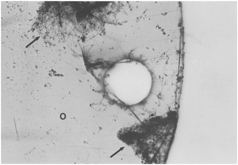
2 56 Fig. 3. The other artificial anterior chamber (arrows) containing a polymethylmethacrylate implant (O) with blue Prolene haptics (P) before implantation. Photograph x 15 Fig. 4. The artificial anterior chamber seen in Fig. 3 after 7 days' implantation in the peritoneal space of a mouse. The chamber is covered with a gray layer and contains a milky fluid. Photography x15 have been seen to be pronounced in association with the presence of naked surfaces of implants. Initially, the concept of the artificial anterior chamber for the study of membranes on implants under experimental conditions is presented in this paper. In the future we think that the use of artificial anterior chambers might be one way to grow clear separating membranes of the patients own monocytes on the surface of his or her lens implant, before this is placed in the eye. Experiment. Artificial anterior chambers were built from the kind of clear plastic tubing used in operating rooms or laboratories and supplied in sterile packages. Two short pieces of this soft tubing were cut and one lens implant of glass and another of polymethylmethacrylate with Prolene haptics was placed in the lumen of the pieces of tubing. 1 The tubing was flattened out in this process and both ends were closed in a flattened position by nylon sutures (Figs. 1 and 2). Thus the lumen of the tubing was closed off at both ends and a central chamber containing the implants was created. In the region of the tied sutures the opposite walls of the tubing were tightly touching. However, there were four narrow openings into the center of the chamber preserved on both sides, next ot the nylon sutures. The size of the pieces of tubing made into artificial anterior chambers was approximately 9 x 6 x 4 mm. Under sterile conditions these artificial anterior chambers were filled with Healon (sodium hyaluronate) and placed into the peritoneal space of two black laboratory mice. The mice measured 50 mm in length without tails. 1 This is to thank IOLAB Corporation of Covina, California and Lynell Medical Technology, Inc., of New York, N.Y. for supplying the lens implants used for this study The implantation of the flat pieces of plastic tubing was a little tight due to the large size of the tubes in relation to the mice. However, the mice remained active, ate, and did not appear sick or in any way incapacitated throughout the week of the experiment. The mice survived the operation without any difficulties. After 7 days the mice were killed. The artificial anterior chambers were found in the peritoneal space without firm adhesions and they could be removed without difficulty. Both pieces of tubing were covered with a smooth layer of slightly yellowish material on the outside. Both contained an exudatelike yellowish fluid in their lumen (Figs. 3 and 4). The artificial anterior chambers were immediately placed into 10% buffered formalin for a day of fixation. The specimens were washed as a whole and then the pieces of tubing were opened with scissors on one side. A yellowish exudatelike layer of mushy substance was attached to the implants as well as to all the surfaces of the plastic tubing. The implants together with the attached layer were removed without difficulty. The lens implant cytology technique was used to stain the layers on both implants with hematoxylin-eosin [3], The glass implant had a thick covering of firmly attached cell layers. These resembled free-moving macrophages and early stages of fibroblastlike cells, mostly of bipolar shape. However, technical difficulties were encountered in the dehydrating and embedding process, after the staining was completed and a preliminary inspection of the specimen had been carried out under the microscope. The plastic implant from the other piece of tubing, however could be dehydrated, embedded, and photographed properly. The polymethylmethacrylate optic portion of the implant was, in the first place, tightly surrounded by a thin eosinophilic film (Figs. 5-7). Fig. 5. Low-power view of the plastic implant removed from the artificial anterior chamber seen in Fig. 4. Its optic part (O) is covered with a thin and regular layer of a slightly eosinophilic proteinaceous substance. Dark staining areas of fibrinous exudate are seen in a somewhat geographical arrangement on parts of the optics. The Prolene haptics (P) show one thick accumulation of cells (arrow). Lens implant cytology technique, H + E stain, photomicrograph x 25 Fig. 6. Portion of the optics of the plastic lens implant with its thin homogenous capsulelike layer (O) and loose additional accumulations of fibrin on its surface (arrows). Lens implant cytology technique, H + E stain, photomicrograph x 100

3 57 1 Fig. 5 Fig. 6

4 58 Fig. 7 Fig. 8
.")
.")
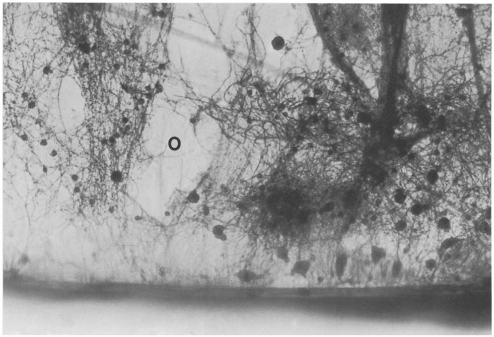
5 59 Fig. 9. High-power view of one of the Prolene haptics (P) of the lens implant seen in Fig. 4 with single macrophages rather evenly distributed all over its surface and an additional nodular accumulation of densely arranged macrophages supported by a fibrin framework (arrow). Lens implant cytology technique, H + E stain, photomicrograph 100 This was of rather homogenous composition and regular structure and contained macrophages in an irregular distribution (Figs. 6 and 7). Another less regular and much looser layer of fibrinous exudate containing more macrophages and occasional polymorphnuclear leukocytes (PMNs) was found on top of the homogenous thin layer on the surface of the implant (Figs. 5-8). The loose network of fibrin had clearly filled some of the space around the implant in the tubing and interconnected the reactive layers of the wall of this tubing and the surface of the implant. The most dense and cellular accumulations of macrophages were seen, in addition to diffusely distributed macrophages, on the Prolene haptics of this implant and presented as rounded bunches of cells with a fibrinous framework (Fig. 9). Discussion This first attempt to create an artificial anterior chamber for the growth of cellular membranes on lens implants in the peritoneal space of the mouse has been quite successful. Just like the anterior chamber of the eye, this artificial anterior chamber was fluid-filled. Healon was used for this purpose with the aim of avoiding the loss of a less viscous fluid from the chamber during the implantation process. Healon has been found not to interfere with the orderly formation of membranes on lens implants by peritoneal mouse macrophages under tissue culture conditions in a recently reported experiment by Wolter and Kunkel, which has been submitted for publication. The peritoneal macrophages were attracted by the implants in both artificial anterior chambers and found their way into their lumen through the narrow openings on both sides of the sutures. They adhered to the implants and within the 7 days of the experiment settled on the surface of the implants. This process was associated with the development of a filmlike, homogeneous, and slightly eosinophilic membrane of a proteinaceous substance all over the surface of both implants. It is suspected that the macrophages produced these membranes, but definite proof of this is not as yet available. On the glass implant fibroblastlike cells were seen in addition to common macrophages, but technical problems with its embedding prevented photographic documentation. The thicker, looser, and more patchy reaction containing fibrin and PMNs superficially attached to the implants probably was part of the more acute inflammatory response elicited by the plastic tubing. This kind of a reaction was found all over the outside and the inside of both pieces of plastic tubing, Of all the plastic materials that we have studied, polymethylmethacrylate causes the least inflammatory reaction, when it is implanted in tissues or tissue fluids. This observation is supported by clinical findings which also show that polymethylmethacrylate is relatively very well tolerated. Supramid and Prolene are both known to elicit more of a cellular response, as well as a thicker proteinaceous capsule, when these substances are used for lens implants [6]. In the present experiment it was observed that the Prolene haptics had much more massive accumulations of cells than the polymethylmethacrylate optics. In comparison to all three of these types of plastics commonly utilized for lens implants, the tubing used for the artificial anterior chamber in the present experiment caused a much more severe reaction with thicker layers of cells, fibrinous exudate, and some PMNs all over its surface. The idea of a fluid-filled artificial anterior chamber for the growing of cells and separating membranes on lens implants will be pursued. Since this first and rather crude model has shown such promise, a better chamber will be built, and obviously this has to be completely composed of polymethylmethacrylate in all parts. Other fluids for filling the chamber and new implantation sites in larger animals will be investigated with the aim of creating a situation resembling the inner eye as far as possible. We foresee a time when intraocular lens implants, within a well designed artificial anterior chamber, will be temporarily placed somewhere in the organism of a patient who will need cataract surgery. The idea would be that all the biochemical and cellular interactions between the patient's phagocyte system and the foreign substance of the implant take place in the safe surroundings of the artificial anterior chamber, and that a permanent separating capsule is created before that implant is placed in its final place in the eye. The haptics could extend out of the fluid-filled artificial anterior chambers for the growth of fibrous attachments. These would be excised at the end of the period to fit an area of the wall of the inner eye for firm attachment. Tissue engineering in the truest sense would thus be used for the separation of the main body, as well as for the fixation of lens implants in the eye. Possible sites for the experimental implantation of artificial anterior Fig. 7. Higher power of the fibrin network (arrow) on the surface of the homogeneous capsule with macrophages (34) on the optic portion (O) of the plastic implant seen in Fig. 4. Lens implant cytology technique, H + E stain, photomicrograph 250 Fig. 8. Border of the plastic implant seen in Fig. 4. The fibrin network on its optic portion (O) exhibits larger macrophages as well as small PMNs, Lens implant cytology technique, H + E stain, photomicrograph 250

6 60 chambers are locations deep in the skin - possibly in the arm pits or on the side of the chest - in the peritoneal space, under the periosteum, or perhaps even in the bone marrow. The latter site has special appeal because it is the primary source of the macrophages. To obtain monocytes from the patient's blood and to grow membranes on lens implants in tissue culture in another possibility, but our experiences with mouse macrophages in tissue culture indicate that continuous supply of new macrophages over a relatively long time is necessary for the formation of useful membranes. The experimental path towards these goals looks open and quite inviting. However, much basic work needs to be done in animal experiments and much new insight will have to be gained before the dream of a lens implant with a pregrown separating membrane in a human eye can become true. References 1. Wolter JR (1982) Cell life on the surface of lens implants. Graefe's Arch Clin Exp Ophthalmol 218: Wolter JR (1982) Foreign body giant cells on intraocular lens implants. Graefe's Arch Clin Exp Ophthalmol 219: Wolter JR (1982) Lens implant cytology. Ophthalmic Surg 13 : Wolter JR (1983) Reactive membrane on lens implant: three month after implantation. Graefe's Arch Clin Exp Ophthalmol 220: Wolter JR (1983) Morphology of the capsulelike portion of the reactive membranes on intraocular lens implants. Graefe's Arch Clin Exp Ophthalmol 220 : Wolter JR (1983) Foreign body giant cells selectively covering haptics of intraocular lens implants: indicators of poor toleration? Ophthalmic Surg (in press) 7. Wolter JR, Kunkel SL (1983) Abdominal implantation of intraocular lenses resulting in the formation of reactive membranes. Graefe's Arch Clin Exp Ophthalmol 220: Wolter JR, Lichter PR (1983) Fibroblastlike cells on intraocular lens implants: phagocytizing erythrocytes. Br J Ophthalmol (in print) Received May 20, 1983
Graefe's Archive. Ophthalmology Springer-Verlag t983. Fusion of maerophages on lens implants resulting in the formation of giant cells
 Graefe's Arch Clin Exp Ophthalmol (1983) 221:1-7 Graefe's Archive for Clinical and Experimental Ophthalmology Springer-Verlag t983 Fusion of maerophages on lens implants resulting in the formation of giant
Cystoid macular edema associated with limbal melanoma*
 Graefe's Arch Clin Exp Ophthalmol (1983) 221:101-105 Graefe's Archive for CliniCal and Experimental Ophthalmology Springer-Verlag 1983 Cystoid macular edema associated with limbal melanoma* J. Reimer Wolter
Foreign Body Giant Cells on Intraocular Lens Implants*
 Graefe's Arch Clin Exp Ophthalmol (1982) 219:103-11I Graefe's Archive for Clinical and Experimental Ophthalmology Springer-Verlag 1982 Foreign Body Giant Cells on Intraocular Lens Implants* J. Reimer Wolter
HYPERPLASIA OF THE ANTERIOR LAYER OF THE IRIS STROMA*t
 Brit. J. Ophthal. (1965) 49, 516 HYPERPLASIA OF THE ANTERIOR LAYER OF THE IRIS STROMA*t BY MALCOLM N. LUXENBERG From the Bascom Palmer Eye Institute, Department of Ophthalmology, University of Miami School
CONSENT FOR CATARACT SURGERY REQUEST FOR SURGICAL OPERATION / PROCEDURE AND ANAESTHETIC
 CONSENT FOR CATARACT SURGERY REQUEST FOR SURGICAL OPERATION / PROCEDURE AND ANAESTHETIC Your doctor has indicated that the condition of your eye appears stable and your cataract surgery and/or implantation
Pathological Findings in Lens Capsule and Silicone Intraocular Lens Extracted From Eye With Chronic Infectious Endophthalmitis
 Pathological Findings in Lens Capsule and Silicone Intraocular Lens Extracted From Eye With Chronic Infectious Endophthalmitis Shizuya Saika,* Yoshiji Kawashima,* Takeshi Miyamoto,* Yuka Okada,* Sai-ichi
PROBABLE HODGKIN'S DISEASE IN A DOG: REPORT OF A CASE 1
 PROBABLE HODGKIN'S DISEASE IN A DOG: REPORT OF A CASE 1 LEONARD K. STALKER, M.D. Fellow in Surgery, The Mayo Foundation CARL F. SCHLOTTHAUER, D.V.M. AND WILLIAM H. FELDMAN, D.V.M., M.S. Division of Experimental
LYMPH GLAND. By : Group 1
 LYMPH GLAND By : Group 1 ANATOMY LYMPH NODE Lymphatic Organs Red bone marrow Thymus gland Lymph nodes Lymph nodules Spleen Primary organs Secondary organs Lymph Nodes Firm, smooth-surfaced, bean-shaped
RECONSTRUCTION OF SUBTOTAL DEFECTS OF THE NOSE BY ABDOMINAL TUBE FLAP. By MICHAL KRAUSS. Plastic Surgery Hospital, Polanica-Zdroj, Poland
 RECONSTRUCTION OF SUBTOTAL DEFECTS OF THE NOSE BY ABDOMINAL TUBE FLAP By MICHAL KRAUSS Plastic Surgery Hospital, Polanica-Zdroj, Poland RECONSTRUCTION of the nose is one of the composite procedures in
Histology of the Eye
 Histology of the Eye Objectives By the end of this lecture, the student should be able to describe: The general structure of the eye. The microscopic structure of:»cornea.»retina. EYE BULB Three coats
Specialist Referral Service Willows Information Sheets. Cataract surgery
 Specialist Referral Service Willows Information Sheets Cataract surgery An operating microscope in use A total cataract - the normally black pupil is bluish white Cataract surgery These notes do not cover
Histopathology: chronic inflammation
 Histopathology: chronic inflammation These presentations are to help you identify, and to test yourself on identifying, basic histopathological features. They do not contain the additional factual information
(From The Rockefeller Institute) Materials and Methods. Observations with the Electron Microscope
 Materials and Methods. Observations with the Electron Microscope") ELECTRON MICROSCOPE STUDY OF THE DEVELOPMENT OF THE PAPILLOMA VIRUS IN THE SKIN OF THE RABBIT* BY ROBERT S. STONE,~ M.D., RICHARD E. SHOPE, M.D., DAN H. MOORE, P,~.D. (From The Rockefeller Institute) PLATES
ELECTRON MICROSCOPE STUDY OF THE DEVELOPMENT OF THE PAPILLOMA VIRUS IN THE SKIN OF THE RABBIT* BY ROBERT S. STONE,~ M.D., RICHARD E. SHOPE, M.D., DAN H. MOORE, P,~.D. (From The Rockefeller Institute) PLATES
Blood and Heart. Student Learning Objectives:
 Blood and Heart Student Learning Objectives: Identify the major components of the blood. Identify the primary structures associated with the heart Follow the blood through the path of the circulation.
Connective Tissue. Consists of two basic elements: Cells and Extra-cellular matrix
 Connective Tissue Consists of two basic elements: Cells and Extra-cellular matrix True Connective Tissue Cells Fibroblasts: Secrete both fibers and ground substance of the matrix (wandering) Macrophages:
Characterization of changes observed in the corneal endothelium with the specular microscope
 Characterization of changes observed in the corneal endothelium with the specular microscope Emil S. Sherrard The specular microscope reveals little of the internal features of the corneal endothelium,
Prelab #4 BLOOD; BONE MARROW; RESPIRATORY; INTEGUEMENT Page 1
 Prelab #4 BLOOD; BONE MARROW; RESPIRATORY; INTEGUEMENT Page 1 Blood Slide 101 This a classic slide of blood cells using a Wright stain. Inspect red blood cells and their appearance. Note the approximate
Tissue Outline (chapter 4) Tissues group of cells that perform structural and roles. List the 4 types:
 Tissues group of cells that perform structural and roles. List the 4 types:") Tissue Outline (chapter 4) Tissues group of cells that perform structural and roles. List the 4 types: 1. 2. 3. 4. I. Epithelial Tissue covers all the surfaces, inside & out. Are the major tissues of,
Tissue Outline (chapter 4) Tissues group of cells that perform structural and roles. List the 4 types: 1. 2. 3. 4. I. Epithelial Tissue covers all the surfaces, inside & out. Are the major tissues of,
Transient Intraocular Pressure Elevation after Trabeculotomy and its Occurrence with Phacoemulsification and Intraocular Lens Implantation
 Transient Intraocular Pressure Elevation after Trabeculotomy and its Occurrence with Phacoemulsification and Intraocular Lens Implantation Masaru Inatani*, Hidenobu Tanihara, Takahito Muto*, Megumi Honjo*,
Bio& 241 Unit 1 / Lecture 4
 Bio& 241 Unit 1 / Lecture 4 Connective Tissue Consists of two basic elements: Cells and Extra-cellular matrix 1 True Connective Tissue Cells Fibroblasts: Secrete both fibers and ground substance of the
CELL AND TISSUE INJURY COURSE-II PATHOLOGY LABORATORY
 CELL AND TISSUE INJURY COURSE-II PATHOLOGY LABORATORY PATHOLOGY of INFECTIOUS DISEASES MICROSCOPY Rengin Ahıskalı Macroscopy samples are shown in the macroscopy presentations of the first two courses.
2/26/2017. Sameh Galal. M.D, FRCS Glasgow. Lecturer of Ophthalmology Research Institute of Ophthalmology
 Sameh Galal M.D, FRCS Glasgow Lecturer of Ophthalmology Research Institute of Ophthalmology No financial interest in the subject presented 1 Managing cataracts in children remains a challenge. Treatment
Glaucoma Glaucoma is a complication which has only recently been confirmed as a feature of
 1.2.4 OPHTHALMOLOGICAL ABNORMALITIES Ocular abnormalities are well documented in patients with NPS 6 62 81 95. 1.2.4.1 Glaucoma Glaucoma is a complication which has only recently been confirmed as a feature
Practical Histology. Lab 3: Connective tissue
 Practical Histology Lab 3: Connective tissue Connective tissues Connective tissue provides structural support for the body by binding cells and tissues together to form organs. It also provides metabolic
CORNEAL CONDITIONS CORNEAL TRANSPLANTATION
 GENERAL INFORMATION CORNEAL CONDITIONS CORNEAL TRANSPLANTATION WHAT ARE CORNEAL CONDITIONS? The cornea is the clear outer layer of the eye. Shaped like a dome, it helps to protect the eye from foreign
PATHWAY OF CENTRIFUGAL FIBRES IN THE HUMAN
 Brit. J. Ophthal. (1965) 49, 246 PATHWAY OF CENTRIFUGAL FIBRES IN THE HUMAN OPTIC NERVE, CHIASM, AND TRACT*t BY J. REIMER WOL-TER AND ROMAN R. KNOBLICH From the Departments of Ophthalmology and Pathology
A adipose cells. B capillary. C epithelium
 EPITHELIA Objective The objective of this class is to observe how different epithelia vary in terms of cell shape, size and number of cell layers enabling them to be well adapted for functions in different
INVESTIGATIVE OPHTHALMOLOGY. Corneal and conjunctival changes in dysproteinemia
 August 1969 Volume 8, Number 4 INVESTIGATIVE OPHTHALMOLOGY Corneal and conjunctival changes in dysproteinemia 7?. M. H. Pinkerton and David M. Robertson A case of dysproteinemia with corneal and conjunctival
Describing and interpreting gross lesions. Prepared for VPM 4600, May 2018; Shannon Martinson
 Describing and interpreting gross lesions Prepared for VPM 4600, May 2018; Shannon Martinson How to Describe (and Interpret) Lesions Step 1 Step 2 Step 3 Step 4 Look at the specimen: Is it normal or abnormal
AP2 Lab 1 - Blood & Heart
 AP2 Lab 1 - Blood & Heart Project 1 - Formed Elements Identification & Recognition See fig. 17.10 and Table 17.2. Instructor may also provide other images. Note: See Fig. 17.11 All formed elements are
Vascular Pattern in Tumours
 Acta Radiologica ISSN: 0001-6926 (Print) (Online) Journal homepage: https://www.tandfonline.com/loi/iaro20 Vascular Pattern in Tumours To cite this article: (1957) Vascular Pattern in Tumours, Acta Radiologica,
EXPERIMENTAL THERMAL BURNS I. A study of the immediate and delayed histopathological changes of the skin.
 EXPERIMENTAL THERMAL BURNS I A study of the immediate and delayed histopathological changes of the skin. RJ Brennan, M.D. and B. Rovatti M.D. The purpose of this study was to determine the progressive
Tissues. Tissues - Overview. Bio211 Laboratory 2. Epithelial and Connective Tissues
 Bio211 Laboratory 2 Epithelial and Connective Tissues 1 Tissues Tissues to be examined under the microscope Epithelial Tissue (p. 79 Lab Manual) [TODAY] Connective Tissue (p. 93 Lab Manual) [TODAY] Muscle/Nervous
EXPERIMENTAL PLEURAL EMPYEMA PATHOLOGIC CHANGES
 Trakia Journal of Sciences, Vol. 3, No. 2, pp 61-65, 2005 Copyright 2005 Trakia University Available online at: http://www.uni-sz.bg ISSN 1312-1723 Original Contribution EXPERIMENTAL PLEURAL EMPYEMA PATHOLOGIC
INTERVERTEBRAL FORAMEN STUDIES
 INTERVERTEBRAL FORAMEN STUDIES I. FORAMEN ENCROACHMENT ASSOCIATED WITH DISC HERNIATION* LEE A. HADLEY, M.D. t Syracuse, New York (Received for publication November ] 8, 1949) T HESE studies are the outgrowth
NOTE ON THE PATHOLOGY OF MORTON'S METATARSALGIA
 NOTE ON THE PATHOLOGY OF MORTON'S METATARSALGIA MAJOR LESTER S. KING, M.C., A.U.S. From the Laboratory Service of the William Beaumont General Hospital, El Paso, Texas Until relatively recently, the immediate
CONTRIBUTION TO THE HISTOPATHOLOGY OF FILARIASIS
 CONTRIBUTION TO THE HISTOPATHOLOGY OF FILARIASIS PHILIP H. HARTZ Public Health Service, Curacao, N.W.I. The histologic changes caused by filariasis (Wucheria Bancrofti) are considered to be non-specific
Corneal Transplantation
 Manchester Royal Eye Hospital Corneal Services Information for Patients Corneal Transplantation A corneal transplant is also known as a corneal graft. What is a corneal graft? The cornea is the curved
Assisting in Ophthalmology. Copyright 2011, 2007, 2003, 1999 by Saunders, an imprint of Elsevier Inc. All rights reserved.
 Assisting in Ophthalmology Learning Objectives Define, spell, and pronounce the terms listed in the vocabulary. Apply critical thinking skills in performing patient assessment and care. Explain the differences
CONNECTIVE TISSUE (C.T.)
") CONNECTIVE TISSUE (C.T.) Objectives: By the end of this lecture, the student should be able to: 1. Enumerate the general characteristics of C.T. 2. Classify C.T into C.T. proper and special types of C.T.
CONNECTIVE TISSUE (C.T.) Objectives: By the end of this lecture, the student should be able to: 1. Enumerate the general characteristics of C.T. 2. Classify C.T into C.T. proper and special types of C.T.
Copyright 2010 Pearson Education, Inc. Blood Vessel Structure
 Blood Vessel Structure Structure of Blood Vessel Walls Arteries and veins Tunica intima, tunica media, and tunica externa Lumen Central blood-containing space Capillaries Endothelium with sparse basal
XUE HUI Department of Histology& Embryology, Basic Medicine College of Jilin University
 SENSE ORGAN XUE HUI Department of Histology& Embryology, Basic Medicine College of Jilin University EYE fibrous globe lens photosensitive cells a system of cells and nerves concentric layers the sclera
Unit I Problem 9 Histology: Basic Tissues of The Body
 Unit I Problem 9 Histology: Basic Tissues of The Body - What is the difference between cytology and histology? Cytology: it is the study of the structure and functions of cells and their contents. Histology:
INTRA-CORNEAL LAMELLAR KERATOPLASTY*
 Brit. J. Ophthal. (1960) 44, 629. INTRA-CORNEAL LAMELLAR KERATOPLASTY* BY TADEUSZ KRWAWICZ Ophthalmological Clinic, Medical Academy, Lublin, Poland THE operative technique of lamellar keratoplasty is still
Biology. Dr. Khalida Ibrahim
 Biology Dr. Khalida Ibrahim BONE TISSUE Bone tissue is a specialized form of connective tissue and is the main element of the skeletal tissues. It is composed of cells and an extracellular matrix in which
Basic Histology. By Mrs. Bailey
 Basic Histology By Mrs. Bailey Primary Tissues 1. Epithelial Tissue 2. Connective Tissue 3. Muscle Tissue 4. Nervous Tissue Very cellular Supported by underlying connective tissue Epithelial & connective
Urinary system. Urinary system
 Distal convoluted tubule (DCT) Highly coiled, ~ 5 mm in length Last part of the nephron. Wall; simple cuboidal epithelium Less metabolically active than the PCT no brush border light eosinophilic cytoplasm
Primary Angle Closure Glaucoma
 www.eyesurgeonlondon.co.uk Primary Angle Closure Glaucoma What is Glaucoma? Glaucoma is a condition in which there is damage to the optic nerve. This nerve carries visual signals from the eye to the brain.
Trabeculectomy - A Short Term Follow-up
 Trabeculectomy - A Short Term Follow-up Pages with reference to book, From 193 To 196 K.S. Hasan, G. Rabbani, S. Hashmani, M.M. Hasan ( Department of Ophthalmology Civil Hospital and Dow Medical College.
OPHTHALMOLOGY AND ULTRASOUND
 Vet Times The website for the veterinary profession https://www.vettimes.co.uk OPHTHALMOLOGY AND ULTRASOUND Author : JAMES OLIVER Categories : Vets Date : April 28, 2008 JAMES OLIVER discusses why ultrasound
EDUCATIONAL COMMENTARY DIFFERENTIATING IMMATURE PERIPHERAL BLOOD CELLS
 Educational commentary is provided through our affiliation with the American Society for Clinical Pathology (ASCP). To obtain FREE CME/CMLE credits click on Continuing Education on the left side of the
COMMUNICATIONS PHOTOCOAGULATION OF THE RETINA* OPHTHALMOSCOPIC AND HISTOLOGICAL FINDINGS. photocoagulation of the rabbit's retina.
 Brit. J. Ophthal. (1963) 47, 577. COMMUNICATIONS PHOTOCOAGULATION OF THE RETINA* OPHTHALMOSCOPIC AND HISTOLOGICAL FINDINGS BY A. LAVYEL Haifa, Israel SINCE the introduction of the photocoagulator by Meyer-Schwickerath
Relationship of Ehrlichia canis-infected Mononuclear Cells to Blood Vessels of Lungs1
 INFECTION AND IMMUNITY, Sept. 1974, p. 590-596 Copyright 0 1974 American Society for Microbiology Vol. 10, No. 3 Printed in U.S.A. Relationship of Ehrlichia canis-infected Mononuclear Cells to Blood Vessels
o A cushion of fat surrounds most of the eye
 Name Period SPECIAL SENSES The Senses of touch o Temperature o Pressure o Pain o Smell o Taste o Sight o Hearing o Equilibrium The Eye and Vision are in the eyes has over a o Most of the eye is enclosed
Inflammation Laboratory 1
 Inflammation Laboratory 1 Lab1 Emphasis: The exudates of acute inflammation Descriptions Morphologic Diagnoses Shannon Martinson: http://people.upei.ca/smartinson VPM 152: February 2012 Describing Lesions
ALLOPLASTIC CLOSURE OF DEFECTS IN GROWING ORGANISMS*
 Arch. Dis. Childh., 1964, 39,161. ALLOPLASTIC CLOSURE OF DEFECTS IN GROWING ORGANISMS* BY R. M. KONRAD From the Chirurgischen Klinik der Medizinischen Akademie, Dusseldorf The importance of operative correction
EDUCATIONAL COMMENTARY MORPHOLOGIC ABNORMALITIES IN LEUKOCYTES
 EDUCATIONAL COMMENTARY MORPHOLOGIC ABNORMALITIES IN LEUKOCYTES Educational commentary is provided through our affiliation with the American Society for Clinical Pathology (ASCP). To obtain FREE CME/CMLE
Variations in the Appearance of Human Elastic Cartilage
 The Ohio State University Knowledge Bank kb.osu.edu Ohio Journal of Science (Ohio Academy of Science) Ohio Journal of Science: Volume 69, Issue 6 (November, 1969) 1969-11 Variations in the Appearance of
BONE TISSUE. Dr. Heba Kalbouneh Associate Professor of Anatomy and Histology
 BONE TISSUE Dr. Heba Kalbouneh Associate Professor of Anatomy and Histology BONE FUNCTION Support Protection (protect internal organs) Movement (provide leverage system for skeletal muscles, tendons, ligaments
Microscopic Anatomy of Inferior Medullary Velum Of Cerebellum
 32 J Anat. Soc. India 51(1) 32-34 (2002) Microscopic Anatomy of Of Cerebellum Arora, N.K. Department of Anatomy, Government Medical College, Chandigarh INDIA. Abstract. A study of the inferior medullary
COURSE DESCRIPTION BASIC FUNDAMENTALS
 TACKLING POSTERIOR CAPSULE RUPTURE AND IOL IMPLANTATION: A VIDEO BASED COURSE TUESDAY - 29 th APRIL, 2014: 1.00 PM-2.30 PM, BCEC, ROOM 258 A ; SESSION 29-308 COURSE DESCRIPTION BASIC FUNDAMENTALS Early
Integumentary System. Integumentary System
 1. General aspects a. The integumentary system consists of several organs major organ of the system is the skin other organs are relatively small and they can be considered as specialized structures of
Sinus trabeculectomy. Preliminary results of IOO operations
 Brit. J. Ophthal. (I 972) 56, 833 Sinus trabeculectomy Preliminary results of IOO operations A. P. NESTEROV, N. V. FEDEROVA, AND Y. E. BATMANOV Department of Ophthalmology, Kazan Medical Institute, Kazan,
_~~~~~~~~~~~~~~... I~~~~~~~~~~~~~~~~~~~~~~~~...x'..'.i ORBITAL BENIGN HAEMANGIO ENDOTHELIOMATA*
 '..,S''''. t ~~~~~~~~~~~~~~~~~~~~~~~. g.# lgl,~~~~~~~~~~~~~~~~~~~~~~~~~~~~~~.%. : Brit. J. Ophthal. (1963) 47, 164. ORBITAL BENIGN HAEMANGIO ENDOTHELIOMATA* BY ALY MORTADA Faculty of Medicine, Cairo University,
Observations on the Pathology of Lesions Associated with Stephanofilaria dinniki Round, 1964 from the Black Rhinoceros (Diceros bicornis)
") Journal of Helminthology, ~ol. XXXVIII, Nos. 1/2, 1964, pp. 171-174. Observations on the Pathology of Lesions Associated with Stephanofilaria dinniki Round, 1964 from the Black Rhinoceros (Diceros bicornis)
Journal of Helminthology, ~ol. XXXVIII, Nos. 1/2, 1964, pp. 171-174. Observations on the Pathology of Lesions Associated with Stephanofilaria dinniki Round, 1964 from the Black Rhinoceros (Diceros bicornis)
Longitudinal Validation Study: Streptozotocin-Induced Diabetes as a Model of Diabetic Retinopathy in Brown Norway Rats
 Longitudinal Validation Study: Streptozotocin-Induced Diabetes as a Model of Diabetic Retinopathy in Brown Norway Rats Robin Dean, Robert Sukhu, Leslie Nemeth, Qin Zhang, Isaac Hakim, Ali Ebramhimnejad,
(Iteceived for publication December 3, 1915)
") TRANSPLANTABLE SARCOMATA OF THE RAT LIVER ARISING IN THE WALLS OF PARASITIC CYSTS G. L. ROHDENBURG, M.D., AND F. D. BULLOCK, M.D. From Colurnbia University, George Crocker Special Re-search Fund, F. C.
TRANSPLANTABLE SARCOMATA OF THE RAT LIVER ARISING IN THE WALLS OF PARASITIC CYSTS G. L. ROHDENBURG, M.D., AND F. D. BULLOCK, M.D. From Colurnbia University, George Crocker Special Re-search Fund, F. C.
Breast Augmentation - Saline Implants
 Breast Augmentation - Saline Implants Breast augmentation, or augmentation mammoplasty, is one of the most common plastic surgery procedures performed today. Over time, factors such as age, genetics, pregnancy,
Neutrophils contribute to fracture healing by synthesizing fibronectin+ extracellular matrix rapidly after injury
 Neutrophils contribute to fracture healing by synthesizing fibronectin+ extracellular matrix rapidly after injury Bastian OW, Koenderman L, Alblas J, Leenen LPH, Blokhuis TJ. Neutrophils contribute to
Eye Examination Techniques in Horses
 Eye Examination Techniques in Horses Dennis E. Brooks DVM, PhD Dip ACVO University of Florida brooksd@mail.vetmed.ufl.edu Basic Instruments How to tell the potential of vision? PLRs (retina, CN 2, chiasm,
Lab Animal Tissue. LEARNING OBJECTIVES: To understand the relationship between the structure and function of different animal tissues
 Name: Bio A.P. PURPOSE: HYPOTHESIS: NONE Lab Animal Tissue BACKGROUND: In animals, groups of closely related cells specialized to perform the same function are called tissues. There are four general classes
Blood Cells Med Terms Quiz
 Blood Cells Med Terms Quiz Question Prompt: 1 Mononuclear white blood cells (agranulocyte) formed in lymph tissue, also a phagocyte and a precursor of macrophages are leukocytes. True False Question Prompt:
Almost any suspected tumor can be aspirated easily and safely. Some masses are more risky to aspirate including:
 DOES THIS PATIENT HAVE CANCER? USING IN-HOUSE CYTOLOGY TO HELP YOU MAKE THIS DIAGNOSIS. Joyce Obradovich, DVM, Diplomate, ACVIM (Oncology) Animal Cancer & Imaging Center, Canton, Michigan Almost every
20 2 Stomach Fig. 2.1 An illustration showing different patterns of the myenteric plexus peculiar to the regions in the guinea-pig stomach stained wit
 Stomach 2 The stomach is unique in that ICC have a different distribution in proximal and distal regions of the same organ. ICC-CM and ICC-LM are densely distributed throughout the thick circular and longitudinal
Urinary system. Urinary system
 INTRODUCTION. Several organs system Produce urine and excrete it from the body Maintenance of homeostasis. Components. two kidneys, produce urine; two ureters, carry urine to single urinary bladder for
The fibrous flexor sheaths of the fingers
 J. Anat. (1988), 156, pp. 185-196 185 With 9 figures Printed in Great Britain The fibrous flexor sheaths of the fingers MARILYN M. JONES AND A. A. AMIS* Division of Anatomy, United Medical and Dental Schools,
BI 121 LAB. WEEK 2: Tissues (continued); Integumentary System
; Integumentary System") BI 121 LAB 2-1 WEEK 2: Tissues (continued); Integumentary System This week you will 1) Review the four major tissue types 2) Review the characteristics of epithelial tissues. 3) Learn the major characteristics
BI 121 LAB 2-1 WEEK 2: Tissues (continued); Integumentary System This week you will 1) Review the four major tissue types 2) Review the characteristics of epithelial tissues. 3) Learn the major characteristics
Intrascleral-fixated intraocular lenses for aphakic correction in the absence of capsular support
 European Journal of Ophthalmology / Vol. 17 no. 5, 2007 / pp. 714-719 Intrascleral-fixated intraocular lenses for aphakic correction in the absence of capsular support R.A. AZNABAYEV, I.S. ZAIDULLIN, M.S.H.
Anatomy: There are 6 muscles that move your eye.
 Thyroid Eye Disease Your doctor thinks you have thyroid orbitopathy. This is an autoimmune condition where your body's immune system is producing factors that stimulate enlargement of the muscles that
A Clinical Study on the Gingival Regeneration with Reference to the Curative Process. Masao K US UNOKI * and Noriko F UJISAKI *
 A Clinical Study on the Gingival Regeneration with Reference to the Curative Process by Masao K US UNOKI * and Noriko F UJISAKI * In 1962 the authors published their study as " 303 Clinical Cases of the
Drusen of the Iris : in Advanced Malignant Choroidal Melanoma*
 Albrecht v. Graefes Arch. klin. exp. Ophthal. 197, 25--30 (1975) 9 by Springer-Verlag 1975 Drusen of the Iris : in Advanced Malignant Choroidal Melanoma* J. l~eimer Wolter and Ronald D. Cox The Departments
B. Classification of epithelium: by number of cell layers present and by shape of the superficial cell layers.
 I. Introduction - tissue: group of cells that are closely associated, similar in structure and function, and perform a common or related function. - four primary tissues: epithelial tissue, connective
Around The Globe in 60 Minutes
 Around The Globe in 60 Minutes Around the GLOBE in Sixty Minutes Basic Ocular Anatomy, Examination, and Diagnostic Techniques Introduction Focusing on canine and feline ocular anatomy and basic examination
The Sense Organs 10/13/2016. The Human Eye. 1. Sclera 2. Choroid 3. Retina. The eye is made up of three layers:
 The human body gathers information from the outside world by using the five senses of: The Sense Organs 12.3 Sight Hearing Taste Smell Touch This information is essential in helping the body maintain homeostasis.
PLATE 34. (Received for publication, June 6, 1921.)
") Published Online: 1 November, 1921 Supp Info: http://doi.org/10.1084/jem.34.5.435 Downloaded from jem.rupress.org on October 18, 2018 REMOTE RESULTS OF COMPLETE HOMOTRANSPLAN- TATION OF THE CORNEA, BY
Published Online: 1 November, 1921 Supp Info: http://doi.org/10.1084/jem.34.5.435 Downloaded from jem.rupress.org on October 18, 2018 REMOTE RESULTS OF COMPLETE HOMOTRANSPLAN- TATION OF THE CORNEA, BY
Ocular and Periocular Trauma. Tina Rutar, MD. Assistant Professor of Ophthalmology and Pediatrics. Director, Visual Center for the Child
 Ocular and Periocular Trauma Tina Rutar, MD Assistant Professor of Ophthalmology and Pediatrics Director, Visual Center for the Child University of California, San Francisco Phone: 415-353-2560 Fax: 415-353-2468
Cell Overview. Hanan Jafar BDS.MSc.PhD
 Cell Overview Hanan Jafar BDS.MSc.PhD THE CELL is made of: 1- Nucleus 2- Cell Membrane 3- Cytoplasm THE CELL Formed of: 1. Nuclear envelope 2. Chromatin 3. Nucleolus 4. Nucleoplasm (nuclear matrix) NUCLEUS
Histopathology: healing
 Histopathology: healing These presentations are to help you identify, and to test yourself on identifying, basic histopathological features. They do not contain the additional factual information that
Cardiovascular System. I. Structures of the heart A. : Pericardium sack that surrounds the heart
 Cardiovascular System I. Structures of the heart A. : Pericardium sack that surrounds the heart 1. : Pericardial Cavity serous fluid filled space between the heart and the pericardium B. Heart Wall 1.
Secondary Intraocular Lens Implantation in University Hospital l, Kuala Lumpur
 Secondary Intraocular Lens Implantation in University Hospital l, Kuala Lumpur Fathilah Jaais, MRCOphth, Department of Ophthalmology, Faculty of Medicine, University of Malaya, Lembah Pantai, 50603 Kuala
Specimen Collection Policies
 Specimen Collection Policies Purpose Great River Medical Center Laboratory is a hospital-based and outreach laboratory with specific standards of excellence. To best serve our patients, all specimens will
REGENERATIVE OF THE BLADDER WALL FOR THE TREATMENT OF A BIOABSORBABLE POLYMER SHEET
 REGENERATIVE OF THE BLADDER WALL FOR THE TREATMENT OF A BIOABSORBABLE POLYMER SHEET Katsuya Okada M.D., Mitsuo Miyazawa M.D.,Ph.D., Masayasu Aikawa M.D.,Ph.D., Isamu Koyama M.D.,Ph.D. Department of Surgery,
We are IntechOpen, the world s leading publisher of Open Access books Built by scientists, for scientists. International authors and editors
 We are IntechOpen, the world s leading publisher of Open Access books Built by scientists, for scientists 4,000 116,000 120M Open access books available International authors and editors Downloads Our
(PSEUDOPHAKOI)*t PERSONAL TECHNIQUES OF PSEUDOPHAKIA
*t PERSONAL TECHNIQUES OF PSEUDOPHAKIA") Brit. J. Ophthal. (1967) 51, 767 IRIS-CLIP AND IRIIDO-CAPSULAR LENS IMPLANTS (PSEUDOPHAKOI)*t PERSONAL TECHNIQUES OF PSEUDOPHAKIA BY C. D. BINKHORST Terneuzen, Netherlands THE iris-clip lens implant or
Brit. J. Ophthal. (1967) 51, 767 IRIS-CLIP AND IRIIDO-CAPSULAR LENS IMPLANTS (PSEUDOPHAKOI)*t PERSONAL TECHNIQUES OF PSEUDOPHAKIA BY C. D. BINKHORST Terneuzen, Netherlands THE iris-clip lens implant or
Original Research Article
 STUDY OF EPITHELIAL PHENOTYPE AFTER PTERYGIUM EXCISION BY USING CONJUNCTIVAL IMPRESSION CYTOLOGY. Dr. Sachin O. Agrawal*, Dr. Sudhir Pendke, Dr. Ravi Chauhan Department of Ophthalmology, Indira Gandhi
CELL AND TISSUE INJURY COURSE-II PATHOLOGY LABORATORY. PATHOLOGY of MASS LESIONS and TISSUE DEFECTS -MACROSCOPY Assoc. Professor Rengin Ahıskalı
 CELL AND TISSUE INJURY COURSE-II PATHOLOGY LABORATORY PATHOLOGY of MASS LESIONS and TISSUE DEFECTS -MACROSCOPY Assoc. Professor Rengin Ahıskalı M1 - RENAL TUBERCULOSIS cavitary areas caseous necrosis fibrous
1/31/2018. Course Objectives. Diagnostic Testing. Optic Nerve Damage ANATOMY AND PHYSIOLOGY OF A GLAUCOMA WORK-UP/TONOMETRY TECHNICIAN: -SDP
 ANATOMY AND PHYSIOLOGY OF A GLAUCOMA WORK-UP/TONOMETRY KNOW THE DISEASE PROCESS TECHNICIAN: EXPLAIN PROCESS OF EXAMINATION Presenters: Dana McMahan, COA Nicole Smith, COA Engage with patient s, help alleviate
Tissues 10/21/2016. Epithelial Tissue
 Tissues This is a generalized cell diagram. It shows the anatomy of a cell, but most cells do not actually look like this. Cells can have a wide variety of shapes and sizes, depending on their function.
Late Intraocular Lens Subluxation in Patients with Uveitis
 Late Intraocular Lens Subluxation in Patients with Uveitis LR Steeples, NP Jones Manchester Royal Eye Hospital Introduction Late in-the-bag IOL subluxation is an unusual complication of phacoemulsification
Case Report: Indocyanine Green Dye Leakage from Retinal Artery in Branch Retinal Vein Occlusion
 Case Report: Indocyanine Green Dye Leakage from Retinal Artery in Branch Retinal Vein Occlusion Hiroki Fujita, Kyoko Ohno-Matsui, Soh Futagami and Takashi Tokoro Department of Visual Science, Tokyo Medical