Biology of Mood & Anxiety Disorders 2012, 2:11
|
|
|
- Jessica Byrd
- 6 years ago
- Views:
Transcription

1 Biology of Mood & Anxiety Disorders This Provisional PDF corresponds to the article as it appeared upon acceptance. Fully formatted PDF and full text (HTML) versions will be made available soon. Neural temporal dynamics of stress in comorbid major depressive disorder and social anxiety disorder Biology of Mood & Anxiety Disorders 2012, 2:11 doi: / Christian E Waugh (waughce@wfu.edu) J. Paul Hamilton (paulhami@stanford.edu) Michael C Chen (mcchen@stanford.edu) Jutta Joormann (jjoormann@psy.miami.edu) Ian H Gotlib (ian.gotlib@stanford.edu) ISSN Article type Research Submission date 27 January 2012 Acceptance date 31 May 2012 Publication date 22 June 2012 Article URL This peer-reviewed article was published immediately upon acceptance. It can be downloaded, printed and distributed freely for any purposes (see copyright notice below). For information about publishing your research in Biology of Mood & Anxiety Disorders or any BioMed Central journal, go to For information about other BioMed Central publications go to Waugh et al. ; licensee BioMed Central Ltd. This is an open access article distributed under the terms of the Creative Commons Attribution License ( which permits unrestricted use, distribution, and reproduction in any medium, provided the original work is properly cited.
2 Neural temporal dynamics of stress in comorbid major depressive disorder and social anxiety disorder Christian E Waugh 1* waughce@wfu.edu J Paul Hamilton 2 paulhami@stanford.edu Michael C Chen 2 mcchen@stanford.edu Jutta Joormann 3 jjoormann@psy.miami.edu Ian H Gotlib 2 ian.gotlib@stanford.edu 1 Department of Psychology, Wake Forest University, P.O. Box 7778, Winston Salem, NC 27109, USA 2 Department of Psychology, Stanford University, Stanford, CA, USA 3 Department of Psychology, University of Miami, Coral Gables, FL, USA Abstract Background *Corresponding author Despite advances in neurobiological research on Major Depressive Disorder and Social Anxiety Disorder, little is known about the neural functioning of individuals with comorbid depression/social anxiety. We examined the timing of neural responses to social stress in individuals with major depression and/or social anxiety. We hypothesized that having social anxiety would be associated with earlier responses to stress, having major depression would be associated with sustained responses to stress, and that comorbid participants would exhibit both of these response patterns. Methods Participants were females diagnosed with pure depression (n = 12), pure social anxiety (n = 16), comorbid depression/social anxiety (n = 17), or as never having had any Axis-I disorder (control; n = 17). Blood oxygenation-level dependent activity (BOLD) was assessed with functional magnetic resonance imaging (fmri). To induce social stress, participants prepared a speech that was ostensibly to be evaluated by a third party.
3 Results Whereas being diagnosed with depression was associated with a resurgence of activation in the medial frontal cortex late in the stressor, having social anxiety was associated with a vigilance-avoidance activation pattern in the occipital cortex and insula. Comorbid participants exhibited activation patterns that generally overlapped with the non-comorbid groups, with the exception of an intermediate level of activation, between the level of activation of the pure depression and social anxiety groups, in the middle and posterior cingulate cortex. Conclusions These findings advance our understanding of the neural underpinnings of major depression and social anxiety, and of their comorbidity. Future research should elucidate more precisely the behavioral correlates of these patterns of brain activation. Keywords Depression, Anxiety, Comorbidity, FMRI, Stress, MPFC Background Although investigators have examined the psychological and neurobiological mechanisms that underlie Major Depressive Disorder (MDD) and Social Anxiety Disorder (SAD), most researchers have assessed these disorders separately. Over 50 percent of people diagnosed with a psychological disorder, however, have additional comorbid disorders [1]. Depression is likely to co-occur with forms of anxiety and, in particular, with social anxiety [2]. Indeed, individuals with comorbid depression/social anxiety (MDD/SAD) have poorer behavioral (e.g. strengthened negative cognitive biases) [3] and life (e.g. likelihood of suicide attempt) [4] outcomes than do people with either diagnosis alone. These deleterious consequences of comorbid depression and social anxiety underscore the importance of elucidating the neural mechanisms that underlie this comorbidity. After decades of research, investigators now have formulated neural models of depression [5] and of social anxiety (e.g.,) [6] based on patterns of neural activation that have been found in these disorders. For example, in response to emotional stimuli, depressed participants exhibit abnormal increases in amygdala [7] and subgenual anterior cingulate cortex activity, [8] and an abnormal lack of dorsolateral prefrontal cortex activity [9]. In response to emotional stimuli, participants with social anxiety exhibit hyperactivity in the amygdala (similar to depressed participants), [6] as well as hyperactivity in the rostral anterior cingulate cortex [10] and insula [11]. These neural models, however, are based on findings of studies conducted with pure, non-comorbid forms of these disorders. We know much less about the neural functioning of individuals with comorbid depression/social anxiety whether comorbid individuals exhibit neural responses that are more similar to the responses of people with one disorder or the other, an additive combination of the disorder-specific responses, or an intermediate blend of the disorder-specific responses.
4 Recent studies highlight the utility of examining the neural functioning of individuals with comorbid disorders. For example, Andreescu et al. [12] found that older participants with diagnosed major depression and high scores on a self-report measure of anxiety exhibited greater activation than did depressed participants with low anxiety scores in the dorsal anterior cingulate cortex in a cognitive interference task. More recently, Etkin and Schatzberg [13] examined the neural functioning of patients diagnosed with depression and/or Generalized Anxiety Disorder while they performed an emotional conflict task and found that, whereas activation deficits in the ventral cingulate and amygdala were shared by all patient groups, activation in the lateral prefrontal cortex was unique to the non-comorbid depression group. These findings demonstrate that examining individuals with comorbid disorders can distinguish patterns of neural functioning that are common to both disorders from those that are unique to each disorder. In the present study, we both extend these findings of studies assessing other comorbid disorders to examine the neural mechanisms that underlie the comorbidity of depression and social anxiety, and advance research on neural functioning in comorbid disorders by examining the temporal dynamics of whole-brain responses to stress. Given the importance of stress in the etiology and maintenance of both depression [14] and anxiety, [15] it is likely that examining the neural correlates of the stress response will prove critical in understanding neural aspects of comorbidity. For example, Young and colleagues found that individuals with comorbid depression and anxiety (including, but not limited to social anxiety) exhibited greater hypothalamic-pituitary-adrenal (HPA) axis responses to social stress than did individuals diagnosed with either depression or anxiety alone [16]. One potentially illustrative difference between anxious and depressed persons is the timing of their responses to stress. Anxious people are characterized by a vigilance-avoidance attentional bias in which they both engage with and disengage from threat-related stimuli relatively quickly [17-19] (although there is mixed evidence for the avoidance from threatrelated stimuli in social anxiety) [20], which may be one mechanism underlying their enhanced responses during the anticipation of stressors [11]. In contrast, depressed individuals are characterized by high levels of rumination about past negative emotional stimuli, [21,22] which may underlie their more sustained responses to stress [23]. Recently, investigators have used social evaluative threat tasks to assess temporal aspects of the neural response to stress in humans. Social evaluative threat tasks induce robust changes in both hypothalamic-pituitary-adrenal axis activity [24] and peripheral nervous system activity [25]. Investigators have demonstrated that the association between social threat and these stress-induced physiological responses is mediated by the rostral medial frontal cortex [26]. This medial frontal region exhibits activation early in the stressor as well as sustained activation throughout the duration of the stressor [26,27]. Although this may represent a normal response profile to stress, the finding that the medial frontal cortex is involved in both the initial generation and subsequent maintenance of the stress response highlights this region as a possible site for activation patterns hypothesized to characterize both social anxiety and depression. Whereas the hyper-vigilance to threat in socially anxious people may affect medial frontal cortex activation early in the stressor, the sustained responses to threat in depressed people may influence medial frontal cortex activation late in the stressor." In the current study, we examined the magnitude and timing of neural responses to social stress in participants diagnosed as having pure depression, pure social anxiety, comorbid depression/social anxiety, or as never having had any Axis-I disorder. We hypothesized that relative to participants without social anxiety, participants diagnosed with social anxiety will
5 exhibit greater changes in medial frontal cortex activation from baseline to early in the stressor (i.e., during instructions or early speech preparation) and less activation late in the stressor (i.e., during late speech preparation or recovery). Alternatively, relative to participants without depression, participants diagnosed with depression will exhibit greater responses in the medial frontal cortex later in the stressor. Finally, it is not clear whether the effects of comorbidity are additive or interactive. We hypothesized that if comorbidity is the additive combination of depression and social anxiety, then comorbid participants should exhibit medial frontal cortex responses both early and late in the stressor. Methods Participants Participants were recruited from local psychiatric outpatient clinics and through website postings. Sixty-four female individuals participated in this study: 16 participants diagnosed with social anxiety disorder; 14 participants diagnosed with major depressive disorder; 17 participants diagnosed with comorbid major depressive disorder and social anxiety disorder; and 17 never-disordered control participants. Diagnostic evaluations were based on DSM-IV- TR criteria using the Structured Clinical Interview for DSM-IV-TR Axis I (SCID-I) [28] administered by trained interviewers. Participants in the comorbid group met diagnostic criteria for both current social anxiety disorder and current major depressive disorder, but did not meet current or lifetime criteria for any other Axis-I disorder. Participants in the two noncomorbid clinical groups met diagnostic criteria for either current social anxiety disorder or major depressive disorder, but did not meet criteria for lifetime comorbidity of anxiety and depression or for any other current or lifetime Axis-I disorder. Finally, controls did not meet criteria for current or past Axis-I psychopathology. Inter-rater reliability kappas ranged from.92 to 1.0 for the above diagnoses. All participants were between 18 and 55 years of age, had no lifetime history of psychotic ideation and no reported substance abuse within the past six months. All aspects of this study complied with the ethical standards for treatment of human participants from the American Psychiatric Association and were approved by the Institutional Review Board. All participants provided informed consent. Due to equipment issues, data from two participants (both from the non-comorbid major depressive disorder group) were excluded from further analyses. Demographic and clinical information Participants provided demographic and clinical information, including age, education, household income, race, symptoms of social anxiety (Social Phobia Anxiety Index) [29] and depression (Beck Depression Inventory - II), [30] treatment status (Table 1), and current medications (Additional file 1: Table S1).
6 Table 1 Demographic characteristics, clinical characteristics, and self-reported responses to the social evaluative threat task of the diagnosed groups Depression (n = 12) Social Anxiety (n = 17) Comorbid (n = 17) Control (n =16) Interaction of depression and Main effect of having depression Main effect of having social anxiety social anxiety G 2 Race (%Caucasian) 75 % 71 % 76 % 50 % Current treatment 33 % 13 % 35 % 0 % 9.82* 7.22*.60 F Age (11.30) (12.34) (10.92) (9.36) Income ab 3.70 (2.11) 4.08 (1.44) 2.60 (1.55) 4.00 (1.75) Education (years) c (2.69) (1.12) (2.25) (2.73) * 4.32* Depression (BDI-II) (13.85) (8.82) (10.83) 2.06 (3.15) * 13.88* Social Anxiety (SPAI) (29.67) (26.11) (17.81) (28.88) * 49.71* Anti-Depressant Load 1.75 (2.01).41 (1.18) 1.12 (2.12).00 (.00) *.08 Anti-Anxiety Load.33 (.65).12 (.33).18 (.39).00 (.00) *.04 Perceived demands 4.78 (1.44) 5.45 (1.18) 5.45 (1.39) 3.74 (1.35) * Perceived resources 3.94 (1.25) 3.73 (1.71) 2.96 (1.37) 5.04 (1.05) * 10.39* Positive affect during speech prep 1.98 (.94) 1.90 (.68) 1.74 (.83) 2.58 (.94) * Negative affect during speech prep 3.02 (1.21) 3.32 (1.12) 3.45 (1.04) 2.26 (.82) * Positive affect after speech prep 2.31 (.88) 2.30 (.82) 2.13 (.59) 2.78 (1.03) Negative affect after speech prep 1.22 (.46) 1.33 (.42) 1.53 (.64) 1.29 (.58) BDI-II = Beck Depression Inventory; SPAI = Social Phobia and Anxiety Inventory; PA = positive affect; NA = negative affect. Standard deviations are in parentheses. * p <.05 a Income was measured on an ordinal scale (1 = less than 10 k, 2 = k, 3 = k, 4 = k, 5 = k, 6 = more than 100 k) b N = 52 (Depression, n = 10; Social Anxiety, n = 13; Comorbid, n = 15; Control, n = 14) c N = 50 (Depression, n = 11; Social Anxiety, n = 16; Comorbid, n = 13; Control, n = 10)
7 Social evaluative threat task In the scanner, participants completed a social evaluative threat task (450 seconds; 7:30) [26]. All instructions were presented visually. For baseline (120 seconds), participants were instructed to relax, to clear their minds of all thoughts and feelings, and to keep their eyes on a fixation cross. In the task instructions period (80 seconds), participants were told that they would have 2 minutes to prepare a 7-minute speech on Why you are a good friend, that they would be audiotaped, that others would evaluate their performance, and that there was a possibility that they would not have to give their speech. In the speech preparation period (120 seconds), participants were instructed to Prepare your speech now, which remained on the screen for the entire period. In the recovery period (130 seconds), participants were notified that they would not have to give a speech and were instructed to wait for the next task to begin; throughout this period, participants viewed a fixation cross. This speech task has been used successfully in several studies to induce robust affective [25], cardiovascular, [25,31] and neural responses [26]. fmri Data Acquisition BOLD data were acquired with a 3.0 T General Electric Signa MR scanner. Following scout scanning, high order shimming was performed over the whole brain until diminishing returns on image distortion correction were met. Next, BOLD data were acquired with a single channel, whole-head imaging coil from 31 axial slices using a spiral pulse sequence [32] [repetition time = 2000 ms, echo time = 40 ms, flip angle = 70, field of view = 220 mm, number of frames = 225]. Axial slices had 3.44 mm 2 in-plane and 4 mm through-plane resolution (with 1 mm between-slice distance). A high resolution structural scan (115 slices, 1 mm 2 in-plane and 1.5 mm through-plane resolution, echo time = min, flip angle = 15, field of view = 22 cm) was performed following BOLD scanning runs. fmri Image Preprocessing Preprocessing was performed with the AFNI imaging analysis suite [33]. BOLD images were slice-time corrected to the fifteenth axial slice and motion corrected to the middle acquisition slice using Fourier interpolation. Data were then spatially smoothed with an 8 mm Gaussian kernel and warped to Talairach template space (voxel size = 3 mm 3 ) [34]. Next, multiple regression was used to remove nuisance effects including six head movement parameters (estimated from motion correction procedure), whole-brain global time series, and linear drift. These images were then converted to statistical parametric mapping (SPM) analyze format and gray-matter masked with the participants averaged structural image. fmri data analysis To determine where and when the four groups differed in their brain activation during the social evaluative threat task, we conducted both standard GLM analyses and change-point analyses [27] on the preprocessed images using a combination of SPM and custom MATLAB (Mathworks, Inc.) scripts. For both analyses, each participant s data were first temporally smoothed (6 s kernel; according to published specifications) [27] with an exponentially weighted moving average.
8 GLM analysis We first conducted a standard GLM analysis to determine whether this simpler analysis could capture temporal differences in activation patterns as a function of diagnosis. The smoothed time-series data were regressed on activation functions that modeled each task period (i.e. 1 s during the task period of interest and 0 s everywhere else). The task periods of interest included the instructions, early speech preparation, late speech preparation, and recovery periods. The participants beta coefficients were then submitted to a multiple regression model. Similar to factorial analyses conducted in previous fmri studies examining the comorbidity of depression and anxiety [13], we entered into this group-level regression model mean-scaled dummy variables representing a) being diagnosed with depression or not (MDD or MDD/SAD [1] vs. CTL or SAD [ 1]); b) being diagnosed with social anxiety or not (SAD or MDD/SAD [1] vs. MDD or CTL [ 1]); and c) their interaction (the product of the first two predictors). We also added into our model medication covariates reflecting both antidepressant and anxiolytic/sedative loads as regressors of non-interest (See Additional file 1: Table S1) [35]. The resulting whole-brain t-maps were corrected (threshold =.0005, k = 14) to derive per-voxel corrected p-values <.05 (based on 1000 Monte Carlo simulations) [36]. Change-point analysis The primary difference between the GLM analysis and the change-point analysis is that instead of estimating activation for an entire task period, the change-point analysis estimates activation for every time-point after baseline. To do this, we first subtracted activity during the baseline period from activity from the task periods of interest (everything after baseline). Next, we conducted weighted multiple regression (using the same group-level regression model mentioned above) on every time-point in each voxel s time-series [27]. Similar to the GLM, activation still reflects changes from baseline, and activation differences still reflects changes in activation between diagnostic groups. Because the change-point analysis conducted hundreds of regressions for each voxel (for each time point), we implemented family-wise error control with a Monte Carlo simulation (5000 iterations) to get corrected p- values across each voxel s time-series. A change-point in activation was defined as the longest series of consecutive time-points exhibiting suprathreshold activation levels or differences in activation as a function of our group-level regressors (minimum of 3 timepoints to capture meaningful changes in activation; corrected p <.05). This created three separate activation maps: 1) a map of the max t within a change-point series, which corresponds roughly to standard GLM contrast maps; 2) a map of the first suprathreshold time-point of the change-point series to determine when these change-points began; and 3) a map of the duration of the change-point series. For ease of presentation, we categorized the change-point onsets as occurring during the instructions ( time-points), early speech preparation ( time-points), late speech preparation ( time-points), or recovery period ( time-points). Next, to derive maps of the simple main effects of diagnoses that were not qualified by the interaction of the two diagnoses, we masked the main effects max-t activation maps with the interaction map. In addition, to limit diagnosis-related differences in activation to those areas that were specifically responsive to the social evaluative threat task, we masked the main effects and interaction max-t activation maps with the max-t activation map from the group intercept. These steps were not necessary for the GLM analyses because we found no main effects or interaction of diagnoses. These change-point max-t activation maps were then thresholded with the same criteria used for the GLM analyses.
9 For post-hoc contrasts tests, we conducted simple-slopes analyses [37] using the average of the beta-coefficients across all the suprathreshold time-points for all the voxels in each cluster. For post-hoc correlations, we averaged each participant s BOLD responses across all the suprathreshold change-point TRs for each voxel in the clusters of interest. We corrected for multiple correlations using Bonferroni correction. Self-report responses to the social evaluative threat task Speech appraisals After participants exited the MRI scanner, they were asked to rate how they would feel right now if you would have had to give the speech on scales assessing appraisals of stress [38]. Participants provided responses from 1 (strongly disagree) to 7 (strongly disagree) on two subscales: one assessing the perceived demands of the social evaluative threat task (e.g. The speech would have been very demanding; The speech would have been very stressful ), and the other assessing the participants perceived resources available to engage in the speech (e.g. I feel that I would have had the abilities to perform well in the speech ). The ratio of perceived demands to perceived resources has been found to underlie feelings of threat [39]. The two subscales exhibited good reliability (demands: α =.86; resources: α =.84). Affect Participants were also asked to rate how they felt when preparing your speech and when you learned that you did not have to give your speech on a shortened Positive and Negative Affect Schedule (PANAS) yielding total positive affect and negative affect scores [40]. Each of the four measures was found to be reliable (speech preparation positive affect: α =.89; speech preparation negative affect: α =.82; recovery positive affect: α =.88; recovery negative affect: α =.78). Results Self-report responses to the social evaluative threat task Speech appraisals We conducted a 2 (depression: present, absent) x 2 (social anxiety: present, absent) x 2 (appraisal type: demands, resources) mixed ANOVA with depression and social anxiety as between-subjects factors and appraisal type as the within-subject factor. This analysis yielded significant interactions of appraisal type and depression, F(1,57) = 7.10, p =.01, and appraisal type and social anxiety, F(1,57) = 17.51, p <.001. In both cases, the presence of a disorder was associated with greater perceived demands than perceived resources (Table 1), indicating that participants with depression and/or social anxiety perceived the social evaluative threat task as threatening. Affect We conducted separate 2 (depression: present, absent) x 2 (social anxiety: present, absent) x 2 (period: speech preparation, recovery) mixed ANOVAs on positive and negative affect, with depression and social anxiety as between-subjects factors and period as a within-subject
10 factor. For positive affect, the ANOVA yielded significant main effects of depression, F(1,57) = 4.14, p =.046, and social anxiety, F(1,57) = 5.12, p =.027. In both cases, the presence of the disorder was associated with lower positive affect than was the absence of the disorder (Table 1). The ANOVA also yielded a significant main effect of period, F(1, 57) = 9.87, p =.003, indicating that participants reported feeling higher positive affect during recovery (Mean = 2.38, SD =.84) than during speech preparation (Mean = 2.03, SD =.85). None of the higher-order interactions was significant. The ANOVA conducted on negative affect yielded significant main effects of social anxiety and period that were qualified by a significant interaction of social anxiety and period, F(1, 57) = 5.56, p =.022. This interaction was due to participants with social anxiety reporting higher levels of negative affect (Mean = 3.40, SD = 1.05) than did participants without social anxiety (Mean = 2.64, SD = 1.06) during speech preparation, F(1,57) = 9.47, p =.003, but not during recovery (social anxiety: Mean = 1.43, SD =.53; no social anxiety: Mean = 1.25, SD =.54; F(1, 57) =.50, p =.48). fmri findings GLM analysis Overall intercept Medial frontal cortex. On average, participants exhibited increased medial frontal cortex activation during the early and late speech preparation periods (Table 2). Other regions. Notably, participants also exhibited decreased activation in a large region of cortex spanning bilateral inferior frontal gyrus and middle cingulate during the instructions period, as well as decreased activation in the anterior cingulate and insula during recovery. Table 2 GLM analysis of BOLD responses to the social evaluative threat task overall intercept 1 Region x y z voxels vol (mm 3 ) Peak t Instructions Positive Activation Occipital cortex Midbrain l. Amygdala r. Middle Temporal G r. Inferior Frontal G l. Inferior Frontal G Early Speech Prep Medial Frontal G./Anterior Cingulate G Late Speech Prep l. Middle Frontal G r. Precentral G Medial Frontal G Recovery l. Middle Temporal G r. Lingual G
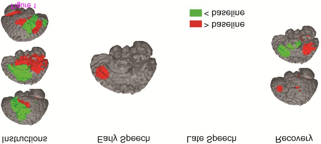
11 Midbrain l. Superior Temporal G l. Middle Frontal G Precuneus Instructions Negative Activation l. Uncus Bilateral Inferior Frontal G./Middle Cingulate G r. Middle Frontal G r. Middle Frontal G Early Speech Occipital Cortex l. Middle Frontal G Late Speech r. Parahippocampal G l. Parahippocampal G Midbrain Recovery Pons Anterior Cingulate G./Caudate/Insula Note. 1 The GLM analysis did not yield any activation differences as a function of diagnosis. l. = left, r. = right Diagnosis differences The GLM analysis did not yield any significant main effects of depression and social anxiety, or significant interaction of depression and social anxiety. fmri findings change-point analysis Overall intercept Medial frontal cortex. On average, participants exhibited increased medial frontal cortex activation during the early speech preparation period (Figure 1; Table 3). Other regions. Notably, participants also exhibited decreased activation in the posterior cingulate during the late speech preparation period and decreased activation in the anterior cingulate during recovery. Figure 1 BOLD responses to social evaluative threat for the overall group intercept. These BOLD responses represent overall changes in activation (intercept from multiple regression equation) from baseline to the four periods of interest (instructions [ timepoints)], early speech preparation [ time-points], late speech preparation [ time-points], and recovery period [ time-points]) Table 3 Change-point analysis of the BOLD responses to the social evaluative threat average (intercept) across all participants
12 Region x y Z voxels vol (mm 3 ) Instructions Activation > Baseline Peak t Changepoint onset Changepoint offset Occipital cortex Midbrain l. Superior Temporal G r. Superior Temporal G. & Insula r. Inferior Frontal G Early Speech Prep Medial Frontal G Late Speech Prep No suprathreshold voxels Recovery r. Fusiform G l. Parahippocampal G Precuneus Posterior Cingulate G l. Superior Temporal G l. Middle Frontal G Activation < Baseline Instructions Middle Cingulate G./Bilateral Insula/Parietal Cortex r. Hippocampus r. Middle Temporal G r. Caudate r. Superior Temporal G Early Speech Prep l. Posterior Cingulate G Late Speech Prep No suprathreshold voxels Recovery r. Thalamus l. Putamen r. Putamen l. Thalamus Anterior Cingulate G Note. vol = volume; G. = Gyrus
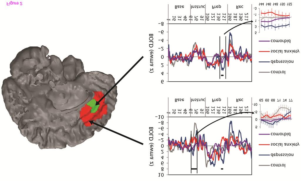
13 Depression vs. No Depression Medial frontal cortex. As hypothesized, the change-point analysis yielded a main effect of being diagnosed with depression on medial frontal cortex activation during the late speech preparation period. Whereas participants diagnosed with depression (MDD, MDD/SAD) exhibited a resurgence of medial frontal cortex activation during the late speech preparation period, participants without depression (SAD, CTL) exhibited a return to baseline during this period (Figure 2). There was also a main effect of being diagnosed with depression on medial frontal cortex activation in the instructions period; participants with diagnosed depression did not exhibit the deactivation that was evident in nondepressed participants (Figure 2; Table 4). Notably, these associations between depression and medial frontal cortex activity remained significant when controlling for depression and anxiety symptoms (both ts > 3.8, ps <.001). Other regions. As with the medial frontal cortex findings, participants diagnosed with depression exhibited a more extensive pattern of activation than did participants without depression during the instructions (e.g., middle cingulate, precentral gyrus) and late speech preparation (e.g., insula, caudate; Table 4) periods. In the early speech preparation period, participants with depression exhibited greater activation than did participants without depression in the inferior frontal gyrus. Figure 2 BOLD responses to social evaluative threat by diagnostic group in the medial frontal cortex. Each graph plots activation across time: EWMA z = exponentially weighted moving average z-statistic (temporally smoothed deviations from baseline period). Base = baseline, Instruc = instructions period, Prep = speech preparation period, and Rec = recovery period. Solid black bars designate the time-points for which there was a main effect of being diagnosed with depression vs. not having depression; insets show these data with standard error bars. BOLD = blood oxygenation level dependent Table 4 Change-point analysis of the BOLD responses to the social evaluative threat task of participants with and without major depressive disorder Region x y z voxels vol (mm 3 ) Peak t changepoint onset changepoint offset Instructions Depression > No Depression comorbid (~,>,<) pure depression 1 r.insula < l. Precentral G.* < l. Precentral G.* < l. Middle Frontal G ~ Medial Frontal G ~ l. Inferior Frontal G Cingulate G.* < Cingulate G Cingulate G < Early Speech Prep l. Precentral G ~ l. Prefrontal G ~ Cingulate G ~ Late Speech Prep
14 l. Insula r. Insula l. Postcentral G ~ r. Caudate Cingulate G l. Sub-gyral ~ l. Sub-gyral* < Recovery r. Cerebellum: Culmen ~ Lingual G.* < Lingual G ~ Instructions No Depression > Depression Posterior Cingulate ~ Cuneus ~ r. Cuneus ~ Early Speech Prep l. Caudate Late Speech Prep l. Lentiform Nucleus Recovery Posterior Cingulate G.* > l. Precentral G ~ Note. These clusters were masked with the clusters that exhibited group average activation differences during the social evaluative threat task. vol = volume; G. = Gyrus. 1 Post-hoc group contrasts that test whether comorbid participants exhibited similar (~; not significantly different from each other, but both groups are different than control participants), greater (>), or less (<) activation than participants with pure depression (p <.05). The lack of a symbol means that comorbid participants did not exhibit significantly different activation than control participants and participants with pure depression * Those regions for which comorbid participants activation significantly differed from both pure depression and pure social anxiety groups Social Anxiety vs. No Social Anxiety Medial frontal cortex. Contrary to predictions, there was no effect of social anxiety diagnosis on medial frontal cortex activation in the speech preparation period or during recovery. Other regions. Instead, participants diagnosed with social anxiety (SAD, MDD/SAD), relative to participants without diagnosed social anxiety (MDD, CTL), exhibited greater activation in the occipital cortex and middle temporal gyrus during instructions as well as less activation in the insula and postcentral gyrus during recovery (Table 5).
15 Table 5 Change-point analysis of the BOLD responses to the social evaluative threat task of participants with and without social anxiety disorder Region x y z voxels vol (mm 3 ) Peak t changepoint onset changepoint offset Instructions Cerebellum: Declive* 8 r. Lingual G.* 26 r. Superior Temporal G. 56 Early Speech Prep No suprathreshold voxels Late Speech Prep No suprathreshold voxels Social Anxiety > No Social Anxiety comorbid (~,>,<) social anxiety < < ~ Recovery r. Cerebellum: Declive* < l. Cerebellum: Declive ~ No Social Anxiety > Social Anxiety Instructions l. Insula r. Precentral G ~ l. Inferior Frontal G.* > r. Middle Frontal G ~ Cingulate G l. Cingulate/ Supramarginal G ~ Early Speech Prep No suprathreshold voxels Late Speech Prep No suprathreshold voxels Recovery r. Insula ~ l. Insula ~ - l. Postcentral G ~ Note. These clusters were masked with the clusters that exhibited group average activation differences during the social evaluative threat task. vol = volume; G. = Gyrus. 1 Post-hoc group contrasts that test whether comorbid participants exhibited similar (~; not significantly different from each other, but both groups are different than control participants), greater (>), or less (<) activation than participants with pure social anxiety (p <.05). The lack of a symbol means that comorbid participants did not exhibit significantly different activation than control participants and participants with pure social anxiety * Those regions for which comorbid participants activation significantly differed from both pure depression and pure social anxiety groups
16 Interaction of Depression and Social Anxiety Medial frontal cortex. There was no interaction of diagnoses of depression and social anxiety on activation in the medial frontal cortex. Other regions. Participants diagnosed with comorbid depression/social anxiety exhibited different patterns of activation than did participants with non-comorbid diagnoses in several regions (Figure 3; Table 6). The comorbid participants exhibited intermediate activation in the middle cingulate cortex and precentral gyrus (less than the pure depression group and more than the pure social anxiety group; Figure 3) as well as the posterior cingulate (less than the pure social anxiety group and more than the pure depression group). The comorbid participants also exhibited activation similar to that exhibited by the control participants in several regions, including greater activation than participants with pure depression or pure social anxiety in the insula (instructions) and middle temporal gyrus (recovery), and less activation than the pure diagnostic groups in the cerebellum (instructions) and cuneus (instructions/recovery). Figure 3 Post-hoc group contrasts that compare the activation of the comorbid group with that of the pure depression and social anxiety groups. A. To have demonstrated similar activation as one of the two pure diagnostic groups, the comorbid group must have exhibited activation that was not significantly different from the pure diagnostic group, and the activation of both the comorbid and pure diagnostic groups must have been significantly different than controls. To have demonstrated different activation than the two pure diagnostic groups, the comorbid group must have exhibited significantly different activation from both the pure depression and social anxiety groups. B. Each group s beta estimates of the EWMA z statistic for selected regions. BOLD = blood oxygenation level dependent. EWMA z = exponentially weighted moving average z-statistic (temporally smoothed deviations from baseline period)
17 Table 6 Change-point analysis of the interaction of depression and social anxiety in BOLD responses to the social evaluative threat task Region x y z voxels vol (mm 3 ) Peak t change-point onset Instructions l. Parahippocampal G l. Insula l. Insula l. Insula r. Insula* l. Precentral G. Early Speech Prep l. Insula Late Speech Prep l. Lentiform Nucleus Recovery change-point offset Comorbid and/or Control > Depression and/or Social Anxiety Control Depression Social Anxiety Co-morbid b a a ab c a ab b b a a ab b a a a b a a 1.00 b b ab a 0.26 b c a b 2.00 b b a a 1.39 ab l. Middle Temporal G.* b a a 2.08 b l. Precentral G b a a a Instructions Depression and/or Social Anxiety > Comorbid and/or Control Cerebellum: Declive* a c d 2.94 b Cuneus* a c c b r. Inferior Frontal G a 7.13 c b ab r. Insula a 8.04 b a a r. Cuneus* a b c 3.66 a Early Speech Prep No suprathreshold voxels
18 Late Speech Prep No suprathreshold voxels Recovery Lingual G a c bc b Precuneus* a c c b Note. These clusters were masked with the clusters that exhibited group average activation differences during the social evaluative threat task. vol = volume; G. = Gyrus. Different subscripts represent significantly differences between the groups, calculated using simple slopes analyses on the beta coefficients (presented here) from the multiple regression models * Those regions for which comorbid participants activation significantly differed from both pure depression and pure social anxiety groups
19 Correlations among brain activation and self-reported responses to the social evaluative threat task Regions associated with depression We examined whether the differential medial frontal cortex responses (MDD > No MDD) during the instructions and late speech preparation periods were related to self-reported responses to the task (α corr =.05/6 =.008). No correlations survived Bonferroni correction. Regions associated with social anxiety We next examined the correlations between self-reported responses to the speech task and differential responses in the lingual gyrus and superior temporal gyrus during the instructions period (SAD > No SAD) and in the insula during the recovery period (SAD < No SAD) regions that may represent the hypothesized vigilance-avoidance pattern of responding (α corr =.05/6 =.008). Across all participants, no correlations survived Bonferroni correction. Across only the participants with social anxiety, activation in the lingual gyrus during the instructions period was negatively correlated with negative affect to preparing the speech, r(32) =.49, p =.004, and perceived demands of the speech, r(32) =.47, p =.006. In addition, left insula activation during the recovery period was positively correlated with perceived demands of the speech, r(32) =.48, p =.005. Discussion The present study was designed to examine magnitude and timing of neural responses to stress in individuals diagnosed with non-comorbid depression, non-comorbid social anxiety, and comorbid depression/social anxiety. All groups exhibited increased medial frontal cortex activation during the early speech preparation, which is consistent with findings from previous studies [26], followed by decreased activation toward the middle of the speech anticipation period. As we predicted, the change-point analysis revealed that participants with depression (both pure and comorbid) exhibited greater medial frontal cortex activation than those without depression late in the stress task. Notably, whereas we had initially hypothesized that depressed participants would exhibit a sustained pattern of activation, we found instead that depressed participants exhibited a resurgence of medial frontal cortex activation late in the stress task. This late resurgence of medial frontal cortex activation suggests that participants with depression re-engaged with the stressor, although it is unclear what psychological functions subserved this re-engagement. This medial frontal cortex activation during the late speech preparation period was not significantly correlated with any self-reported responses to the speech task, suggesting that depressed participants reengagement with the stressor was not due to a more intense negative emotional response to the stressor. One possible explanation for this finding is that this late activation in the medial prefrontal cortex for the depressed participants was due to rumination about negative aspects of the stress task. Indeed, activation in the medial frontal cortex has been found to be associated with functions that subserve rumination about negative events: self-referential processing, [41] self-conscious emotional reactivity, [42] and extended duration of negative emotions [43]. Alternatively, this region has also been found to be associated with generating the arousal necessary for anticipated effort, [26,44] suggesting the possibility that this pattern of activation was due to depressed participants re-exerting the effort they anticipated it would take to perform the upcoming speech. Future investigations that explicitly assess ruminative
20 thoughts as well as motivation/anticipated effort in depression are necessary to test the veracity of these formulations. The vigilance-avoidance hypothesis for participants with social anxiety was only partially supported. Participants without depression (i.e., those with either social anxiety or no disorder) exhibited decreased activation in the medial frontal cortex during the instructions period. Consistent with the vigilance hypothesis, deactivation in this region co-occurs with cognitive engagement to external stimuli, [45] suggesting that participants with pure social anxiety were paying relatively more attention to external stimuli (the speech instructions) than were participants with depression. Further supporting this formulation, participants with both pure social anxiety and comorbid social anxiety/depression exhibited greater activation in other regions associated with attending to external stimuli, including the occipital cortex and superior temporal gyrus [46], than did participants without social anxiety. Consistent with the avoidance hypothesis, during recovery, social anxiety and comorbid participants exhibited decreased activation in the insula, a region associated with monitoring internal states [47], especially those associated with threat [48]. Together, this pattern of findings suggest that participants with social anxiety exhibited heightened engagement with the stressor when first learning about it (during instructions period) and significant disengagement from the stressor when learning that they would not have to give a speech after all. In addition, we found evidence that this vigilance-avoidance pattern of brain activation may have been adaptive. Across socially anxious participants, the tendency to exhibit greater lingual gyrus activation during the instructions period and decreased insula activation during recovery predicted participants tendency to perceive the speech task as less demanding. Contrary to predictions, however, having social anxiety did not predict greater medial frontal cortex activation during the early part of the stressor. One possibility that should be tested in future investigations is that the preparation of the speech was too threatening for participants. Indeed, individual differences in social anxiety have been found to be better predictors of differential response to mildly threatening social situations than to highly threatening social situations [49]. As predicted, the comorbid participants responded similarly to the participants with pure depression and pure social anxiety in a majority of the suprathreshold voxels. Notably, however, during the speech period, the comorbid participants exhibited activation patterns that more closely matched the participants with pure depression than the participants with pure social anxiety (Figure 3). In future studies, investigators should examine whether this asymmetrical influence of these two diagnoses on brain responses to social threat extend to other facets of neural and behavioral functioning. In the few brain regions in which the comorbid participants exhibited different activations than did the participants with pure diagnoses, two patterns emerged. First, the comorbid participants exhibited intermediate activation levels in the middle cingulate cortex, precentral gyrus, and posterior cingulate. This finding raises the intriguing possibility that in these regions the psychological and neural effects of having both depression and social anxiety interact. Second, compared with the pure diagnostic groups, the comorbid group and never-disordered control participants exhibited more tempered responses in the insula (less deactivation) and cuneus (less activation). Future investigations should target these regions as possible sites of dysregulation for individuals with comorbid depression and social anxiety in which neural reactivity to stress is dampened despite heightened self-reported reactivity. In addition to unpacking the psychological mechanisms that underlie these differences in activation among the diagnostic groups, future studies that replicate and extend this work
21 should address other elements of the current design. For example, investigators should recruit larger samples of both male and female medication-free participants to replicate and extend the present findings to determine whether they generalize to males and to ensure that they are not influenced by medication status. Also, although the speech stressor we used induced similar levels of neural activation during the early-stress period in the depressed and socially anxious participants, the social threat inherent in this task is theoretically more relevant to social anxiety than to depression. Indeed, the participants with social anxiety did report a slightly higher increase in negative affect in response to the social threat stressor than did the participants with depression. By exploring more depression-relevant stressors, future studies should be able to distinguish activation patterns that generalize to other types of stressors from patterns that are specific to the speech stressor. Conclusions In sum, the present study is among the first to delineate the neural functioning of comorbidity, and is the first to examine specifically the neural functioning of comorbid depression and social anxiety. We used innovative statistical and methodological techniques to probe temporal aspects of stress-induced neural responding and demonstrated that changepoint analyses are more sensitive to temporal differences in activation than standard GLM analyses. This change-point approach allowed us to identify both when and where neural activation differences occurred as a function of diagnosis and yielded several intriguing patterns of activation. Future investigations should assess more explicitly the psychological mechanisms that might underlie these neural activation patterns, as well as possible diagnosis-related differences in functional connectivity among these regions [50] during exposure to stress. Elucidating these unique and shared neural responses to social evaluative threat in individuals with pure depression, pure social anxiety, and comorbid depression/social anxiety may facilitate the selection or development of effective interventions for these debilitating disorders. Competing interests The authors declare that, except for income received from our primary employer, no financial support or compensation has been received from any individual or corporate entity over the past three years for research or professional service and there are no personal financial holdings that could be perceived as constituting a potential conflict of interest. Authors contributions CW helped conceive of the design of the study, conducted the data analyses, and took the lead on writing the manuscript. JPH and MCC helped conceive of the design of the study, conducted the fmri sessions, consulted on data analyses, and contributed to writing the manuscript. JJ helped conceive of the design of the study and contributed to writing the manuscript. IHG helped conceive of the design of the study, consulted on data analyses, and contributed to writing the manuscript. All authors have read and approve of this manuscript.
22 Acknowledgments The authors declare that, except for income received from our primary employer, no financial support or compensation has been received from any individual or corporate entity over the past three years for research or professional service and there are no personal financial holdings that could be perceived as constituting a potential conflict of interest. The authors thank Juliana Gonzales, Kalpa Bhattacharjee, Arkadiy Maksimovskiy, and Maria Lemus for their help in data collection and preparation. This research was funded by NIMH Grant MH awarded to Ian H. Gotlib. Dr. Waugh had full access to all of the data in the study and takes responsibility for the integrity of the data and the accuracy of the data analysis. References 1. Kessler RC, Berglund P, Demler O, Jin R, Walters EE: Lifetime prevalence and age-ofonset distributions of DSM-IV disorders in the national comorbidity survey replication. Arch Gen Psychiatry 2005, 62(6): Brown TA, Campbell LA, Lehman CL, Grisham JR, Mancill RB: Current and lifetime comorbidity of the DSM-IV anxiety and mood disorders in a large clinical sample. J Abnorm Psychol 2001, 110(4): Gilboa-Schechtman E, Presburger G, Marom S, Hermesh H: The effects of social anxiety and depression on the evaluation of facial crowds. Behav Res Ther 2005, 43: Stein MB, Fuetsch M, Muller N, Hofler M, Lieb R, Wittchen H-U: Social anxiety disorder and the risk of depression. Arch Gen Psychiatry 2001, 58: Gotlib IH, Hamilton JP: Neuroimaging and depression: Current status and unresolved issues. Curr Dir Psychol Sci 2008, 17(2): Phan KL, Fitzgerald DA, Nathan PJ, Tancer ME: Association between amygdala hyperactivity to harsh faces and severity of social anxiety in generalized social phobia. Biol Psychiatry 2006, 59: Sheline YI, Barch DM, Donnelly JM, Ollinger JM, Snyder AZ, Mintun MA: Increased amygdala response to masked emotional faces in depressed subjects resolves with antidepressant treatment: An fmri study. Biol Psychiatry 2001, 50: Gotlib IH, Sivers H, Gabrieli JDE, et al: Subgenual anterior cingulate activation to valenced emotional stimuli in major depression. Neuroreport 2005, 16: Siegle GJ, Thompson W, Carter CS, Steinhauer SR, Thase ME: Increased amygdala and decreased dorsolateral prefrontal BOLD responses in unipolar depression: Related and independent features. Biol Psychiatry 2007, 61: Blair K, Shaywitz J, Smith BW, et al: Response to emotional expressions in generalized social phobia and generalized anxiety disorder: Evidence for separate disorders. Am J Psychiatry 2008, 165(9):
23 11. Lorberbaum JP, Kose S, Johnson MR, et al: Neural correlates of speech anticipatory anxiety in generalized social phobia. Neuroreport 2004, 15(18): Andreescu C, Butters M, Lenze EJ, et al: fmri activation in late-life anxious depression: A potential biomarker. Int J Geriatr Psychiatry 2009, 24(8): Etkin A, Schatzberg AF: Common abnormalities and disorder-specific compensation during implicit regulation of emotional processing in Generalized Anxiety and Major Depressive Disorders. Am J Psychiatry 2011, 168(9): Monroe SM, Simons AD: Diathesis-stress theories in the context of life stress research: Implications for the depressive disorders. Psychol Bull 1991, 110(3): Allen JL, Rapee RM, Sandberg S: Severe life events and chronic adversities as antecedents to anxiety in children: A matched control study. J Abnorm Child Psychol 2008, 36: Young EA, Abelson JL, Cameron OG: Effect of comorbid anxiety disorders on the Hypothalamic-Pituitary-Adrenal Axis response to a social stressor in major depression. Biol Psychiatry 2004, 56: Amir N, Foa EB, Coles ME: Automatic activation and strategic avoidance of threatrelevant information in social phobia. J Abnorm Psychol 1998, 107(2): Koster EHW, Verschuere B, Crombez G, Van Damme S: Time-course of attention for threatening pictures in high and low trait anxiety. Behav Res Ther 2005, 43(8): Mogg K, Bradley B, Miles F, Dixon R: Time course of attentional bias for threat scenes: Testing the vigilance-avoidance hypothesis. Cognition & Emotion 2004, 18(5): Amir N, Elias J, Klumpp H, Przeworski A: Attentional bias to threat in social phobia: facilitated processing of threat or difficulty disengaging attention from threat? Behav Res Ther 2003, 41(11): McCullough ME, Orsulak P, Brandon A, Akers L: Rumination, fear, and cortisol: An in vivo study of interpersonal transgressions. Health Psychol 2007, 26(1): Nolen-Hoeksema S: The Role of Rumination in Depressive Disorders and Mixed Anxiety/Depressive Symptoms. J Abnorm Psychol 2000, 109(3): Young EA, Lopez JF, Murphy-Weinberg V, Watson SJ, Akil H: Hormonal evidence for altered responsiveness to social stress in major depression. Neuropsychopharmacology 2000, 23(4): Mikolajczak M, Roy E, Luminet O, de Timary P: Resilience and hypothalamicpituitary-adrenal axis reactivity under acute stress in young men. Stress 2008, 11(6):
24 25. Waugh CE, Panage S, Mendes WB, Gotlib IH: Cardiovascular and affective recovery from anticipatory threat. Biol Psychol 2010, 84: Wager TD, Waugh CE, Lindquist M, Noll DC, Fredrickson BL, Taylor SF: Brain mediators of cardiovascular responses to social threat, Part I: Reciprocal dorsal and ventral sub-regions of the medial prefrontal cortex and heart-rate reactivity. NeuroImage 2009, 47: Lindquist M, Waugh C, Wager TD: Modeling state-related fmri activity using change-point theory. NeuroImage 2007, 35: : User's guide for the structured clinical interview for DSM-IV Axis 1 disorders [computer program]. Version 20th edition.: ; Turner SM, Beidel DC, Dancu CV, Stanley MA: An empirically derived inventory to measure social fears and anxiety: The Social Phobia and Anxiety Inventory. Psychological Assessment: A Journal of Consulting and Clinical Psychology 1989, 1(1): Beck AT, Steer RA, Brown GK: Manual for the Beck Depression Inventory-II. San Antonio, TX: Psychological Corporation; Fredrickson BL, Mancuso RA, Branigan C, Tugade MM: The undoing effect of positive emotions. Motiv Emotion 2000, 24(4): Glover GH, Law CS: Spiral-in/out BOLD fmri for increased SNR and reduced susceptibility artifacts. Magn Reson Med 2001, 46(3): Cox RW: AFNI: Software for analysis and visualization of functional magnetic resonance neuroimages. Comput Biomed Res 1996, 29: Talairach J, Tournoux P: Co-Planar Stereotaxic Atlas of the Human Brain. Stuttgart, Germany: Thieme; Sackheim HA: The definition and meaning of treatment-resistant depression. J Clin Psychiatry 2001, 62: Ward BD: Simultaneous inference for fmri data. Accessed August 2007, () Jaccard J, Turrisi R: Interaction effects in multiple regression. In Thousand Oaks. Vol nd edition. CA: Sage University Press; Mendes WB, Gray HM, Mendoza-Denton R, Major B, Epel ES: Why egalitarianism might be good for your health: Physiologial thriving during stressful intergroup encounters. Psychol Sci 2007, 18(11): Tomaka J, Blascovich J, Kelsey RM, Leitten CL: Subjective, physiological, and behavioral effects of threat and challenge appraisal. J Pers Soc Psychol 1993, 65(2):
25 40. Watson D, Clark LA, Tellegen A: Development and validation of brief measures of positive and negative affect: The PANAS scales. J Pers Soc Psychol 1988, 54(6): Amodio DM, Frith CD: Meeting of minds: The medial frontal cortex and social cognition. Nat Rev Neurosci 2006, 7: Sturm VE, Sollberger M, Seeley WW, et al: Role of right pregenual anterior cingulate cortex in self-conscious emotional reactivity. Social Cognitive and Affective Neuroscience. 2012,. 43. Waugh CE, Hamilton JP, Gotlib IH: The neural temporal dynamics of the intensity of emotional experience. NeuroImage 2010, 49: Critchley HD, Mathias CJ, Josephs O, et al: Human cingulate cortex and autonomic control: Converging neuroimaging and clinical evidence. Brain 2003, 126(10): Shulman GL, Fiez JA, Corbetta M, et al: Common blood flow changes across visual tasks: II decreases in cerebral cortex. J Cogn Neurosci 1997, 9(5): Fiez JA, Peterson SE: Neuroimaging studies of word reading. Proc Natl Acad Sci U S A 1998, 95(3): Craig AD: Opinion: How do you feel? Interoception: the sense of the physiological condition of the body. Nat Rev Neurosci 2002, 3(8): Nitschke JB, Sarinopoulos I, Mackiewicz KL, Schaefer HS, Davidson RJ: Functional neuroanatomy of aversion and its anticipation. NeuroImage 2006, 29(1): Gramer M, Saria K: Effects of social anxiety and evaluative threat on cardiovascular responses to active performance situations. Biol Psychol 2007, 74(1): Carballedo A, Scheuerecker J, Meisenzahl E, et al: Functional connectivity of emotional processing in depression. J Affect Disord 2011, 134(1 3): Additional files Additional_file_1 as DOCX Additional file 1 Supplementary information and tables.
26
27
Resistance to forgetting associated with hippocampus-mediated. reactivation during new learning
 Resistance to Forgetting 1 Resistance to forgetting associated with hippocampus-mediated reactivation during new learning Brice A. Kuhl, Arpeet T. Shah, Sarah DuBrow, & Anthony D. Wagner Resistance to
Supplemental Data. Inclusion/exclusion criteria for major depressive disorder group and healthy control group
 1 Supplemental Data Inclusion/exclusion criteria for major depressive disorder group and healthy control group Additional inclusion criteria for the major depressive disorder group were: age of onset of
Supplementary Information Methods Subjects The study was comprised of 84 chronic pain patients with either chronic back pain (CBP) or osteoarthritis
 or osteoarthritis") Supplementary Information Methods Subjects The study was comprised of 84 chronic pain patients with either chronic back pain (CBP) or osteoarthritis (OA). All subjects provided informed consent to procedures
Supplementary Information Methods Subjects The study was comprised of 84 chronic pain patients with either chronic back pain (CBP) or osteoarthritis (OA). All subjects provided informed consent to procedures
Supplementary Information
 Supplementary Information The neural correlates of subjective value during intertemporal choice Joseph W. Kable and Paul W. Glimcher a 10 0 b 10 0 10 1 10 1 Discount rate k 10 2 Discount rate k 10 2 10
SUPPLEMENT: DYNAMIC FUNCTIONAL CONNECTIVITY IN DEPRESSION. Supplemental Information. Dynamic Resting-State Functional Connectivity in Major Depression
 Supplemental Information Dynamic Resting-State Functional Connectivity in Major Depression Roselinde H. Kaiser, Ph.D., Susan Whitfield-Gabrieli, Ph.D., Daniel G. Dillon, Ph.D., Franziska Goer, B.S., Miranda
Cover Page. The handle holds various files of this Leiden University dissertation
 Cover Page The handle http://hdl.handle.net/1887/32078 holds various files of this Leiden University dissertation Author: Pannekoek, Nienke Title: Using novel imaging approaches in affective disorders
Supporting Online Material for
 www.sciencemag.org/cgi/content/full/324/5927/646/dc1 Supporting Online Material for Self-Control in Decision-Making Involves Modulation of the vmpfc Valuation System Todd A. Hare,* Colin F. Camerer, Antonio
Supplementary Online Content
 Supplementary Online Content Redlich R, Opel N, Grotegerd D, et al. Prediction of individual response to electroconvulsive therapy via machine learning on structural magnetic resonance imaging data. JAMA
Hippocampal brain-network coordination during volitionally controlled exploratory behavior enhances learning
 Online supplementary information for: Hippocampal brain-network coordination during volitionally controlled exploratory behavior enhances learning Joel L. Voss, Brian D. Gonsalves, Kara D. Federmeier,
Supporting online material. Materials and Methods. We scanned participants in two groups of 12 each. Group 1 was composed largely of
 Placebo effects in fmri Supporting online material 1 Supporting online material Materials and Methods Study 1 Procedure and behavioral data We scanned participants in two groups of 12 each. Group 1 was
Supplementary Online Material Supplementary Table S1 to S5 Supplementary Figure S1 to S4
 Supplementary Online Material Supplementary Table S1 to S5 Supplementary Figure S1 to S4 Table S1: Brain regions involved in the adapted classification learning task Brain Regions x y z Z Anterior Cingulate
Supplementary Online Content
 1 Supplementary Online Content Miller CH, Hamilton JP, Sacchet MD, Gotlib IH. Meta-analysis of functional neuroimaging of major depressive disorder in youth. JAMA Psychiatry. Published online September
Neural activity to positive expressions predicts daily experience of schizophrenia-spectrum symptoms in adults with high social anhedonia
 1 Neural activity to positive expressions predicts daily experience of schizophrenia-spectrum symptoms in adults with high social anhedonia Christine I. Hooker, Taylor L. Benson, Anett Gyurak, Hong Yin,
Hallucinations and conscious access to visual inputs in Parkinson s disease
 Supplemental informations Hallucinations and conscious access to visual inputs in Parkinson s disease Stéphanie Lefebvre, PhD^1,2, Guillaume Baille, MD^4, Renaud Jardri MD, PhD 1,2 Lucie Plomhause, PhD
Supplementary Online Content
 Supplementary Online Content Green SA, Hernandez L, Tottenham N, Krasileva K, Bookheimer SY, Dapretto M. The neurobiology of sensory overresponsivity in youth with autism spectrum disorders. Published
Text to brain: predicting the spatial distribution of neuroimaging observations from text reports (submitted to MICCAI 2018)
") 1 / 22 Text to brain: predicting the spatial distribution of neuroimaging observations from text reports (submitted to MICCAI 2018) Jérôme Dockès, ussel Poldrack, Demian Wassermann, Fabian Suchanek, Bertrand
1 / 22 Text to brain: predicting the spatial distribution of neuroimaging observations from text reports (submitted to MICCAI 2018) Jérôme Dockès, ussel Poldrack, Demian Wassermann, Fabian Suchanek, Bertrand
Identification of Neuroimaging Biomarkers
 Identification of Neuroimaging Biomarkers Dan Goodwin, Tom Bleymaier, Shipra Bhal Advisor: Dr. Amit Etkin M.D./PhD, Stanford Psychiatry Department Abstract We present a supervised learning approach to
Procedia - Social and Behavioral Sciences 159 ( 2014 ) WCPCG 2014
 WCPCG 2014") Available online at www.sciencedirect.com ScienceDirect Procedia - Social and Behavioral Sciences 159 ( 2014 ) 743 748 WCPCG 2014 Differences in Visuospatial Cognition Performance and Regional Brain Activation
Available online at www.sciencedirect.com ScienceDirect Procedia - Social and Behavioral Sciences 159 ( 2014 ) 743 748 WCPCG 2014 Differences in Visuospatial Cognition Performance and Regional Brain Activation
Distinct Value Signals in Anterior and Posterior Ventromedial Prefrontal Cortex
 Supplementary Information Distinct Value Signals in Anterior and Posterior Ventromedial Prefrontal Cortex David V. Smith 1-3, Benjamin Y. Hayden 1,4, Trong-Kha Truong 2,5, Allen W. Song 2,5, Michael L.
A possible mechanism for impaired joint attention in autism
 A possible mechanism for impaired joint attention in autism Justin H G Williams Morven McWhirr Gordon D Waiter Cambridge Sept 10 th 2010 Joint attention in autism Declarative and receptive aspects initiating
Title of file for HTML: Supplementary Information Description: Supplementary Figures, Supplementary Tables and Supplementary References
 Title of file for HTML: Supplementary Information Description: Supplementary Figures, Supplementary Tables and Supplementary References Supplementary Information Supplementary Figure 1. The mean parameter
Supporting online material for: Predicting Persuasion-Induced Behavior Change from the Brain
 1 Supporting online material for: Predicting Persuasion-Induced Behavior Change from the Brain Emily Falk, Elliot Berkman, Traci Mann, Brittany Harrison, Matthew Lieberman This document contains: Example
Supplementary Digital Content
 Supplementary Digital Content Contextual modulation of pain in masochists: involvement of the parietal operculum and insula Sandra Kamping a, Jamila Andoh a, Isabelle C. Bomba a, Martin Diers a,b, Eugen
Supplemental Information. Triangulating the Neural, Psychological, and Economic Bases of Guilt Aversion
 Neuron, Volume 70 Supplemental Information Triangulating the Neural, Psychological, and Economic Bases of Guilt Aversion Luke J. Chang, Alec Smith, Martin Dufwenberg, and Alan G. Sanfey Supplemental Information
Topographical functional connectivity patterns exist in the congenitally, prelingually deaf
 Supplementary Material Topographical functional connectivity patterns exist in the congenitally, prelingually deaf Ella Striem-Amit 1*, Jorge Almeida 2,3, Mario Belledonne 1, Quanjing Chen 4, Yuxing Fang
For better or for worse: neural systems supporting the cognitive down- and up-regulation of negative emotion
 For better or for worse: neural systems supporting the cognitive down- and up-regulation of negative emotion Kevin N. Ochsner, a, * Rebecca D. Ray, b Jeffrey C. Cooper, b Elaine R. Robertson, b Sita Chopra,
Methods to examine brain activity associated with emotional states and traits
 Methods to examine brain activity associated with emotional states and traits Brain electrical activity methods description and explanation of method state effects trait effects Positron emission tomography
Supplementary Material S3 Further Seed Regions
 Supplementary Material S3 Further Seed Regions Figure I. Changes in connectivity with the right anterior insular cortex. (A) wake > mild sedation, showing a reduction in connectivity between the anterior
How to report my result using REST slice viewer?
 How to report my result using REST slice viewer? Han Zhang Center for Cognition and Brain Disorders, Hangzhou Normal University napoleon1982@gmail.com 2013/12/30 Commonly, you got an activation for functional
Theory of mind skills are related to gray matter volume in the ventromedial prefrontal cortex in schizophrenia
 Theory of mind skills are related to gray matter volume in the ventromedial prefrontal cortex in schizophrenia Supplemental Information Table of Contents 2 Behavioral Data 2 Table S1. Participant demographics
QUANTIFYING CEREBRAL CONTRIBUTIONS TO PAIN 1
 QUANTIFYING CEREBRAL CONTRIBUTIONS TO PAIN 1 Supplementary Figure 1. Overview of the SIIPS1 development. The development of the SIIPS1 consisted of individual- and group-level analysis steps. 1) Individual-person
SUPPLEMENTARY MATERIAL. Table. Neuroimaging studies on the premonitory urge and sensory function in patients with Tourette syndrome.
 SUPPLEMENTARY MATERIAL Table. Neuroimaging studies on the premonitory urge and sensory function in patients with Tourette syndrome. Authors Year Patients Male gender (%) Mean age (range) Adults/ Children
Classification and Statistical Analysis of Auditory FMRI Data Using Linear Discriminative Analysis and Quadratic Discriminative Analysis
 International Journal of Innovative Research in Computer Science & Technology (IJIRCST) ISSN: 2347-5552, Volume-2, Issue-6, November-2014 Classification and Statistical Analysis of Auditory FMRI Data Using
Study No: Title : Rationale: Phase: Study Period: Study Design: Centres: Indication: Treatment: Objectives: Statistical Methods: Sample Size
 The study listed may include approved and non-approved uses, formulations or treatment regimens. The results reported in any single study may not reflect the overall results obtained on studies of a product.
Supporting Information
 Supporting Information Newman et al. 10.1073/pnas.1510527112 SI Results Behavioral Performance. Behavioral data and analyses are reported in the main article. Plots of the accuracy and reaction time data
Proactive and reactive control during emotional interference and its relationship to trait anxiety
 brain research 1481 (2012) 13 36 Available online at www.sciencedirect.com www.elsevier.com/locate/brainres Research Report Proactive and reactive control during emotional interference and its relationship
The Impact of Anxiety-Inducing Distraction on Cognitive Performance: A Combined Brain Imaging and Personality Investigation
 The Impact of Anxiety-Inducing Distraction on Cognitive Performance: A Combined Brain Imaging and Personality Investigation Ekaterina Denkova 1, Gloria Wong 2, Sanda Dolcos 3, Keen Sung 1, Lihong Wang
Nature Neuroscience: doi: /nn Supplementary Figure 1. Task timeline for Solo and Info trials.
 Supplementary Figure 1 Task timeline for Solo and Info trials. Each trial started with a New Round screen. Participants made a series of choices between two gambles, one of which was objectively riskier
Supplementary information Detailed Materials and Methods
 Supplementary information Detailed Materials and Methods Subjects The experiment included twelve subjects: ten sighted subjects and two blind. Five of the ten sighted subjects were expert users of a visual-to-auditory
Functional topography of a distributed neural system for spatial and nonspatial information maintenance in working memory
 Neuropsychologia 41 (2003) 341 356 Functional topography of a distributed neural system for spatial and nonspatial information maintenance in working memory Joseph B. Sala a,, Pia Rämä a,c,d, Susan M.
Supplementary Online Content
 Supplementary Online Content Devenney E, Bartley L, Hoon C, et al. Progression in behavioral variant frontotemporal dementia: a longitudinal study. JAMA Neurol. Published online October 26, 2015. doi:10.1001/jamaneurol.2015.2061.
Supplementary Figure 1. Histograms of original and phase-randomised data
 Log Supplementary Figure 1. Histograms of original and phase-randomised data BOLD signals histogram Denoised signals histogram Activity-inducing signals histogram Innovation signals histogram 20 15 10
2017, Joule Inc. or its licensors Online appendices are unedited and posted as supplied by the authors.
 Results Validation: Reproducibility Figure S1. Reproducibility of the results of small-world parameters. Differences in topological properties of functional brain networks between bulimia nervosa (BN)
Personal Space Regulation by the Human Amygdala. California Institute of Technology
 Personal Space Regulation by the Human Amygdala Daniel P. Kennedy 1, Jan Gläscher 1, J. Michael Tyszka 2 & Ralph Adolphs 1,2 1 Division of Humanities and Social Sciences 2 Division of Biology California
AN fmri EXAMINATION OF VISUAL INTEGRATION IN SCHIZOPHRENIA
 Journal of Integrative Neuroscience, Vol. 8, No. 2 (2009) 175 202 c Imperial College Press Research Report AN fmri EXAMINATION OF VISUAL INTEGRATION IN SCHIZOPHRENIA STEVEN M. SILVERSTEIN,,, SARAH BERTEN,,
The Social Regulation of Emotion
 The Social Regulation of Emotion James Coan Virginia Affective Neuroscience Laboratory Department of Psychology University of Virginia http://www.affectiveneuroscience.org All Materials 2011, James A Coan,
Functional MRI Mapping Cognition
 Outline Functional MRI Mapping Cognition Michael A. Yassa, B.A. Division of Psychiatric Neuro-imaging Psychiatry and Behavioral Sciences Johns Hopkins School of Medicine Why fmri? fmri - How it works Research
Supporting Information
 Supporting Information Carhart-Harris et al. 10.1073/pnas.1119598109 Fig. S1. Slices for arterial spin labeling (ASL) result. Lightbox display of slices showing regions where there were significant decreases
Malisza et al. Journal of Neurodevelopmental Disorders 2012, 4:12
 Malisza et al. Journal of Neurodevelopmental Disorders 2012, 4:12 RESEARCH Open Access Comparison of spatial working memory in children with prenatal alcohol exposure and those diagnosed with ADHD; A functional
Involvement of both prefrontal and inferior parietal cortex. in dual-task performance
 Involvement of both prefrontal and inferior parietal cortex in dual-task performance Fabienne Collette a,b, Laurence 01ivier b,c, Martial Van der Linden a,d, Steven Laureys b, Guy Delfiore b, André Luxen
Behavioral Task Performance
 Zacks 1 Supplementary content for: Functional Reorganization of Spatial Transformations After a Parietal Lesion Jeffrey M. Zacks, PhD *, Pascale Michelon, PhD *, Jean M. Vettel, BA *, and Jeffrey G. Ojemann,
Source Memory is not Properly Differentiated from Object Memory in Early Schizophrenia An fmri study using virtual reality
 Source Memory is not Properly Differentiated from Object Memory in Early Schizophrenia An fmri study using virtual reality Colin S. Hawco, PhD Lisa Buchy Michael Bodnar, Sarah Izadi, Jennifer Dell Elce,
Attention Response Functions: Characterizing Brain Areas Using fmri Activation during Parametric Variations of Attentional Load
 Attention Response Functions: Characterizing Brain Areas Using fmri Activation during Parametric Variations of Attentional Load Intro Examine attention response functions Compare an attention-demanding
GENDER-SPECIFIC SENSITVITY TO TIME-DISCREPANT TASK CONDITIONS OF REASONING DURING fmri
 GENDER-SPECIFIC SENSITVITY TO TIME-DISCREPANT TASK CONDITIONS OF REASONING DURING fmri by Joshua M. Roberts A Thesis Submitted to the Graduate Faculty of George Mason University in Partial Fulfillment
ORIGINAL ARTICLE. Selective Abnormal Modulation of Hippocampal Activity During Memory Formation in First-Episode Psychosis
 ORIGINAL ARTICLE Selective Abnormal Modulation of Hippocampal Activity During Memory Formation in First-Episode Psychosis Amélie M. Achim, PhD; Marie-Claude Bertrand, PhD; Hazel Sutton, BSc; Alonso Montoya,
Neural Processing of Emotional Musical and Nonmusical Stimuli in Depression
 RESEARCH ARTICLE Neural Processing of Emotional Musical and Nonmusical Stimuli in Depression Rebecca J. Lepping 1 *, Ruth Ann Atchley 2, Evangelia Chrysikou 2, Laura E. Martin 1,3, Alicia A. Clair 4, Rick
Distinct neural bases of disruptive behavior and autism symptom severity in boys with autism spectrum disorder.
 Himmelfarb Health Sciences Library, The George Washington University Health Sciences Research Commons Pediatrics Faculty Publications Pediatrics 1-1-2017 Distinct neural bases of disruptive behavior and
Do women with fragile X syndrome have problems in switching attention: Preliminary findings from ERP and fmri
 Brain and Cognition 54 (2004) 235 239 www.elsevier.com/locate/b&c Do women with fragile X syndrome have problems in switching attention: Preliminary findings from ERP and fmri Kim Cornish, a,b, * Rachel
Estimation of Statistical Power in a Multicentre MRI study.
 Estimation of Statistical Power in a Multicentre MRI study. PhD Student,Centre for Neurosciences Ninewells Hospital and Medical School University of Dundee. IPEM Conference Experiences and Optimisation
Reasoning and working memory: common and distinct neuronal processes
 Neuropsychologia 41 (2003) 1241 1253 Reasoning and working memory: common and distinct neuronal processes Christian C. Ruff a,b,, Markus Knauff a,c, Thomas Fangmeier a, Joachim Spreer d a Centre for Cognitive
NeuroImage 45 (2009) Contents lists available at ScienceDirect. NeuroImage. journal homepage:
 Contents lists available at ScienceDirect. NeuroImage. journal homepage:") NeuroImage 45 (2009) 614 626 Contents lists available at ScienceDirect NeuroImage journal homepage: www.elsevier.com/locate/ynimg Functional connectivity of the human amygdala using resting state fmri
NeuroImage 45 (2009) 614 626 Contents lists available at ScienceDirect NeuroImage journal homepage: www.elsevier.com/locate/ynimg Functional connectivity of the human amygdala using resting state fmri
Supplementary materials. Appendix A;
 Supplementary materials Appendix A; To determine ADHD diagnoses, a combination of Conners' ADHD questionnaires and a semi-structured diagnostic interview was used(1-4). Each participant was assessed with
We are IntechOpen, the world s leading publisher of Open Access books Built by scientists, for scientists. International authors and editors
 We are IntechOpen, the world s leading publisher of Open Access books Built by scientists, for scientists 4,000 116,000 120M Open access books available International authors and editors Downloads Our
Voxel-based morphometry in clinical neurosciences
 Voxel-based morphometry in clinical neurosciences Ph.D. Thesis Ádám Feldmann Department of Behavioural Sciences Leader of Doctoral School: Prof. Dr.Sámuel Komoly, D.Sc. Program leader: Prof. Dr.Sámuel
A Common Neural Network for Cognitive Reserve in Verbal and Object Working Memory in Young but not Old
 Cerebral Cortex Advance Access published August 3, 2007 Cerebral Cortex doi:10.1093/cercor/bhm134 A Common Neural Network for Cognitive Reserve in Verbal and Object Working Memory in Young but not Old
Supplementary Materials for
 Supplementary Materials for Folk Explanations of Behavior: A Specialized Use of a Domain-General Mechanism Robert P. Spunt & Ralph Adolphs California Institute of Technology Correspondence may be addressed
WHAT DOES THE BRAIN TELL US ABOUT TRUST AND DISTRUST? EVIDENCE FROM A FUNCTIONAL NEUROIMAGING STUDY 1
 SPECIAL ISSUE WHAT DOES THE BRAIN TE US ABOUT AND DIS? EVIDENCE FROM A FUNCTIONAL NEUROIMAGING STUDY 1 By: Angelika Dimoka Fox School of Business Temple University 1801 Liacouras Walk Philadelphia, PA
Neural correlates of recognition memory of social information in people with schizophrenia
 Research Paper Neural correlates of recognition memory of social information in people with schizophrenia Philippe-Olivier Harvey, PhD; Martin Lepage, PhD Department of Psychiatry, McGill University, Montréal,
APNA 25th Annual Conference October 19, Session 1022
 When Words Are Not Enough The Use of Sensory Modulation Techniques to Replace Self- Injurious Behaviors in Patients with Borderline Personality Disorder General Organization of the Brain Lita Sabonis,
DIFFERENT BOLD RESPONSES TO EMOTIONAL FACES AND EMOTIONAL FACES AUGMENTED BY CONTEXTUAL INFORMATION. Kyung Hwa Lee
 DIFFERENT BOLD RESPONSES TO EMOTIONAL FACES AND EMOTIONAL FACES AUGMENTED BY CONTEXTUAL INFORMATION by Kyung Hwa Lee B.A., Chungnam National University, 1997 M.A., Chungnam National University, 1999 Submitted
Self-related neural response to tailored smoking-cessation messages. predicts quitting
 Neural response to tailored messages and quitting Page 1 Supporting Online Material Self-related neural response to tailored smoking-cessation messages predicts quitting Hannah Faye Chua 1, S. Shaun Ho
Facial Emotion Processing in Paranoid and Non-Paranoid Schizophrenia
 FACIAL EMOTION PROCESSING IN SCHIZOPHRENIA 1 Running head: FACIAL EMOTION PROCESSING IN SCHIZOPHRENIA Facial Emotion Processing in Paranoid and Non-Paranoid Schizophrenia Sophie Jacobsson Bachelor Degree
Title of file for HTML: Peer Review File Description:
 Title of file for HTML: Supplementary Information Description: Supplementary Figure, Supplementary Tables, Supplementary Methods and Supplementary References Title of file for HTML: Peer Review File Description:
ORIGINAL ARTICLE. Disrupted Amygdalar Subregion Functional Connectivity and Evidence of a Compensatory Network in Generalized Anxiety Disorder
 ORIGINAL ARTICLE Disrupted Amygdalar Subregion Functional Connectivity and Evidence of a Compensatory Network in Generalized Anxiety Disorder Amit Etkin, MD, PhD; Katherine E. Prater, BA; Alan F. Schatzberg,
200 CHAPTER 5: TOPOGRAPHIC MAPPING OF VOICE CROSSMODAL PLASTICITY
 200 CHAPTER 5: TOPOGRAPHIC MAPPING OF VOICE CROSSMODAL PLASTICITY Introduction Sensory substitution neural imaging studies have focused on the presence of crossmodal plasticity, and the functional association
Supplementary Material. Functional connectivity in multiple cortical networks is associated with performance. across cognitive domains in older adults
 Supplementary Material Functional connectivity in multiple cortical networks is associated with performance across cognitive domains in older adults Emily E. Shaw 1,2, Aaron P. Schultz 1,2,3, Reisa A.
doi: /brain/aws024 Brain 2012: 135; Altered brain mechanisms of emotion processing in pre-manifest Huntington s disease
 doi:10.1093/brain/aws024 Brain 2012: 135; 1165 1179 1165 BRAIN A JOURNAL OF NEUROLOGY Altered brain mechanisms of emotion processing in pre-manifest Huntington s disease Marianne J. U. Novak, 1 Jason D.
The Cognitive Control of Memory: Age Differences in the Neural Correlates of Successful Remembering and Intentional Forgetting
 The Cognitive Control of Memory: Age Differences in the Neural Correlates of Successful Remembering and Intentional Forgetting Avery A. Rizio, Nancy A. Dennis* The Pennsylvania State University, Department
Supporting Information
 Revisiting default mode network function in major depression: evidence for disrupted subsystem connectivity Fabio Sambataro 1,*, Nadine Wolf 2, Maria Pennuto 3, Nenad Vasic 4, Robert Christian Wolf 5,*
Twelve right-handed subjects between the ages of 22 and 30 were recruited from the
 Supplementary Methods Materials & Methods Subjects Twelve right-handed subjects between the ages of 22 and 30 were recruited from the Dartmouth community. All subjects were native speakers of English,
Common Neural Substrates for Ordinal Representation in Short-Term Memory, Numerical and Alphabetical Cognition
 Common Neural Substrates for Ordinal Representation in Short-Term Memory, Numerical and Alphabetical Cognition Lucie Attout 1 *, Wim Fias 2, Eric Salmon 3, Steve Majerus 1 1 Department of Psychology -
Neuroimaging in Clinical Practice
 Neuroimaging in Clinical Practice John Gabrieli Department of Brain and Cognitive Sciences & Martinos Imaging Center at the McGovern Institute for Brain Research, MIT Disclosures Neither I nor my spouse/partner
Task-Related Functional Connectivity Analysis of Emotion Discrimination in a Family Study of Schizophrenia
 Schizophrenia Bulletin doi:10.1093/schbul/sbx004 Task-Related Functional Connectivity Analysis of Emotion Discrimination in a Family Study of Schizophrenia Vina M. Goghari*,1, Nicole Sanford 2,3, Michael
The Neural Signature of Social Norm Compliance Manfred Spitzer, Urs Fischbacher, Bärbel Herrnberger, Georg Grön, Ernst Fehr
 Neuron, volume 56 Supplemental Data The Neural Signature of Social Norm Compliance Manfred Spitzer, Urs Fischbacher, Bärbel Herrnberger, Georg Grön, Ernst Fehr Supplemental Data In order to examine the
Face-specific resting functional connectivity between the fusiform gyrus and posterior superior temporal sulcus
 HUMAN NEUROSCIENCE Original Research Article published: 24 September 2010 doi: 10.3389/fnhum.2010.00176 Face-specific resting functional connectivity between the fusiform gyrus and posterior superior temporal
How does adversity in childhood get under the skin
 How does adversity in childhood get under the skin What can we learn from neuroscience and epigenetics? Eamon McCrory Professor of Developmental Neuroscience & Psychopathology, UCL Consultant Clinical
Distinct valuation subsystems in the human brain for effort and delay
 Supplemental material for Distinct valuation subsystems in the human brain for effort and delay Charlotte Prévost, Mathias Pessiglione, Elise Météreau, Marie-Laure Cléry-Melin and Jean-Claude Dreher This
Altered effective connectivity network of the thalamus in post traumatic stress disorder: a resting state FMRI study with granger causality method
 DOI 10.1186/s40535-016-0025-y RESEARCH Open Access Altered effective connectivity network of the thalamus in post traumatic stress disorder: a resting state FMRI study with granger causality method Youxue
Investigations in Resting State Connectivity. Overview
 Investigations in Resting State Connectivity Scott FMRI Laboratory Overview Introduction Functional connectivity explorations Dynamic change (motor fatigue) Neurological change (Asperger s Disorder, depression)
Neural evidence for cultural differences in the valuation of positive facial expressions
 Social Cognitive and Affective Neuroscience, 2016, 243 252 doi: 10.1093/scan/nsv113 Advance Access Publication Date: 4 September 2015 Original Article Neural evidence for cultural differences in the valuation
Supplementary Online Content
 Supplementary Online Content Gregg NM, Kim AE, Gurol ME, et al. Incidental cerebral microbleeds and cerebral blood flow in elderly individuals. JAMA Neurol. Published online July 13, 2015. doi:10.1001/jamaneurol.2015.1359.
fmri and Voxel-based Morphometry in Detection of Early Stages of Alzheimer's Disease
 fmri and Voxel-based Morphometry in Detection of Early Stages of Alzheimer's Disease Andrey V. Sokolov 1,3, Sergey V. Vorobyev 2, Aleksandr Yu. Efimtcev 1,3, Viacheslav S. Dekan 1,3, Gennadiy E. Trufanov
Psychiatry Research: Neuroimaging
 Psychiatry Research: Neuroimaging 172 (2009) 83 91 Contents lists available at ScienceDirect Psychiatry Research: Neuroimaging journal homepage: www.elsevier.com/locate/psychresns Cortico-limbic response
The rewarding nature of provocationfocused rumination in women with borderline personality disorder: a preliminary fmri investigation
 Peters et al. Borderline Personality Disorder and Emotion Dysregulation (2018) 5:1 DOI 10.1186/s40479-018-0079-7 RESEARCH ARTICLE Open Access The rewarding nature of provocationfocused rumination in women
Childhood maltreatment, latent vulnerability and the shift to preventative help:
 Anna Freud National Centre for Children and Families Childhood maltreatment, latent vulnerability and the shift to preventative help: Understanding the link between childhood maltreatment and long-term
Brain Network Activity During Face Perception: The Impact of Perceptual Familiarity and Individual Differences in Childhood Experience
 Cerebral Cortex Advance Access published August 13, 2016 Cerebral Cortex, 2016; 1 13 doi: 10.1093/cercor/bhw232 Original Article ORIGINAL ARTICLE Brain Network Activity During Face Perception: The Impact
The neural correlates of visuo-spatial working memory in children with autism spectrum disorder: effects of cognitive load
 Vogan et al. Journal of Neurodevelopmental Disorders 2014, 6:19 RESEARCH Open Access The neural correlates of visuo-spatial working memory in children with autism spectrum disorder: effects of cognitive
An f MRI study of facial emotion processing in children and adolescents with 22q11.2 deletion syndrome.
 Royal College of Surgeons in Ireland e-publications@rcsi Psychiatry Articles Department of Psychiatry 1-1-2015 An f MRI study of facial emotion processing in children and adolescents with 22q11.2 deletion
Funding: NIDCF UL1 DE019583, NIA RL1 AG032119, NINDS RL1 NS062412, NIDA TL1 DA
 The Effect of Cognitive Functioning, Age, and Molecular Variables on Brain Structure Among Carriers of the Fragile X Premutation: Deformation Based Morphometry Study Naomi J. Goodrich-Hunsaker*, Ling M.