著者最終稿 (author final version) post.
|
|
|
- Christina Rosamund Hicks
- 5 years ago
- Views:
Transcription
1 Title Superior Segmental Optic Hypoplas Health Care Project Participants( Author(s) YAMAMOTO, Tetsuya Citation [Japanese journal of ophthalmolog [583] Issue Date Rights Version 著者最終稿 (author final version) post URL この資料の著作権は 各資料の著者 学協会 出版社等に帰属します
2 1/17 Full title: Superior Segmental Optic Hypoplasia Found in the Eye Health Care Project in Tajimi Running title: Superior Segmental Optic Hypoplasia in Tajimi Names of the authors: Tetsuya Yamamoto 1, MD, Miho Sato 1, MD, & Aiko Iwase 1,2, MD Affiliation: 1: Department of Ophthalmology, Gifu University Graduate School of Medicine 2: Department of Ophthalmology, Tajimi Municipal Hospital Corresponding author: Tetsuya Yamamoto, MD, Department of Ophthalmology, Gifu University Graduate School of Medicine, 1-1 Yanagido, Gifu-shi, , Japan Tel: Fax: Contents of manuscript Title page Abstract/key words Text References Figures Tables 1 page 1 page 7 pages 2 pages 1 figure 5 tables Financial support: Supported by a grant from the Ministry of Education, Culture, Sports, Science and Technology, Japan (grant number: ) Proprietary interest statement: Authors have no proprietary interest in the development or marketing of any device mentioned in the article or in any competing device.
3 2/17 Abstract Purpose: To investigate the prevalence and characteristics of superior segmental optic hypoplasia in Japanese. Methods: We studied 14,779 subjects, aged 40 years or older, who underwent IMAGEnet fundus photography for a large-scale eye disease screening project conducted in Tajimi, Japan. A single researcher reviewed all of the photographs for the presence of ocular abnormality in the optic nerve head and retina, paying special attention to the presence of superior segmental optic hypoplasia. Results: Fundus photographs of 28,396 eyes of 14,431 cases were successfully reviewed. We found superior segmental optic hypoplasia in 54 eyes of 37 cases (0.2% per eye and 0.3% per case). Of the 37 cases, 23 (0.2%) showed the corresponding visual field defect in at least one eye. Conclusion: The prevalence of superior segmental optic hypoplasia is 0.3% in Japanese. Key words: superior segmental optic hypoplasia, prevalence, optic disc anomaly, glaucomatous optic neuropathy
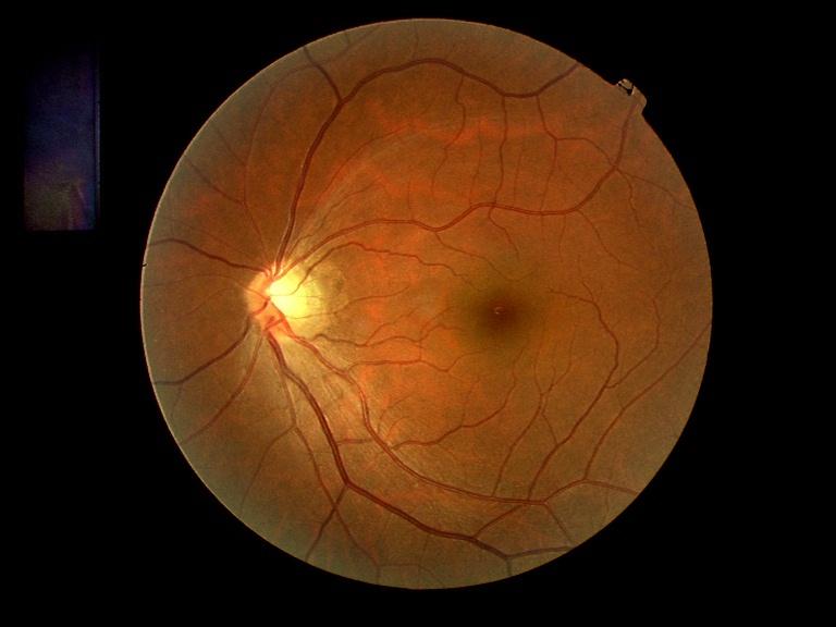
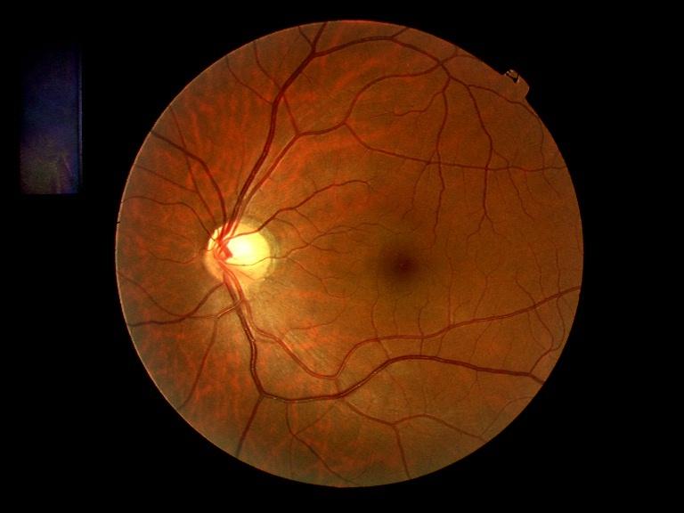
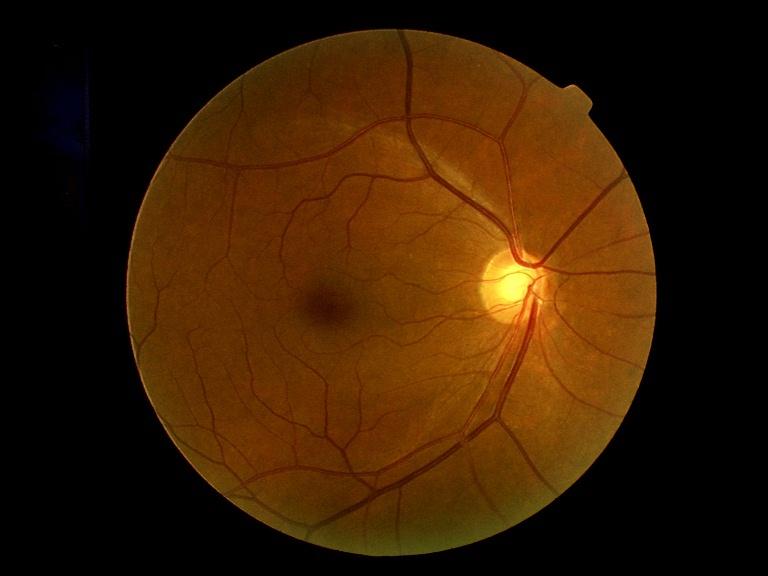
4 3/17 Introduction Superior segmental optic hypoplasia is a congenital anomaly affecting the optic nerve head and the retina. This condition is ophthalmoscopically characterized by a relatively superior entrance of the central retinal artery, pallor of the superior optic disc, a superior peripapillary scleral halo, and thinning of the superior peripapillary nerve fiber layer. 1-8 Perimetry reveals inferior altitudinal defect or inferior sector defect connecting to the blind spot. Visual acuity is not affected in most cases. In an optical coherence tomographic study of this condition, Unoki et al. suggested the usefulness of optical coherence tomography for detecting mild cases of superior segmental optic hypoplasia. 1 Superior segmental optic hypoplasia was originally reported as a kind of optic nerve hypoplasia. The term, superior segmental optic hypoplasia, was generally accepted after Kim et al. investigated an association of this condition with maternal type I diabetes mellitus. 2 Hoyt coined the term, topless optic disc, 3 to emphasize the appearance of the optic disc. Although the association with maternal diabetes is suspected, the etiology of this congenital disorder is not well understood. Unoki et al. reported familial cases of superior segmental optic hypoplasia and suggested the involvement of a genetic factor in this condition. 1 Because thinning of the neuroretinal rim of the optic nerve head occurs in superior segmental optic hypoplasia, predominantly in the nasal superior region, differentiation of this condition from glaucomatous optic neuropathy is of clinical importance. In particular, normal-tension glaucoma resembles superior segmental optic hypoplasia in terms of its localized rim thinning and the lack of elevated intraocular pressure. To investigate the basic features of superior segmental optic hypoplasia, especially its prevalence, we conducted the present study in a large-scale eye disease screening project.
5 4/17 Materials and Methods The data used in the present study were obtained at the Eye Health Care Project in Tajimi and were provided by the Japan Glaucoma Society, which conducted the large-scale eye disease screening project. Our usage of the data was approved by the Japan Glaucoma Society, which has contracted with Tajimi City so that the society can use the data strictly for scientific purposes as long as subject anonymity is preserved. Additionally, informed consent was obtained from all participants before participating in the eye disease screening project and all of them gave the Japan Glaucoma Society permission to use the individual data for scientific research on condition of anonymity. The details of the Eye Health Care Project in Tajimi will be published elsewhere. 9 In short, the study was based on a large-scale eye disease screening project conducted in Tajimi City, Gifu, Japan between September 2000 and October The project was comprised of two study populations: one consisted of 4,000 randomly selected citizens aged 40 or older who underwent a detailed ophthalmological check-up, and the other consisted of a general screening for the general population aged 40 or older. In the latter, the following ophthalmological examinations were conducted for both eyes: visual acuity testing, refractometry with a refractometer (KP-8100PA, Topcon, Tokyo, Japan), corneal thickness measurement with an SP-2000P (Topcon, Tokyo, Japan), perimetry with an FDT (frequency doubling technology) screener (Humphrey Instruments, San Leandro, CA) with the C-20-1 screening program, 45-degree fundus photography with an IMAGEnet 6S (Topcon, Tokyo, Japan), applanation tonometry with a Goldmann tonometer, slit-lamp biomicroscopy, and van Herick testing. In addition, the participants were requested to fill out a questionnaire including their medical history, and their systemic blood pressure, body weight, and height were also measured. Since most of the participants were unfamiliar with FDT testing, the test was repeated after repeated full explanations of the testing and the second result was adopted if the first result was abnormal or unreliable. While the subject s
6 5/17 ethnicity was not determined in the questionnaire, practically speaking, all of the participants were Japanese. The general screening targeted 50,165 citizens aged 40 or older. Of the 50,165 citizens, 14,779 participated in the screening, yielding a response rate of 29.5%. Additionally, we offered a second, voluntary definitive examination on another day to participants who were suspected to have any ocular diseases. At the definitive examination, visual field testing with a Humphrey Field Analyzer (HFA) using the 30-2 SITA Fast program was conducted for subjects with suspected optic disc abnormalities of any type. In the present study, all of the 45-degree IMAGEnet fundus photographs of the above-mentioned 14,779 participants were reviewed for the presence of ocular abnormality in the optic nerve head and the retina of the posterior pole by one of the authors (TY), who was also one of the chief investigators for the Eye Health Care Project in Tajimi. The reviewer paid special attention to the presence of superior segmental optic hypoplasia. The subject information, except for the bilateral fundus photographs, was masked to the reviewer. The definition of superior segmental optic hypoplasia in the present study was: rim thinning of the optic nerve head most prominent in the superior nasal region with corresponding nerve fiber layer defects in the superior nasal region in at least one eye. In cases with superior segmental optic hypoplasia, visual function, i.e., visual acuity, the FDT screener result, and the HFA result, if available, was reviewed after reading the fundus photographs. In addition, demographic and other ophthalmological data were reviewed. An FDT abnormality was defined as the presence of test points with a p value less than 5% in the probability map. An HFA abnormality was defined based on the criteria proposed by Anderson and Patella 10 : when the pattern standard deviation probability plot showed a cluster of three or more nonedge contiguous points having sensitivity with probability of less than 5%, with one of these having a probability of less than 1% in one hemifield, the hemifield was rated as abnormal. If the corresponding inferior defect in the FDT screening or the HFA testing was present, the eye was rated
7 6/17 as superior segmental optic hypoplasia definite; if not, it was rated as superior segmental optic hypoplasia suspect. The data were analyzed using StatView version 5.0 (SAS Institute Inc., Cary, USA) on a personal computer. Differences among the groups were evaluated using chi-square test when applicable. Results Of the 14,779 participants in the screening project, 45-degree IMAGEnet fundus photographs of 28,396 eyes of 14,431 cases were successfully reviewed. A total of 1,162 eyes of 814 cases were not used because the fundus photos were unavailable for 312 eyes and because the photos were of poor quality for 850 eyes. The age and gender distribution of the study population is shown in Table 1. Of the 14,431 cases reviewed, we found 54 eyes of 37 cases of superior segmental optic hypoplasia (0.2% per eye and 0.3% per case): 23 cases (0.2%) were superior segmental optic hypoplasia definite in at least 1 eye and the remaining 14 cases (0.1%) were suspect. Of the 23 definite cases, 5 were definite bilaterally, 5 were definite in 1 eye and suspect in the other eye, and 13 were unilateral. Of the 14 suspect cases, 7 were bilateral and 7 unilateral. The age and gender distribution of superior segmental optic hypoplasia found in the present study is shown in Table 2 (Table 2A for the overall cases and Table 2B for the definite cases). There was a statistically significant difference in the prevalence among age groups (p= and p=0.0425, for overall and definite cases, respectively). No significant gender difference was found (p= and p=0.8942, for overall and definite cases, respectively).
8 7/17 Tables 1A & 1B, Tables 2A & 2B The data for the 37 superior segmental optic hypoplasia cases found in the present study are summarized in Table 3 and example fundus photos are shown in Fig 1. Of the 37 superior segmental optic hypoplasia cases, 10 were male and the remaining 27 were female. The age was 53.1 ± 10.3 years (mean ± standard deviation) and ranged from 40 to 76 years. The visual acuity was better than or equal to 20/20 in 40 eyes (74%) and worse than 20/25 in 2 eyes (4%) of the 54 eyes with superior segmental optic hypoplasia. When the left eyes were selected in bilateral cases and the affected eyes were selected for unilateral cases, the refractive error in spherical equivalent was ± 2.72 D (mean ± standard deviation) and ranged from -8.25D to +1.88D; the intraocular pressure was 14.2 ± 2.5 mmhg (mean ± standard deviation) and ranged from 9 mmhg to 19 mmhg. not identified because the medical questionnaire did not address this issue. Maternal diabetes was One case of superior segmental optic hypoplasia was reported to have diabetes mellitus but the remaining 36 cases were not. Table 3 and Figure 1 Discussion The definition of superior segmental optic hypoplasia employed in the present study was partly different from others. Originally, this anomaly is characterized by a relatively superior entrance of the central retinal artery, pallor of the superior optic disc, a superior peripapillary scleral halo, and thinning of the superior peripapillary nerve fiber layer. 1-8 However, optic disc pallor and superior peripapillary scleral halo are not necessarily seen in all patients, at least in Japanese. According to
9 8/17 Unoki et al. 1 superior scleral halo was not seen in 5 of 7 affected eyes in Japanese. In a study subjected four Japanese cases by Hashimoto et al. 7 only one eye showed superior scleral halo and disc pallor. Additionally, optic disc pallor was not found in Japanese cases with highly suspected superior segmental optic hypoplasia. 11,12 The present study revealed that the prevalence of superior segmental optic hypoplasia is 0.3% in Japanese. This is the first report on the prevalence of superior segmental optic hypoplasia based on a large-scale eye disease screening. About two-thirds of the cases had the corresponding visual field defect. Because a diagnosis of superior segmental optic hypoplasia is based on optic disc appearance and the corresponding visual field, diagnostic measures including IMAGEnet fundus photography and an FDT screener seem to be appropriate for the detection of this disease. It should be emphasized that, in contradistinction to an epidemiological study, the present study was based on a large-scale screening and, thus, the 0.3% prevalence rate needs further validation. Additionally, the reason for age difference found in the present study should be sought though we cannot explain it at this moment. Superior segmental optic hypoplasia resembles glaucomatous optic neuropathy in terms of localized rim thinning when the optic disc has a cupping. In glaucoma cases with normal intraocular pressure, i.e., normal-tension glaucoma, differentiation with superior segmental optic hypoplasia is important since both conditions lack elevated intraocular pressure. The key point of differentiation is the localization of the rim thinning and the characteristic visual field changes: inferior altitudinal defect or inferior sector defect connecting to the blind spot. Since the 0.3% prevalence of superior segmental optic hypoplasia is substantial and represents about one-tenth of that of normal-tension glaucoma in Japanese, 9,13 awareness of this condition should increase in clinical practice. In conclusion, we found that the prevalence of superior segmental optic hypoplasia is 0.3% in Japanese based on data from a large-scale eye disease screening.
10 9/17 Acknowledgement The authors wish to express their deep gratitude to Ms. Megumi Onda for her devoted secretarial assistance.
11 10/17 References 1. Unoki K, Ohba N, Hoyt WF. Optical coherence tomography of superior segmental optic hypoplasia. Br J Ophthalmol 2002;86: Kim RY, Hoyt WF, Lessell S, Narahara MH. Superior segmental optic hypoplasia. A sign of maternal diabetes mellitus. Am J Ophthalmol 1989;107: Landau K, Djahanshahi-Bajka J, Kirchschläger BM. Topless optic disks in children of mothers with type I diabetes mellitus. Am J Ophthalmol 1998;125: Nelson M, Lessell S, Sadun AA. Optic nerve hypoplasia and maternal diabetes mellitus. Arch Neurol 1986;43: Petersen RA, Walton DS. Optic nerve hypoplasia with good visual acuity and visual field defects. Arch Ophthalmol 1997;95: Bjork A, Laurell CG, Laurell U. Bilateral optic nerve hypoplasia with good visual acuity. Am J Ophthalmol 1978;86: Hashimoto M, Ohtsuka K, Nakagawa T, Hoyt WF. Topless optic disk syndrome without maternal diabetes mellitus. Am J Ophthalmol 1999;128: Brodsky MC, Shroeder GT, Ford R. Superior segmental optic hypoplasia in identical twins. J Clin Neuro-ophthalmol 1993;13: Iwase A, Suzuki Y, Araie M, et al. The prevalence and intraocular pressure of primary open-angle glaucoma in Japanese. The Tajimi Study. Ophthalmology in press 10. Anderson DR, Patella VM. Automated Static Perimetry, 2nd ed. St. Louis: Mosby; Namba T, Wakakura M, Shirakawa S, Ishikawa S. Sectorial hypoplasia of the optic nerve. Neuro-ophthalmol Jpn 1987;4: Okazaki S, Miyazawa H, Sekiya Y, et al. Partial hypoplasia of the optic nerve.
12 11/17 Neuro-ophthalmol Jpn 1987;4: Shiose Y, Kitazawa Y, Tsukahara S, et al. Epidemiology of glaucoma in Japan. A nationwide glaucoma survey. Jpn J Ophthalmol. 1991;35:
13 12/17 Table 1. The age and gender distribution of the study population A. Population targeted and actually screened age (yrs.) male female total /7,024 2,024/7,143 3,020/14, ,531/7,931 3,131/7,880 4,662/15, ,895/5,287 2,486/5,279 4,381/10, ,162/3,822 1,554/5,799 2,716/9,621 total 5,584/24,064 9,195/26,101 14,779/50,165 Population actually screened / population targeted
14 13/17 Table 1. Continued B. Population whose fundus photos were examined and subjected in the present study age (yrs.) male female total ,009 2, ,513 3,101 4, ,861 2,427 4, ,094 1,441 2,535 total 5,453 8,978 14,431
15 14/17 Table 2. The age and gender distribution of superior segmental optic hypoplasia found A. Overall cases age (yrs.) male female total /985 (0.4) 11/2,009 (0.5) 15/2,994 (0.5) /1,513 (0.2) 10/3,101 (0.3) 13/4,614 (0.3) /1,861 (0.1) 5/2,427 (0.2) 6/4,288 (0.1) 70-2/1,094 (0.2) 1/1,441 (0.1) 3/2,535 (0.1) total 10/5,453 (0.2) 27/8,978 (0.3) 37/14,431 (0.3) Number of cases / population studied (prevalence in percent) p= for age and p= for gender (chi-square test)
16 15/17 Table 2. Continued B. Definite cases only age (yrs.) male female total /985 (0.3) 7/2,009 (0.3) 10/2,994 (0.3) /1,513 (0.2) 4/3,101 (0.1) 7/4,614 (0.2) /1,861 (0.1) 2/2,427 (0.1) 3/4,288 (0.1) 70-2/1,094 (0.2) 1/1,441 (0.1) 3/2,535 (0.1) total 9/5,453 (0.2) 14/8,978 (0.2) 23/14,431 (0.2) Number of cases / population studied (prevalence in percent) p= for age and p= for gender (chi-square test)
17 16/17 Table 3. Superior segmental optic hypoplasia found in the present study. Refractive error is expressed in spherical equivalent. The right eye of case 28 was rated as optic hypoplasia rather than superior segmental optic hypoplasia. Abbreviations: FDT: frequency doubling technology screener, HFA: Humphrey Field Analyzer, D: diopter, SSOH: superior segmental optic hypoplasia, IOP: intraocular pressure, DM: history of diabetes mellitus, RE: right eye, LE: left eye, f: female, m: male, I: inferior defects; the number represents the number of defects, S: superior defects, ITD: inferior temporal defect, IAD: inferior altitudinal defect, GD: generalized depression, NA: not available, D: definite, S: suspect. See attached sheet.
18 17/17 Figure legends Fig. 1. Fundus photographs of superior segmental optic hypoplasia. Upper left: the right eye of case 4; upper right: the left eye of case 12; lower left: the left eye of case 18; lower right: the right eye of case 34.
19 Table 3 visual acuity FDT defects HFA defects refractive error (D) IOP (mmhg) type of SSOH case sex age (years) DM RE LE RE LE RE LE RE LE RE LE RE LE 1 f 58-20/20 20/20 l-1 S-2 l-1 NA NA D D 2 f 65-20/15 20/ NA NA S S 3 m 73-20/30 20/20 0 l-2 NA NA D 4 f 54-20/15 20/ ITD normal D - 5 m 58-20/25 20/ NA NA S S 6 m /15 20/15 l-1 0 NA NA D - 7 f 51-20/40 20/ NA NA S - 8 f 68-20/20 20/ NA NA S S 9 f 50-20/15 20/ NA NA S 10 f 42-20/20 20/ NA NA S - 11 f 64-20/20 20/ NA NA S - 12 f 47-20/15 20/15 0 l-1 NA NA S D 13 f 50-20/20 20/15 l-2 S-1 ITD normal D - 14 f 42-20/20 20/ NA NA S S 15 m 46-20/15 20/ ITD normal D - 16 f 42-20/20 20/ NA NA S 17 f 42-20/20 20/20 l-1 0 NA NA D S 18 m 42-20/15 20/15 0 l-1 NA NA S D 19 f 40-20/15 20/15 0 l-3 NA NA D 20 f 51-20/15 20/ ITD normal D - 21 f 76-20/40 20/50 l-4 0 IAD normal D - 22 f 47-20/20 20/20 l-1 0 NA NA D - 23 f 66-20/25 20/25 l-1 0 ITD ITD D D 24 f 47-20/20 20/ NA NA S S 25 m 45-20/15 20/ NA NA S S 26 f 40-20/20 20/20 0 l-2 normal IAD D 27 f 52-20/15 20/15 0 S-4 NA NA S 28 m 57-20/15 20/15 l-1 l-4 S-2 GD IAD D 29 f 50-20/15 20/ NA NA S 30 f 56-20/25 20/ NA NA S S 31 m 74-20/25 20/25 0 l-2 NA NA S D 32 f 68-20/25 20/ ITD ITD D D 33 f 43-20/15 20/ normal ITD D 34 m 61-20/20 20/25 l-1 0 NA NA D S 35 f 46-20/15 20/20 0 S-2 ITD ITD D D 36 f 50-20/25 20/20 l-1 S-1 S-2 NA NA D - 37 m 47-20/15 20/15 0 l-2 ITD IAD D D

20 4R 12L 18L 34R
S uperior segmental optic hypoplasia (SSOH), also termed
, also termed") 910 CLINICAL SCIENCE Optical coherence tomography of superior segmental optic hypoplasia K Unoki, N Ohba, W F Hoyt... See end of article for authors affiliations... Correspondence to: Kazuhiko Unoki, MD,
910 CLINICAL SCIENCE Optical coherence tomography of superior segmental optic hypoplasia K Unoki, N Ohba, W F Hoyt... See end of article for authors affiliations... Correspondence to: Kazuhiko Unoki, MD,
Jong Chul Han, Da Ye Choi, and Changwon Kee. 1. Introduction
 Ophthalmology Volume 2015, Article ID 641204, 7 pages http://dx.doi.org/10.1155/2015/641204 Clinical Study The Different Characteristics of Cirrus Optical Coherence Tomography between Superior Segmental
Prevalence and risk factors of superior segmental optic hypoplasia in a Korean population: the Korea National Health and Nutrition Examination Survey
 Seo et al. BMC Ophthalmology 2014, 14:157 RESEARCH ARTICLE Open Access Prevalence and risk factors of superior segmental optic hypoplasia in a Korean population: the Korea National Health and Nutrition
Macular Ganglion Cell Complex Measurement Using Spectral Domain Optical Coherence Tomography in Glaucoma
 Med. J. Cairo Univ., Vol. 83, No. 2, September: 67-72, 2015 www.medicaljournalofcairouniversity.net Macular Ganglion Cell Complex Measurement Using Spectral Domain Optical Coherence Tomography in Glaucoma
Retinal nerve fiber layer thickness in Indian eyes with optical coherence tomography
 Original articles in Indian eyes with optical coherence tomography Malik A, Singh M, Arya SK, Sood S, Ichhpujani P Department of Ophthalmology Government Medical College and Hospital, Sector 32, Chandigarh,
Noel de Jesus Atienza, MD, MSc and Joseph Anthony Tumbocon, MD
 Original Article Philippine Journal of OPHTHALMOLOGY Diagnostic Accuracy of the Optical Coherence Tomography in Assessing Glaucoma Among Filipinos. Part 1: Categorical Outcomes Based on a Normative Database
Patterns of Subsequent Progression of Localized Retinal Nerve Fiber Layer Defects on Red-free Fundus Photographs in Normal-tension Glaucoma
 pissn: 1011-8942 eissn: 2092-9382 Korean J Ophthalmol 2014;28(4):330-336 http://dx.doi.org/10.3341/kjo.2014.28.4.330 Original Article Patterns of Subsequent Progression of Localized Retinal Nerve Fiber
Spontaneous Intraocular Pressure Reduction in Normal-Tension Glaucoma and Associated Clinical Factors
 CLINICAL INVESTIGATIONS Spontaneous Intraocular Pressure Reduction in Normal-Tension Glaucoma and Associated Clinical Factors Akihiro Oguri, Tetsuya Yamamoto and Yoshiaki Kitazawa Department of Ophthalmology,
Seiji T. Takagi, Yoshiyuki Kita, Asuka Takeyama, and Goji Tomita. 1. Introduction. 2. Subjects and Methods
 Ophthalmology Volume 2011, Article ID 914250, 5 pages doi:10.1155/2011/914250 Clinical Study Macular Retinal Ganglion Cell Complex Thickness and Its Relationship to the Optic Nerve Head Topography in Glaucomatous
Retinal Nerve Fiber Layer Measurements in Myopia Using Optical Coherence Tomography
 Original Article Philippine Journal of OPHTHALMOLOGY Retinal Nerve Fiber Layer Measurements in Myopia Using Optical Coherence Tomography Dennis L. del Rosario, MD and Mario M. Yatco, MD University of Santo
53 year old woman attends your practice for routine exam. She has no past medical history or family history of note.
 Case 1 Normal Tension Glaucoma 53 year old woman attends your practice for routine exam. She has no past medical history or family history of note. Table 1. Right Eye Left Eye Visual acuity 6/6 6/6 Ishihara
Intro to Glaucoma/2006
 Intro to Glaucoma/2006 Managing Patients with Glaucoma is Exciting Interesting Challenging But can often be frustrating! Clinical Challenges To identify patients with risk factors for possible glaucoma.
Study of Retinal Nerve Fiber Layer Thickness Within Normal Hemivisual Field in Primary Open-Angle Glaucoma and Normal-Tension Glaucoma
 Study of Retinal Nerve Fiber Layer Thickness Within Normal Hemivisual Field in Primary Open-Angle Glaucoma and Normal-Tension Glaucoma Chiharu Matsumoto, Shiroaki Shirato, Mai Haneda, Hiroko Yamashiro
Ability of Scanning Laser Polarimetry (GDx) to Discriminate among Early Glaucomatous, Ocular Hypertensive and Normal Eyes in the Korean Population
 to Discriminate among Early Glaucomatous, Ocular Hypertensive and Normal Eyes in the Korean Population") Korean J Ophthalmol Vol. 18:1-8, 2004 Ability of Scanning Laser Polarimetry (GDx) to Discriminate among Early Glaucomatous, Ocular Hypertensive and Normal Eyes in the Korean Population Sun Young Lee, MD,
Korean J Ophthalmol Vol. 18:1-8, 2004 Ability of Scanning Laser Polarimetry (GDx) to Discriminate among Early Glaucomatous, Ocular Hypertensive and Normal Eyes in the Korean Population Sun Young Lee, MD,
PRESCRIBING IN GLAUCOMA: GUIDELINES FOR NZ OPTOMETRISTS
 PRESCRIBING IN GLAUCOMA: GUIDELINES FOR NZ OPTOMETRISTS Introduction Independent prescribing relates to the capacity to use clinical judgement in respect of diagnosis and treatment. It does not mean working
10/27/2013. Optic Red Herrings
 Optic Red Herrings 1 Optic neuropathy Compressive Inflammatory Toxic Glaucomatous Ischemic Post traumatic GLAUCOMATOUS OPTIC NEUROPATHY Glaucoma: Traditionally defined as a progressive optic neuropathy
PREVALENCE OF GLAUCOMA AMONG FISHERMEN COMMUNITY OF MUNDRA TALUKA OF KUTCH DISTRICT- A CROSS- SECTIONAL STUDY
 ORIGINAL RESEARCH PREVALENCE OF GLAUCOMA AMONG FISHERMEN COMMUNITY OF MUNDRA TALUKA OF KUTCH DISTRICT- A CROSS- SECTIONAL STUDY Sanjay Upadhyay 1, Jayantilal Shah 2 1 Assistant Professor, 2 Associate Professor,
NERVE FIBER LAYER THICKNESS IN NORMALS AND GLAUCOMA PATIENTS
 Nerve fiber layer thickness in normals and glaucoma patients 403 NERVE FIBER LAYER THICKNESS IN NORMALS AND GLAUCOMA PATIENTS HIROTAKA SUZUMURA, KAYOKO HARASAWA, AKIKO KOBAYASHI and NARIYOSHI ENDO Department
S Morishita, T Tanabe, S Yu, M Hangai, T Ojima, H Aikawa, N Yoshimura. Clinical science
 Department of Ophthalmology and Visual Sciences, Kyoto University Graduate School of Medicine, Kyoto, Japan Correspondence to: Dr T Tanabe, Department of Ophthalmology, The Tazuke Kofukai Medical Institute,
Differences between Non-arteritic Anterior Ischemic Optic Neuropathy and Open Angle Glaucoma with Altitudinal Visual Field Defect
 pissn: 1011-8942 eissn: 2092-9382 Korean J Ophthalmol 2015;29(6):418-423 http://dx.doi.org/10.3341/kjo.2015.29.6.418 Original Article Differences between Non-arteritic Anterior Ischemic Optic Neuropathy
The Effect of Pupil Dilation on Scanning Laser Polarimetry With Variable Corneal Compensation
 C L I N I C A L S C I E N C E The Effect of Pupil Dilation on Scanning Laser Polarimetry With Variable Corneal Compensation Amjad Horani, MD; Shahar Frenkel, MD, PhD; Eytan Z. Blumenthal, MD BACKGROUND
Diagnostic Accuracy of OCT with a Normative Database to Detect Diffuse Retinal Nerve Fiber Layer Atrophy: Diffuse Atrophy Imaging Study METHODS
 Glaucoma Diagnostic Accuracy of OCT with a Normative Database to Detect Diffuse Retinal Nerve Fiber Layer Atrophy: Diffuse Atrophy Imaging Study Jin Wook Jeoung, 1,2 Seok Hwan Kim, 1,3 Ki Ho Park, 1,2
Relationship Between Structure
 Original Article Relationship Between Structure and Function of the Optic Nerve Head-Glaucoma versus Normal Dr Savita Bhat, Dr Anna Elias, Dr Siddharth Pawar, Dr S.J. Saikumar, Dr Alpesh Rajput, superior,
Parafoveal Scanning Laser Polarimetry for Early Glaucoma Detection
 Yamanashi Med. J. 18(1), 15~ 20, 2003 Original Article Parafoveal Scanning Laser Polarimetry for Early Glaucoma Detection Satoshi KOGURE, Yoshiki TODA, Hiroyuki IIJIMA and Shigeo TSUKAHARA Department of
Glaucoma: Diagnostic Modalities
 Glaucoma: Diagnostic Modalities - Dr. Barun Kumar Nayak, Dr. Sarika Ramugade Glaucoma is a leading cause of blindness in the world, especially in older people. Early detection and treatment by ophthalmologist
A vailability of the scanning laser polarimeter now permits
 70 CLINICAL SCIENCE Specificity and sensitivity of glaucoma detection in the Japanese population using scanning laser polarimetry Shigeo Funaki, Motohiro Shirakashi, Kiyoshi Yaoeda, Haruki Abe, Shiho Kunimatsu,
Study of correlation of cup disc ratio with visual field loss in primary open angle glaucoma
 Original Research Article DOI: 10.18231/2395-1451.2017.0003 Study of correlation of cup disc ratio with visual field loss in primary open angle glaucoma Pankaj Soni 1,*, Ashwani Srivastava 2, Akash Srivastava
Comparison of Optic Disc Topography Measured by Retinal Thickness Analyzer with Measurement by Heidelberg Retina Tomograph II
 Comparison of Optic Disc Topography Measured by Retinal Analyzer with Measurement by Heidelberg Retina Tomograph II Noriko Itai*, Masaki Tanito*, and Etsuo Chihara* *Senshokai Eye Institute, Uji, Kyoto,
Clinical Study Visual Field Loss Morphology in High- and Normal-Tension Glaucoma
 Journal of Ophthalmology Volume 2012, Article ID 327326, 8 pages doi:10.1155/2012/327326 Clinical Study Visual Field Loss Morphology in High- and Normal-Tension Glaucoma Michele Iester, 1, 2 Fabio De Feo,
Scanning Laser Tomography to Evaluate Optic Discs of Normal Eyes
 Scanning Laser Tomography to Evaluate Optic Discs of Normal Eyes Hiroshi Nakamura,* Toshine Maeda,* Yasuyuki Suzuki and Yoichi Inoue* *Eye Division of Olympia Medical Clinic, Tokyo, Japan; Department of
Comparative evaluation of time domain and spectral domain optical coherence tomography in retinal nerve fiber layer thickness measurements
 Original article Comparative evaluation of time domain and spectral domain optical coherence tomography in retinal nerve fiber layer thickness measurements Dewang Angmo, 1 Shibal Bhartiya, 1 Sanjay K Mishra,
Clinical decision making based on data from GDx: One year observations
 Washington University School of Medicine Digital Commons@Becker Open Access Publications 2002 Clinical decision making based on data from GDx: One year observations James C. Bobrow Washington University
A Formula to Predict Spectral Domain Optical Coherence Tomography (OCT) Retinal Nerve Fiber Layer Measurements Based on Time Domain OCT Measurements
 Retinal Nerve Fiber Layer Measurements Based on Time Domain OCT Measurements") pissn: 1011-8942 eissn: 2092-9382 Korean J Ophthalmol 2012;26(5):369-377 http://dx.doi.org/10.3341/kjo.2012.26.5.369 Original Article A Formula to Predict Spectral Domain Optical Coherence Tomography (OCT)
pissn: 1011-8942 eissn: 2092-9382 Korean J Ophthalmol 2012;26(5):369-377 http://dx.doi.org/10.3341/kjo.2012.26.5.369 Original Article A Formula to Predict Spectral Domain Optical Coherence Tomography (OCT)
Analysis of Fundus Photography and Fluorescein Angiography in Nonarteritic Anterior Ischemic Optic Neuropathy and Optic Neuritis
 pissn: 1011-8942 eissn: 2092-9382 Korean J Ophthalmol 2016;30(4):289-294 http://dx.doi.org/10.3341/kjo.2016.30.4.289 Original Article Analysis of Fundus Photography and Fluorescein Angiography in Nonarteritic
Scanning Laser Polarimetry and Optical Coherence Tomography for Detection of Retinal Nerve Fiber Layer Defects
 접수번호 : 2008-105 Korean Journal of Ophthalmology 2009;23:169-175 ISSN : 1011-8942 DOI : 10.3341/kjo.2009.23.3.169 Scanning Laser Polarimetry and Optical Coherence Tomography for Detection of Retinal Nerve
Scanning Laser Polarimetry in Patients with Acute Attack of Primary Angle Closure
 Scanning Laser Polarimetry in Patients with Acute Attack of Primary Angle Closure Jimmy S. M. Lai*, Clement C. Y. Tham, Jonathan C. H. Chan*, Nelson K. F. Yip, Wilson W. T. Tang, Patrick S. H. Li*, Jane
Influence of Myopic Disc Shape on the Diagnostic Precision of the Heidelberg Retina Tomograph
 Influence of Myopic Disc Shape on the Diagnostic Precision of the Heidelberg Retina Tomograph Yoshio Yamazaki,* Keiji Yoshikawa, Shiho Kunimatsu, Nobuyuki Koseki, Yasuyuki Suzuki, Shun Matsumoto and Makoto
CHAPTER 13 CLINICAL CASES INTRODUCTION
 2 CHAPTER 3 CLINICAL CASES INTRODUCTION The previous chapters of this book have systematically presented various aspects of visual field testing and is now put into a clinical context. In this chapter,
The optic disc in glaucoma, III: diffuse optic disc pallor with raised intraocular pressure
 British Journal of Ophthalmology, 1978, 62, 670-675 The optic disc in glaucoma, III: diffuse optic disc pallor with raised intraocular pressure R. A. HITCHINGS From the Institute of Ophthalmology, Moorfields
Sensitivity and specificity of new GDx parameters Colen TP, Tang NEML, Mulder PGH and Lemij HG Submitted for publication CHAPTER 7
 Sensitivity and specificity of new GDx parameters Colen TP, Tang NEML, Mulder PGH and Lemij HG Submitted for publication CHAPTER 7 61 Abstract Purpose The GDx is a scanning laser polarimeter that assesses
3/16/2018. Perimetry
 Perimetry The normal visual field extends further away from fixation temporally and inferiorly than superiorly and nasally. From the center of the retina this sensitivity decreases towards the periphery,
CLINICAL SCIENCES. Screening for Glaucoma With Frequency-Doubling Technology and Damato Campimetry
 CLINICAL SCIENCES Screening for Glaucoma With Frequency-Doubling Technology and Damato Campimetry Noriko Yamada, MD; Philip P. Chen, MD; Richard P. Mills, MD; Martha M. Leen, MD; Marc F. Lieberman, MD;
Glaucoma at a tertiary referral eye hospital in Nepal
 Original article Glaucoma at a tertiary referral eye hospital in Nepal Paudyal I 1,Thapa S S 1, Paudyal G 2, Gurung R 2, Ruit S 2 1 Nepal Glaucoma Eye Clinic, Tilganga Institute of Ophthalmology, Kathmandu,
T he retinal ganglion cells of different sizes have distinct
 604 CLINICAL SCIENCE Agreement between frequency doubling perimetry and static perimetry in eyes with high tension glaucoma and normal tension glaucoma S Kogure, Y Toda, D Crabb, K Kashiwagi, F W Fitzke,
To assess the glaucoma diagnostic ability of Fourier Domain Optical Coherence Tomography
 American Journal of Engineering Research (AJER) e-issn : 2320-0847 p-issn : 2320-0936 Volume-02, Issue-11, pp-104-110 www.ajer.org Research Paper Open Access To assess the glaucoma diagnostic ability of
Research Article The Pattern of Retinal Nerve Fiber Layer and Macular Ganglion Cell-Inner Plexiform Layer Thickness Changes in Glaucoma
 Hindawi Ophthalmology Volume 2017, Article ID 78365, 8 pages https://doi.org/10.1155/2017/78365 Research Article The Pattern of Retinal Nerve Fiber Layer and Macular Ganglion Cell-Inner Plexiform Layer
Prevalence Of Primary Open Angle Glaucoma in Diabetic Patients
 IOSR Journal of Dental and Medical Sciences (IOSR-JDMS) e-issn: 2279-0853, p-issn: 2279-0861.Volume 16, Issue 6 Ver. III (June. 2017), PP 147-151 www.iosrjournals.org Prevalence Of Primary Open Angle Glaucoma
EXPERIMENTAL AND THERAPEUTIC MEDICINE 6: , 2013
 268 Comparison of optic nerve morphology in eyes with glaucoma and eyes with non-arteritic anterior ischemic optic neuropathy by Fourier domain optical coherence tomography YUXIN YANG 1, HAITAO ZHANG 1,
Evaluating Optic Nerve Damage: Pearls and Pitfalls
 54 The Open Ophthalmology Journal, 9, 3, 54-58 Evaluating Optic Nerve Damage: Pearls and Pitfalls Open Access Paul J. Mackenzie * and Frederick S. Mikelberg Division of Glaucoma, Department of Ophthalmology
Effect of brimonidine on intraocular pressure in normal tension glaucoma: A short term clinical trial
 European Journal of Ophthalmology / Vol. 13 no. 7, 2003 / pp. 611-615 Effect of brimonidine on intraocular pressure in normal tension glaucoma: A short term clinical trial S.A. GANDOLFI, L. CIMINO, P.
GLAUCOMA REPEAT READINGS PATHWAY
 GLAUCOMA REPEAT READINGS PATHWAY Level 1a: Goldmann style applanation tonometry repeat readings A first level community service for IOP refinement where other signs of glaucoma are not present will reduce
Question 1: Comment on the optic nerve appearance of each eye.
 Case 2 - Right Optic Nerve Head Drusen (ONHD) A 41 year old female was referred by her optometrist for a workup for unilateral optic disc drusen, OCT, and visual field changes. The patient was otherwise
Disc Hemorrhages in Patients with both Normal Tension Glaucoma and Branch Retinal Vein Occlusion in Different Eyes
 Korean Journal of Ophthalmology 21(4):222-227, 2007 DOI : 10.3341/kjo.2007.21.4.222 Disc Hemorrhages in Patients with both Normal Tension Glaucoma and Branch Retinal Vein Occlusion in Different Eyes Young
The Common Clinical Competency Framework for Non-medical Ophthalmic Healthcare Professionals in Secondary Care
 The Common Clinical Competency Framework for Non-medical Ophthalmic Healthcare Professionals in Secondary Care Glaucoma November 2016 Association of Health Professions in Ophthalmology General basic competences
Correlation of Blue Chromatic Macular Sensitivity with Optic Disc Change in Early Glaucoma Patients
 Correlation of Blue Chromatic Macular Sensitivity with Optic Disc Change in Early Glaucoma Patients Yoshio Yamazaki, Kenji Mizuki, Fukuko Hayamizu and Chizuru Tanaka Department of Ophthalmology, Nihon
Central Corneal Thickness-An important variable for prognostication in Primary Open Angle glaucoma; A Kolkata based study in Eastern India
 Original article: Central Corneal Thickness-An important variable for prognostication in Primary Open Angle glaucoma; A Kolkata based study in Eastern India 1Dr. Apala Bhattacharya, 2 Dr Gautam Bhaduri,
Science & Technologies
 STANDARD COMPUTERIZED PERIMETRY IN FUNCTION OF DIAGNOSTIC GLAUCOMA Iljaz Ismaili, 1 Gazepov Strahil, 2, Goshevska Dashtevska Emilija 1 1 University Eye Clinic,Skopje 2 Clinical Hospital, Shtip Abstract
Bitemporal visual field defects mimicking chiasmal compression in eyes with tilted disc syndrome
 Optometry (2009) 80, 232-242 Bitemporal visual field defects mimicking chiasmal compression in eyes with tilted disc syndrome Joseph W. Sowka, O.D., a and Vincent V. Luong, B. Optom. b a Nova Southeastern
Method for comparing visual field defects to local RNFL and RGC damage seen on frequency domain OCT in patients with glaucoma.
 Method for comparing visual field defects to local RNFL and RGC damage seen on frequency domain OCT in patients with glaucoma. Donald C. Hood 1,2,* and Ali S. Raza 1 1 Department of Psychology, Columbia
Sequential non-arteritic anterior ischemic optic neuropathy (NAION) Jonathan A. Micieli, MD Valérie Biousse, MD
 Jonathan A. Micieli, MD Valérie Biousse, MD") Sequential non-arteritic anterior ischemic optic neuropathy (NAION) Jonathan A. Micieli, MD Valérie Biousse, MD A 68 year old white woman had a new onset of floaters in her right eye and was found to have
Sequential non-arteritic anterior ischemic optic neuropathy (NAION) Jonathan A. Micieli, MD Valérie Biousse, MD A 68 year old white woman had a new onset of floaters in her right eye and was found to have
Evaluation of retinal nerve fiber layer thickness parameters in myopic population using scanning laser polarimetry (GDxVCC)
") Dada T et al Original article Evaluation of retinal nerve fiber layer thickness parameters in myopic population using scanning laser polarimetry (GDxVCC) Dada T1, Aggarwal A1, Bali SJ2, Sharma A1, Shah
Dada T et al Original article Evaluation of retinal nerve fiber layer thickness parameters in myopic population using scanning laser polarimetry (GDxVCC) Dada T1, Aggarwal A1, Bali SJ2, Sharma A1, Shah
International Journal of Scientific & Engineering Research, Volume 4, Issue 12, December ISSN
 International Journal of Scientific & Engineering Research, Volume 4, Issue 12, December-2013 108 Name of Chief and Corresponding Author : Dr Chandrima Paul TITLE : Comparison of glaucoma diagnostic ability
Visual Field Interpretation Anthony B. Litwak, OD, FAAO VA Medical Center Baltimore, MD
 Visual Field Interpretation Anthony B. Litwak, OD, FAAO VA Medical Center Baltimore, MD Dr. Litwak is on the speaker bureau and advisory panel for Alcon and Zeiss Meditek o Confirms Glaucoma Diagnosis!
Optical Coherence Tomograpic Features in Idiopathic Retinitis, Vasculitis, Aneurysms and Neuroretinitis (IRVAN)
") Columbia International Publishing Journal of Ophthalmic Research (2014) Research Article Optical Coherence Tomograpic Features in Idiopathic Retinitis, Vasculitis, Aneurysms and Neuroretinitis (IRVAN)
Columbia International Publishing Journal of Ophthalmic Research (2014) Research Article Optical Coherence Tomograpic Features in Idiopathic Retinitis, Vasculitis, Aneurysms and Neuroretinitis (IRVAN)
Retinal Nerve Fiber Analysis: the Role It Plays in Assessing Glaucoma Grace Martin, CPOT
 Retinal Nerve Fiber Analysis: the Role It Plays in Assessing Glaucoma Grace Martin, CPOT Every day, patients experiencing visual difficulties are seen in the optometric practice. This can range from a
The evaluation of retinal nerve fiber layer in pigment dispersion syndrome and pigmentary glaucoma using scanning laser polarimetry
 European Journal of Ophthalmology / Vol. 13 no. 4, 2003 / pp. 377-382 The evaluation of retinal nerve fiber layer in pigment dispersion syndrome and pigmentary glaucoma using scanning laser polarimetry
Evolving glaucoma management True diagnostic integration for the preservation of vision
 Evolving glaucoma management True diagnostic integration for the preservation of vision // GLAUCOMA MANAGEMENT MADE BY ZEISS The moment you are certain it is glaucoma. This is the moment we work for. There
This study was limited to those discs in which all 3 THE CRESCENT
 British Journal of Ophthalmology, 1978, 62, 16-20 The tilted disc DAVID DORRELL From the Department of Neuro-Ophthalmology, National Hospitals for Nervous Diseases, Queen Square, London SUMMARY Sixty tilted
CLINICAL SCIENCES. Felipe A. Medeiros, MD; Linda M. Zangwill, PhD; Christopher Bowd, PhD; Robert N. Weinreb, MD
 CLINICAL SCIENCES Comparison of the GDx VCC Scanning Laser Polarimeter, HRT II Confocal Scanning Laser Ophthalmoscope, and Stratus OCT Optical Coherence Tomograph for the Detection of Glaucoma Felipe A.
International Journal of Health Sciences and Research ISSN:
 International Journal of Health Sciences and Research www.ijhsr.org ISSN: 2249-9571 Original Research Article Conversion of Ocular Hypertensives into Glaucoma: A Retrospective Study Aditi Singh 1, Shibi
Available online at Pelagia Research Library. Advances in Applied Science Research, 2013, 4(6):
:") Available online at www.pelagiaresearchlibrary.com Advances in Applied Science Research, 2013, 4(6):201-206 ISSN: 0976-8610 CODEN (USA): AASRFC Comparison of glaucoma diagnostic ability of retinal nerve
Available online at www.pelagiaresearchlibrary.com Advances in Applied Science Research, 2013, 4(6):201-206 ISSN: 0976-8610 CODEN (USA): AASRFC Comparison of glaucoma diagnostic ability of retinal nerve
Is Posner Schlossman Syndrome Benign?
 Is Posner Schlossman Syndrome Benign? Aliza Jap, FRCS (G), 1 Meenakshi Sivakumar, FRCS (Ed), M Med (Ophth), 2, Soon-Phaik Chee, FRCS (Ed), FRCOphth 2 Purpose: To determine the clinical course of patients
RETINAL NERVE FIBER LAYER
 CLINICAL SCIENCES The Effect of Scan Diameter on Retinal Nerve Fiber Layer Thickness Measurement Using Stratus Optic Coherence Tomography Giacomo Savini, MD; Piero Barboni, MD; Michele Carbonelli, MD;
Research Article Repeatability of Perimacular Ganglion Cell Complex Analysis with Spectral-Domain Optical Coherence Tomography
 Ophthalmology Volume 2015, Article ID 605940, 5 pages http://dx.doi.org/10.1155/2015/605940 Research Article Repeatability of Perimacular Ganglion Cell Complex Analysis with Spectral-Domain Optical Coherence
New Concepts in Glaucoma Ben Gaddie, OD Moderator Murray Fingeret, OD Louis Pasquale, MD
 New Concepts in Glaucoma Ben Gaddie, OD Moderator Murray Fingeret, OD Louis Pasquale, MD New Concepts in Glaucoma Optical Coherence Tomography: Is it necessary and needed to diagnose and monitor glaucoma?
Correspondence should be addressed to Kui Dong Kang;
 Journal of Ophthalmology, Article ID 931738, 9 pages http://dx.doi.org/10.1155/2014/931738 Clinical Study Correlation between Optic Nerve Parameters Obtained Using 3D Nonmydriatic Retinal Camera and Optical
International Journal Of Basic And Applied Physiology
 A STUDY TO CORRELATE OPTIC CUP/DISC RATIO WITH VISUAL FIELD DEFECTS IN PRIMARY OPEN ANGLE GLAUCOMA Nilay B. Patel, Jayendrasinh M. Jadeja 2, Purvi Bhagat, Jagdeepkaur S. Dani 4, Arjunkumar Jakasania Harsiddh
Role of Central Corneal Thickness in Circadian Intraocular Pressure Fluctuations among Patients with Primary Open Angle Glaucoma
 Role of Central Corneal Thickness in Circadian Intraocular Pressure Fluctuations among Patients with Primary Open Angle Glaucoma Mohannad Albdour MD*, Karanjit Kooner MD, PHD** ABSTRACT Objectives: To
Case report: bilateral optic nerve head drusen and glaucoma
 Romanian Journal of Ophthalmology, Volume 61, Issue 4, October-December 2017. pp:310-314 CASE REPORT Case report: bilateral optic nerve head drusen and glaucoma Mănoiu Mihaela-Roxana*, Amri Jade Amine*,
A comparison of HRT II and GDx imaging for glaucoma detection in a primary care eye clinic setting
 (2007) 21, 1050 1055 & 2007 Nature Publishing Group All rights reserved 0950-222X/07 $30.00 www.nature.com/eye CLINICAL STUDY A comparison of HRT II and GDx imaging for glaucoma detection in a primary
CLINICAL SCIENCES. Steven L. Mansberger, MD; Pamela A. Sample, PhD; Linda Zangwill, PhD; Robert N. Weinreb, MD
 CLINICAL SCIENCES Achromatic and Short-Wavelength Automated Perimetry in Patients With Glaucomatous Large Cups Steven L. Mansberger, MD; Pamela A. Sample, PhD; Linda Zangwill, PhD; Robert N. Weinreb, MD
 Title Neuromyelitis Optica in Japanese Author(s) TANAKA, Yuji Citation [Internal Medicine] vol.[50] no.[ Issue Date 2011 Rights The Japanese Society of Internal 内科学会 ) Version 出版社版 (publisher version)
Discrimination between normal and glaucomatous eyes with visual field and scanning laser polarimetry measurements
 586 Glaucoma Service, Department of Ophthalmology, University of Campinas, Campinas, Brazil R Lauande-Pimentel R A Carvalho H C Oliveira D C Gonçalves L M Silva V P Costa Glaucoma Service, Department of
Risk Factors for Open-Angle Glaucoma in a Japanese Population
 Risk Factors for Open-Angle Glaucoma in a Japanese Population The Tajimi Study Yasuyuki Suzuki, MD, PhD, 1 Aiko Iwase, MD, PhD, 2 Makoto Araie, MD, PhD, 3 Tetsuya Yamamoto, MD, PhD, 4 Haruki Abe, MD, PhD,
Reports. Macular Thickness as a Potential Biomarker of Mild Alzheimer s Disease
 Reports Macular Thickness as a Potential Biomarker of Mild Alzheimer s Disease Although several postmortem findings in the retina of patients with Alzheimer s disease (AD) are available, 1 new biomarkers
GLAUCOMA SUMMARY BENCHMARKS FOR PREFERRED PRACTICE PATTERN GUIDELINES
 SUMMARY BENCHMARKS FOR PREFERRED PRACTICE PATTERN GUIDELINES Introduction These are summary benchmarks for the Academy s Preferred Practice Pattern (PPP) guidelines. The Preferred Practice Pattern series
Research Article Peripapillary Retinoschisis in Glaucoma Patients
 Ophthalmology Volume 216, Article ID 161272, 8 pages http://dx.doi.org/1.1155/216/161272 Research Article Peripapillary Retinoschisis in laucoma Patients Serife Bayraktar, Zafer Cebeci, Melis Kabaalioglu,
Reproducibility of Nerve Fiber Layer Thickness Measurements by Use of Optical Coherence Tomography
 Reproducibility of Nerve Fiber Layer Thickness Measurements by Use of Optical Coherence Tomography Eytan Z. Blumenthal, MD, 1 Julia M. Williams, BS, 1 Robert N. Weinreb, MD, 1 Christopher A. Girkin, MD,
CLINICAL SCIENCES. Comparison of Glaucoma Diagnostic Capabilities of Cirrus HD and Stratus Optical Coherence Tomography
 CLINICAL SCIENCES Comparison of Glaucoma Diagnostic Capabilities of Cirrus HD and Stratus Optical Coherence Tomography Seong Bae Park, MD; Kyung Rim Sung, MD, PhD; Sung Yong Kang, MD; Kyung Ri Kim, BS;
Ganglion cell complex scan in the early prediction of glaucoma
 Original article in the early prediction of glaucoma Ganekal S Nayana Super Specialty Eye Hospital and Research Center, Davangere, Karnataka, India Abstract Objective: To compare the macular ganglion cell
Clinical Guidance and Monitoring for Change. Cecilia Fenerty MD FRCOphth Manchester Royal Eye Hospital
 Clinical Guidance and Monitoring for Change Cecilia Fenerty MD FRCOphth Manchester Royal Eye Hospital Glaucoma Referral Criteria 2000 Original referral scheme Simple criteria based on IOP/Disc/Field Solitary
Factors Associated With Visual Field Progression in Cirrus Optical Coherence Tomography-guided Progression Analysis: A Topographic Approach
 ORIGINAL STUDY Factors Associated With Visual Field Progression in Cirrus Optical Coherence Tomography-guided Progression Analysis: A Topographic Approach Joong Won Shin, MD, Kyung Rim Sung, MD, PhD, Jiyun
CLINICAL SCIENCES. optic neuropathy characterized
 CLINICAL SCIENCES Spectral-Domain Optical Coherence Tomography for Detection of Localized Retinal Nerve Fiber Layer Defects in Patients With Open-Angle Glaucoma Na Rae Kim, MD; Eun Suk Lee, MD, PhD; Gong
MEDICAL POLICY. Proprietary Information of Excellus Health Plan, Inc. A nonprofit independent licensee of the BlueCross BlueShield Association
 MEDICAL POLICY SUBJECT: OPHTHALMOLOGIC TECHNIQUES PAGE: 1 OF: 7 If the member's subscriber contract excludes coverage for a specific service it is not covered under that contract. In such cases, medical
Reliability analyses of the GDx nerve-fiber analyzer
 VOL. 29 NO. 2 PHILIPPINE JOURNAL OF Ophthalmology APRIL ORIGINAL ARTICLE - JUNE 2004 1, 2, 3 Patricia M. Khu, MD, MS Edgardo U. Dorotheo, MD 1 Lawrence Tinio, MD 2 Cynthia P. Cordero, MS 4 1, 2, 3 Manuel
Kobe University Repository : Kernel
 Kobe University Repository : Kernel タイトル Title 著者 Author(s) 掲載誌 巻号 ページ Citation 刊行日 Issue date 資源タイプ Resource Type 版区分 Resource Version 権利 Rights DOI JaLCDOI URL Agreement of Rebound Tonometer in Measuring
MEDICAL POLICY SUBJECT: CORNEAL ULTRASOUND PACHYMETRY. POLICY NUMBER: CATEGORY: Technology Assessment
 MEDICAL POLICY SUBJECT: CORNEAL ULTRASOUND,, PAGE: 1 OF: 5 If a product excludes coverage for a service, it is not covered, and medical policy criteria do not apply. If a commercial product, including
Optical coherence tomography (OCT) is a noninvasive,
 is a noninvasive,") Ability of Stratus OCT to Detect Progressive Retinal Nerve Fiber Layer Atrophy in Glaucoma Eun Ji Lee, 1,2 Tae-Woo Kim, 1,2 Ki Ho Park, 2 Mincheol Seong, 3 Hyunjoong Kim, 4 and Dong Myung Kim 2 PURPOSE.
Ability of Stratus OCT to Detect Progressive Retinal Nerve Fiber Layer Atrophy in Glaucoma Eun Ji Lee, 1,2 Tae-Woo Kim, 1,2 Ki Ho Park, 2 Mincheol Seong, 3 Hyunjoong Kim, 4 and Dong Myung Kim 2 PURPOSE.
LARGE DISCS WITH LARGE CUPS A DIAGNOSTIC CHALLENGE IN AFRICAN PATIENTS. Darshana Soma
 LARGE DISCS WITH LARGE CUPS A DIAGNOSTIC CHALLENGE IN AFRICAN PATIENTS Darshana Soma A research report submitted to the Faculty of Health Sciences, University of the Witwatersrand, Johannesburg, in partial
Elevated intraocular pressure (IOP) is a major risk factor for
 is a major risk factor for") Glaucoma The Association between Retinal Vessel Diameter and Retinal Nerve Fiber Layer Thickness in Asymmetric Normal Tension Glaucoma Patients Joon Mo Kim, 1 Mo Sae Kim, 2 Hyo Ju Jang, 1 Ki Ho Park, 3
Glaucoma The Association between Retinal Vessel Diameter and Retinal Nerve Fiber Layer Thickness in Asymmetric Normal Tension Glaucoma Patients Joon Mo Kim, 1 Mo Sae Kim, 2 Hyo Ju Jang, 1 Ki Ho Park, 3