THE TAMILNADU DR.M.G.R. MEDICAL UNIVERSITY CHENNAI, TAMILNADU
|
|
|
- Tyler Fitzgerald
- 5 years ago
- Views:
Transcription

1 A STUDY ON COMPARISON OF CENTRAL CORNEAL THICKNESS IN NORMALS, PRIMARY OPEN ANGLE GLAUCOMA AND OCULAR HYPERTENSIVES DISSERTATION SUBMITTED FOR MASTER OF SURGERY DEGREE BRANCH III - OPHTHALMOLOGY APRIL 2012 THE TAMILNADU DR.M.G.R. MEDICAL UNIVERSITY CHENNAI, TAMILNADU 1
2 Dept. of Ophthalmology, Govt. Rajaji Hospital, Madurai. CERTIFICATE This is to certify that this dissertation entitled A STUDY ON COMPARISON OF CENTRAL CORNEAL THICKNESS IN NORMALS, PRIMARY OPEN ANGLE GLAUCOMA, AND OCULAR HYPERTENSIVES has been done by DR. M. SHERIN HAROON under my guidance in DEPARTMENT OF OPHTHALMOLOGY, Madurai Medical College, Madurai. I certify regarding the authenticity of the work done to prepare this dissertation. DR. P.THIYAGARAJAN. M.S., D.O., PROFESSOR & H.O.D. DEPARTMENT OF OPHTHALMOLOGY GOVT. RAJAJI HOSPITAL & MADURAI MEDICAL COLLEGE MADURAI. 2
3 DECLARATION I, Dr. M. SHERIN HAROON, solemnly declare that the dissertation titled A STUDY ON COMPARISON OF CENTRAL CORNEAL THICKNESS IN NORMALS, PRIMARY OPEN ANGLE GLAUCOMA AND OCULAR HYPERTENSIVES has been prepared by me. This is submitted to The Tamil Nadu Dr. M.G.R. Medical University, Chennai, in partial fulfillment of the requirement for the award of M.S.,(Ophthalmology) Branch-III degree Examination to be held in APRIL Place : Madurai Date : Dr.M. SHERIN HAROON 3
4 ACKNOWLEDGEMENT I am deeply indebted to Dr. P. THIYAGARAJAN. M.S., D.O, Professor and Head of the department of Ophthalmology, Madurai Medical college, Madurai for the able guidance, inspiration and encouragement he rendered at every stage of this study. I am extremely thankful to Prof. Dr. G.S. SRINIVASAN, M.S., DO., for his immense support and guidance in doing this study. I acknowledge with gratitude the dynamic guidance and persistent encouragement given to me by my guide Dr. N. PARVATHA SUNDARI, M.S., DO, Assistant Professor in Ophthalmology, Department of Ophthalmology, Madurai Medical College, Madurai for having taken keen interest in sharing her ideas throughout the study period. I thank all my Assistant Professors for their encouragement, advice and guidance. My Sincere thanks to Dr.A. EDWIN JOE, M.D (FM) B.L.., Dean, Madurai Medical College, & Govt Rajaji Hospital, Madurai for permitting me to utilize the clinical materials of the hospital. 4
5 Last, but not the least, my profound gratitude to all the patients, to whom I owe everything because, this venture would not have been possible with out them. 5
6 CONTENTS PART ONE PAGE NO 1. INTRODUCTION 1 2. TONOMETERS AND TONOMETRY 2 3. GOLDMANN APPLANATION TONOMETRY 6 4. CENTRAL CORNEAL THICKNESS AND APPLANATION TONOMETRY PACHYMETRY IMPORTANCE OF CENTRAL CORNEAL THICKNESS IN GLAUCOMA REVIEW OF LITERATURE 38 PART TWO 1. AIMS AND OBJECTIVES MATERIALS AND METHODS STATISTICAL ANALYSIS RESULTS AND OBSERVATIONS DISCUSSION SUMMARY CONCLUSION 68 APPENDIX 1. BIBLIOGRAPHY 2. PROFORMA 3. KEY TO MASTER CHART & MASTER CHART 6
7 A STUDY ON COMPARISON OF CENTRAL CORNEAL THICKNESS IN NORMALS, PRIMARY OPEN ANGLE GLAUCOMA AND OCULAR HYPERTENSIVES ABSTRACT AIM: To compare the central corneal thickness (CCT) and determine the range and distribution in controls, primary open angle glaucoma (POAG) patients and ocular hypertensives. Also to predict any differences in central corneal thickness in relation to age and sex variation. METHODS : One hundred and fifty patients (50 controls, 50 POAG patients, 50 ocular hypertensives) aged 40 years or more were enrolled in our study and all subjects underwent a complete ophthalmic examination that included applanation tonometry and CCT measurements with an ultrasonic pachymeter. RESULTS : Mean central corneal thickness in 50 control subjects was 533µm (SD 29µm), 50 POAG patients was 536µm (SD 29µm), 50 ocular hypertensives was 561µm (SD 28µm ).There were no differences between sexes in our study in
8 POAG and ocular hypertension,though CCT was higher in females in control group. CONCLUSION : Mean central corneal thickness was similar to that found in clinical studies, and was significantly higher in patients with ocular hypertension. A negative association with age and a positive association with intraocular pressure were seen. CCT can be a confounding factor in the measurement of intraocular pressure. KEY WORDS: central corneal thickness, applanation tonometry, primary open angle glaucoma, ocular hypertension.
9 INTRODUCTION Glaucoma is one of the most important causes of irreversible bilateral blindness. Its estimated prevalence in India is about 3% to 4%. Because early detection and treatment may slow the rate of visual field loss and consequent blindness, there have been efforts to develop screening methods for the early diagnosis of the disease. Screening for glaucoma is however a daunting problem. Patients need to be screened appropriately referred, advised and treated if glaucomatous optic disc damage and field loss has to be avoided. The traditional approaches to screening include Tonometry, optic disc evaluation, assessment of nerve fiber layer and visual field tests, all of which have marked limitations. Goldmann applanation Tonometry is the current gold standard for the measurement of IOP. Various studies have shown that variations in central corneal thickness affect the accuracy of measurement in intraocular pressure by applanation Tonometry. The present study is aimed at comparing central corneal thickness in open angle glaucoma, ocular hypertension and controls. 7
10 TONOMETERS AND TONOMETRY IOP MEASUREMENT DIRECT METHOD To measure IOP directly in a living eye using a manometric technique. A needle is inserted into anterior chamber via paracentesis site and is connected to fluid filled tubing. The height of fluid in tube corresponds to IOP. Needle can also be connected to fluid filled reservoir with pressure sensitive membrane. Movement of membrane recorded optically or electronically is a measure of IOP. Not applicable clinically. INDIRECT METHOD Based on eye response to an applied force IOP measurement is performed by deforming the globe and correlating the force responsible for it to the pressure in the eye. Palpation Method: IOP estimated by response of eye to pressure applied by finger pulp (indents easily / firm to touch). 8
11 CLASSFICATION OF TONOMETERS All clinical tonometers measure the intraocular pressure by relating a deformation of globe to the force responsible for deformation. Two basic types differ according to the shape and magnitude of the deformation. 1. Indentation tonometry and 2. Applanation (flattening) tonometry. Indentation Tonometers The shape of the deformation with this type of Tonometer is a truncated cone (fig 1A). The precise shape, however is variable and unpredictable. In addition, these instruments displace a relatively large intraocular volume. As a result of these characteristics conversion tables based on empirical data from in vitro and in vivo studies must be used to estimate the IOP. The prototype of this group, the Schiotz Tonometer, was introduced in Applanation Tonometers The shape of the deformation with these tonometers is a simple flattening (fig 1B), and because the shape is constant, its relationship to the IOP can, in most cases, be derived from mathematical calculations. The applanation tonometers are further differentiated on the basis of the variable that is measured. 9
12 Fig 1.Corneal deformation created by (A) indentation tonometers (a truncated cone) and (B) applanation tonometers (simple flattening) 10
13 Variable force This type of Tonometer measures the force that is required to applanate (flatten) a standard area of the corneal surface. The prototype is the Goldmann applanation Tonometer, which was introduced in Others include: 1. Hand held Goldmann type tonometers - Perkins - Draegers 2. Mackay marg tonometer 3. Tonopen 4. Pneumatic tonometer. Variable area Other applanation tonometers measure the area of cornea that is flattened by a known force (weight). The prototype in this group is the Maklakov Tonometer, which was introduced in Others include: 1. Applanometer 2. Tonomat 3. Halberg tonometer 4. Glaucotest. The division between indentation and applanation Tonometers, however, does not correlate entirely with the magnitude of intraocular 11
14 volume displacement. Goldmann type tonometers have relatively minimal displacement, whereas that with Maklakov-type tonometers is sufficiently large to require the conversion tables. Non contact Tonometer A third type of Tonometer uses a puff of air to deform the cornea and measures either the time or force of the air puff that is required to create a standard amount of corneal deformation. The prototype was introduced by Grolman in Eg. Pulsair tonometer. 12
15 GOLDMANN APPLANATION TONOMETRY Basic concept Goldmann applanation Tonometry is the international clinical standard for measuring intraocular pressure. Goldmann based his concept of Tonometry on a modification of the Maklakov-Fick law (also referred to as the Imbert-Fick law). 1,2 This law states that an external force (W) against a sphere equals the pressure in the sphere ( P t ) times the area flattened (applanated) by the external force (A) (fig 2A) W =P t x A The validity of the law requires that the sphere be a) perfectly spherical, b) dry, c) perfectly flexible, and d) infinitely thin. The cornea fails to satisfy any of these requirements, in that it is aspherical and wet, and neither perfectly flexible nor infinitely thin. The moisture creates a surface tension (S), and the lack of flexibility requires a force to bend the cornea (B), which is independent of the internal pressure. In addition because the cornea has a central thickness of approximately 550µm, the outer area of flattening (A) is not the same as the inner area (A1) It was, therefore, necessary to modify the Imbert-Fick law in the following manner to account for these characteristics of the cornea.(fig 2 B) W + S = P t A1 +B 13
16 Fig 2 A: The Imbert Fick law (W = P t x A) B: modification of Imbert-Fick law for cornea (W+S = P t xa 1 + B) 14
17 When A1 = 7.35mm 2 S balances B and W = P t This internal area of applanation is obtained when the diameter of the external area of corneal applanation is 3.06 mm, which is used in the standard instrument. The volume of displacement produced by applanating an area with a diameter of 3.06mm is approximately 0.50mm 3 so that P t is very close to P 0 and ocular rigidity does not significantly influence the measurement. Description of Tonometer The instrument is mounted on a standard slit-lamp in such a way that the examiner s view is directed through the center of a plastic biprism, which is used to applanate the cornea. Two beam-splitting prisms with in the applanating unit optically convert the circular area of corneal contact into semicircles. The prisms are adjusted so that the inner margins of the semicircles overlap when 3.06 mm of cornea is applanated. The biprism is attached by a rod to a housing, which contains a coil spring and series of levers that are used to adjust the force of the biprism against the cornea.( Fig 3) Technique The cornea is anesthetized with a topical preparation, and the tear film is stained with sodium fluorescein. The tonometer tip is cleaned with a sterilizing solution 3,4-7 and the tip and prism are set in correct position on the slit lamp. Sterile tonometer tip covers may be used rather 15
18 Fig 3 Goldmann-type applanation tonometer A: Basic features of tonometer shown in contact with patients cornea B: Enlargement shows tear film meniscus created by contact of biprism and cornea C: View through biprism (1) reveals circular meniscus (2),which is converted into semicircles (3) by prisms 16
19 than a disinfecting solution, if preferred 8. The tension knob is set at 1g. If the knob is set at 0, the prism may vibrate when it touches the eye and damages the corneal epithelium. The 1g position is used before each measurement. As a rule, it is more accurate to measure IOP by increasing rather than decreasing the force of applanation. The 0 graduation mark of the prism is set at the white line on the prism holder. If the patient has more than 3D of corneal astigmatism, the area of contact between the cornea and the prism is elliptic rather than circular. In this situation the prism should be rotated to about 45 degrees from the long axis of the ellipse that is, the prism graduation corresponding to the least curved meridian of cornea should be set at the red mark on the prism holder 9. An alternative approach is to average the IOP readings obtained with the axis of the prism horizontal and then vertical 10,11. The cobalt blue filter is used with the slit beam opened maximally. The angle between the illumination and the microscope should be approximately 60 degrees. The room illumination is reduced. Biprism is brought into gentle contact with the apex of cornea. The clinician observes the applanation through the biprism at low power. A monocular view is obtained of the central applanated zone and the surrounding Fluorescein stained tear film. Semicircles touch when 3.06 mm area is applanated. Using the control 17

20 MEASUREMENT OF IOP WITH APPLANATION TONOMETER 18
21 stick the observer raises lowers and centers the assembly until two equal semicircles is seen in the center of the field of view. If two semicircles are not equal in size IOP is over estimated. The clinician turns the tension knob in both directions to ensure that the instrument is in good position. If the semicircles cannot be made too small the instrument is too far forward. If the semicircles cannot be made too large the instrument is too far from the eye. The Fluorescein rings should be approximately 0.25 to 0.35 mm in thickness or about one- tenth the diameter of the flattened area. If the rings are too narrow, the patient should blink two or three times to replenish the Fluorescein, additional Fluorescein may be needed if necessary. If the Fluorescein rings are too narrow, IOP is underestimated. If the Fluorescein rings are too wide, the patient s eyelids should be blotted carefully with a tissue, and the front surface of the prism should be dried with lint free material. An excessively wide Fluorescein ring is less of a problem than a very narrow ring, but can cause IOP to be over-estimated. The Fluorescein rings normally undergo a rhythmic movement in response to the cardiac cycle. The tension knob is rotated until the inner borders of the fluorescein rings touch each other at the mid point of their pulsations. IOP is measured in the right eye until 3 successive readings are within 1 mmhg. IOP is then measured in the left eye. The reading 19
22 obtained in grams is multiplied by 10 to give the IOP in millimeters of mercury. The value is recorded along with the date, time of day, list of ocular medications, and time of last instillation of ocular medication. Calibration It is essential that Goldmann applanation tonometer be calibrated periodically at least monthly. Sterilization It is possible to transfer bacteria, viruses, and the other infectious agents with the Tonometer head 12 including such potentially serious infections as epidemic keratoconjuctivitis, hepatitis B, and theoretically, acquired immunodeficiency syndrome. The biprism should be rinsed and dried immediately after use. Between uses, the prism head should be soaked in a solution such as diluted bleach or 3% hydrogen peroxide. 70% ethanol and 70% isopropanol are effective as sterilizing solutions but were shown in one study to cause mild damage to the Tonometer tip after one month of immersion 13,14. Sources of error with Goldmann Tonometry Although the Goldmann Tonometer is reliable and accurate through a wide range of IOP s, errors in measurement can arise from a number of factors, including those that follow: 20
23 1. The alignment and thickness of the mires affects the reading as thin mires overestimate IOP and thick mires underestimate IOP. 2. Inadequate Fluorescein staining of the tear film causes an underestimation of IOP. This commonly occurs when too much time elapses between instillation of fluorescein and the measurement of pressure. To avoid this problem the IOP should be measured within the first minute or so after instilling the Fluorescein. 3. Elevating the eyes more than 15 degrees above the horizontal causes an over estimation of IOP. 4. Widening the lid fissure excessively causes an over estimation of IOP. 5. Repeated Tonometry reduces IOP, causing an underestimation of the true level. This effect is greatest between the first and second readings, but the trend continues through a number of repetitions. A scarred, irregular cornea distorts the Fluorescein rings and makes it difficult to estimate IOP. 6. The thickness of the cornea affects IOP readings 15. If the cornea is thick because of edema, IOP is underestimated 15. If the cornea is thick because of additional tissue, IOP is overestimated 15,27. The Goldmann Tonometer is accurate after epikeratophakia 37. Central corneal pressures 21
24 have been shown to be lower than peripheral corneal readings following Photorefractive keratectomy Decreased corneal thickness leads to underestimation of the IOP. This is seen following excimer laser ablation (LASIK, PRK etc). Correction nomograms have been developed to correct for the same. In general a decrease in the corneal thickness by 100 microns will lead to an underestimation of the IOP by 5-7mmHg. 8. If the examiner presses on the globe, or if the patient squeezes his eyelids, IOP is overestimated. Taking a time to assure the patient and taking care to avoid causing pressure against the globe can help guard against these problems. 9. If corneal astigmatism is greater than 3 D, IOP is underestimated for with- the- rule astigmatism and overestimated for against- the - rule astigmatism 10. The IOP is inaccurate 1mmHg for every 3D of astigmatism Goldmann applanation tonometer can also be used over a soft (high water content) contact lens, but the lens power is a factor in the readings taken, and conversion tables are required. Hyperopic lenses causing the maximum variation. 12. Factors that alter the ocular rigidity affect the indentation tonometry readings, and to a lesser extent the applanation readings also. 22
25 Falsely Low IOP Too little fluorescein Thin cornea Corneal edema With the rule astigmatism > 1 mmhg per 3D. Prolonged contact Inaccurate with irregular corneal surface. Falsely High IOP Too much fluorescein Thick cornea Steep cornea >l mmhg per 3 D of against the rule astigmatism Merits of Goldmann applanation Tonometry Gold standard Accurate Reproducible Ease of use Demerits of Goldmann applanation Tonometry Expensive Needs slit lamp 23
26 COMPARISON OF TONOMETERS Type Advantages Disadvantages Schiotz Indentation Simple construction Extensive clinical use Portability Ease of application Low cost Goldman Non Contact Tonopen (Mackay marg type) Pneumo tonometer Mackay marg Perkins Applanation (Variable force) Independent of corneal curvature, ocular rigidity, Most accurate clinical measure of IOP Slit lamp required Applanation Non contact with eye Reliable with the physiologic range No anaesthesia Mass screening Applanation Portable Irregular corneas Soft CL Modified applanation Modified applanation Diseased corneas, Soft CL Continuous monitoring Instantaneous recording Most accurate in scarred and edematous corneas, Soft CL Applanation Portable, in any position Pediatric use, correlates well with Goldmann Affected by ocular rigidity Corneal curvature and thickness Conversion nomograms Reads lower than Goldmann Supine Position Instrument error Multiple reading required Contact method Sterilization, Mose effect Subjective (Technical expertise required) Sitting position Ocular pulsation Corneal thickness and Irregularities Calibration required Squeezing, lid touch contact method, Sterilization Ocular pulsation Abnormal cornea Poor fixation Variable reading in glaucomatous eyes Multiple reading required Subepithelial bubbles Overestimates low IOP Under estimates high IOP Contact method May overestimate IOP Reads higher (Less reliable than Goldmann) No longer available As for Goldmann (Except position) 24
27 The Pascal Dynamic Contour Tonometer The Dynamic contour tonometer (DCT, Pascal tonometer) is a new digital contact tonometer designed to measure intraocular pressure independent of corneal properties. The slit-lamp mounted device furnishes a numeric output of intraocular pressure and of ocular pulse amplitude upon touching the cornea for a few seconds. It measures pulsatile IOP directly and continuously ( dynamically). The Pascal Dynamic Contour Tonometer utilizes a curved tip to match the corneal surface, thereby minimizing corneal distortion. This device is based on Pascal s Law of Pressure, which states that pressure applied to a confined fluid is transmitted undiminished throughout the confining vessel of the system. The concave shape of the tip generates minimal corneal distortion and theoretically eliminates errors in measuring IOP induced by ocular rigidity when the cornea is applanated with a flat- tipped tonometer. The Pascal Dynamic Contour Tonometer is a slit-lamp mounted device that is operated in a fashion similar to a Goldmann applanation tonometer. A microchip enabled, solid-state sensor embedded within the tip records 100 IOP measurements per second and averages them over fluctuations in ocular pulse amplitude (ie, the range in IOP change during the cardiac cycle). Auditory feedback provides the operator with instant 25
28 clues about the quality of the measurements, and a digital display shows the final averaged IOP as well as a Q-value that may be used objectively to judge the quality of the final measurement. Distinctive features of the PASCAL Dynamic Contour Tonometer: Unlike applanation tonometers, which are influenced by corneal thickness and other characteristics of the cornea and hence may produce misleading estimates of IOP, a contour tonometer provides an accurate direct measurement of true IOP, which is independent of inter-individual variations in corneal properties. PASCAL detects and accurately measures the dynamic pulsatile fluctuations in IOP and thus permits a more detailed assessment of the pressure range to which the eye is subjected due to pulsatile ocular blood flow. No fluorescein staining is required for the measurement. Convenient documentation with optional wireless printer: Prints all the values shown on the LCD display and the actual IOP curve. 26
29 CENTRAL CORNEAL THICKNESS AND APPLANATION TONOMETRY Because the mathematical calculation for Goldmann applanation Tonometry is based on a presumed average Central corneal thickness (CCT), variations in this parameter can lead to errors in this measurement. Ehlers and co-workers noted that corneal thickening due to edema causes an underestimation of true IOP higher pressure, whereas variations of CCT in normal corneas can lead to falsely higher readings with thicker corneas and falsely lower with thinner corneas 15. The clinical importance of the latter observations has subsequently been highlighted by numerous studies, which have also attempted to answer several questions. What is the mean and range of CCT s in general populations and in subgroups, including various glaucoma groups? What correction factor for the adjusted IOP measurement should be used when the CCT deviates from the mean? And, how does refractive surgery influence the IOP measurements? In their modification of the Imbert-Fick Law, Goldmann and Schmidt assigned an average corneal thickness of 520µm. More recent studies have provided slightly higher mean values of 537µ to 554µ in 27
30 normal subjects 16,17 with a wide range of 427µ to 620µ in one study 16. These values may be influence by ethnicity, with thinner mean CCTs of 530µ to 53lµ in one African-American population 18 and 495µ to 514µ in a Mongolian population 19, whereas a study in Japan revealed a mean of 552µ among normal subjects 20. Of even greater clinical importance is the observation that individuals with ocular hypertension have significantly thicker CCTs, with reported means ranging from 570µ to 606µ 21-25, whereas patients with Normal tension glaucoma have thinner mean CCTs of 514µ to 521µ 17,22. Patients with chronic open angle glaucoma, pseudoexfoliation syndrome and chronic angleclosure glaucoma appear to have mean CCTs similar to that in the normal population 17. There is a lack of agreement regarding the correction factor that should be used for adjusting the IOP, measured by Goldmann Tonometry, when the CCT deviates from the normal. Ehlers and colleagues have published a table in which the average error is 0.7 mm Hg per 10µ of deviation from the mean of 520µ 15, which has been supported by others 27. Still other studies, however, have revealed smaller errors of 0.19 mm Hg per 10µ 16, which is consistent with a direct canulation study 28. IOP measurements with another applanation tonometer, the Tono-pen, 28
31 have also been shown to be influenced by CCT, with reported errors of 0.29mmHg per 10µ in men and 0.12 mmhg per 10µ in women 29. Refractive surgery for Myopia, which produces a thinning of CCT, also influences IOP measurements and must be considered when following up the patients after the surgery. This is true for both photorefractive keratectomy and laser assisted in-situ keratomileusis (LASIK) Both procedures may lead to underestimation of the IOP because of central corneal thinning. A critical amount of thinning may be necessary to have a clinically significant influence on the measured IOP 36, although there is no reliable correction factor for adjusting the post-operative pressure measurements, and it may be best to use the difference between the preoperative and postoperative readings as the correction factor for each patient. 29
32 PACHYMETRY Corneal thickness is shown to influence the accuracy of applanation Tonometry 28. Corneal thickness can be measured with an optical device called pachymeter. The normal cornea has a central thickness of about 0.52mm and becomes thicker in the paracentral zone (from about 0.52mm inferiorly to 0.57mm superiorly) and peripheral zone (from 0.63 mm inferiorly to 0.67mm superiorly). The thinnest zone is about 1.5mm temporal to the geographic center. Currently, the most common approaches to corneal thickness measurement include optical and ultrasound pachymetry. Methods of Pachymetry Various methods of pachymetry include Optical pachymetry Ultrasound pachymetry Specular microscopy Confocal microscopy Ultrasound Biomicroscopy Scanning Slit topography system and Anterior Segment OCT 30
33 Optical Pachymetry Optical methods of pachymetry were first described as early as 1951 by Maurice & Giardini 41. Optical Pachymetry can be performed using a device that attaches to the slit lamp beam. It consists of measuring the oblique section of the cornea by means of a split prism and aligning the split images so the epithelial layer coincides with the endothelial layer. While the Haag-Streit pachymeter achieves alignment by manual control, automated instruments such as specular microscope and orbscan obtain a digitalized image and calculate the number of pixels. Ultrasound pachymetry The first ultrasound pachymtery was introduced by Kremer in Ultrasound pachymetry is the most efficient and accurate way to measure corneal thickness. Ultrasound pachymetry uses high-frequency sound waves to detect the epithelial and endothelial layers, both of which are highly reflective surfaces. Knowing the velocity of sound in corneal tissue, the distance between the 2 reflecting surfaces can be calculated by detecting the time lapse between the reflected sound waves from the 2 surfaces. 31

34 ULTRASOUND PACHYMETER 32
35 Pachymeter is calibrated at the beginning of each session. Unlike the optical Pachymeter, it must touch the corneal surface and thus requires topical anaesthesia. The applanating tip must be perpendicular to the surface because tilting induces errors. This was confirmed by an audible beep produced by the instrument. Ultrasound pachymetry is precise, easy to use, and relatively inexpensive. Traditionally, optical pachymetry had been performed using the Haag-Streit pachymeter, whose measurements are reported to be less reproducible and less reliable than the ultrasound pachymeter 43. Ultrasound pachymetry measurements have demonstrated high intraobserver reproducibility 44. However results among observers vary significantly 45. A new high frequency ultrasound technique incorporating digital signal processing is non -invasive and can measure both epithelial and corneal thickness with precision. A speed sound of 1640 m/sec was used. Disadvantages Requirement of physical contact with the cornea Technician error Specular microscopy Pachymetry can also be obtained with specular microscopy. Some specular microscopes designed to evaluate the corneal endothelial 33
36 cell count also measures corneal thickness using electromechanical devices. These are designed to measure central and apical readings only. The measurement derived is based on the distance from the posterior surface of the tear film to the posterior surface of the descemet s membrane, thus reducing the error of as much as 20 to 30µm. In the contact mode, corneal touch is involved and compression may be another source of error. Because the specular microscope functions as an optical pachymeter, it requires clear reflections of the epithelial and endothelial surfaces to obtain a reliable thickness instrument. As such, its clinical use is limited to corneas that are free of edema, scarring, deposits or opacities that may distort light transmission. Although the machine automated the process of centralizing the image for the measurement, there was no means to ensure perpendicularity. However, it has the distinct advantages of being operator independent and noninvasive. Improved signal processing and other methods such as Laser interferometry allow the examiner to map the corneal thickness very precisely. 34
37 Ultrasound Biomicroscopy Though it operates by same principle as ultrasound pachymetry, it affords a higher resolution at a transduction frequency of 50MHZ than conventional ultrasound pachymetry at 20MHZ. UBM transducer fluid is immersed in transduction fluid and is not in contact with cornea. An advantage of this measurement method is the ability to visualize the reflecting surfaces easily. However, it is also a much more lab- intensive procedure for the operator, who must manually adjust the transducer head centrally and perpendicularity of the image. For the subject, it means a longer time for the procedure, discomfort from the eye cup, and the transiently blurred vision from the transducer gel. The Scanning Slit topography system The Scanning slit topography system is an optical scanning-slit instrument that provides topographic analysis and pachymetry measurements of the cornea. Scanning-slit topography requires the patient to fixate for 1.0 to 1.5 seconds. Orbscan elevation topography techniques, recently introduced into wider clinical practices allow the creation of pachymetry maps obtained by a slit-scanning optical device. 35
38 Orbscan pachymetry overestimates corneal thickness compared to ultrasound pachymetry. The noncontact Orbscan system measures the hydrated mucous component of the tear film over the cornea; contact ultrasonic pachymetry does not. Thus, Orbscan readings are higher than ultrasonic readings and require the use of the acoustic equivalent correction factor (0.92). Nonetheless, a major advantage of this technique is the creation of an instantaneous picture of wide-field corneal thickness that allows for classification of different corneal patterns. Advantages over ultrasound are that it is non-invasive and non-contact. Instantaneous results are accessible for up to 9600 points on the cornea, a global or wide-field representation of corneal thickness is provided as a colour map, and there are several analysis and presentation options from proprietary software. The option of a 3-dimensional representation of acquired data and digital storage of images for comparison over time is clearly advantageous. The main disadvantage of elevation- based pachymetry, in addition to the overestimation of the corneal thickness compared to ultrasound, is dependence on various optical phenomena. Orbscan readings are higher than ultrasonic readings and require the use of the acoustic equivalent correction factor (0.92). If the cornea is 36
39 unusually thick (>600 microns) or thin (less than 500 microns) as measured with ultrasonic pachymetry, then an Orbscan could be performed to confirm the measurements. Anterior Segment Optical coherence Tomography (OCT) OCT is a relatively newer technique used to measure corneal thickness. OCT is a non- invasive, non-contact imaging technique that uses infrared light to obtain high-resolution cross-sectional images in vivo. Although the technology has been used primarily in the diagnosis of optic nerve and retinal pathology, more recently it has been shown to be valuable for study of the cornea. Advantages over ultrasound Direct Non-invasive Non-contact Another advantage is the ability to fixate the patient s gaze during testing. In vitro video monitoring of the scanned surface allows for more accurate placement. Precision of placement is further enhanced with patient s fixation on a light target. In contrast, Ultrasound pachymetry lacks a method to fixate the patient s gaze during repeated measurements. Probe placement is therefore difficult to reproduce and is affected by the test operator. In addition to corneal thickness, OCT measurement also provides high resolution cross-sectional imaging of the 37

40 CCT MEASUREMENT WITH ANTERIOR SEGMENT OCT 38
41 cornea in vivo. UBM and confocal microscopy also provides highresolution images in vivo. However UBM requires immersion in saline solution and confocal microscopy requires direct contact with transducer. THE IMPORTANCE OF CENTRAL CORNEAL THICKNESS IN GLAUCOMA Glaucoma is defined as an optic neuropathy characterized by typical appearance of optic nerve head and characteristic visual field loss 40. The diagnosis of glaucoma is based on a combination of factors including IOP (intraocular pressure), optic disc (and nerve fibre layer ) damage and specific field defects. Raised IOP is the only known causal risk factor. IOP is the only factor that is accessible for manipulation by the ophthalmologist to treat glaucoma. In routine practice IOP is one of the most important factors in gauging the progression of the disease, the response to treatment and possibly still used in the diagnosis of glaucoma. Goldmann applanation Tonometry, the current gold standard for the measurement of IOP 46, is based on Imbert -Fick law. Goldmann observed that when the area applanated was 7.35mm 2 the surface tension due to the tear film counterbalanced the resistance to indentation of the cornea, thus making it unnecessary to consider the rigidity of the globe and the surface tension of the tear film in applanation Tonometry 47. More recent evidence indicates that these, as well as a number of other 39
42 factors (e.g. Significant astigmatism, corneal curvature) do affect the accuracy of applanation Tonometry. Variations in corneal thickness change the resistance of the cornea to indentation so that this is no longer balanced exactly by the tear film surface tension. This may affect the accuracy of the measurement of IOP. A thinner cornea may require less force to applanate it, leading to underestimation of true IOP. While a thicker cornea would need more force thus giving an artifactually high lop reading. Goldmann himself discussed the influence of variations of CCT on IOP measured by applanation 9. However, he felt that significant variations in CCT occurred only rarely and hence assumed a normal CCT of 520µm for his instrument. A positive correlation between increased corneal thickness and IOP has been reported earlier 15,27,48. Studies in eyes with manometrically controlled IOP have demonstrated a significant disparity between the actual IOP and simultaneous applanation Tonometry readings. This disparity was related to the CCT. The underestimation of IOP was as much as 4.9mmHg in thin corneas, while thick corneas produced an overestimation of about 6.8mmHg. 16,21,22 Accordingly it has been suggested that measurement of corneal thickness is necessary for the accurate interpretation of applanation Tonometry. 40
43 It has been calculated that applanation Tonometry over/ underestimated IOP by 5mmHg for every 70µm corneal thickness 15. A correction factor for IOP, to adjust for CCT measurements that differ from Normal CCT was proposed as follows: Corrected IOP = Applanation IOP + [5mmHg (Mean normal CCT - measured CCT/ 70µm)] Since a cornea can only be so thin before it becomes pathological, the normal variation of CCT in the population (like the IOP) is skewed to the right. The possibility exists that the measured IOP in patients at either end of the spectrum could be an under/over estimation of the actual hydrostatic IOP and this could be a confounding factor in the terminology of glaucoma based only on the cut-off value of 21mmHg. This study was undertaken to determine whether, in our population, patients classified as Ocular Hypertensives did indeed have thicker corneas compared to normal subjects or true glaucoma patients. The role of CCT in glaucoma is still confounding and is under scrutiny. CCT is important in glaucoma by two means: 1) by altering the accuracy of applanation Tonometry readings and 2) as a predictive factor in the development of POAG as shown by the OHTS (ocular hypertensive study). 41
44 CENTRAL CORNEAL THICKNESS IN NORMALS Various studies have estimated the central corneal thickness in normal subject as a control population. The number of control subjects belong to different population depending on where the study was done. In 2000, Doughty and Zaman 49 presented an extensive review, including a meta-analysis, on the subject of human corneal thickness and its impact on tonometry. They found a significant association between CCT and IOP readings using GAT. They found that the average CCT measured by optical methods was 530 µm and that the average normal CCT measured using ultrasound was 544 µm. The Rotterdam study performed a cross-sectional study on the distribution of central corneal thickness and its association with intraocular pressure. The Rotterdam study itself is a population-based cohort study of 7,983 residents, aged 55 years and more, of a suburb of Rotterdam, the Netherlands, which aimed at investigating determinants of chronic disabling ophthalmologic, cardiovascular, neurogeriatric and locomotor disease. The study enrolled 352 control subjects all aged more than 55 years, with normal corneas on slit lamp examination and having had no previous eye surgery. The mean central corneal thickness in the control 42

45 NORMAL OPTIC DISC 43
46 subject was 537.4µm (95% confidence interval to 540.9µm, range 427 to 620µm). There was no significant difference between right and left eyes or between men and women. Central corneal thickness did not change significantly with age, and it was similar for right and left eyes and for men and women. There was no association between central corneal thickness and time of examination 16. The Chennai glaucoma study, a population-based study of adults aged 40 years and older residing in the southern Indian state of Tamil Nadu found that the mean CCT for the population was ±33.5µm (range, µm). Males (515.6 ±33.8µm) had significantly (P = ) thicker corneas than females (508.0±32.8 µm) 26. CENTRAL CORNEAL THICKNESS IN PRIMARY OPEN ANGLE GLAUCOMA Primary open angle glaucoma (POAG) is a chronic progressive condition with characteristic changes at the optic disc (glaucomatous excavation), where it is usually possible to identify reduced visual function related to the disc changes. In most patients, the IOP is above the normal range (i.e. over 21 mmhg) at some time of the day, usually being highest in the morning. In addition, there is a 44

47 ADVANCED GLAUCOMATOUS CUPPING 45
48 gonioscopically open angle indistinguishable from normal and, in those eyes with elevated IOP, a reduced facility of outflow. Numerous studies of central corneal thickness in glaucomatous patients have suggested that the central corneal thickness in ocular hypertensives is thicker than in normals and in Normal tension glaucoma thinner than in normals. Most studies have reported there is no significant difference between primary open angle glaucoma and 24, 50,51 normals. The Rotterdam study enrolled 30 patients of Primary open angle glaucoma to study central corneal thickness and its association with IOP. It was found that the mean central corneal thickness was significantly lower than in the control subjects (21.5µm; 95% confidence interval, µm; P=. 001) Past surgical or different medical treatments in the primary open angle glaucoma group had no effect on the central corneal thickness. The central corneal thickness being significantly lower in Primary open angle glaucoma in this study in contrast to finding in other studies was attributed to too low a power or the use of less accurate optical pachymetry in those studies 16. The central corneal thickness of patients with POAG was 515µm (range µm) in a study using optical low coherence 46
49 reflectometry, which is a new and more precise method than ultrasonic pachymetry. The design of the optical reflectometer is based on a Michelson reflectometer. The criterion for inclusion for primary open angle glaucoma in this study was an untreated IOP of 22 mm of Hg or higher, an open angle, a glaucomatous optic disc and visual field defects. Patients with primary open angle glaucoma and controls were included in this observational, concurrent case-control study. The mean central corneal thickness of normals in this study was found to be 524µm (range µm). The sample size being relatively low, it is difficult to rule out if any differences could have become manifest with a larger sample size 53. In the Chennai glaucoma study the mean CCT in glaucoma subjects was 514.1±33.6 µm (range, µm) and was similar to the population mean. CENTRAL CORNEAL THICKNESS IN OCULAR HYPERTENSION (OHT) Ocular hypertension (OHT) is a term reserved for eyes in which the intraocular pressure (IOP) lies above the normal population range, the optic nerve and visual field show no signs of glaucomatous damage, and there is no ocular co-morbidity. 47


50 RED FREE IMAGE SHOWING NERVE FIBRE LAYER DEFECT 48
51 Excluded from this definition are eyes with raised IOP from demonstrable causes such as pseudoexfoliation and pigment dispersion syndrome. Most population studies on the over 40-year age group indicate that IOPs measured with Goldmann Tonometry are distributed in a manner similar to a normal distribution (mean Approximately 16 mmhg). Persons with IOPs greater than two standard deviations above the mean can be labelled Ocular Hypertensive. This gives an upper limit for normal IOPs in Caucasians of 21 mmhg. It is of note however that this figure is statistically derived and does not imply that disease is present if measured IOP levels exceed this value. Risk factors for the development of OHT Epidemiological studies have identified those individuals in a population most at risk: - 1. Increasing age 2. Individuals of black African or Caribbean origin. 3. Female gender 4. Systemic hypertension 5. Current use of oral and/or inhaled corticosteroids 6. Diabetes (especially those on insulin) 7. Family history of glaucoma 49
52 Risk factors for conversion to glaucoma Estimates vary as to the conversion rate from OHT to POAG, depending on subject selection and diagnostic criteria. It is likely that approximately 10% of individuals with persistent OHT will convert to POAG over a ten-year period. Risk factors for the conversion of OHT to POAG can be divided into ocular and systemic. The most important are listed below. A. Ocular risk factors Height of the IOP - the greater the IOP the greater the risk Large vertical cup/disc ratio (indicating reduced neuroretinal rim area/volume) Cup/disc (C/D) ratio asymmetry >0.2 Previous history of disc hemorrhage Retinal nerve fibre layer defect in the absence of morphometric optic nerve head changes Thinner than average central corneal thickness (note excimer laser procedures on the cornea can result in artifactually lowered IOPs on measurement) B. Systemic risk factors Increasing age Family history Individuals of black African or Caribbean origin 50
53 OHT was defined as IOP greater than 21 mmhg or use of IOP lowering medications, with a cup-disc ratio of less than 0.5, and with no glaucomatous visual field defect in the base line phase of the Rotterdam study. The Rotterdam study enrolled 13 patients of OHT to study CCT & its association with IOP. It was found that mean CCT was slightly, not significantly higher than in the control subjects (=l6µm; 95% confidence limit, to µm; P = 0.093) which has also been found by others. CCT of patients with OHT was 553µm in a study using optical coherence reflectometry. CCT as a predictive factor in the development of POAG as shown in the OHTS (Ocular hypertensive treatment study) 23,56 The OHTS was a multicentric randomized trial designed to evaluate the safety and efficacy of topical ocular hypotensive medications in delaying or preventing the onset of POAG in individuals with OHT. In this study a thinner CCT measurement predicted the development of POAG in both univariate and multivariate models. Among participants who developed POAG, the mean CCT was 553.lµm ± 38.8µm when compared to 574µm ± 37.8µm among those who did not develop POAG. The entire participants were divided into three groups. CCT < 555µm; CCT µm and CCT > 588µm. Compared with the 51
54 participants with the thickest corneas (>588µm), participants with intermediate thickness ( µm) had a hazard ratio of 1.7 and participants with the thinnest CCT (<555µm) had a hazard ratio of 3.4.The risk of developing POAG was found to be highest among participants with the thinnest corneas in any IOP range and also in any range of C/D ratios. The OHTS is the first study to prospectively document that a thinner CCT measurement predicts the development of POAG. Corneal thickness appears to be strong predictive factor for the development of POAG even after adjusting for the effects of baseline age, IOP, vertical C/D ratio & PSD, which are the other risk factors identified in the OHTS. Participants with a CCT of less than 555µm had a three fold greater risk of developing POAG compared to participants with CCT> 588µm. The predictive power of CCT may be due to its effect on the measurement of IOP. However the OHTS study states that the possibility that CCT is related to other factors affecting susceptibility to glaucomatous damage cannot be excluded. The OHTS concludes that CCT provides new information about the risk of developing POAG and recommends its measurement in the clinical evaluation of patient with OHT. 52
55 A REVIEW OF LITERATURE A literature search using Pubmed was performed using the key words central corneal thickness and primary open angle glaucoma. There were 40 results out of which the important ones are highlighted below. The ocular hypertension treatment study aimed to describe baseline demographic and clinical factors that predict which participants in the ocular hypertension treatment study (OHTS) developed primary open glaucoma. This was based on the background that the ocular hypertension study (OHTS) has shown that topical ocular hypotensive medication is effective in delaying or preventing the onset of primary open angle glaucoma in individuals with elevated intraocular pressure (ocular hypertension) and no evidence of glaucomatous damage. Univariate and multivariate analyses showed that thinner central corneal measurements were an important predictor for the development of primary open angle glaucoma. It was concluded that the central corneal thickness was found to be a powerful predictor for the development of primary open angle glaucoma 50. Wu L et al in their study on corneal thickness and intraocular pressure in case with ocular hypertension and glaucoma concluded that there is a large variation in central corneal thickness of normal subjects, 53
56 which is significantly positively correlated with intraocular pressure. The central corneal thickness is significantly thicker in ocular hypertension subjects, which should be considered as an important variable in follow up. No significant differences in central corneal thickness are shown among normal tension glaucoma, primary open angle glaucoma and normal groups. It is suggested that central corneal thickness has little influence on the diagnosis of normal tension glaucoma and primary open angle glaucoma 52. Singh RP, Goldberg L et al studied central corneal thickness, Tonometry, and ocular dimensions in glaucoma and ocular hypertension and concluded that in primary open angle glaucoma and healthy eyes, the central corneal thickness was not very different when compared to eyes with ocular hypertension which had thicker corneas and eyes with normal tension glaucoma which had thinner corneas 24. Central Corneal Thickness was significantly higher (p 0.001) in patients with ocular hypertension than in normal individuals or in subjects with either normal tension glaucoma, primary open angle glaucoma, or pseudoexfoliation glaucoma, there being no significant differences between the latter four groups in a study by Ventura AC et al of the University of Berns, Switzerland
57 Central corneal thickness was significantly higher in ocular hypertensive subjects [593 (SD 35) microns, p<0.0001] than in the controls [530 (SD 32) microns] whereas patients with Normal tension glaucoma [482 (SD 28) microns, p<0.000l], Pseudoexfoliation glaucoma [493(SD 33) microns, p < ], and primary open angle glaucoma [512 (SD 30) microns, p<0.05] showed significantly lower reading in study of central corneal thickness determined with optical coherence tomography in various types of glaucoma 51. A study comparing the relationship between corneal thickness and measured intraocular pressure in a general ophthalmology clinic concluded that in glaucoma suspect eyes with modest elevation of intraocular pressure and thick cornea may be at low risk of progression to primary open angle glaucoma as in this study most glaucoma suspect eye had thick corneas, which would tend to increase the tonometrically recorded IOP. Thus many patients with high intraocular pressure and a thick central corneal thickness do not necessarily have high intra ocular pressure and may not need to be followed as Glaucoma suspect eye 17. The Rotterdam study performed a cross-sectional study on the distribution of central corneal thickness and its association with intraocular pressure. The mean corneal thickness was significantly lower in POAG subjects than in the control subjects (-21.5m thinner, 95% 55
58 confidence interval, to -8.8m ; p=0.001) in the Rotterdam study as mentioned previously 16. The Chennai glaucoma study reports the distribution and factors associated with CCT among individuals from a rural and an urban south Indian population. In this the mean CCT for the population was µm, and male corneas were 8µm thicker than female corneas. The CCT was 18µm greater in the urban population & Central corneal thickness was negatively correlated with increasing age and positively with intraocular pressure
59 AIMS AND OBJECTIVES The main objectives of this study comparing central corneal thickness in primary open angle glaucoma, ocular hypertension and control group was to 1. Determine the range and distribution of central corneal thickness in normals, primary open angle glaucoma (POAG) and ocular hypertension (OHT) in South Indian population. 2. Determine if there were significant differences in central corneal thickness measurement in normals, POAG and OHT. 3. Predict if there were differences in central corneal thickness in relation to age and sex variation in controls, POAG and OHT. 57
60 MATERIALS AND METHODS The design of the study was a prospective, masked, controlled study. A total of 150 patients were enrolled in this study during the period from July 2010 to July There are three groups of patients, controls, primary open angle glaucoma (POAG) and ocular hypertension. All patients were aged 40 and above keeping in mind the age distribution in POAG and ocular hypertension. A total of 50 controls were enrolled in the study after getting informed consent. Patients who met the inclusion criteria were chosen in consecutive manner from the ophthalmology outpatient department of our hospital. Controls were matched for age and other demographic factors like gender and ethnicity. The inclusion criteria for Control group included 1. Intraocular pressures < 21mm Hg in both the eyes measured by Goldmann s applanation Tonometer. 2. Normal optic discs 3. Normal visual fields 4. Open angles on Gonioscopy and 5. No family history of glaucoma, no suspicion of any form of glaucoma, or any other eye disease. 58
61 Inclusion criteria for POAG included the following 1. Intraocular pressures (IOP) prior to treatment > 21mmHg or current IOP on treatment < 21mmHg measured by Goldmann s applanation Tonometer. 2. Glaucomatous optic disc with or without nerve fiber layer defects. 3. Glaucomatous visual field defects atleast in one hemifield not within 5 degrees of fixation OR field defects in both hemifields and / or loss within 5 degrees of fixation in atleast one hemifield (As per Preferred Practice Pattern POAG AAO Guidelines) Open angles on Gonioscopy Inclusion criteria for ocular hypertension included the following 1. IOP > 21 mmhg on atleast 2 occasions 2. Healthy optic discs with no glaucomatous features 3. No field defects and 4. Open angles on Gonioscopy. 50 POAG and 50 ocular hypertension patients were enrolled in the study period after informed consent. Patients who met the inclusion criteria were screened in a consecutive manner from glaucoma clinic of ophthal OPD in our hospital. Both newly diagnosed cases and those on treatment were included as the use of topical glaucoma medications were 59

62 FIELD DEFECTS IN THE ARCUATE AREA 60
63 not found to cause any significant changes in central corneal thickness in clinical studies. Exclusion Criteria were as follows 1. Evidence of any anterior segment pathology including corneal opacities 2. Previous intraocular or corneal surgery 3. Diabetes mellitus, use of contact lenses or any other conditions that may affect corneal thickness 4. Corneal edema 5. Corneal astigmatism > 2 D and sphere > 4D 6. Any Retinal, optic nerve or intracranial disease 7. Evidence of pseudoexfoliation 8. Angle closure and secondary open angle glaucomas METHODS All patients enrolled in this study underwent 1. Determination of best corrected visual acuity 2. Slitlamp Biomicroscopy to exclude corneal pathology 3. Applanation Tonometry 4. Gonioscopy 61
64 5. Dilated fundus examination and stereoscopic examination of the optic discs and the nerve fiber layer using +90D lens with the slit lamp. All glaucomatous patients underwent automated perimetry prior to dilatation using G1 Normal program of the Octopus 300 Field Analyzer if it had not been done within three months of the study. Central corneal thickness was measured in both the eyes of all the patients after informed consent. The readings were taken using PACSCAN 300P model of SONOMED Inc. The corneal velocity was preset at 1636ms. A calibration check was performed before actual measurements. A measurement accuracy test is also performed to ensure the functionality of PACSCAN. This performs an internal calibration check, which should generate a reading of 500 = / - 1m. Topical proparacaine 0.5% was instilled in both the eyes. The patients were seated, erect and were all asked to look at a target fixed 3m away when the measurements were made. Three consecutive readings were taken for each eye by a single observer, an ophthalmologist, who was masked to the diagnosis. The observer was not aware of the group to which the patient belonged and the numerical value of the readings taken which was recorded silently by a technician to avoid any bias. All readings were taken between 9 am and 62

65 MEASURING CCT WITH ULTRASOUND PACHYMETER 63
66 12 noon. Though some studies report a slight variation in the central corneal thickness during a 24 hour period, some studies like the Rotterdam study that studied this aspect concluded that there was no association between central corneal thickness and time of examination. 64
67 STATISTICAL ANALYSIS The student t test linear regression test were used for comparing means between groups. For categorical data, chi-square test was used. The effect of central corneal thickness on intraocular pressure was evaluated with linear regression analysis. All analyses were performed with the Stata 8.1 software (Stata Corporation, College Station, Texas, USA) p value < 0.05 was considered to be statistically significant. 65
68 RESULTS AND OBSERVATIONS The following observations were made from measurements taken in the three study groups. Table 1 A AGE WISE DISTRIBUTION OF THE SUBJECTS Age in years Number of Number of Number of Controls POAG patients OHT patients Studying the age distribution in the study groups, it was found that the maximum numbers of patients in all three groups were between the ages of 51 to 60 years. Maximum number of patients in controls also fell between 40 to 60 years and this was purely incidental. Thus three groups were comparable. 66
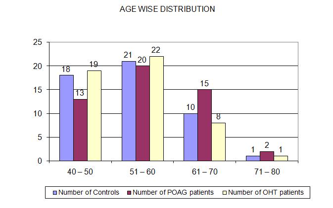
69 67
70 Table 1B AGE RANGE Age in years Minimum Maximum Controls POAG OHT Controls ranged between 41 to 72 years, in POAG and OHT ranges were 40 to 80 years and are thus comparable. Descriptive Statistics (p = 0.130) Group N Range Minimum Maximum Mean SD Control Age POAG Age OHT Age
71 Table 2 SEX DISTRIBUTION AMONG STUDY GROUPS Number of Subjects Males Female Controls POAG OHT The gender distribution with in the study group showed a near equal distribution among the POAG and OHT, but slightly lesser number of males in control group. No measures were taken to ensure that equal number of males and females participated in the study. 69
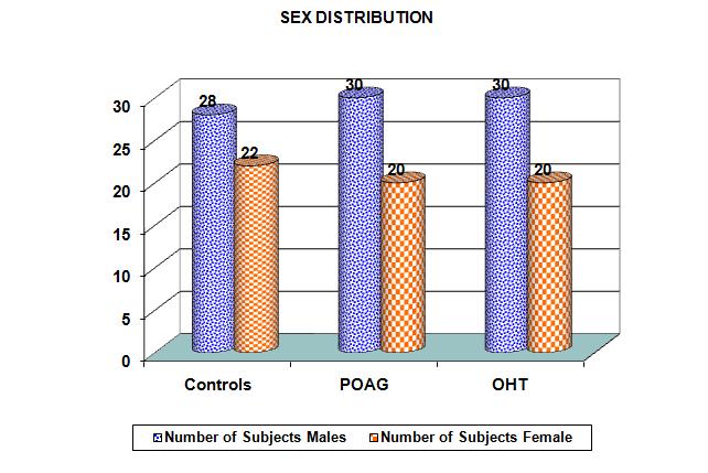
72 70
73 Table 3 MEAN CCT AND MEAN IOP CCT Mean (µm) IOP Mean (mmhg) Normal POAG OHT T Test Group Number of Mean SD P value eyes CCT Controls POAG CCT Controls <0.001 OHT CCT POAG <0.001 OHT Using t test, there was no statistically significant difference of mean CCT of POAG when compared to control. (p = 0.448) In the ocular hypertension group, we saw a significantly higher central corneal thickness than in the control subjects. (p<0.001) Comparing OHT with POAG group, OHT group was found to have significantly higher CCT than in POAG (p < 0.001) 71
74 72
75 T Test Group Number of Mean SD P value eyes IOP Controls <0.001 POAG IOP Controls <0.001 OHT IOP Controls <0.001 OHT Using T test, mean IOP was found to be significantly higher in POAG group when compared to controls (p < 0.001) Mean IOP was significantly higher in OHT group when compared to controls (p<0.001) Mean IOP was significantly higher in OHT group when compared to POAG. (p<0.001) 73
76 Table 4 MEAN CCT AND RANGE Central Corneal Thickness (µm) Mean Range Normal POAG OHT The mean CCT in the control group was 533 µm with a range µm In the POAG group, the mean was 536 µm with a range of µm And in the OHT group, the mean was 561 µm with a range µm, which were again comparable. 74
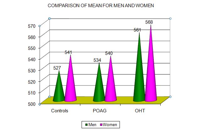
77 Table 5 COMPARISON OF MEAN FOR MEN AND WOMEN Men Women Controls POAG OHT T Test Group Sex Number Mean SD P value Controls CCT Male Female POAG CCT Male Female OHT CCT Male Female There was no statistical difference in CCT values between men and women in POAG & ocular hypertension group. In controls group CCT of women was higher. Using T test, p value for 3 groups are Controls POAG OHT
78 76
Glaucoma: Diagnostic Modalities
 Glaucoma: Diagnostic Modalities - Dr. Barun Kumar Nayak, Dr. Sarika Ramugade Glaucoma is a leading cause of blindness in the world, especially in older people. Early detection and treatment by ophthalmologist
1/31/2018. Course Objectives. Diagnostic Testing. Optic Nerve Damage ANATOMY AND PHYSIOLOGY OF A GLAUCOMA WORK-UP/TONOMETRY TECHNICIAN: -SDP
 ANATOMY AND PHYSIOLOGY OF A GLAUCOMA WORK-UP/TONOMETRY KNOW THE DISEASE PROCESS TECHNICIAN: EXPLAIN PROCESS OF EXAMINATION Presenters: Dana McMahan, COA Nicole Smith, COA Engage with patient s, help alleviate
Intro to Glaucoma/2006
 Intro to Glaucoma/2006 Managing Patients with Glaucoma is Exciting Interesting Challenging But can often be frustrating! Clinical Challenges To identify patients with risk factors for possible glaucoma.
Corporate Medical Policy
 Corporate Medical Policy Optical Coherence Tomography (OCT) Anterior Segment of the Eye File Name: Origination: Last CAP Review: Next CAP Review: Last Review: optical_coherence_tomography_(oct)_anterior_segment_of_the_eye
MEDICAL POLICY SUBJECT: CORNEAL ULTRASOUND PACHYMETRY. POLICY NUMBER: CATEGORY: Technology Assessment
 MEDICAL POLICY SUBJECT: CORNEAL ULTRASOUND,, PAGE: 1 OF: 5 If a product excludes coverage for a service, it is not covered, and medical policy criteria do not apply. If a commercial product, including
The Role of the RNFL in the Diagnosis of Glaucoma
 Chapter 1. The Role of the RNFL in the Diagnosis of Glaucoma Introduction Glaucoma is an optic neuropathy characterized by a loss of of retinal ganglion cells and their axons, the Retinal Nerve Fiber Layer
Interpretation of corneal tomography
 Interpretation of corneal tomography Presented by Chameen Samarawickrama - Westmead Hospital - Liverpool Hospital - University of Sydney - University of New South Wales The University of Sydney Page 1
INTRODUCTION S. HERDENER, D. HAFIZOVIC, M. PACHE, S. LAUTEBACH, J. FUNK. University Eye Hospital, Freiburg - Germany
 European Journal of Ophthalmology / Vol. 18 no. 1, 2008 / pp. 39-43 Is the PASCAL -Tonometer suitable for measuring intraocular pressure in clinical routine? Long- and short-term reproducibility of dynamic
Central corneal thickness determined with optical coherence tomography in various types of glaucoma
 Br J Ophthalmol 2000;84:1233 1237 1233 Department of Ophthalmology, Ludwig-Maximilians- University, Munich, Germany M Bechmann M J Thiel B Roesen S Ullrich M W Ulbig K Ludwig Correspondence to: Martin
True IOP No Doubt Facts and Figures Figures. Ziemer Ophthalmic Systems AG a Ziemer Group Company Allmendstrasse 11 CH-2562 Port, Switzerland
 True IOP No Doubt Facts and Figures Figures and Facts Ziemer Ophthalmic Systems AG a Ziemer Group Company Allmendstrasse 11 CH-2562 Port, Switzerland There is increasing evidence that DCT measures IOP
3/16/2018. Ultrasound Biomicroscopy in Glaucoma By Ahmed Salah Abdel Rehim. Prof. of Ophthalmology Al-Azhar University
 Ultrasound Biomicroscopy in Glaucoma By Ahmed Salah Abdel Rehim Prof. of Ophthalmology Al-Azhar University 1 Ultrasound biomicroscopy (UBM) is a recent technique to visualize anterior segment with the
Dr. Harvey Richman, OD, FAAO, FCOVD Diplomate American Board of Optometry Executive Committee AOA Third Party Center Founder Ask the AOA Coding
 Dr. Harvey Richman, OD, FAAO, FCOVD Diplomate American Board of Optometry Executive Committee AOA Third Party Center Founder Ask the AOA Coding Experts 92000 Codes Special Ophthalmological Services Describe
Learn Connect Succeed. JCAHPO Regional Meetings 2017
 Learn Connect Succeed JCAHPO Regional Meetings 2017 Faculty Biometry and IOL Calculations ASCRS and ASOA Symposium and Congress Los Angeles, CA Daniel H. Chang, M.D. - Empire Eye and Laser Center Bakersfield,
LARGE DISCS WITH LARGE CUPS A DIAGNOSTIC CHALLENGE IN AFRICAN PATIENTS. Darshana Soma
 LARGE DISCS WITH LARGE CUPS A DIAGNOSTIC CHALLENGE IN AFRICAN PATIENTS Darshana Soma A research report submitted to the Faculty of Health Sciences, University of the Witwatersrand, Johannesburg, in partial
Assisting in Ophthalmology. Copyright 2011, 2007, 2003, 1999 by Saunders, an imprint of Elsevier Inc. All rights reserved.
 Assisting in Ophthalmology Learning Objectives Define, spell, and pronounce the terms listed in the vocabulary. Apply critical thinking skills in performing patient assessment and care. Explain the differences
NIIOS. Cornea Specialist, Melles Cornea Clinic, NIIOS, Rotterdam, the Netherlands Naval Hospital, Athens, Greece VASILIS S.
 Cornea Specialist, Melles Cornea Clinic, NIIOS, Rotterdam, the Netherlands Naval Hospital, Athens, Greece Goldman applanation tonometry is still the gold standard for measuring IOP, but its accuracy is
Correlating central corneal thickness and intraocular pressure in ocular hypertension and glaucoma
 VOL. 3 NO. 1 PHILIPPINE JOURNAL OF Ophthalmology JANUARY ORIGINAL ARTICLE JUNE 07 Jonathan G. Soriano, MD 1 Ma. Margarita L. Lat-Luna, MD 1, 3 Patricia M. Khu, MD 1, 1 Department of Ophthalmology and Visual
Corporate Medical Policy
 Corporate Medical Policy Glaucoma, Evaluation by Ophthalmologic Techniques File Name: Origination: Last CAP Review: Next CAP Review: Last Review: glaucoma_evaluation_by_ophthalmologic_techniques 3/2001
Training Checking Vision Tonometry
 Training 101-2 Checking Vision Tonometry Checking Vision The classic example of an eye chart is the Snellen eye chart, in general they show 11 rows of capital letters. The top row contains one letter (usually
GLAUCOMA SUMMARY BENCHMARKS FOR PREFERRED PRACTICE PATTERN GUIDELINES
 SUMMARY BENCHMARKS FOR PREFERRED PRACTICE PATTERN GUIDELINES Introduction These are summary benchmarks for the Academy s Preferred Practice Pattern (PPP) guidelines. The Preferred Practice Pattern series
The pinnacle of refractive performance.
 The pinnacle of refractive performance. WaveLight REFRACTIVE PORTFOLIO Advancing REFRACTIVE SURGERY Contoura Vision sets a new standard in LASIK outcomes More than 98% of patients would choose it again.
Accuracy and Repeatability of a New Portable Ultrasound Pachymeter
 Accuracy and Repeatability of a New Portable Ultrasound Pachymeter Queirós A 1, González-Méijome JM 1, Fernandes P 1, Jorge J 1, Almeida J B 1, Parafita MA 2 1 Department of Physics (Optometry), School
PREVALENCE OF GLAUCOMA AMONG FISHERMEN COMMUNITY OF MUNDRA TALUKA OF KUTCH DISTRICT- A CROSS- SECTIONAL STUDY
 ORIGINAL RESEARCH PREVALENCE OF GLAUCOMA AMONG FISHERMEN COMMUNITY OF MUNDRA TALUKA OF KUTCH DISTRICT- A CROSS- SECTIONAL STUDY Sanjay Upadhyay 1, Jayantilal Shah 2 1 Assistant Professor, 2 Associate Professor,
Role of Central Corneal Thickness in Circadian Intraocular Pressure Fluctuations among Patients with Primary Open Angle Glaucoma
 Role of Central Corneal Thickness in Circadian Intraocular Pressure Fluctuations among Patients with Primary Open Angle Glaucoma Mohannad Albdour MD*, Karanjit Kooner MD, PHD** ABSTRACT Objectives: To
Goals. Glaucoma PARA PEARL TO DO. Vision Loss with Glaucoma
 Glaucoma Janet R. Fett, OD Drs. Kincaid, Fett and Tharp So Sioux City, NE eyewear21@hotmail.com Goals Understand Glaucoma Disease process Understand how your data (objective and subjective) assists in
5/18/2014. Fundamentals of Gonioscopy Workshop Aaron McNulty, OD, FAAO Walt Whitley, OD, MBA, FAAO
 1 Fundamentals of Gonioscopy Workshop Aaron McNulty, OD, FAAO Walt Whitley, OD, MBA, FAAO 2 3 4 5 6 Optometry s Meeting 2014 The Most Valuable Glaucoma Tool Glaucoma Diagnosis Gonioscopy Central corneal
Cataract Surgery in the Patient with a History of LASIK or PRK
 Cataract Surgery in the Patient with a History of LASIK or PRK #56996-RS April 2018 Sebastian Lesniak, MD Matossian Eye Associates None Disclosures Bio Matossian Eye Associates, Hopewell NJ, 7/2015 Present
THE CHRONIC GLAUCOMAS
 THE CHRONIC GLAUCOMAS WHAT IS GLAUCOMA? People with glaucoma have lost some of their field of all round vision. It is often the edge or periphery that is lost. That is why the condition can be missed until
Medical Policy An independent licensee of the Blue Cross Blue Shield Association
 Pachymetry Page 1 of 8 Medical Policy An independent licensee of the Blue Cross Blue Shield Association Title: Pachymetry Professional Institutional Original Effective Date: March 11, 2004 Original Effective
Macular Ganglion Cell Complex Measurement Using Spectral Domain Optical Coherence Tomography in Glaucoma
 Med. J. Cairo Univ., Vol. 83, No. 2, September: 67-72, 2015 www.medicaljournalofcairouniversity.net Macular Ganglion Cell Complex Measurement Using Spectral Domain Optical Coherence Tomography in Glaucoma
THE CHRONIC GLAUCOMAS
 THE CHRONIC GLAUCOMAS WHAT IS GLAUCOMA People with glaucoma have lost some of their field of all round vision. It is often the edge or periphery that is lost. That is why the condition can be missed until
Comparison of a New Non-Contact Tonometer with Goldmann Applanation
 Eye (1989) 3, 332-337 Comparison of a New Non-Contact Tonometer with Goldmann Applanation M. J. MOSELEY, N. M. EVANS and A. R. FIELDER Leicester Summary A comparison of a new non-contact tonometer (Keeler
LOCSU Community Services. Glaucoma Repeat Readings & OHT Monitoring Community Service Pathway. Issued by Local Optical Committee Support Unit May 2009
 LOCSU Community Services Glaucoma Repeat Readings & OHT Monitoring Community Service Pathway Issued by Local Optical Committee Support Unit May 2009 [Revised November 2013] Contents Page Executive Summary...
Comparison of Central Corneal Thickness Measurements with Pentacam, Orbscan II, and Ultrasound Pachymeter
 Comparison of Central Corneal Thickness Measurements with Pentacam, Orbscan II, and Ultrasound Pachymeter Abbas-Ali Yekta, PhD 1 Hassan Hashemi, MD 2,3 Mehdi KhabazKhoob, MSc 3 Asghar Dostdar, MSc 4 Shiva
RECENT ADVANCES IN ANTERIOR SEGMENT IMAGING OF THE EYE. by Eszter Szalai, M.D. Supervisor: László Módis, M.D., Ph.D.
 SHORT THESIS FOR THE DEGREE OF DOCTOR OF PHILOSOPHY (Ph.D.) RECENT ADVANCES IN ANTERIOR SEGMENT IMAGING OF THE EYE by Eszter Szalai, M.D. Supervisor: László Módis, M.D., Ph.D. UNIVERSITY OF DEBRECEN DOCTORAL
Written by Administrator Wednesday, 13 January :27 - Last Updated Thursday, 21 January :34
 angle closure glaucoma A type of glaucoma caused by a sudden and severe rise in eye pressure. Occurs when the pupil enlarges too much or too quickly, and the outer edge of the iris blocks the eye s drainage
3/23/2016. Regina Smolyak, MD Glaucoma Service Flaum Eye Institute
 Under the Affordable Care Act providers are pressured to maintain a high level of medical care, while reducing the cost of the treatment delivered Regina Smolyak, MD Glaucoma Service Flaum Eye Institute
Immersion Vs Contact Biometery for Axial Length Measurement before Phacoemulsification
 Original Article Immersion Vs Contact Biometery for Axial Length Measurement before Phacoemulsification with Foldable IOL Irum Abbas, Atif Mansoor Ahmad, Tahir Mahmood Pak J Ophthalmol 2009, Vol. 25 No.
Peretyagin O.A., Cand. Med. Sc., Ass. Prof.; Dmitriev S.K., Dr. Med. Sc., Prof.; Lazar Yu.M., Cand. Med. Sc.; Tatarina Yu.A., Junior Research Fellow
 Changes in corneoscleral rigidity and corneal thickness at various target intraocular pressures in patients with stabilized primary open-angle glaucoma Peretyagin O.A., Cand. Med. Sc., Ass. Prof.; Dmitriev
53 year old woman attends your practice for routine exam. She has no past medical history or family history of note.
 Case 1 Normal Tension Glaucoma 53 year old woman attends your practice for routine exam. She has no past medical history or family history of note. Table 1. Right Eye Left Eye Visual acuity 6/6 6/6 Ishihara
Effect of corneal parameters on measurements using the pulsatile ocular blood flow tonograph and Goldmann applanation tonometer
 518 EXTENDED REPORT Effect of corneal parameters on measurements using the pulsatile ocular blood flow tonograph and Goldmann applanation tonometer P Gunvant, M Baskaran, L Vijaya, I S Joseph, R J Watkins,
L. Spadea, R. Ferrante, F. Romani, A. Di Gregorio
 University of L Aquila Eye Clinic Head: Prof. Leopoldo Spadea ULTRAFAST EXCIMER LASER FOR TRANS-EPITHELIAL CUSTOMIZED PHOTOREFRACTIVE SURGERIES: CLINICAL RESULTS WITH 6 MONTHS FOLLOW UP L. Spadea, R. Ferrante,
GLAUCOMA REPEAT READINGS PATHWAY
 GLAUCOMA REPEAT READINGS PATHWAY Level 1a: Goldmann style applanation tonometry repeat readings A first level community service for IOP refinement where other signs of glaucoma are not present will reduce
The Common Clinical Competency Framework for Non-medical Ophthalmic Healthcare Professionals in Secondary Care
 The Common Clinical Competency Framework for Non-medical Ophthalmic Healthcare Professionals in Secondary Care Glaucoma November 2016 Association of Health Professions in Ophthalmology General basic competences
Kathryn L. Wotman, DVM, DACVIM Jeffrey E. Bowersox, DVM, DACVO. Veterinary Specialty Center of Delaware
 Kathryn L. Wotman, DVM, DACVIM Jeffrey E. Bowersox, DVM, DACVO Veterinary Specialty Center of Delaware Contact Lensesuse in Veterinary Ophthalmology -Corneal erosions Spontaneous Chronic Corneal Epithelial
PRECISION PROGRAM. Injection Technique Quick-Reference Guide. Companion booklet for the Video Guide to Injection Technique
 Injection Technique Quick-Reference Guide PRECISION PROGRAM Companion booklet for the Video Guide to Injection Technique Available at www.ozurdexprecisionprogram.com Provides step-by-step directions with
CLINICAL SCIENCES. Effect of Central Corneal Thickness, Corneal Curvature, and Axial Length on Applanation Tonometry
 CLINICAL SCIENCES Effect of Central Corneal Thickness, Corneal Curvature, and Axial Length on Applanation Tonometry Markus Kohlhaas, MD; Andreas G. Boehm, MD; Eberhard Spoerl, PhD; Antje Pürsten, Dipl-Ing
PRE-DESCEMET S ENDOTHELIAL KERATOPLASTY (PDEK) DR ASHVIN AGARWAL
 DR ASHVIN AGARWAL") PRE-DESCEMET S ENDOTHELIAL KERATOPLASTY (PDEK) DR ASHVIN AGARWAL Endothelial keratoplasty (EK) has evolved at a brisk pace and the volume of data accumulated over the past 10 years has demonstrated that
PRE-DESCEMET S ENDOTHELIAL KERATOPLASTY (PDEK) DR ASHVIN AGARWAL Endothelial keratoplasty (EK) has evolved at a brisk pace and the volume of data accumulated over the past 10 years has demonstrated that
The Egyptian Journal of Hospital Medicine (January 2018) Vol. 70 (8), Page
 Vol. 70 (8), Page") The Egyptian Journal of Hospital Medicine (January 2018) Vol. 70 (8), Page 1371-1376 Relationship Between Corneal Astigmatism and Intraocular Pressure Abdulrahman E. Algarni 1, Mohammad Abahussin 2, Nuha
The Egyptian Journal of Hospital Medicine (January 2018) Vol. 70 (8), Page 1371-1376 Relationship Between Corneal Astigmatism and Intraocular Pressure Abdulrahman E. Algarni 1, Mohammad Abahussin 2, Nuha
The Sense Organs 10/13/2016. The Human Eye. 1. Sclera 2. Choroid 3. Retina. The eye is made up of three layers:
 The human body gathers information from the outside world by using the five senses of: The Sense Organs 12.3 Sight Hearing Taste Smell Touch This information is essential in helping the body maintain homeostasis.
Informed Consent for Excimer Laser Surface Ablation Surgery (PRK, LASEK, epi-lasik, and others)
") Informed Consent for Excimer Laser Surface Ablation Surgery (PRK, LASEK, epi-lasik, and others) Patient name (printed): Patient date of birth: Please review this information so you can make an informed
Informed Consent for Excimer Laser Surface Ablation Surgery (PRK, LASEK, epi-lasik, and others) Patient name (printed): Patient date of birth: Please review this information so you can make an informed
Noel de Jesus Atienza, MD, MSc and Joseph Anthony Tumbocon, MD
 Original Article Philippine Journal of OPHTHALMOLOGY Diagnostic Accuracy of the Optical Coherence Tomography in Assessing Glaucoma Among Filipinos. Part 1: Categorical Outcomes Based on a Normative Database
Exceptional versatility without compromise
 Introducing the VICTUS femtosecond laser platform Exceptional versatility without compromise FEMTOSECOND TECHNOLOGY that empowers Introducing VICTUS the first femtosecond laser capable of exceptional performance
PRESCRIBING IN GLAUCOMA: GUIDELINES FOR NZ OPTOMETRISTS
 PRESCRIBING IN GLAUCOMA: GUIDELINES FOR NZ OPTOMETRISTS Introduction Independent prescribing relates to the capacity to use clinical judgement in respect of diagnosis and treatment. It does not mean working
Central corneal thickness (CCT) measurement is a critical
 measurement is a critical") ARTICLE Topographic Paracentral Corneal Thickness With Pentacam and Orbscan: Effect of Acoustic Factor Javier González-Pérez, O.D., M.Sc., Ph.D., Jose M. González-Méijome, O.D., Ph.D., María T. Rodríguez
ARTICLE Topographic Paracentral Corneal Thickness With Pentacam and Orbscan: Effect of Acoustic Factor Javier González-Pérez, O.D., M.Sc., Ph.D., Jose M. González-Méijome, O.D., Ph.D., María T. Rodríguez
Central Corneal Thickness-An important variable for prognostication in Primary Open Angle glaucoma; A Kolkata based study in Eastern India
 Original article: Central Corneal Thickness-An important variable for prognostication in Primary Open Angle glaucoma; A Kolkata based study in Eastern India 1Dr. Apala Bhattacharya, 2 Dr Gautam Bhaduri,
Role of central corneal thickness measurement in management of open angle glaucoma and glaucoma suspects in Calabar, Nigeria
 Original Research Article Role of central corneal thickness measurement in management of open angle glaucoma and glaucoma suspects in Calabar, Nigeria Nkanga DG 1,2, Ibanga AA 1,2, Nkanga ED 2, Etim BA
LOCSU Community Services. Glaucoma Repeat Readings & OHT Monitoring Community Service Pathway. Issued by Local Optical Committee Support Unit May 2009
 LOCSU Community Services Glaucoma Repeat Readings & OHT Monitoring Community Service Pathway Issued by Local Optical Committee Support Unit May 2009 [Revised May 2016, Version 3.3] Contents Page Executive
FITTING GUIDE & TIPS FOR ACHIEVING SUCCESS
 AT M hybrid contact lens for astigmatism FITTING GUIDE & TIPS FOR ACHIEVING SUCCESS AT M hybrid contact lens for astigmatism Contents Overview 2 Patient Candidates 2 Key Features of SynergEyes A 3 SynergEyes
CORNEAL CONDITIONS CORNEAL TRANSPLANTATION
 GENERAL INFORMATION CORNEAL CONDITIONS CORNEAL TRANSPLANTATION WHAT ARE CORNEAL CONDITIONS? The cornea is the clear outer layer of the eye. Shaped like a dome, it helps to protect the eye from foreign
The Effect of Pupil Dilation on Scanning Laser Polarimetry With Variable Corneal Compensation
 C L I N I C A L S C I E N C E The Effect of Pupil Dilation on Scanning Laser Polarimetry With Variable Corneal Compensation Amjad Horani, MD; Shahar Frenkel, MD, PhD; Eytan Z. Blumenthal, MD BACKGROUND
Targeting Intraocular Pressure in Glaucoma: a Teaching Case Report 1
 Targeting Intraocular Pressure in Glaucoma: a Teaching Case Report 1 By: Andrew Kemp, OD, Marcus Gonzales, OD, FAAO, Joe DeLoach, OD, FAAO, and Zanna Kruoch, OD FAAO Background Glaucoma is a range of conditions
3/23/2016. Diagnostic Services Taylor Pannell CRA, OCT-C. Services Available. Important info for the Tech to know. Visual Fields
 Services Available Diagnostic Services Taylor Pannell CRA, OCT-C Static and Kinetic Visual Fields Pachymetry Anterior and Posterior Segment OCT Fundus Photos FAF,FA,ICG Slit Lamp Photography Confocal HRT
Are traditional assessments a waste of time? NZAO 2015
 Are traditional assessments a waste of time? NZAO 2015 Disclosures No financial interests other than Optometry Practice owner Full time optometrist Not a glaucoma prescriber ODOB Board Chair Previously
PHOTOREFRACTIVE KERATECTOMY (PRK) PATIENT INFORMATION BOOKLET
 PATIENT INFORMATION BOOKLET") 616.365.5775 www.keillasik.com PHOTOREFRACTIVE KERATECTOMY (PRK) PATIENT INFORMATION BOOKLET Please read this entire booklet. Discuss its contents with your doctor so that questions are answered to your
616.365.5775 www.keillasik.com PHOTOREFRACTIVE KERATECTOMY (PRK) PATIENT INFORMATION BOOKLET Please read this entire booklet. Discuss its contents with your doctor so that questions are answered to your
Populations Interventions Comparators Outcomes Individuals: Who are being evaluated for angleclosure
 Protocol Optical Coherence Tomography of the Anterior Eye Segment (90318) Medical Benefit Effective Date: 07/01/14 Next Review Date: 05/18 Preauthorization No Review Dates: 07/11, 07/12, 07/13, 05/14,
Clinical Study Evaluation of the New Digital Goldmann Applanation Tonometer for Measuring Intraocular Pressure
 Ophthalmology, Article ID 461681, 5 pages http://dx.doi.org/1.1155/214/461681 Clinical Study Evaluation of the New Digital Goldmann Applanation Tonometer for Measuring Intraocular Pressure Yuta Sakaue,
Collaboration in the care of glaucoma patients and glaucoma suspects. Barry Emara MD FRCS(C) Nico Ristorante November 29, 2012
 Nico Ristorante November 29, 2012") Collaboration in the care of glaucoma patients and glaucoma suspects Barry Emara MD FRCS(C) Nico Ristorante November 29, 2012 Goals of Collaboration Patient-centred and evidence based approach Timely access
Collaboration in the care of glaucoma patients and glaucoma suspects Barry Emara MD FRCS(C) Nico Ristorante November 29, 2012 Goals of Collaboration Patient-centred and evidence based approach Timely access
Early Detection Of Glaucoma Clinical Clues. Points To Live By. Glaucoma Risk Factors 10/3/2014
 Early Detection Of Glaucoma Clinical Clues Eric E. Schmidt, O.D. Omni Eye Specialists Wilmington, NC schmidtyvision@msn.com Points To Live By 25% of G pxs NEVER have IOP >21mm 50% of G pxs have trough
A Review Of Risk Factors. Early Detection Of Glaucoma Clinical Clues. A risk factor analysis is critical. Points To Live By
 A Review Of Risk Factors Early Detection Of Glaucoma Clinical Clues Eric E. Schmidt, O.D. Omni Eye Specialists Wilmington, NC schmidtyvision@msn.com FINDACAR Family history IOP Nearsightedness Diabetes/Vascular
Clinical Guidance and Monitoring for Change. Cecilia Fenerty MD FRCOphth Manchester Royal Eye Hospital
 Clinical Guidance and Monitoring for Change Cecilia Fenerty MD FRCOphth Manchester Royal Eye Hospital Glaucoma Referral Criteria 2000 Original referral scheme Simple criteria based on IOP/Disc/Field Solitary
FEP Medical Policy Manual
 FEP Medical Manual 9.03.05 Corneal Topography/Computer-Assisted Corneal Topography/ Photokeratoscopy Last Review: September 2016 Next Review: September 2017 Related Policies 9.03.28 Corneal Collagen Cross-linking
PRACTICAL APPROACH TO MEDICAL MANAGEMENT OF GLAUCOMA
 PRACTICAL APPROACH TO MEDICAL MANAGEMENT OF GLAUCOMA DR. RAVI THOMAS, DR. RAJUL PARIKH, DR. SHEFALI PARIKH IJO MAY 2008 PRESENTER AT JDOS : DR. RAHUL SHUKLA T.N. SHUKLA EYE HOSPITAL TERMINOLOGY POAG: PRIMARY
Dina H. Erickson, O.D., a Denise Goodwin, O.D., a Michael Rollins, O.D., b Amber Belaustegui, O.D., c and Chad Anderson a
 Optometry (2009) 80, 169-174 Comparison of dynamic contour tonometry and Goldmann applanation tonometry and their relationship to corneal properties, refractive error, and ocular pulse amplitude Dina H.
OPHTHALMOLOGY AND ULTRASOUND
 Vet Times The website for the veterinary profession https://www.vettimes.co.uk OPHTHALMOLOGY AND ULTRASOUND Author : JAMES OLIVER Categories : Vets Date : April 28, 2008 JAMES OLIVER discusses why ultrasound
Correspondence should be addressed to Fusako Fujimura;
 BioMed Research International Volume 2013, Article ID 370592, 5 pages http://dx.doi.org/10.1155/2013/370592 Research Article Repeatability and Reproducibility of Measurements Using a NT-530P Noncontact
Keratoconus Clinic. Optometric Co-management Opportunities
 Keratoconus Clinic Optometric Co-management Opportunities The Bochner Eye Institute established the first Keratoconus Clinic in Canada in 2008. The consultation and advanced imaging are OHIP covered. All
Central corneal thickness among glaucoma patients attending Menelik II Hospital, Addis Ababa, Ethiopia
 Original article Central corneal thickness among glaucoma patients attending Menelik II Hospital, Addis Ababa, Ethiopia Zelalem Gizachew 1, Abiy Mulugeta 1* Abstract Background: Glaucoma is one of the
Meet Libby. Corneal Dysgenesis, Degeneration, and Dystrophies Definitions. Dr. Victor Malinovsky
 Meet Libby Corneal Dysgenesis, Degeneration, and Dystrophies 2006 Dr. Victor Malinovsky Definitions Dysgenesis: (congenital anomalies) A development disorder that results in a congenital malformation of
Around The Globe in 60 Minutes
 Around The Globe in 60 Minutes Around the GLOBE in Sixty Minutes Basic Ocular Anatomy, Examination, and Diagnostic Techniques Introduction Focusing on canine and feline ocular anatomy and basic examination
Effect of brimonidine on intraocular pressure in normal tension glaucoma: A short term clinical trial
 European Journal of Ophthalmology / Vol. 13 no. 7, 2003 / pp. 611-615 Effect of brimonidine on intraocular pressure in normal tension glaucoma: A short term clinical trial S.A. GANDOLFI, L. CIMINO, P.
Tonometry Through The Ages
 Tonometry Through The Ages Tonometrie im Wandel der Zeit Habilitationsschrift zur Erlangung der venia legendi auf dem Gebiet der Ophthalmologie vorgelegt von Dr. Christoph Kniestedt Zürich 2007 Kniestedt,
Abdel Rahman ElSebaey, MD, PhD.
 Surface Ablation Refractive Surgery Abdel Rahman ElSebaey, MD, PhD. Menoufia University History Correction of optical defects of human eye started 1200 AD. Spherical error corrected by spectacle on 13
Andrew Francis, MSIII Boston University School of Medicine Unite For Sight Chapter President and Global Impact Volunteer ACCRA, GHANA
 Andrew Francis, MSIII Boston University School of Medicine Unite For Sight Chapter President and Global Impact Volunteer ACCRA, GHANA Acknowledgements Thank you My project is funded by a grant from the
NEW YORK UNIVERSITY SCHOOL OF MEDICINE DEPARTMENT OF OPHTHALMOLOGY EDUCATIONAL OBJECTIVES AND GOALS
 NEW YORK UNIVERSITY SCHOOL OF MEDICINE DEPARTMENT OF OPHTHALMOLOGY EDUCATIONAL OBJECTIVES AND GOALS Revision Date: 6/30/06 Distribution Date: 7/6/06 The Department of Ophthalmology at the NYU Medical Center
Retinal Nerve Fiber Analysis: the Role It Plays in Assessing Glaucoma Grace Martin, CPOT
 Retinal Nerve Fiber Analysis: the Role It Plays in Assessing Glaucoma Grace Martin, CPOT Every day, patients experiencing visual difficulties are seen in the optometric practice. This can range from a
The Relative Frequency and Risk Factors of Primary Open Angle Glaucoma and Angle Closure Glaucoma
 Original Article The Relative Frequency and Risk Factors of Primary Open Angle Glaucoma and Angle Closure Glaucoma Naila Ali, Syed Ali Wajid, Nasir Saeed, Muhammad Daud Khan Pak J Ophthalmol 07, Vol. 23
Glaucoma. Cornea. Iris
 Glaucoma Introduction Glaucoma is a group of eye diseases that can lead to blindness if not treated. Openangle glaucoma, the most common form of glaucoma, affects about 3 million Americans. Half of those
Ocular Studies of EMF Exposure at the MMW M. Kojima 1,2,3), Y. Suzuki 4)
, Y. Suzuki 4)") Ocular Studies of EMF Exposure at the MMW M. Kojima 1,2,3), Y. Suzuki 4) 1. Division of Vision Research for Environmental Health, Medical Research Institute, Kanazawa Medical University 2. Department of
Ocular Studies of EMF Exposure at the MMW M. Kojima 1,2,3), Y. Suzuki 4) 1. Division of Vision Research for Environmental Health, Medical Research Institute, Kanazawa Medical University 2. Department of
Glaucoma Clinical Update. Barry Emara MD FRCS(C) Giovanni Caboto Club October 3, 2012
 Giovanni Caboto Club October 3, 2012") Glaucoma Clinical Update Barry Emara MD FRCS(C) Giovanni Caboto Club October 3, 2012 Objectives Understand the different categories of glaucoma Recognize the symptoms and signs of open angle and angle-closure
Glaucoma Clinical Update Barry Emara MD FRCS(C) Giovanni Caboto Club October 3, 2012 Objectives Understand the different categories of glaucoma Recognize the symptoms and signs of open angle and angle-closure
Comparison of icare Tonometer with Pulsair and Tonopen in Domiciliary Work
 Optometry in Practice Vol 6 (2005) 33 39 Comparison of icare Tonometer with Pulsair and Tonopen in Domiciliary Work Dawn E C Roberts BSc(Hons) MSc MCOptom Healthcall Optical Services, Luton, Bedfordshire,
RETINAL CONDITIONS RETINAL CONDITIONS
 GENERAL INFORMATION RETINAL CONDITIONS RETINAL CONDITIONS WHAT ARE RETINAL CONDITIONS? Retinal conditions affect the light-sensitive tissue at the back of eye known as the retina. They include diseases
he Role of UBM and Anterior Segment OCT in Anterior Segment Imaging
 Ophthalmic Instrumentation T he Role of UBM and Anterior Segment OCT in Anterior Segment Imaging M. Chockalingam DNB FRCS PGDHM N. V. Arulmozhi Varman MS Since its development and usage, Ultrasound biomicroscopy
3/16/2018. Perimetry
 Perimetry The normal visual field extends further away from fixation temporally and inferiorly than superiorly and nasally. From the center of the retina this sensitivity decreases towards the periphery,
Ultrasound Evaluation of the Posterior Segment of the Eye A Ready Reckoner
 180 Kerala Journal of Ophthalmology Vol. XX, No. 2 OPHTHALMIC INSTRUMENTATION Ultrasound Evaluation of the Posterior Segment of the Eye A Ready Reckoner Dr. Mahesh G. MS DO DNB FRCSEd., Dr. A. Giridhar
Jose M. Martinez-de-la-Casa, Julian Garcia-Feijoo, Ana Fernandez-Vidal, Carmen Mendez-Hernandez, and Julian Garcia-Sanchez METHODS
 Ocular Response Analyzer versus Goldmann Applanation Tonometry for Intraocular Pressure Measurements Jose M. Martinez-de-la-Casa, Julian Garcia-Feijoo, Ana Fernandez-Vidal, Carmen Mendez-Hernandez, and
CLINICAL SCIENCES. Clinical Significance of Central Corneal Thickness in the Management of Glaucoma
 CLINICAL SCIENCES Clinical Significance of Central Corneal Thickness in the Management of Glaucoma Carolyn Y. Shih, MD; Joshua S. Graff Zivin, PhD; Stephen L. Trokel, MD; James C. Tsai, MD Objective: To
The second most common causes of blindness worldwide. ( after cataract) The commonest cause of irreversible blindness in the world Estimated that 3%
 The commonest cause of irreversible blindness in the world Estimated that 3%") The second most common causes of blindness worldwide. ( after cataract) The commonest cause of irreversible blindness in the world Estimated that 3% of our population age > 40 have glaucoma In the past:
The second most common causes of blindness worldwide. ( after cataract) The commonest cause of irreversible blindness in the world Estimated that 3% of our population age > 40 have glaucoma In the past:
CLASS-y Laser Treats Glaucoma
 Article # 404 Comments About the Author Released: Author: Category: March 12th, 2014 Issue #0314 Ehud Assia Feature S S S S S CLASS-y Laser Treats Glaucoma Transforming complex, invasive and risky glaucoma
Pearls for the Refractive Technician Fadiah Alkhawaldeh, IMBA, COT, ROUB
 Pearls for the Refractive Technician Fadiah Alkhawaldeh, IMBA, COT, ROUB Cleveland Clinic Cole Eye Institute OOS, Columbus, OH February, 2014 alkhawf@ccf.org NO FINANCIAL DISCLOSURES A Puzzle of an Eye