Glomerular Aging and Focal Global Glomerulosclerosis: A Podometric Perspective
|
|
|
- Leo Johnson
- 6 years ago
- Views:
Transcription
1 Glomerular Aging and Focal Global Glomerulosclerosis: A Podometric Perspective Jeffrey B. Hodgin,* Markus Bitzer, Larysa Wickman, Farsad Afshinnia, Su Q Wang, Christopher O Connor,* Yan Yang, Chrysta Meadowbrooke, Mahboob Chowdhury, Masao Kikuchi, Jocelyn E. Wiggins, and Roger C. Wiggins *Departments of Pathology, Internal Medicine, and Pediatrics and Communicable Diseases, University of Michigan, Ann Arbor, Michigan ABSTRACT Kidney aging is associated with an increasing proportion of globally scarred glomeruli, decreasing renal function, and exponentially increasing ESRD prevalence. In model systems, podocyte depletion causes glomerulosclerosis, suggesting age-associated glomerulosclerosis could be caused by a similar mechanism. We measured podocyte number, size, density, and glomerular volume in 89 normal kidney samples from living and deceased kidney donors and normal poles of nephrectomies. Podocyte nuclear density decreased with age due to a combination of decreased podocyte number per glomerulus and increased glomerular volume. Compensatory podocyte cell hypertrophy prevented a change in the proportion of tuft volume occupied by podocytes. Young kidneys had high podocyte reserve (podocyte density.300 per 10 6 mm 3 ), but by years of age, average podocyte nuclear density decreased to,,100 per 10 6 mm 3, with corresponding podocyte hypertrophy. In older age podocyte detachment rate (urine podocin mrna-to-creatinine ratio) was higher than at younger ages and podocytes were stressed (increased urine podocin-to-nephrin mrna ratio). Moreover, in older kidneys, proteinaceous material accumulated in the Bowman space of glomeruli with low podocyte density. In a subset of these glomeruli, mass podocyte detachment events occurred in association with podocytes becoming binucleate (mitotic podocyte catastrophe) and subsequent wrinkling of glomerular capillaries, tuft collapse, and periglomerular fibrosis. In kidneys of young patients with underlying glomerular diseases similar pathologic events were identified in association with focal global glomerulosclerosis. Podocyte density reduction with age may therefore directly lead to focal global glomerulosclerosis, and all progressive glomerular diseases can be considered superimposed accelerators of this underlying process. J Am Soc Nephrol 26: , doi: /ASN Biologic aging, the process by which time-associated changes take place in living cells and tissues, is amenable to scientific analysis. With such analysis it can be broken down into its constituent elements and manipulated to change its rate and, thereby, the aging process itself. Chronologic age, on the other hand, is a fixed time dimension that for practical purposes cannot be altered. Aging-associated structural changes in the kidney and glomerulus have been repeatedly noted for nearly 100 years. 1 5 Glassock and Rule provide a comprehensive contemporary review. 6 The prevalence of glomerulosclerosis increases with age. 2 6 Renal pathologists use an equation ([age/2]210) as a guide to determine whether the proportion of sclerotic glomeruli present in a biopsy exceeds a normal expected range. 4 As glomeruli become increasingly sclerosed, downstream tubules atrophy/ involute so that glomerular density (number per remaining volume of cortex) increases, interstitial Received August 7, Accepted February 8, J.B.H., M.B., and L.W. contributed equally to this work. Published online ahead of print. Publication date available at. Correspondence: Dr. Roger C. Wiggins, 1570B MSRB2, 1150 West Medical Center Drive, Ann Arbor, MI rwiggins@umich.edu Copyright 2015 by the American Society of Nephrology 3162 ISSN : / JAmSocNephrol26: , 2015

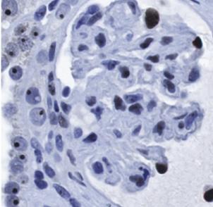
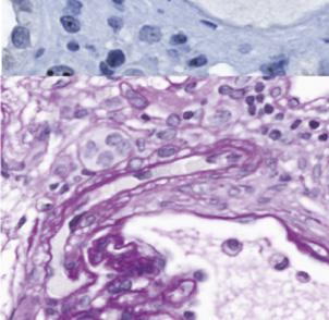
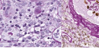
2 fibrosis accumulates, and kidneys eventually lose mass. 3,6 In 1949, Davies and Shock reported that kidney function decreased with age, 7 as has been confirmed by most subsequent studies. 5,8,9 The egfr decreases with age at an average rate of about 0.8% per year detectable after age 30 years. 5 9 With the advent of RRTs in combination with increasing life expectancy, the true effect of the aging kidney has become all too obvious. ESRD prevalence increases exponentially with age, and age is the strongest known risk factor for ESRD by far. 10,11 This age-associated structurefunction-outcome triad implies a direct biologic effect of aging on the kidney, particularly on the glomerulus, whose underlying mechanism(s) are not well understood. It has become clear from model systems that depletion of the long-lived postmitotic neuron-like cell of the kidney glomerulus, the podocyte, causes glomerulosclerosis in direct proportion to the degree of depletion FSGS resulting from podocyte injury and loss can occur via a series of pathologic steps defined by Kriz and colleagues Glomerular tuft podocyte density (number per volume), the determinant of whether glomerulosclerosis occurs, can become reduced through (1) loss of podocytes per se (via detachment or cell death), (2) glomerular tuft enlargement (so that each podocyte becomes responsible for an increased area of filtration surface), and (3) podocyte dysfunction or phenotype switch (wherein the podocyte no longer performs its normal functions). 19 Observational studies in human glomerular diseases also demonstrate an association between podocyte density decrease and outcome in diabetic glomerulosclerosis, IgA nephropathy, hypertensive glomerulosclerosis, and transplant glomerulopathy We previously reported age-related reduction in podocyte density (shown as increased glomerular volume per podocyte) associated with podocyte hypertrophic stress and failure leading to glomerulosclerosis in Fischer344 rats. 28 Prevention of age-associated glomerular enlargement achieved by reduced calorie intake prevented development of glomerulosclerosis, thereby demonstrating that age-related glomerulosclerosis could potentially be modulated. 28 To determine whether simple podocyte depletion per se could cause glomerulosclerosis, we developed the hdtr transgenic rat, in which a predetermined number of podocytes could be selectively eliminated at a predetermined time. 13 Using this model we proved that podocyte depletion per se quantitatively caused all features of glomerulosclerosis with an appearance similar to that seen in humans. 13 To form an alternative way of causing relative podocyte depletion that would be more analogous to aging, we developed the podocin promoter-driven AA-4EBP1 transgenic rat. In this model, podocytes are selectively prevented from undergoing normal hypertrophy while all other cells can undergo hypertrophy normally. 15 Using this model we proved that glomerular enlargement itself drives glomerulosclerosis in a podocyte-dependent manner. 15 These model studies therefore tell us that glomerulosclerosis can be caused by podocyte depletion initiated by apparently completely different mechanisms that do not involve the immune system and that have in common reduced podocyte density and increased podocyte stress. In the rat these processes appear to drive aging-associated glomerulosclerosis. 28 Because reduced podocyte density causes glomerulosclerosis in models and is associated with glomerulosclerosis in human glomerular diseases, the question arises as to whether glomerular aging in humans might also be associated with decreased podocyte density. A newly developed morphometric approach to measure podocyte parameters ( podometrics ) applicable to archival kidney biopsy material offers the opportunity to test this hypothesis. 29 RESULTS Figure 1 illustrates methods used to estimate podocyte density, number, size, glomerular volume, and other morphometric variables. To span the human age range, we analyzed 89 kidneys that were within normal pathologic limits identified by conventional light microscopic pathologic analysis. The cohort was composed of deceased kidney transplant donors age 4 18 years (n=20) and age years (n=11), living kidney transplant donors age years (n=29), and the normal pole of nephrectomies for cancer age years (n=29). Figure 1. Podometric methodology. The upper panels show TLE4-red fluorescent podocyte nuclei (red) in glomerular profiles from young (A), middle-aged (B), and older (C) people. The lower panels show the same sections for which the coverslip has been removed and peroxidase immunocytochemistry performed to delineate the Glepp1 peroxidase positive area (brown) occupied by podocytes. Calibrated photomicrographs of the TLE4 immunofluorescent sections are used to measure podocyte nuclear mean caliper diameter. Photomicrographs of the Glepp1-stained sections are used to measure glomerular tuft area and the percentage of the tuft area occupied by podocytes. These primary data measured in the histologic section are then used to estimate podocyte nuclear density, glomerular volume, number of podocyte nuclei per tuft, Glepp1-positive tuft volume, Glepp1- negative tuft volume, and podocyte cell volume. The bar shows 100 mm. J Am Soc Nephrol 26: , 2015 Glomerular Aging 3163
3 Podocyte nuclear density decreased about 0.9% per year from.300 per 10 6 mm 3 at,20 years of age to, mm 3 by years of age (P,0.001) (Figure 2A). Glomerular volume per podocyte, the average territory each podocyte is responsible for managing, increased by 2.7% per year (P,0.001) (Figure 2B). These changes with aging resulted from a combination of (1) reduced podocyte nuclear number per glomerulus (slope, 20.34% per year; P=0.002) (Figure 2C) and (2) increased glomerular tuft volume (slope, 1.6% per year; P,0.001) (Figure 2D). Table 1 shows slope data for these variables. Table 2 shows data by age group across all cohorts to emphasize the continuous change with aging and to provide ranges. Table 3 shows that excluding the normal kidney pole data, the data from older deceased donors, or both of these groups did not significantly change the podocyte density slope or correlation. Although podocyte nuclear density decreased with age, the Glepp1-positive podocyte cytoplasmic area expressed as percentage of the tuft area occupied by podocytes did not change with age (Figure 3A). Furthermore, when glomerular volume is considered, the total podocyte cell volume (mass) significantly increased with age at a rate of 1.8% per year (P,0.001) (Figure 3B). The increased total podocyte cell mass and decreased podocyte nuclear number per glomerulus mean that Figure 2. Aging-associated changes in podocyte density resulting from changes in podocyte nuclear number per tuft and glomerular volume. (A) Podocyte nuclear density (number per tuft volume). (B) Reciprocal glomerular volume per podocyte. The underlying causes of the density change are a reduction in podocyte number per glomerular tuft (C) and an increase in glomerular volume (D). Table 1 provides slopes and statistical information for these data using bivariate linear regression models Journal of the American Society of Nephrology J Am Soc Nephrol 26: , 2015
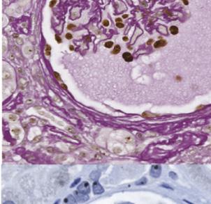
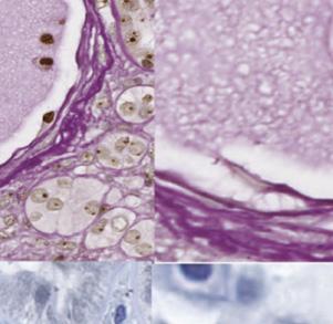
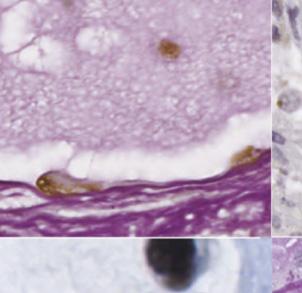
4 Table 1. Glomerular morphometric variables by age Variable c at Age = 0 m Error of Estimate P Value Podocyte density (n per 10 6 mm 3 ) ,0.001 Glomerular volume per podocyte (310 3 ) ,0.001 Podocytes per glomerular tuft (n) Glomerular volume (310 6 mm 3 ) ,0.001 Glepp1-positive tuft area (%) Glepp1-positive tuft volume (310 6 mm 3 ) ,0.001 Glepp1-negative tuft volume (310 6 mm 3 ) ,0.001 Mean podocyte cell volume (310 3 mm 3 ) ,0.001 Mean podocyte nuclear volume (310 3 mm 3 ) ,0.001 Data shown as the linear relationship with age for each measured parameter to allow a normal range for any parameter at any age to be calculated according to y=mx+c, where y is the parameter of interest, x is the age, m is the coefficient of x, and c is the intercept at age=0. The error of the estimate is shown. the average podocyte cell volume increased with age (increase of 3.2% per year; P,0.001) (Figure 3C). Podocyte nuclear volume also increased with age at a rate of 2% per year (P,0.001) (Figure 3D), being highly correlated with podocyte cell volume (R 2 =0.63) (Figure 3E). This relationship between cell volume and nuclear volume is well known as the karyoplasmic ratio 30,31 and independently confirms the age-related changes noted above. Podocyte nuclear size is therefore a surrogate for cell size. In contrast nonpodocyte cell nuclei did not increase in size with age (Figure 3F) reflecting the fact that glomerular endothelial cells and mesangial cells proliferate in response to hypertrophic stress whereas podocytes have to hypertrophy. Figure 4 shows how as podocyte nuclear density decreases from.300 to,100 per 10 6 mm 3,podocytecellsizemust increase by as much as 6-fold. At low podocyte density, small further decrements require larger and ever-increasing podocyte hypertrophic responses that would be projected to eventually result in podocyte hypertrophic stress. To test this projection, we measured the amount of podocyte detachment in urine pellet samples from 277 people age 3 92 years with normal urine/kidney function and urine proteinto-creatinine ratio #0.18 (Figure 5). The group older than age 60 years had significantly higher urine pellet podocin mrnato-creatinine ratio (3.3-fold; P,0.001). The urine podocinto-aquaporin2 mrna ratio was also higher in this age group, demonstrating preferential podocyte loss versus distal nephron cells (expressing aquaporin2). Therefore, the increased podocin mrna is the result of preferential podocyte detachment rather than overall nephron loss in older age. Furthermore, podocyte stress was also increased in the older group, as assessed by comparing two mrna species from the same cell that we have previously reported as reflecting podocyte stress (the urine podocin-to-nephrin mrna ratio). 32 Thus, in older age, podocytes are more stressed and podocytes are detaching in greater amounts. We used Glepp1 peroxidase-stained histologic sections to search for putative short-lived intermediates between normal glomeruli and globally sclerosed glomeruli that from the preceding data would be expected to be present in old but not young kidneys. Several potential intermediates were identified (Figure 6, Table 4). Proteinaceous matrix in the Bowman space (PMBS) was present in a proportion of.60-year-old glomeruli (Figures 6A and 7B) and rarely present in young glomeruli. This suggests the possibility that glomeruli with hypertrophic stressed podocytes might be leaking protein into the Bowman space. To test this concept, we measured the podocyte nuclear density in kidneys with a high proportion of glomeruli with PMBS and found that the average density was significantly lower in these kidneys compared with other old kidneys (Figure 8A). The average podocyte nuclear volume in glomeruli containing PMBS was also significantly larger than in neighboring glomeruli in the same kidney section that did not contain PMBS (Figure 8B). These data are compatible with PMBS reflecting higher level podocyte hypertrophy and stress in the subset of glomeruli with leaky glomeruli. Some glomeruli with PMBS also had podocyte-derived subcellular fragments trapped within the proteinaceous matrix identified by being Glepp1 positive (Figure 6B). A subset of these glomeruli also had podocyte-derived homogeneous periodic acid Schiff (PAS) positive glycocalyx material associated with the PMBS that was both Glepp1 and podocalyxin positive by immunoperoxidase (Figure 6, C E). A further subset of glomeruli with PMBS had multiple detached podocytes present within the PMBS (mass podocyte detachment events [MPDEs]) as demonstrated by the cells being Glepp1 and podocalyxin positive and containing WT1-positive nuclei (Figures 6, H F, and 7, C H). On average, 50% of these detached podocytes were binucleate or multinucleate. This result is compatible with a catastrophic mitosis-driven podocyte detachment process as suggested by Lasagni and colleagues. 33 Evaluation of PMBS glomeruli undergoing mass podocyte detachment events showed that glomerular capillaries had become wrinkled and appeared to be in various stages of tuft collapse in association with development of periglomerular fibrosis (Figure 9, C K). Five kidney biopsy specimens from younger people with diverse underlying clinical conditions in which we had also noted MPDEs were reviewed (Tables 5 and 6). All specimens showed low podocyte density and binucleate podocytes in the Bowman space (Figure 9, Table 6), together with wrinkled GBM, glomerular tuft collapse, and focal global glomerulosclerosis (FGGS), but not FSGS. None of the biopsy specimens showed arteriosclerosis (Figure 9, A and P). Therefore, the same pattern of glomerular injury occurred in these nonaging biopsy specimens in association with MPDEs. The term benign nephrosclerosis has historically been used to describe pathologic changes associated with aging and culminating in FGGS. 3,34 The mechanism behind this J Am Soc Nephrol 26: , 2015 Glomerular Aging 3165
5 Table 2. Glomerular morphometric characteristics by age group Variable Age 18 yr (Column A) Comparison Age yr (Column B) Comparison Age yr (Column C) Comparison Age >60 yr (Column D) Comparison All Ages Average age (yr) Patients (n) Podocyte density (n per 10 6 mm 3 ) Mean B, a C, b D b A, a C, a D b A, b B, a D c A, b B, b C c Range Glomerular volume per podocyte (310 3 ) Mean B, C, b D b A, C, b D b A, b B b A, b B b Range Podocytes per glomerular tuft (n) Mean C, c D a D a A c A, a B c Range Glomerular volume (310 6 mm 3 ) Mean C, b D b C, a D b A, b B a A, b B b Range Glepp1-positive tuft area (%) Mean Range Glepp1-positive tuft volume (310 6 mm 3 ) Mean C, b D b C, a D b A, b B a A, b B b Range Glepp1-negative tuft volume (310 6 mm 3 ) Mean C, c D b C, c D b A, a B c A, b B a Range Mean podocyte cell volume (310 3 mm 3 ) Mean B, a C, b D b A, a C, b D b A, b B, b D b A, b B b Range Mean podocyte nuclear volume (310 3 mm 3 ) Mean C, b D b C, b D b A, b B, b D a A, b B, b C a Range Data are shown as the mean61 SD and range. ANOVA with Bonferroni correction for multiple comparisons was applied to compare means. Statistical comparisons are shown by column designation (below) where each age group is compared with all other age groups. The average number of glomerular tufts evaluated per biopsy was (total of 2154 glomeruli). a P,0.01. b P, c P, Journal of the American Society of Nephrology J Am Soc Nephrol 26: , 2015
6 Table 3. Podocyte density decrease with age persists in subgroup analysis Group designation Samples (n) Slope Correlation r P Value All samples 89 y= x 20.67,0.001 All: nephrectomy samples 60 y= x 20.66,0.001 All: older deceased donor kidneys 78 y= x 20.67,0.001 All: both nephrectomy and older deceased donor 49 y= x 20.53,0.001 Changes in podocyte density with age persist after elimination from analysis either the nephrectomy normal pole samples, which may have been affectedbykidney cancer related effects (All: nephrectomy samples), or by elimination of the older deceased donor samples, which might have been affected by vascular diseaseor other factors (All: older deceased donor kidneys), or both of these groups (All: both nephrectomy and older decreased donors) from the analysis. Slope dataare shown in a y=mx+c format where y is the podocyte density per 10 6 mm 3 and x is the age in years. The Pearson correlation r is shown. process has been thought to be ischemic and is generally assumed to be secondary to arteriosclerosis. To better define these relationships, we therefore evaluated PAS-stained sections from 44 nephrectomy samples aged.60 years containing 8570 glomerular profiles. Sections were scored for arteriosclerosis, FGGS, ischemic-like glomerulopathy (see Concise Methods), and podocyte stress/detachment events (PMBS and MPDEs). Multivariate analysis (Figure 10) showed that the ischemic-like glomerulopathy phenotype did not correlate with arteriosclerosis but did correlate significantly with both podocyte detachment events and FGGS. This result supports the concept developed from two independent data sets shown above (Figures 8 and 9) that podocyte detachment events are mechanistically related to glomerulopathy with wrinkled GBM, collapsing tufts, and periglomerular fibrosis, which, in turn, results in FGGS. Figure 10 also shows that arteriosclerosis correlates significantly with FGGS independent of podocyte detachment events. From bivariate analysis shown in Figure 10, we conclude that in this older kidney cohort the relative contributions of arteriosclerosis and podocyte depletion to net FGGS were approximately equal. DISCUSSION This report shows that as normal human glomeruli age, the average podocyte (nuclear) density progressively decreases from.300 per 10 6 mm 3 at young age to,100 per 10 6 mm 3 by about years of age because of the combined effects of reduced podocyte number per glomerulus and increasing glomerular volume. As podocyte density decreases, further density decrements mandate larger and ever-increasing podocyte hypertrophic responses. For a cell as complex as the podocyte, with its requirement to maintain contiguous interdigitating foot processes with its neighbors to completely cover the filtration surface area, this represents an important logistic challenge that at some point will result in podocyte hypertrophic stress. Consistent with this concept, the amount of podocyte detachment increased in older age. This increase was not merely due to loss of whole nephrons because the urine podocin-to-aquaporin2 mrna ratio was also increased, thereby demonstrating that podocytes were being preferentially lost relative to distal tubule and collecting duct cells that contain aquaporin2 mrna. Furthermore, the urine pellet podocin-to-nephrin mrna ratio of two podocyte-specific markersfromthesamecell,whichwehave previously reported as an index of podocyte stress, 32 was also increased in older kidneys. This represents preferential downregulation of nephrin mrna expression previously documented to occur in aging rat glomeruli and in association with hypertrophic podocyte stress. 14,28 Microscopic examination of old kidneys (but not young kidneys) showed candidates for direct evidence for podocyte stress and accelerated detachment in glomeruli. A subset of glomeruli could be identified with PMBS to form an apparently semi-stable matrix. Kidneys with large numbers of PBMS glomeruli had lower podocyte density, and within these kidneys the PMBS glomeruli had larger podocyte nuclei than neighboring glomeruli without PMBS. This finding suggests that compensatory podocyte hypertrophy had reached especially high levels in these leaky glomeruli. Some PMBS glomeruli also showed detached podocyte-derived subcellular particles (containing the apical podocyte markers podocalyxin and Glepp1) trapped within the proteinaceous cast, suggesting that stressed podocytes can detach subcellular elements, as has previously been identified in glomerular disease urine. 35,36 Additionally, podocytes themselves detached and were retained in the proteinaceous matrix. Some glomeruli exhibited large numbers of detached podocytes (MPDEs) wherein about 50% of the detached podocytes were binucleate. In many glomeruli binucleate podocytes were also present in glomeruli, potentially accounting for the increase in average podocyte nuclear size. Prior reports have also identified binucleate/multinucleate podocytes in glomerular disease urine. 37,38 Glomeruli that were undergoing MPDEs also had wrinkled GBM and collapsing glomerular tufts associated with periglomerular fibrosis, compatible with the concept that mass podocyte detachment itself resulted in glomerular tuft collapse. In a small cohort of younger kidneys with diverse underlying glomerular diseases we similarly identified MPDEs associated with low podocyte density, mitotic cells in the Bowman space, glomerular tuft collapse, and the FGGS phenotype (but not the FSGS phenotype). Therefore, glomerular tuft collapse appears to result from podocyte depletion occurring (1) mass podocyte detachment (as described above) or (2) as a result of a changed podocyte phenotype. In the latter scenario, HIV infection (or other mechanisms) causes podocytes to lose their normal markers and foot processes and to J Am Soc Nephrol 26: , 2015 Glomerular Aging 3167
7 Figure 3. Podocyte cell hypertrophy compensates for reduction in podocyte density with age. Glepp1 area density does not change with successful aging (A). However, the volume occupied by podocytes (Glepp1 percentage area3glomerular volume) does increase with aging (B). The average podocyte cell volume (Glepp1 volume/podocyte nuclear number per tuft) increases with age (C). Average podocyte nuclear volume also increases with aging (D). Podocyte cell volume and nuclear volume are highly correlated (E). In contrast, 3168 Journal of the American Society of Nephrology J Am Soc Nephrol 26: , 2015
8 Figure 4. Podocyte density/size plot providing a mechanistic explanation for podocyte hypertrophic failure associated with aging. A plot of podocyte density against cell size with the mean and 95% confidence limits demonstrates how a reduction in podocyte density with aging necessitates an exponential (inversely proportional) increase in cell size to maintain the filtration barrier. At high podocyte densities (younger age), a density decrease is easily accommodated by small increases in podocyte size. However, at a density,100 per 10 6 mm 3 (by older age), small decrements in podocyte density require large and ever-increasing compensatory changes in cell size, thereby driving hypertrophic stress and detachment. Best fit curve estimation revealed a power curve with R 2 =0.93. become proliferative cells that cannot serve normal podocyte functions (collapsing FSGS variant). 39,40 Tuft collapse is therefore a common downstream event caused by loss of normal podocytes resulting from podocyte detachment (detachmentassociated collapse) or proliferation (proliferation-associated collapse). These data are consistent with the concept that within a glomerular population at any one time, a subset of glomeruli can develop critically low podocyte density as a consequence of aging or glomerular injury. As podocytes become hypertrophically stressed, they leak protein across the filter to form a matrix-like structure in the Bowman space, detach subcellular fragments, and/or attempt to divide. Lasagni and colleagues 33 have suggested that the rearrangement of the actin cytoskeleton required for podocyte cell division is incompatible with podocytes maintaining their normal function and adhesion to the GBM. Mitotic podocytes therefore detach (catastrophic mitotic podocyte detachment). 33 Cho and colleagues used a theoretical construct to predict that detachment of one podocyte would result in sequential detachment of neighboring podocytes through a cooperative mechanism. 41 Support for this concept is provided by experimental models from Ichikawa and colleagues (podocyte damage damages podocytes) and Cui and colleagues (innocent bystander hypothesis) and by our studies showing that once a critical proportion of podocytes are lost the remaining podocytes autonomously detach in an angiotensin II dependent manner leading to global glomerulosclerosis. 14,42,43 The mechanism of FGGS associated with aging (so-called benign nephrosclerosis) and its relationship to BP has been controversial. 3,34,44 The concept that glomerular lesions were ischemic was not supported by the India ink perfusion studies of autopsied kidneys performed by Shapiro; these studies found that ischemic glomeruli were particularly well perfused, thereby proving that obstructive arteriopathy could not be the cause of ischemic changes. 45 However, age-associated glomerular lesions are commonly called ischemic, with a presumed arteriosclerotic association, 34 although the precise mechanism by which ischemia occurs is not well understood. To evaluate these concepts we scored 8570 glomeruli from 44 old kidneys and found that the presumptive ischemic-like glomeruli did not correlate with arteriosclerosis, as would have been expected if the lesions were caused by underlying arterial disease. Instead, the ischemia-like phenotype correlated with podocyte detachment events. The relationship between podocyte detachment and collapsing FGGS is supported in three ways from these data: (1) glomeruli with detaching podocytes showed wrinkling of glomerular tufts and collapsing glomerulopathy; (2)inasubsetof youngerkidneys weidentified the same association of mass podocyte detachment and collapsing glomerulopathy in the absence of arterial disease; and (3) by multivariate analysis we showed that arteriosclerosis did not correlate with the ischemic-like phenotype. In our analysis arteriosclerosis appeared to independently contribute to the FGGS phenotype. We speculate that NFkB signaling (which we previously identified as a major pathway in old glomeruli and is known to be a driver of atherosclerosis) could contribute to net FGGS, but further studies are required to evaluate this possibility. 44,46 This would be analogous to Alzheimer disease and vascular disease as the two major average glomerular nonpodocyte nuclear volumes do not change with aging (F). Table 1 provides slopes and statistical information for these data. The data shown in Figures 2 and 3 are for male and female patients combined, using linear regression modeling. Analysis by sex showed no statistical difference between sexes (or trends toward differences) for any of the preceding variables (data not shown). J Am Soc Nephrol 26: , 2015 Glomerular Aging 3169
9 Figure 5. Urine pellet podocyte mrna markers by age showing accelerated detachment and podocyte stress in the group older than age 60 years. (A) The urine podocin mrna-to-creatinine ratio is a measure of the rate of podocyte detachment relative to urine creatinine excretion analogous to the urine protein-to-creatinine ratio. The urine podocin mrna-to-creatinine ratio in the.60- year-old group is increased 3.3-fold above the mean of younger people (,60 years; P,0.001). (B) The urine podocin-to-aquaporin2 mrna ratio is a measure of the rate of podocyte detachment relative to detachment of another kidney cell type (distal tubule and collecting duct cells that express aquaporin2 mrna) demonstrating that podocytes are lost preferentially to other kidney cells in the.60-year-old age group. (C) The urine pellet podocin-tonephrin mrna ratio is a ratio of two podocyte-specific markers that contributors to dementia often working in parallel but contributing variable amounts to net brain dysfunction. 47 Kidney aging must also involve other vascular, genetic, epigenetic, and environmental processes as outlined by Striker. 48 The data suggest that glomeruli can be considered to have a life cycle (Figure 11). During normal life, if an individual glomerulus density decreases enough (because of podocyte loss or glomerular volume increase), it eventually reaches a critical stage at which podocytes are forced into mitosis and thereby undergo podocyte detachment followed by glomerular tuft collapse, resulting in FGGS. In normal kidneys the MPDE intermediate is rare and short-lived (observable in only 0.2% of 11,358 old glomeruli in the two old kidney cohorts examined by different methods, and probably much rarer in younger glomeruli), but in pathologic conditions can be much higher (mean of 8.7% in five biopsy cases with low podocyte density shown in Table 5). The rare MPDE in a normal kidney cortex is therefore analogous to a supernova in the life cycle of stars. It is a rarely observed but very important event toward understanding the star life cycle. MPDEs seem to result in subsequent glomerular tuft collapse through presumed vasoconstrictor mechanisms, which rapidly limit blood flow into a glomerulus and thereby protein loss into the filtrate. In contrast, segmental sclerosis may occur when podocyte depletion occurs at a slower rate, allowing remaining podocytes to migrate together (regroup) to protect regions (capillary loops) of the glomerular filter while other segments are abandoned to sclerosis. This latter scenario occurs in many glomerular diseases commonly affecting younger people, whose podocytes will be less hypertrophied and therefore more agile and adaptable. Although these longerlived semi-stable intermediates (e.g., diabetic glomerulosclerosis and FSGS variants) do preserve glomerular filtration, their disorganized glomerular structure is unable to efficiently prevent protein leaking from blood into the filtrate. Steffes and colleagues also reported an increase in average glomerular volume with age but did not report a decreased podocyte number per tuft. 22 The average slope of reduced podocyte density about 0.9% per year is similar to the average rate of reduced egfr reported in apparently normal humans (about 0.8% per year). 5 9 Variation in the density slope between individuals would explain differences in agingassociated rates of progression, as has been reported by Fliser and colleagues. 9 Huber and colleagues report that podocyte replacement capacity is minimal in the adult mouse. 49 Interpretation of the data presented requires the assumption that the kidneys analyzed are representative of the we have previously reported as a measure of podocyte stress. 32 Distribution of male and female patients did not significantly differ in the various age groups. The box plots show median and interquartile ranges, and the corresponding means were compared using ANOVA with Bonferroni correction. Outliers outside the mean62sd range are shown as circles and those outside the mean63sd range are shown as asterisks Journal of the American Society of Nephrology J Am Soc Nephrol 26: , 2015
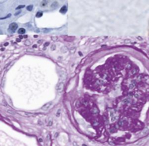
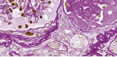
 A subset of old glomeruli develop Glepp1-negative proteinaceous material in the Bowman space (arrowhead).")
10 Figure 6. Old kidneys show stages of podocyte stress and detachment by Glepp1 and podocalyxin immunoperoxidase histochemistry. Images from.60-year-old kidneys. (A) A subset of old glomeruli develop Glepp1-negative proteinaceous material in the Bowman space (arrowhead). (B) Glepp1-positive subcellular cell particles detach from podocytes and are present within the proteinaceous matrix in the Bowman space (arrowhead). (C) The proteinaceous matrix in the Bowman space becomes Glepp1 positive, suggesting loss of glycocalyx material from podocytes. (D F) Glepp1-positive podocytes detach from the glomerulus and appear within proteinaceous matrix material in the Bowman space. (G) Glomeruli become globally scarred but still contain remnants of Glepp1. (H) Similar staining is present for podocalyxin, as confirmation that two independent podocyte antibodies show the same patterns. (I) Control immunoperoxidase staining showing that the observed peroxidase product was specific. normal population. Potential limitations include the following: (1) Deceased donors do not necessarily reflect a random sample of the population; (2) living donors are carefully selected and therefore do not represent a random population sample; and (3) the unaffected pole of a kidney containing cancer could theoretically be affected by predisposing genetic, environmental, or growth factors that lead to cancer or the cancer milieu itself. Furthermore, these kidneys were selected as lacking major stigmata of kidney disease at the light microscopic level and therefore may not have been representative of a random population sample. Nevertheless, the overall study represents a reasonable approximation of a normal kidney sample over a wide age range, and the density slope was not significantly altered by excluding the normal kidney pole or the older deceased donor groups from the analysis. The association of podocyte detachment events with FGGS was also confirmed in non-nephrectomy samples. In summary, the data provided support a hypothesis wherein reduction in podocyte density throughout life in humans causes hypertrophic podocyte stress in some glomeruli, eventually resulting in glomerular tuft collapse and FGGS. This FGGS mechanism may be a significant cause of ESRD in both the aging kidney and diverse glomerular diseases. According to this scenario, whether adequate numbers of glomeruli successfully survive for a lifetime will depend on (1) cumulative podocyte loss, (2) glomerular volume increase, and (3) the ability of podocytes to successfully adapt to hypertrophic stress. Each of these variables could potentially be modulated by diet and therapeutic intervention. All glomerular diseases can be seen as superimposed upon this underlying mechanism. CONCISE METHODS Use of archival kidney tissues from biopsies from deceased donors, living donors, and the uninvolved pole from nephrectomies for cancer was approved by the University of Michigan Institutional Review Board (HUM , HUM ). Kidney Sample Selection Pathologic light microscopic analysis was used as the gold standard for inclusion or exclusion of samples. Samples were included if kidney structure in all tissue compartments available for analysis was within normal limits, as assessed by conventional light microscopy in the Table 4. (n=89) Proportion of globally sclerotic and transitional glomeruli per age group observed in Glepp1-peroxidase sections Variable Age <19 yr (Column A) Age yr (Column B) Age yr (Column C) Age >60 yr (Column D) Glomeruli evaluated (n) FGGS (%) a b b c Transitional glomeruli (%) a a d d Podocyte mass detachment events (%) Podocyte glycocalyx detachment (%) Values expressed with a plus/minus sign are the mean6sd. FGGS was definedaccordingto Figure 6I. Transitional glomeruliwere defined accordingto Figure 6, B F. Transitional glomeruli were any glomerulus that had proteinaceous material and/or detached cells in the Bowman space. Podocyte mass detachment events contained at least three detached Glepp1-positive cells in the Bowman space. Podocyte glycocalyx detachment events contained definitely Glepp1-positive material in the proteinaceous cast in the Bowman space, as shown in Figure 7. a P,0.05 vs. columns C and D. b P,0.05 vs. column D. c P,0.05 vs. columns A, B, and C. d P,0.05 vs. columns A and B. J Am Soc Nephrol 26: , 2015 Glomerular Aging 3171


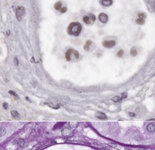
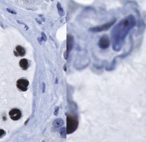
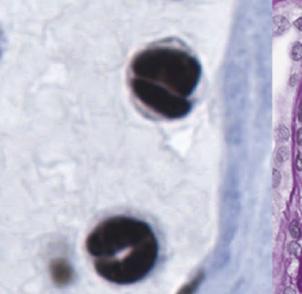
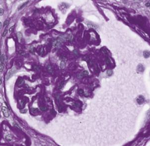
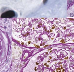

11 Figure 7. Detaching podocytes are multinucleate and associated with wrinkling of the GBM, glomerular collapse, and periglomerular fibrosis. (A) Normal PAS-stained old glomerulus with peroxidase-stained WT1-positive podocyte nuclei. (B) Proteinaceous material in the Bowman space is PAS positive (arrowhead) and the glomerulus shows a wrinkled appearance with early periglomerular layering. (C) WT1-positive nuclei present within the PAS-positive proteinaceous material in the Bowman space (arrowhead). (D and E) WT1-positive 3172 Journal of the American Society of Nephrology J Am Soc Nephrol 26: , 2015

12 kidney biopsy for transplants or deidentified kidney sections from the uninvolved poles of kidneys removed for cancer. All kidney transplants were biopsied at the time of implantation. Kidney biopsy specimens from living donors were from people screened and selected to ensure that underlying diseases that could compromise the health of the donor or recipient were absent (age years; n=29). To extend the age range above and below the living donor group, we used groups of deceased donors from younger (4 18 years, n=20) and older (48 64 years; n=11) ages. Deceased donors are approved as kidney donors if they had normal kidney function before death (n=31). To extend the older age range further, we used kidney sections obtained from the normal pole of nephrectomies performed for cancer (age range, years; n=29). These deidentified kidney samples (for which only age and sex information was available) were selected as having no pathologic stigmata of kidney disease. Additional clinical information about preexisting diseases in this cohort was not available. Therefore, we cannot exclude the possibility that some deidentified samples could have been from people with diabetes or hypertension but without kidney abnormalities. Because the Glepp1 area density did not change with age (as shown in Figure 3, Table 1), we used it to define a lower range of normal for Glepp1 percentage area density (mean22sd=33.0). One sample fell below this normal range and was excluded from further analysis. One additional biopsy specimen had fewer than eight glomerular profiles present and was therefore excluded on the basis of criteria for adequate estimate of glomerular volume, as outlined below and previously reported. 29 Podometric Analysis The TLE4 immunofluorescent method for estimating podocyte nuclear density 29 was combined with a Glepp1 immunoperoxidase method for measuring podocyte cell area density in the same section, as previously reported. 27 A total of 2154 glomeruli were evaluated in 89 biopsy specimens ( per biopsy). For PAS-stained sections ( ; Sigma-Aldrich, St. Louis, MO), podocyte nulcei were identified by immunoperoxidase using a murine monoclonal anti-wt1 antibody (LS-C105719) from LifeSpan BioSciences Inc. (Seattle, WA). Podocyte Nuclear Density by TLE4 Immunofluorescence The method used was as reported elsewhere. 27 Glepp1 Positive Percentage Area by Immunoperoxidase This immunoperoxidase method was used as previously reported 27 to identify podocyte cell area. It is piggy-backed onto the TLE4 Figure 8. Glomeruli with proteinaceous material in the Bowman space have reduced podocyte density and larger podocyte nuclei. (A) Podocyte density. Kidney sections in which a large proportion of glomeruli had PMBS (n=5) (PMBS.30%) had significantly lower podocyte density than glomeruli that did not have a large proportion of glomerular tufts with protein in the Bowman space (n=15). (B) Podocyte nuclear volume as a surrogate for podocyte cell volume: WT1-positive podocyte nuclear volume was measured in glomeruli that contained Bowman space proteinaceous cast material (PMBS) versus neighboring glomeruli in the same kidney section that did not have PMBS. The average podocyte nuclear volume in glomeruli containing PMBS was larger ( mm 3 ; n=609) than in neighboring glomeruli in the same kidney sections that did not contain PMBS ( mm 3 ; n=554) (P,0.01), compatible with greater podocyte hypertrophy in the subset of glomeruli associated with PMBS. immunofluorescent method outlined earlier so that podocyte nuclear data from the identical slide can be combined with the peroxidase method for estimation of podocyte cell size. Because the sections are podocyte nuclei within the Bowman space (arrowhead). Podocyte nuclei of cells remaining attached to the glomerular tuft (arrowheads) are shown at higher power to be binucleate (E, arrowheads). (F) Detached podocytyes are binucleate (arrowheads). (G) Glomerulus with binucleate detached WT1-positive cells are present in the Bowman space (G, arrowhead) and remain attached to the glomerular tuft (H, arrowheads). (H) High-power view of panel G to illustrate binucleate cells (arrowheads) and wrinkled glomerular capillary loops (arrow). (I) Glomerulus with detached podocytes in the Bowman space (shown by arrowheads) that is wrinkled and collapsed (arrow). (J) PAS-stained section showing a glomerulus at an earlier stage with detached podocytes in the Bowman space (arrowheads) at left and a glomerulus with major tuft collapse (arrow) at right. (K) PAS-stained section with WT1 peroxidase again showing three glomeruli. The upper glomerulus appears normal. The left glomerulus shows detached podocytes (arrowhead), proteinaceous material in the Bowman space, and periglomerular layering. The right glomerulus shows major glomerular tuft collapse and global glomerulosclerosis. The average glomerular tuft diameter is 137 micrometers. J Am Soc Nephrol 26: , 2015 Glomerular Aging 3173


 PAS-stained kidney biopsy from a 52-year-old kidney")


, proteinaceous")
")
 in panel D.")
 space and proteionaceous material (arrow) in")
 Immunofluorescent images using TLE4 to identify")


 Masson trichrome stained section from a 38-year-old")
.")

 Sections from a 9-year-old kidney.")

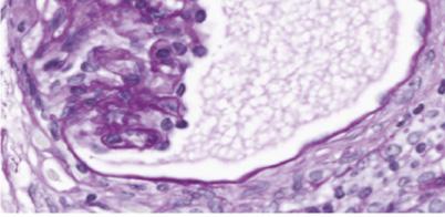
13 Figure 9. Glomeruli from diverse forms of glomerular injury not associated with old age also show mass podocyte detachment events, multinucleate podocytes in the Bowman space, wrinkled GBM, glomerular collapse, and FGGS in the absence of arteriosclerosis. See Tables 5 and 6 for clinical information. (A) PAS-stained kidney biopsy from a 52-year-old kidney showing two normal glomeruli, one glomerulus with proteinaceous material and cells in the Bowman space, and one glomerulus with a collapsed tuft, global sclerosis, and periglomerular fibrosis. (B D) Higher-power views of glomeruli from panel A. Note a normal glomerulus (panel A), proteinaceous material (arrow) and detached cells (arrowhead) in the Bowman space of a glomerulus with wrinkled GBM (panel C) and a collapsed glomerulus with periglomerular fibrosis (arrowhead) in panel D. (E) Glepp1-peroxidase showing that both cells (arrowhead) space and proteionaceous material (arrow) in Bowman space contain Glepp1. (F H) Immunofluorescent images using TLE4 to identify podocyte nuclei (red in panel F) and DAPI to identify nuclei (blue with overlap between TLE4 and DAPI showing as purple). Note that detached cells contain nuclei staining for TLE4 and some nuclei are binucleate (gray arrowheads). Parietal podocytes are present. (I K) PASstained sections showing detached cells in the Bowman space in biopsy specimens from 9-, 16-, and 31-year old kidneys, respectively (arrowheads). (L) Masson trichrome stained section from a 38-year-old showing detached podocytes (arrowhead) and globally sclerosed glomerulus at right (arrow). (M) PAS staining of the same glomerulus as shown in panel L showing an apparently patent arteriole feeding the collapsed glomerulus (arrow). (N P) Sections from a 9-year-old kidney. (N) Three glomeruli, including one glomerulus with PMBS, one glomerulus with numerous detached cells in the Bowman space (arrowhead), and one collapsed globally sclerotic glomerulus (arrow) (silver stain). (O) Collapsing glomerulus (arrow) associated with cells in the Bowman space (arrowhead). (P) Normal artery in this biopsy specimen (PAS). The average glomerular tuft diameter is 137 micrometers Journal of the American Society of Nephrology J Am Soc Nephrol 26: , 2015
14 Table 5. Patient Clinical characteristics associated with MPDEs Kidney Type Kidney Age (yr) Patient Age (yr) Sex Clinical Diagnosis egfr (ml/min per 1.73 m 2 ) 1 Native M SDNS / Native 9 9 M SRNS / TP F Diabetes TP M ANCA/diabetes TP M FSGS Mean6SD The two sets of values shown in patients 1 and 2 for UPCR are variations present at the time of biopsy when patients were in relapse and shortly afterwards when in remission after treatment with calcineurin. inhibitors. UPCR, urinary protein-to-creatinine ratio; M, male; TP, transplant; SDNS, steroid-dependent nephrotic syndrome; SRNS, steroid resistant nephrotic syndrome. UPCR Table 6. Patient Pathologic characteristics associated with MPDEs Foot Process Effacement Podocyte Density per 10 6 mm 3 Normal (%) Glomerular Number FGGS (%) FSGS (%) MPDE (%) PGDE (%) Ischemic (%) 1 Focal Partial artial ND ND % Mean6SD In all five biopsy specimens, the Bowman space contained multinucleate podocytes as judged by TLE4 immunostaining with DAPI counterstain to identify nuclei (Figure 9). PGDE, podocyte glycocalyx detachment event; ND, not done. affixed to the slide, no changes in relative structure or size occur between the two measurements, even though sections are in aqueous phase for the immunofluorescent TLE4 measurements and dehydrated for the immunoperoxidase measurements. After imaging of the TLE4 immunofluorescent sections, the coverslip is removed by incubation in xylene. After rehydration, sections were stained for immunoperoxidase using Vectastain Mouse IgG Kit (PK-6102; Vector Laboratories Inc., Burlingame, CA) as previously reported. 27 Glomerular Volume Estimation The area of all glomerular tuft profiles present in the Glepp1-stained section (including every small profile) is measured. The average radius (r) of all tuft profiles is then calculated assuming glomeruli are spherical. The average maximal radius (R) is then calculated as R=r34/p according to Weibel. 50 Glomerular tuft volume is given as 4/3pR 3. If fewer than eight tuft profiles were present in the biopsy specimen, then glomerular volume estimates are considered to be insufficient to reliably make the estimate, as previously described. 29 Globally sclerotic glomeruli were excluded from analysis. Mean Podocyte Cell Volume Estimation The average Glepp1 positive percentage area3glomerular volume estimates average total podocyte volume (mass) per glomerular tuft. Similarly, the Glepp1-negative glomerular tuft volume represents scarred tuft, mesangial matrix expansion, mesangial cells, endothelial cells, and open capillary loops containing blood products. The average total glomerular volume divided by the average number of podocyte nuclei per tuft estimates average individual podocyte volume. Methodologic Assumptions The correction factor morphometric method allows analysis of archival formalin-fixed biopsy specimens, thereby facilitating unbiased evaluation of all glomeruli per sample (total 2154 glomeruli in 89 biopsy specimens). The glomerular and podocyte morphometric variables obtained are as reported by others The method relies on identifying podocyte nuclei by immunostaining for the transcription factor TLE4 rather than the traditional podocyte transcription factor WT1. 29,51 TLE4 provides a robust podocyte nuclear signal that allows software to estimate the podocyte nuclear size (mean caliper diameter) and thus quantitate them in tissue sections. 29 Under pathologic circumstances, podocytes can sometimes lose their normal transcription factor complement and have duplicated nuclei. We have focused on quantitating podocytes that can perform normal podocyte functions. If podocyte nuclei do not express TLE4 (or WT1) then we assume that they will have changed their phenotype and no longer are functioning as normal podocytes; thus they would not be counted by this method. If two nuclei are present within a single cell, we anticipate that this will reflect the unsuccessful attempt of that podocyte to divide in response to hypertrophic signals. We rationalize that such a cell will be larger than a cell with a single nucleus, and therefore that counting both nuclei will appropriately represent the larger filtration surface area represented by that podocyte. As with any method, assumptions must be understood and critically evaluated within the context of the task at hand. Urine Sample Analysis Urine samples were collected (n=291) as previously described from deidentified people age 3 89 years who had no known kidney disease J Am Soc Nephrol 26: , 2015 Glomerular Aging 3175
FSGS as an Adaptive Response to Growth-Induced Podocyte Stress
 FSGS as an Adaptive Response to Growth-Induced Podocyte Stress Ryuzoh Nishizono,* Masao Kikuchi,* Su Q. Wang,* Mahboob Chowdhury,* Viji Nair,* John Hartman,* Akihiro Fukuda,* Larysa Wickman, Jeffrey B.
The Two Kidney to One Kidney Transition and Transplant Glomerulopathy: A Podocyte Perspective
 The Two Kidney to One Kidney Transition and Transplant Glomerulopathy: A Podocyte Perspective Yan Yang,* Jeffrey B. Hodgin, Farsad Afshinnia,* Su Q. Wang,* Larysa Wickman, Mahboob Chowdhury,* Ryuzoh Nishizono,*
Ordering Physician. Collected REVISED REPORT. Performed. IgG IF, Renal MCR. Lambda IF, Renal MCR. C1q IF, Renal. MCR Albumin IF, Renal MCR
 RenalPath Level IV Wet Ts IgA I Renal IgM I Renal Kappa I Renal Renal Bx Electron Microscopy IgG I Renal Lambda I Renal C1q I Renal C3 I Renal Albumin I Renal ibrinogen I Renal Mayo Clinic Dept. of Lab
Surgical Pathology Report
 Louisiana State University Health Sciences Center Department of Pathology Shreveport, Louisiana Accession #: Collected: Received: Reported: 6/1/2012 09:18 6/2/2012 09:02 6/2/2012 Patient Name: Med. Rec.
RENAL HISTOPATHOLOGY
 RENAL HISTOPATHOLOGY Peter McCue, M.D. Department of Pathology, Anatomy & Cell Biology Sidney Kimmel Medical College There are no conflicts of interest. 1 Goals and Objectives! Goals Provide introduction
Case # 2 3/27/2017. Disclosure of Relevant Financial Relationships. Clinical history. Clinical history. Laboratory findings
 Case # 2 Christopher Larsen, MD Arkana Laboratories Disclosure of Relevant Financial Relationships USCAP requires that all planners (Education Committee) in a position to influence or control the content
Glomerular Pathology- 1 Nephrotic Syndrome. Dr. Nisreen Abu Shahin
 Glomerular Pathology- 1 Nephrotic Syndrome Dr. Nisreen Abu Shahin The Nephrotic Syndrome a clinical complex resulting from glomerular disease & includes the following: (1) massive proteinuria (3.5 gm /day
Renal Pathology 1: Glomerulus. With many thanks to Elizabeth Angus PhD for EM photographs
 Renal Pathology 1: Glomerulus With many thanks to Elizabeth Angus PhD for EM photographs Anatomy of the Kidney http://www.yalemedicalgroup.org/stw/page.asp?pageid=stw028980 The Nephron http://www.beltina.org/health-dictionary/nephron-function-kidney-definition.html
Dr Ian Roberts Oxford. Oxford Pathology Course 2010 for FRCPath Illustration-Cellular Pathology. Oxford Radcliffe NHS Trust
 Dr Ian Roberts Oxford Oxford Pathology Course 2010 for FRCPath Present the basic diagnostic features of the commonest conditions causing proteinuria & haematuria Highlight diagnostic pitfalls Nephrotic
3. PODOCYTE INJURY IN GLOMERULAR DISEASES
 How to Cite this article: Podocyte Injury in Glomerular Diseases - ejifcc 20/01 2009 http://www.ifcc.org 3. PODOCYTE INJURY IN GLOMERULAR DISEASES Mirjana Sabljar Matovinović Podocytes are injured in diabetic
Glomerular pathology in systemic disease
 Glomerular pathology in systemic disease Lecture outline Lupus nephritis Diabetic nephropathy Glomerulonephritis Associated with Bacterial Endocarditis and Other Systemic Infections Henoch-Schonlein Purpura
Overview of glomerular diseases
 Overview of glomerular diseases *Endothelial cells are fenestrated each fenestra: 70-100nm in diameter Contractile, capable of proliferation, makes ECM & releases mediators *Glomerular basement membrane
CHAPTER 2. Primary Glomerulonephritis
 2nd Report of the PRIMARY GLOMERULONEPHRITIS CHAPTER 2 Primary Glomerulonephritis Sunita Bavanandan Lee Han Wei Lim Soo Kun 21 PRIMARY GLOMERULONEPHRITIS 2nd Report of the 2.1 Introduction This chapter
RENAL EVENING SPECIALTY CONFERENCE
 RENAL EVENING SPECIALTY CONFERENCE Harsharan K. Singh, MD The University of North Carolina at Chapel Hill Disclosure of Relevant Financial Relationships No conflicts of interest to disclose. CLINICAL HISTORY
Interesting case seminar: Native kidneys Case Report:
 Interesting case seminar: Native kidneys Case Report: Proximal tubulopathy and light chain deposition disease presented as severe pulmonary hypertension with right-sided cardiac dysfunction and nephrotic
CHAPTER 2 PRIMARY GLOMERULONEPHRITIS
 CHAPTER 2 Sunita Bavanandan Lim Soo Kun 19 5th Report of the 2.1: Introduction This chapter covers the main primary glomerulonephritis that were reported to the MRRB from the years 2005-2012. Minimal change
Use of mycophenolate mofetil in steroid-dependent and -resistant nephrotic syndrome
 Pediatr Nephrol (2003) 18:833 837 DOI 10.1007/s00467-003-1175-4 BRIEF REPORT Gina-Marie Barletta William E. Smoyer Timothy E. Bunchman Joseph T. Flynn David B. Kershaw Use of mycophenolate mofetil in steroid-dependent
Diabetes, Obesity and Heavy Proteinuria
 Diabetes, Obesity and Heavy Proteinuria Clinical Case 41 yo Black woman with heavy proteinuria History 2014: noted to have proteinuria on routine lab testing (1.1g/g). 1+ edema. Blood pressure has been
Histopathology: Hypertension and diabetes in the kidney These presentations are to help you identify basic histopathological features.
 Histopathology: Hypertension and diabetes in the kidney These presentations are to help you identify basic histopathological features. They do not contain the additional factual information that you need
Classification of Glomerular Diseases and Defining Individual Glomerular Lesions: Developing International Consensus
 Classification of Glomerular Diseases and Defining Individual Glomerular Lesions: Developing International Consensus Mark Haas MD, PhD Department of Pathology & Laboratory Medicine Cedars-Sinai Medical
Year 2004 Paper one: Questions supplied by Megan
 QUESTION 53 Endothelial cell pathology on renal biopsy is most characteristic of which one of the following diagnoses? A. Pre-eclampsia B. Haemolytic uraemic syndrome C. Lupus nephritis D. Immunoglobulin
Histopathology: Glomerulonephritis and other renal pathology
 Histopathology: Glomerulonephritis and other renal pathology These presentations are to help you identify basic histopathological features. They do not contain the additional factual information that you
Urinary CD80 as a Replacement for Renal Biopsy for Diagnosis of Pediatric Minimal Change Disease
 KIDNEY DISEASES Urinary CD80 as a Replacement for Renal Biopsy for Diagnosis of Pediatric Minimal Change Disease Heba Mostafa Ahmed, 1 Dina Ahmed Ezzat, 1 Noha A Doudar, 2 Mai Adel 1 1 Departement of Pediatrics,
Lab 3, case 1. Is this an example of nephrotic or nephritic syndrome? Why? Which portion of the nephron would you expect to be abnormal?
 Lab 3, case 1 12-year-old Costa Rican boy is brought into clinic by his parents because of dark brownish-red urine over the last 24 hours. The family has been visiting friends in Indianapolis for two weeks.
C1q nephropathy the Diverse Disease
 C1q nephropathy the Diverse Disease Danica Galešić Ljubanović School of Medicine, University of Zagreb Dubrava University Hospital Zagreb, Croatia Definition Dominant or codominant ( 2+), mesangial staining
Glomerular diseases mostly presenting with Nephritic syndrome
 Glomerular diseases mostly presenting with Nephritic syndrome 1 The Nephritic Syndrome Pathogenesis: proliferation of the cells in glomeruli & leukocytic infiltrate Injured capillary walls escape of RBCs
Case Presentation Turki Al-Hussain, MD
 Case Presentation Turki Al-Hussain, MD Director, Renal Pathology Chapter Saudi Society of Nephrology & Transplantation Consultant Nephropathologist & Urological Pathologist Department of Pathology & Laboratory
Podocyte Biology and clinical applications Dr. F. Ahmadi Professor Of Nephrology TUMS
 Podocyte Biology and clinical applications Dr. F. Ahmadi Professor Of Nephrology TUMS Proteinuria is a major healthcare problem that affects several hundred million people worldwide. Proteinuria is a cardinal
Basic Urinary Tract Anatomy and Histology
 Basic Urinary Tract Anatomy and Histology The two kidneys are located in the retroperitoneum on either side of the vertebral bladder and the contraction of the detrusor muscle. Any mechanical barrier,
The organs of the human body were created to perform ten functions among which is the function of the kidney to furnish the human being with thought.
 The organs of the human body were created to perform ten functions among which is the function of the kidney to furnish the human being with thought. Leviticus Rabba 3 Talmud Berochoth 6 1 b Outline &
University of Bristol - Explore Bristol Research. Peer reviewed version. Link to published version (if available): 10.
: 10.") Prasad, R., Hadjidemetriou, I., Maharaj, A., Meimaridou, E., Buonocore, F., Saleem, M.,... Metherell, L. A. (2017). Sphingosine-1-phosphate lyase mutations cause primary adrenal insufficiency and steroid-resistant
Prasad, R., Hadjidemetriou, I., Maharaj, A., Meimaridou, E., Buonocore, F., Saleem, M.,... Metherell, L. A. (2017). Sphingosine-1-phosphate lyase mutations cause primary adrenal insufficiency and steroid-resistant
The kidneys are excretory and regulatory organs. By
 exercise 9 Renal System Physiology Objectives 1. To define nephron, renal corpuscle, renal tubule, afferent arteriole, glomerular filtration, efferent arteriole, aldosterone, ADH, and reabsorption 2. To
renoprotection therapy goals 208, 209
 Subject Index Aldosterone, plasminogen activator inhibitor-1 induction 163, 164, 168 Aminopeptidases angiotensin II processing 64 66, 214 diabetic expression 214, 215 Angiotensin I intrarenal compartmentalization
Light and electron microscopical studies of focal glomerular sclerosis
 J. clin. Path., 1971, 24, 846-850 Light and electron microscopical studies of focal glomerular sclerosis A. H. NAGI, F. ALEXANDER, AND R. LANNIGAN From the Department of Pathology, Queen's University of
FIBRILLARY GLOMERULONEPHRITIS DIAGNOSTIC CRITERIA, PITFALLS, AND DIFFERENTIAL DIAGNOSIS
 FIBRILLARY GLOMERULONEPHRITIS DIAGNOSTIC CRITERIA, PITFALLS, AND DIFFERENTIAL DIAGNOSIS Guillermo A. Herrera MD Louisiana State University, Shreveport Fibrils in bundles 10-20 nm d Diabetic fibrillosis
Elevated Serum Creatinine, a simplified approach
 Elevated Serum Creatinine, a simplified approach Primary Care Update Creighton University School of Medicine. April 27 th, 2018 Disclosure Slide I have no disclosures and have no conflicts with this presentation.
RECURRENT AND DE NOVO RENAL DISEASES IN THE ALLOGRAFT. J. H. Helderman,MD,FACP,FAST
 RECURRENT AND DE NOVO RENAL DISEASES IN THE ALLOGRAFT J. H. Helderman,MD,FACP,FAST Vanderbilt University Medical Center Professor of Medicine, Pathology and Immunology Medical Director, Vanderbilt Transplant
The topic of normal vascular and glomerular anatomy is introduced
 Normal Vascular and Glomerular Anatomy Arthur H. Cohen Richard J. Glassock The topic of normal vascular and glomerular anatomy is introduced here to serve as a reference point for later illustrations of
Hasan Fattah 3/19/2013
 Hasan Fattah 3/19/2013 AASK trial Rational: HTN is a leading cause of (ESRD) in the US, with no known treatment to prevent progressive declines leading to ESRD. Objective: To compare the effects of 2 levels
Progressive renal failure inability of podocytes to replicate and the consequences for development of glomerulosclerosis
 Nephrol Dial Transplant (1996) 11: 1738-1742 Special Feature IMephrology Dialysis Transplantation Progressive renal failure inability of podocytes to replicate and the consequences for development of glomerulosclerosis
Focal-Segmental Glomerulosclerosis The Relationship Between Tubular Atrophy and Segmental Sclerosis
 Anatomic Pathology / TUBULAR ATROPHY IN FOCAL-SEGMENL GLOMERULOSCLEROSIS Focal-Segmental Glomerulosclerosis The Relationship Between Tubular Atrophy and Segmental Sclerosis Stephen M. Bonsib, M D Key Words:
Nephritic vs. Nephrotic Syndrome
 Page 1 of 18 Nephritic vs. Nephrotic Syndrome Terminology: Glomerulus: A network of blood capillaries contained within the cuplike end (Bowman s capsule) of a nephron. Glomerular filtration rate: The rate
CASE OF THE WEEK 1
 www.nephro-pathology.com CASE OF THE WEEK 1 Clinical Presentation: A 17 year old Indian boy presented with anasarca, decreased urine output and episodes of nausea and vomiting over the last three weeks.
Recurrent Idiopathic Membranous Glomerulonephritis After Kidney Transplantation and Successful Treatment With Rituximab
 TRANSPLANTATION Recurrent Idiopathic Membranous Glomerulonephritis After Kidney Transplantation and Successful Treatment With Rituximab Khadijeh Makhdoomi, 1,2 Saeed Abkhiz, 1,2 Farahnaz Noroozinia, 1,3
RECURRENT AND DE NOVO RENAL DISEASES IN THE ALLOGRAFT
 RECURRENT AND DE NOVO RENAL DISEASES IN THE ALLOGRAFT HISTOPATHOLOGIC DISORDERS AFFECTING THE ALLOGRAFT OTHER THAN REJECTION RECURRENT DISEASE DE NOVO DISEASE TRANSPLANT GLOMERULOPATHY Glomerular Non-glomerular
General introduction. General introduction
 General introduction 1 Chapter 1 Proteinuria is the excretion of proteins into the urine. Presence of abnormal proteinuria, the urinary excretion of abnormal amounts of serum proteins (briefly called proteinuria),
The podocyte, a key cell involved in glomerulosclerosis,
 Podocyte Hypertrophy, Adaptation, and Decompensation Associated with Glomerular Enlargement and Glomerulosclerosis in the Aging Rat: Prevention by Calorie Restriction Jocelyn E. Wiggins,* Meera Goyal,
Glomerular pathology-2 Nephritic syndrome. Dr. Nisreen Abu Shahin
 Glomerular pathology-2 Nephritic syndrome Dr. Nisreen Abu Shahin 1 The Nephritic Syndrome Pathogenesis: inflammation proliferation of the cells in glomeruli & leukocytic infiltrate Injured capillary walls
Secondary IgA Nephropathy & HSP
 Secondary IgA Nephropathy & HSP Anjali Gupta, MD 1/11/11 AKI sec to Hematuria? 65 cases of ARF after an episode of macroscopic hematuria have been reported in the literature in patients with GN. The main
STEROID-RESISTANT NEPHROTIC SYNDROME (SRNS)
") MARIO NEGRI INSTITUTE FOR PHARMACOLOGICAL RESEARCH CLINICAL RESEARCH CENTRE FOR RARE DISEASES ALDO E CELE DACCO' Villa Camozzi - 24020 Ranica (Bergamo) Italy Telephone 39-35-4535304 fax 39-35-4535373 STEROID-RESISTANT
MARIO NEGRI INSTITUTE FOR PHARMACOLOGICAL RESEARCH CLINICAL RESEARCH CENTRE FOR RARE DISEASES ALDO E CELE DACCO' Villa Camozzi - 24020 Ranica (Bergamo) Italy Telephone 39-35-4535304 fax 39-35-4535373 STEROID-RESISTANT
Dr Ian Roberts Oxford
 Dr Ian Roberts Oxford Oxford Pathology Course 2010 for FRCPath Present the basic diagnostic features of the commonest conditions causing renal failure Highlight diagnostic pitfalls. Crescentic GN: renal
A clinical syndrome, composed mainly of:
 Nephritic syndrome We will discuss: 1)Nephritic syndrome: -Acute postinfectious (poststreptococcal) GN -IgA nephropathy -Hereditary nephritis 2)Rapidly progressive GN (RPGN) A clinical syndrome, composed
Ectopic Notch Activation in Developing Podocytes Causes Glomerulosclerosis
 Ectopic Notch Activation in Developing Podocytes Causes Glomerulosclerosis Aoife M. Waters,* Megan Y.J. Wu,* Tuncer Onay,* Jacob Scutaru,* Ju Liu, Corrinne G. Lobe, Susan E. Quaggin, and Tino D. Piscione*
Chapter 4: Steroid-resistant nephrotic syndrome in children Kidney International Supplements (2012) 2, ; doi: /kisup.2012.
 2, ; doi: /kisup.2012.") http://www.kidney-international.org & 2012 KDIGO Chapter 4: Steroid-resistant nephrotic syndrome in children Kidney International Supplements (2012) 2, 172 176; doi:10.1038/kisup.2012.17 INTRODUCTION This
http://www.kidney-international.org & 2012 KDIGO Chapter 4: Steroid-resistant nephrotic syndrome in children Kidney International Supplements (2012) 2, 172 176; doi:10.1038/kisup.2012.17 INTRODUCTION This
Renal System Physiology
 M58_MARI0000_00_SE_EX09.qxd 7/18/11 2:37 PM Page 399 E X E R C I S E 9 Renal System Physiology Advance Preparation/Comments 1. Prior to the lab, suggest to the students that they become familiar with the
Prof. Rosanna Coppo Director of the Nephrology, Dialysis and Transplantation Department Regina Margherita Hospital Turin, Italy. Slide 1.
 ROLE OF PATHOLOGY AND CLINICAL FEATURES IN PREDICTING PROGRESSION OF IGA NEPHROPATHY: RESULTS FROM THE ERA-EDTA RESEARCH VALIGA Rosanna Coppo, Turin, Italy Chairs: François Berthoux, Saint-Etienne, France
Dr Ian Roberts Oxford. Oxford Pathology Course 2010 for FRCPath Illustration-Cellular Pathology. Oxford Radcliffe NHS Trust
 Dr Ian Roberts Oxford Oxford Pathology Course 2010 for FRCPath Plan of attack: Diagnostic approach to the renal biopsy Differential diagnosis of the clinical syndromes of renal disease Microscopy Step
Idiopathic focal segmental glomerulosclerosis: a favourable prognosis in untreated patients?
 O R I G N A L A R T I C L E Idiopathic focal segmental glomerulosclerosis: a favourable prognosis in untreated patients? J.K.J. Deegens 1* K.J.M. Assmann 2, E.J. Steenbergen 2, L.B. Hilbrands 1, P.G.G.
Case Presentation Turki Al-Hussain, MD
 Case Presentation Turki Al-Hussain, MD Director, Renal Pathology Chapter Saudi Society of Nephrology & Transplantation Consultant Nephropathologist & Urological Pathologist Department of Pathology & Laboratory
Dr Michael Rayment Chelsea and Westminster Hospital, London
 17 TH ANNUAL CONFERENCE OF THE BRITISH HIV ASSOCIATION (BHIVA) Dr Michael Rayment Chelsea and Westminster Hospital, London 6-8 April 2011, Bournemouth International Centre A decade of renal biopsies in
41B. Metabolism produces wastes that must be eliminated from the body. This. Renal System Physiology: Computer Simulation
 41B E X E R C I S E Renal System Physiology: Computer Simulation O B J E C T I V E S 1. To define the following terms: glomerulus, glomerular capsule, renal corpuscle, renal tubule, nephron, proximal convoluted
PHGY210 Renal Physiology
 PHGY210 Renal Physiology Tomoko Takano, MD, PhD *Associate Professor of Medicine and Physiology McGill University *Nephrologist, McGill University Health Centre Lecture plan Lecture 1: Anatomy, basics
A. Correct! Flushing acids from the system will assist in re-establishing the acid-base equilibrium in the blood.
 OAT Biology - Problem Drill 16: The Urinary System Question No. 1 of 10 1. Which of the following would solve a drop in blood ph? Question #01 (A) Decreased retention of acids. (B) Increased excretion
As outlined under External contributions (see appendix 7.1), the group of Prof. Gröne at the
, the group of Prof. Gröne at the") 3 RESULTS As outlined under External contributions (see appendix 7.1), the group of Prof. Gröne at the DKFZ in Heidelberg (Dept. of Cellular and Molecular pathology) contributed to this work by performing
3 RESULTS As outlined under External contributions (see appendix 7.1), the group of Prof. Gröne at the DKFZ in Heidelberg (Dept. of Cellular and Molecular pathology) contributed to this work by performing
THE KIDNEY AND SLE LUPUS NEPHRITIS
 THE KIDNEY AND SLE LUPUS NEPHRITIS JACK WATERMAN DO FACOI 2013 NEPHROLOGY SIR RICHARD BRIGHT TERMINOLOGY RENAL INSUFFICIENCY CKD (CHRONIC KIDNEY DISEASE) ESRD (ENDSTAGE RENAL DISEASE) GLOMERULONEPHRITIS
Case 3. ACCME/Disclosure. Laboratory results. Clinical history 4/13/2016
 Case 3 Lynn D. Cornell, M.D. Mayo Clinic, Rochester, MN Cornell.Lynn@mayo.edu USCAP Renal Case Conference March 13, 2016 ACCME/Disclosure Dr. Cornell has nothing to disclose Clinical history 57-year-old
Prof. Andrzej Wiecek Department of Nephrology, Endocrinology and Metabolic Diseases Medical University of Silesia Katowice, Poland.
 What could be the role of renal denervation in chronic kidney disease? Andrzej Wiecek, Katowice, Poland Chairs: Peter J. Blankestijn, Utrecht, The Netherlands Jonathan Moss, Glasgow, UK Prof. Andrzej Wiecek
Renal Physiology - Lectures
 Renal Physiology 2011 Lisa M. Harrison-Bernard, PhD lharris@lsuhsc.edu Renal Physiology - Lectures Physiology of Body Fluids 2. Structure & Function of the Kidneys 3. Renal Clearance & Glomerular Filtration
Index. electron microscopy, 81 immunofluorescence microscopy, 80 light microscopy, 80 Amyloidosis clinical setting, 185 etiology/pathogenesis,
 A Acute antibody-mediated rejection (Acute AMR) clinical features, 203 clinicopathologic correlations, 206 pathogenesis, 205 206 204 205 light microscopy, 203 204 Acute cellular rejection (ACR) clinical
Prof. Armando Torres Nephrology Section Hospital Universitario de Canarias University of La Laguna Tenerife, Canary Islands, Spain.
 Does RAS blockade improve outcomes after kidney transplantation? Armando Torres, La Laguna, Spain Chairs: Hans De Fijter, Leiden, The Netherlands Armando Torres, La Laguna, Spain Prof. Armando Torres Nephrology
Pathology of Complement Mediated Renal Disease
 Pathology of Complement Mediated Renal Disease Mariam Priya Alexander, MD Associate Professor of Pathology GN Symposium Hong Kong Society of Nephrology July 8 th, 2017 2017 MFMER slide-1 The complement
RNPDC CCNP Anatomy and Physiology: Renal System Pre-Quiz 2015
 RNPDC CCNP Anatomy and Physiology: Renal System Pre-Quiz 2015 1. In which abdominal cavity do the kidneys lie? a) Peritoneum. b) Anteperitoneal. c) Retroperitoneal. d) Parietal peritoneal 2. What is the
Clinical and pathologic characteristics of focal segmental glomerulosclerosis pathologic variants
 original article http://www.kidney-international.org & 2006 International Society of Nephrology Clinical and pathologic characteristics of focal segmental glomerulosclerosis pathologic variants DB Thomas
Actualités néphrologiques Jean Hamburger 23 Avril Marie Courbebaisse, Service de Physiologie Hôpital Européen Georges Pompidou, Paris
 Single nephron GFR Actualités néphrologiques Jean Hamburger 23 Avril 2018 Marie Courbebaisse, Service de Physiologie Hôpital Européen Georges Pompidou, Paris Introdution Glomerular filtration and SNGFR
Monoclonal Gammopathies and the Kidney. Tibor Nádasdy, MD The Ohio State University, Columbus, OH
 Monoclonal Gammopathies and the Kidney Tibor Nádasdy, MD The Ohio State University, Columbus, OH Monoclonal gammopathy of renal significance (MGRS) Biopsies at OSU (n=475) between 2007 and 2016 AL or AH
Review of Rituximab and renal transplantation. Dr.E Nemati. Professor of Nephrology
 Review of Rituximab and renal transplantation Dr.E Nemati Professor of Nephrology Introductio n Rituximab is a chimeric anti-cd20 monoclonal antibody. The CD20 antigen is a transmembrane nonglycosylated
Plasma soluble urokinase receptor levels are increased but do not distinguish primary from secondary focal segmental glomerulosclerosis
 http://www.kidney-international.org & 213 International Society of Nephrology see commentary on page 235 Plasma soluble urokinase receptor levels are increased but do not distinguish primary from secondary
An unusual association between focal segmental sclerosis and lupus nephritis: a distinct concept from lupus podocytopathy?
 CEN Case Rep (2015) 4:70 75 DOI 10.1007/s13730-014-0142-1 CASE REPORT An unusual association between focal segmental sclerosis and lupus nephritis: a distinct concept from lupus podocytopathy? Hironari
Management of Rejection
 Management of Rejection I have no disclosures Disclosures (relevant or otherwise) Deborah B Adey, MD Professor of Medicine University of California, San Francisco Kidney and Pancreas Transplant Center
Urinary system. Urinary system
 INTRODUCTION. Several organs system Produce urine and excrete it from the body Maintenance of homeostasis. Components. two kidneys, produce urine; two ureters, carry urine to single urinary bladder for
Histology Urinary system
 Histology Urinary system Urinary system Composed of two kidneys, two ureters, the urinary bladder, and the urethra, the urinary system plays a critical role in: 1- Blood filtration,(filtration of cellular
Special Challenges and Co-Morbidities
 Special Challenges and Co-Morbidities Renal Disease/ Hypertension/ Diabetes in African-Americans M. Keith Rawlings, MD Medical Director Peabody Health Center AIDS Arms, Inc Dallas, TX Chair, Internal Medicine
Biopsy Features of Kidney Allograft Rejection Banff B. Ivanyi, MD Department of Pathology, University of Szeged, Szeged, Hungary
 Biopsy Features of Kidney Allograft Rejection Banff 2017 B. Ivanyi, MD Department of Pathology, University of Szeged, Szeged, Hungary Treatment of allograft dysfunction should rely on the biopsy findings
Accepted Manuscript. Glomerulopathy Associated with Moderate Obesity
 Accepted Manuscript Glomerulopathy Associated with Moderate Obesity Yusuke Okabayashi, M.D., Nobuo Tsuboi, M.D. Ph.D., Takaya Sasaki, M.D., Kotaro Haruhara, M.D., Go Kanzaki, M.D., Kentaro Koike, M.D.
Donor Quality Assessment
 Donor Quality Assessment Francesc Moreso, MD, PhD Renal Transplant Unit Hospital Universitari Vall d Hebron Barcelona. Spain 4/9/2017 Donor Quality Assessment 1 What is the problem? Across all age ranges,
CASE 3 AN UNUSUAL CASE OF NEPHROTIC SYNDROME
 CASE 3 AN UNUSUAL CASE OF NEPHROTIC SYNDROME Dr Seethalekshmy N.V., Dr.Annie Jojo, Dr Hiran K.R., Amrita institute of Medical Sciences, Kochi, Kerala Case history 34 year old gentleman Nephrotic range
Urinary System Laboratory
 Urinary System Laboratory 1 Adrenal gland Organs of The Urinary System Renal artery and vein Kidney Ureter Urinary bladder Figure 26.1 2 Urethra Functions of the urinary system organs: Urethra expels urine
Kidney Physiology. Mechanisms of Urine Formation TUBULAR SECRETION Eunise A. Foster Shalonda Reed
 Kidney Physiology Mechanisms of Urine Formation TUBULAR SECRETION Eunise A. Foster Shalonda Reed The purpose of tubular secrection To dispose of certain substances that are bound to plasma proteins. To
BCH 450 Biochemistry of Specialized Tissues
 BCH 450 Biochemistry of Specialized Tissues VII. Renal Structure, Function & Regulation Kidney Function 1. Regulate Extracellular fluid (ECF) (plasma and interstitial fluid) through formation of urine.
Glomerulosclerosis is the major pathologic process responsible
 Podocyte Depletion Causes Glomerulosclerosis: Diphtheria Toxin Induced Podocyte Depletion in Rats Expressing Human Diphtheria Toxin Receptor Transgene Bryan L. Wharram,* Meera Goyal,* Jocelyn E. Wiggins,
19. RENAL PHYSIOLOGY ROLE OF THE URINARY SYSTEM THE URINARY SYSTEM. Components and function. V BS 122 Physiology II 151 Class of 2011
 19. RENAL PHYSIOLOGY THE URINARY SYSTEM Components and function The urinary system is composed of two kidneys, the functionally filtering apparatus, which connect through two tubular structures called
Diabetic Nephropathy in Spontaneously Diabetic Torii (SDT) Rats
 Rats") The Open Diabetes Journal, 2011, 4, 45-49 45 Diabetic Nephropathy in Spontaneously Diabetic Torii (SDT) Rats Takeshi Ohta * and Tomohiko Sasase Open Access Biological/Pharmacological Research Laboratories,
The Open Diabetes Journal, 2011, 4, 45-49 45 Diabetic Nephropathy in Spontaneously Diabetic Torii (SDT) Rats Takeshi Ohta * and Tomohiko Sasase Open Access Biological/Pharmacological Research Laboratories,
Keisuke Suzuki Naoto Miura Hirokazu Imai
 Clin Exp Nephrol (2014) 18:606 612 DOI 10.1007/s10157-013-0867-8 ORIGINAL ARTICLE Estimated glomerular filtration rate and daily amount of urinary protein predict the clinical remission rate of tonsillectomy
The evolution of the classification of nephrotic syndrome Laura Barisoni, MD
 The evolution of the classification of nephrotic syndrome Laura Barisoni, MD Department of Pathology and Medicine, Division of Nephrology New York University Old classification schemes: Proteinuria and
Introduction. Acute sodium overload produces renal tubulointerstitial inflammation in normal rats
 Acute sodium overload produces renal tubulointerstitial inflammation in normal rats MI Roson, et al. Kidney International (2006) Introduction Present by Kanya Bunnan and Wiraporn paebua Tubular sodium
The organs of the human body were created to perform ten functions among which is the function of the kidney to furnish the human being with thought.
 The organs of the human body were created to perform ten functions among which is the function of the kidney to furnish the human being with thought. Leviticus Rabba 3 Talmud Berochoth 6 1 b Nephrology
The UK Renal Registry collects national data about the causes and treatment of kidney failure.
 1 Kidney failure is a serious condition. Many kidney patients receive some form of renal replacement therapy (RRT) such as dialysis. The UK Renal Registry collects national data about the causes and treatment
New Ideas. Better Medicines. Third Quarter Financial Results Conference Call
 New Ideas. Better Medicines. Third Quarter 2018 Financial Results Conference Call Forward-Looking Statements 2 This presentation contains forward-looking statements that involve substantial risks and uncertainties.
Crescentic Glomerulonephritis (RPGN)
") Crescentic Glomerulonephritis (RPGN) Background Rapidly progressive glomerulonephritis (RPGN) is defined as any glomerular disease characterized by extensive crescents (usually >50%) as the principal histologic
Crescentic Glomerulonephritis (RPGN) Background Rapidly progressive glomerulonephritis (RPGN) is defined as any glomerular disease characterized by extensive crescents (usually >50%) as the principal histologic
Physiology Lecture 2. What controls GFR?
 Physiology Lecture 2 Too much blood is received by the glomerular capillaries, this blood contains plasma, once this plasma enters the glomerular capillaries it will be filtered to bowman s space. The