Getting Paid for Technology
|
|
|
- Patience Stafford
- 5 years ago
- Views:
Transcription
1 Getting Paid for Technology Harvey Richman, OD Rebecca Wartman, OD Optometry s Meeting June 2018 Disclaimers for Presentation 1.All information was current at time it was prepared 2.Drawn from national policies, with links included in the presentation for your use 3.Prepared as a tool to assist doctors and staff and is not intended to grant rights or impose obligations 4.Prepared and presented carefully to ensure the information is accurate, current and relevant 5.No conflicts of interest exist for presenters- financial or otherwise. However, Rebecca and Harvey write for Optometric Journals Disclaimers for Presentation 6. Of course the ultimate responsibility for the correct submission of claims and compliance with provider contracts lies with the provider of services 7. AOA, AOA-TPC, Optometry s Meeting, its presenters, agents, and staff make no representation, warranty, or guarantee that this presentation and/or its contents are error-free and will bear no responsibility or liability for the results or consequences of the information contained herein 8. The content of the COPE Accredited CE activity was prepared with assistance from Kara Webb (AOA Staff) and Doug Morrow OD 1


2 Disclaimers for Presentation Cornea and Anterior Segment Eye Review Maps 2


3 Ocular higher order wavefront maps Ocular higher order wavefront maps 3D view Settings 3


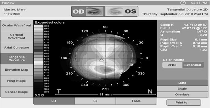
4 Corneal wavefront allows direct comparison of corneal and ocular wavefronts Compare corneal with total WF Corneal wavefront allows direct comparison of corneal and ocular wavefronts Tangential and 4


5 Axial Curvature Measurments Topography color scales Elevation maps Various overlay and fit zone options Topography Computerized corneal topography, unilateral or bilateral with interpretation and report Detection of subtle corneal surface irregularity and astigmatism 16 5

6 Indications & Limitations of Coverage: Post penetrating keratoplasty Post kerato refractive complications Post op irregular astigmatism Corneal dystrophy, bullous keratopathy Complications of transplanted cornea, Keratoconus Reasons For Denial: Non covered for refractive procedures Often billable privately for contact lens evaluations or included in examination fee Corneal Hysteresis Uses air impulse stimulation Unilateral or bilateral I Interpretation and report 6

7 Low Corneal Hysteresis Optic nerve damage Visual field loss Functional progression of GLC Larger magnitude of IOP reduction Dynamic finding that may increase after medications are implemented No utilization guidelines No published LCD Some Medical Policy list as E/I/U Anterior segment imaging interpretation and report Specular microscopy and endothelial cell analysis Bilateral Service Justified Slit lamp evidence of endothelial dystrophy (guttata) Slit lamp evidence of corneal edema Undergoing secondary intraocular lens implant Previous ocular surgery now requires cataract surgery Fitting with extended wear contact after ocular surgery Limitations If only visual problem is cataract not eligible considered part of presurgical examination. Not covered in preoperative evaluation of refractive keratoplasty 7
 Reduced Corneal NFL length & Corneal sensitivity = increased severity diabetic peripheral neuropathy Previous studies")

8 Corneal Confocal Microscopy Examines unmyelinated corneal nerve high magnification, using laser scanning CCM to image corneal sub basal nerve plexus Can predict insipient peripheral neuropathy in Type 1 DM (63% Sensitive; 74% Specific) Reduced Corneal NFL length & Corneal sensitivity = increased severity diabetic peripheral neuropathy Previous studies demonstrate utility for CCM in other neuropathies Corneal Confocal Microscopy Blepharoplasty Guidelines Visual field examinations used determine medical necessity for blepharoplasty Often performed twice, taped lids and untapped lids Repeated service should be submitted with CPT modifier 76 on a separate detail line but often denied External Photos now often used Review carrier LCD 8

 Medicare Fees National Non Facility Fee")
9 New Blepharoplasty Guidelines CGS: Complaint Physical findings Visual fields Noridan: Complaint Physical finding Photos WPS: Complaint Physical findings Visual fields Photos Cahaba: Complaint Physical findings Visual fields Photos Palmetto: Complaint Physical findings Photos 5 Carriers with LCD for Blepharoplasty 2 have eliminated Visual Fields requirement Blepharoplasty Photos Anterior Segment Photography External ocular photography with interpretation and report for documentation of medical progress (eg, close up photography, slit lamp photography, gonio photography, stereo photography) Medicare Fees National Non Facility Fee $
10 External Ocular Photography Bilateral Code Check carrier for limitations or restrictions of coverage External ocular photography is covered when a special camera is used to obtain magnified photographs of lesions (e.g., the cornea, iris or lids) for purpose of following the patient's condition Medical quality images may be of digital, Polaroid Macro 3 SLR or equivalent Photographs for purpose of documenting for medicolegal purposes or preauthorization (e.g., gross trauma, amount of ptosis or redundant lid tissue) are not separately reimbursed not medically necessary SCANNING COMPUTERIZED OPHTHALMIC DIAGNOSTIC IMAGING, ANTERIOR SEGMENT, WITH INTERPRETATION AND REPORT, UNILATERAL OR BILATERAL Narrow angle, suspected narrow angle, and mixed narrow and open angle glaucoma Determining the proper intraocular lens for a patient who has had prior refractive surgery and now requires cataract extraction Iris tumor Presence of corneal edema or opacity that precludes visualization or study of the anterior chamber SCANNING COMPUTERIZED OPHTHALMIC DIAGNOSTIC IMAGING, ANTERIOR SEGMENT, WITH INTERPRETATION AND REPORT, UNILATERAL OR BILATERAL No using 52 modifier or LT or RT modifier if only CPT codes not covered with SCODI: 76512,


11 Pupilometry Pupilometry Pupilometry 11

12 Pupilometry CPT 0341T Pupilometry Quantitative pupillometry with interpretation and report, unilateral or bilateral RVU=0 Local Carrier Priced MGD Dysfunction 12


13 MGD Imaging Ocular Surface Interferomoter CPT III CODES 0330T Tear film Imaging unilateral or bilateral report 0207T Clear eyelid gland w/ heat/intermittent pressure 13
: Type 2 DM Detection (via AGEs on lens) Sensitivity = 67%, Specificity")
 Takes 6 secs, non invasive, doesn t require fasting Lens Autofluorescence Integral to General Ophthalmologic or E&M Now No LCD/")
14 Lens Autofluorescence FDA Market Clearance 2013 Lens Autofluorescence Using Lens Fluorescence Biomicroscope calibrated with standards traceable to National Institute of Standards and Technology (NIST): Type 2 DM Detection (via AGEs on lens) Sensitivity = 67%, Specificity = 94% (Hemoglobin A1C: Sens.= 44%, Spec.= 79%) (Fasting Plasma Gluc.: Sens.=50%, Spec.= 95%) Takes 6 secs, non invasive, doesn t require fasting Lens Autofluorescence Integral to General Ophthalmologic or E&M Now No LCD/ Medical Policy could be considered but not recommended 14

15 92072 Fitting of a contact lens for management of keratoconus, initial fitting. For subsequent fittings, use either the 9921X or 9201X codes. Report materials in addition to this code, using either or the appropriate HCPCS Level II material code. Question: At what point after the initial fitting of a keratoconus lens is a new lens (not a replacement) billable with code due to the fact that the lens no longer fits the patient s need? Answer: Description of work for initial fittings includes the results of diagnostic tests done prior to contact lens fitting to assess the corneal ectasia, which and are used in concert with slit lamp examination to assess corneal shape and determine initial contact lens parameters (eg, diameter, base curve, and secondary curves). Lens designs can include corneal, scleral, hybrid, or piggyback systems. Keratometry, lid anatomy, tear film, and refraction are also performed and/or rechecked. If the lens need to be changed because it no longer fits the patient s needs, the fitting of new lens is considered an initial fitting and should include all of the services noted above. CPT Assistant September


16 Glaucoma Gonioscopy Used to diagnose injury or disease in anterior chamber of eye, performed under local anesthetic due to necessity of placing specialized lens directly on the eye to obtain a clear image Bilateral Procedure Code LCD Utilization Visual Field Examinations Limited, unilateral or bilateral, with interpretation and report; examination Intermediate, unilateral or bilateral, with interpretation and report Extended, unilateral or bilateral, with interpretation and report 16
 E")
 G VFI Bar: Remaining useful vision H")

17 Glaucoma Management 5 0 A Baseline visual fields B Glaucoma Hemifield Test C Current visual field D GPA / Deviation from Baseline (Event Analysis) E GPA Alert F VFI plot: Visual Field Index Display of Trend (3 5yrs) under current conditions (Trend Analysis) G VFI Bar: Remaining useful vision H Rate of Progression / Significance Guided Progression Analysis B A F H G C D E Guided Progression Analysis Change of baseline intuitive & quick operation 17


18 Serial Visual Field Overview Visual Field Overview Serial Visual Field Overview Indications & Limitations of Coverage Necessary to establish diagnosis Monitor course for treatment Determine change in therapeutic plan 18


19 Indications & Limitations of Coverage medically necessary to diagnose and follow retinal disorders diagnosis or follow up of glaucoma or neurologic disease MILD visual field abnormality (inner circle = 10 degrees, outer circle = 20 degrees) MODERATE visual field abnormality (inner circle = 10 degrees, outer circle = 20 degrees) 19
 Coding Guidelines VF")

 Optical")
20 SEVERE visual field abnormality (inner circle = 10 degrees, outer circle = 20 degrees) Coding Guidelines VF All services are considered bilateral 50 modifier not appropriate 52 modifier if only doing one eye 76 modifier if doing repeat procedure Scanning Laser Tests Confocal laser scanning ophthalmoscopy (topography) Optical Coherence tomography 20


21 Versatile Multi Modality Imaging Glaucoma Versatile Multi Modality Imaging Coding guidelines Scanning computerized ophthalmic diagnostic imaging (e.g., scanning laser) with interpretation and report, unilateral or bilateral No using either a 52 LT or RT modifier if reduced CPT codes not covered with SCODI: 92225, 92226, modifier usage GA modifier usage with ABN 21
22 92133 Glaucoma Indications Scanning Computerized Ophthalmic Diagnostic Imaging, posterior segment, with interpretation and report, unilateral or bilateral; optic nerve Technological improvements have rendered SCODI as valuable diagnostic tool in diagnosis and treatment of glaucoma. These improvements enable discernment of changes of nerve fiber even in advanced cases of glaucoma. Expected that only two exams/eye/year would be required to manage the patient who has glaucoma or is suspected of having glaucoma. Glaucoma Severity/Staging Level Scanning Laser Frequency Current frequency limitations for Scanning Laser for most regions: Mild or Suspect Glaucoma Moderate Glaucoma Advanced or Severe Glaucoma 1 Time per year 2 Times per year NO Scanning laser but up to 4 Visual Fields / year Utilization Guidelines GLC Although CMS guidelines state Only two exams/eye/year are allowed for patient who has or is suspected of having glaucoma Most LCD state once per year to follow pre glaucoma patients or those with mild stage One or two tests per year for patients with moderate staging, followed with SLT or visual fields if both SLT and visual fields are used, only one of each tests Advanced stage, field testing preferred by Medicare guidance 22


23 Ganglion Cell Analysis Isolates Ganglion Cell Layer Measures thickness for sum of GCL and IPL layers using data from Macular cube scans. RNFL distribution in the macula depends on individual anatomy, while the GCL+IPL appears regular and elliptical for most normal individuals Propriety algorithms are adapted for specific anatomy, use GCL and IPL thickness Excludes RNFL Thickness Map Show thickness measurements of the GCL + IPL in the 6mm by 6mm cube and Deviation contains Maps an elliptical Show Sector a maps annulus - centered comparison divide the about elliptical of the fovea. GCL annulus + IPL of the thickness Thickness to Map tableinto normative Shows 6 regions: average data. 3 equally and minimum sized sectors thickness in the within superior the region elliptical and annulus. Horizontal and 3 equally sized Vertical B-scans. sectors in the inferior region. Values are compared to normative data. Ganglion Cell Analysis Key Elements Diagnostic CPT's Pachymetry: CPT Bilateral. Billable for Corneal Problems and Glaucoma. Requires Interpretation and Report. 23

 Macular")
24 Visual Field Technology 0378T Visual field assessment, with concurrent real time data analysis and accessible data storage with patient initiated data transmitted to a remote surveillance center for up to 30 days; review and interpretation with report by a physician or other qualified health care professional 0464T 0464T Visual Evoked Potential, testing for glaucoma, with interpretation and report (For visual evoked potential screening for visual acuity, use 0333T) Macular Diseases 24


25 All relevant information on 1 screen First visit Prior visit Todays visit Screen Layout 1 glance to see change Visualization of change Navigate multiple visits all at once Synchronized navigation 25


26 HD Line raster in MultiMode Navigator Completes the combined information between OCT data and fundus images Versatile Multi Modality Imaging RPE Detachment Versatile Multi Modality Imaging Proliferative Diabetic Retinopathy 26


27 Versatile Multi Modality Imaging Versatile Multi Modality Imaging Diagnostic Insight 27


28 Greater Diagnostic Insight Review Integrated Images and Registered OCT Scans Scanning Computerized Ophthalmic Dagnostic Imaging, posterior segment, with interpretation and report, unilateral or bilateral; retina Retinal disorders are most common causes of severe and permanent vision loss. These technologies are valuable tools for evaluation and treatment of patients with retinal disease, especially macular abnormalities. These imaging techniques are useful tools to measure effectiveness of therapy, and in determining need for ongoing therapy, or safety of cessation of therapy Scanning Computerize Ophthalmic Diagnostic Imaging, posterior segment, with interpretation and report, unilateral or bilateral; retina Utilization Guidelines AMD/DR Only one exam/eye/2 months is allowed for the patient whose primary ophthalmological diagnosis is related to a retinal disease One exam/eye/month is allowed for the patient who is undergoing active treatment for macular degeneration or diabetic retinopathy Glaucoma? 28


29 Fundus Autofluorescence (FAF) Potential info health & function of entire retina Photoreceptors contain light sensing molecules susceptible to damage/x linking, & shed their damaged outer segments RPE phagocytize the segments & molecules stored in liposomes, forming lipofuscin (LF) Disease states & oxidative damage = LF Hyper fluorescence = excess LF accumulation Hypo fluorescence = RPE cells die/are absent LASER Speckle Flowgraphy Noninvasive way to assess ocular blood flow CRVO study completed 29


30 Angiography software OCTA Non invasive, dyeless Hi res, 3 D visualization of retinal vasculature Images motion of scattering particles such as RBCs using sequential OCT x sectional scans :Fundus Photography Fundus photography with interpretation and report Bilateral Code 90 Photography Document abnormalities Check carrier s medical policy for limitations or restrictions of coverage Obtain filing requirements from carrier for bilateral or multiple procedures 91 30
31 92250 Utilization Guidelines Fundus photography. Generally, it is not medically necessary to repeat fundus photography more often than every 2 years for follow up of stable glaucoma. Repeat photographs for retinopathy are rarely necessary. Wide Field Retinal Screening S9886 Not Medically Necessary Service Patient is aware not medically necessary Screenings are not covered in most cases 31


32 Macular Pigment Densitometers No Current LCD No Current Defined Code Studies Inconclusive Image from Marco MPOD Assesment 92081? Heterochromic Flicker Photometry 92081? 32


33 Preferential Hyperacuity Perimeter Discontinued 92082? ERG Electroretinography with interpretation and report Bilateral Code LCDs Changing Most TPP experimental except for plaquenil Not for EOMs New code coming?? Visual Evoked Potential UPDATE 2018 Visual Evoked Potential (VEP) checkerboard or flash testing, central nervous system, except glaucoma, with interpretation and report Bilateral Code General Supervision Special Training? Utilization Guidelines HERE 33
 Developed with RBRVS 2003 Insures proper")
 policy manual o 92133 or 92134 and 92250 but MAY use 59 modifier o 92133 and 92134 may NOT be used together even with 59 modifier")
34 0333T 0469T Retinal polarization scan, ocular screening with on site automated results, bilateral Retinal Birefringence scanners (RBS) hand held instruments measure the changes in the polarization of light detect eye misalignment or strabismus No LCD Medical Policy E/I/U National Correct Coding Initiative (NCCI) Developed with RBRVS 2003 Insures proper Medicare payments (Resource Based Relative Value System) Identify pairs of services not billed together (same physician for same patient on same day) Component element edits o and Medically Unlikely Edits (MUE) policy manual o or and but MAY use 59 modifier o and may NOT be used together even with 59 modifier 34

35 NCCI Edits MUE together, column 1 code is paid MUE MAY be allow together o 0 not allowed o 1 allowed o 9 non applicable o If clinical circumstances justify appending a modifier to column 2 code of code pair, payment for both codes may be allowed MUST READ AND UNDERSTAND WHAT CAN BE DONE TOGETHER AND WHEN Cannot use a modifier just to get paid NCCI Edits Relevant to Optometry Fundus photography (CPT code 92250) and scanning ophthalmic computerized diagnostic imaging (e.g., CPT codes 92132, 92133, 92134) are generally mutually exclusive of one another in that a provider would use one technique or the other to evaluate fundal disease. However, there are a limited number of clinical conditions where both techniques are medically reasonable and necessary on the ipsilateral eye. In these situations, both CPT codes may be reported appending modifier 59 to CPT code (CPT code was deleted January 1, 2011.) CPT code (fitting of contact lens for treatment of ocular surface disease) should not be reported with a corneal procedure CPT code for a bandage contact lens applied after completion of a procedure on the cornea. Modifier 59 Definition Distinct Procedural Service identifies procedures/services not normally reported together, but appropriately billable under the circumstances Modifier 59 should not be used to bypass a Procedure to Procedure (PTP) edit unless the proper criteria for use of the modifier are met. Documentation in the medical record must satisfy the criteria required by any NCCI (National Correct Coding Initiative) associated modifier that is used. 35
36 Modifier 59 History Most widely used modifier according to the Centers for Medicare & Medicaid Services (CMS) Associated with considerable abuse High levels of manual audit Triggers reviews and appeals Results in civil fraud and abuse cases Fundus Photography & SCODI Continued confusion on billing photography and SCODI on same date of service They are mutually exclusive as defined by current NCCI Mutually exclusive is defined as procedures that cannot reasonably be performed at the same anatomic site or same encounter. Fundus Photography & SCODI There has been no specific document defining when you can use and with This means there is no official CMS guidance on using mutually exclusive codes on the same date of service. 36
37 Modifier 59 New CMS Guidance Treatment of posterior segment structures in the eye constitutes treatment of a single anatomic site. (See example 5 Modifier 59) Modifier 59 should not be used if both procedures are performed during the same operative session because the retina and choroid are contiguous structures of the same organ Clinical Decision We know that using modifier 59 has potential to trigger audits NCCI policy statement seems to give provider some wiggle room to use 59 modifier Providers need to use caution if choose to use modifier 59 Coding Experts recommend that scans be performed on different visits to avoid potential for audits. Modifier 59 Changes Since January 5, 2015 new X code modifiers Intended to more clearly define Distinct Procedural Service Rules for use have not been written 37
38 New Modifiers Further Defining Modifier 59 X modifiers meant to define subsets of Modifier 59 X modifiers provide more precise coding options CMS acknowledges that increased education is needed CMS only modifiers CPT manual has not been changed New Modifiers X(EPSU) Modifiers XE Separate Encounter: Service that is distinct because it occurred during a separate encounter New Modifiers X(EPSU) Modifiers XS Separate Structure: Service that is distinct because it was performed on a separate organ/structure 38
39 New Modifiers X(EPSU) Modifiers XP Separate Practitioner: Service that is distinct because it was performed by a different practitioner New Modifiers X(EPSU) Modifiers XU Unusual Non Overlapping Service: Use of a service that is distinct because it does not overlap usual components of the main service. New Modifiers X(EPSU) Modifiers CMS continues to recognize Modifier 59 Instructions state that 59 should not be used when a more descriptive modifier is available Providers should not use modifier 59 and a new X modifier together for the same code 39
40 YOU SURVIVED Questions? Thank You! 40
Dr. Harvey Richman, OD, FAAO, FCOVD Diplomate American Board of Optometry Executive Committee AOA Third Party Center Founder Ask the AOA Coding
 Dr. Harvey Richman, OD, FAAO, FCOVD Diplomate American Board of Optometry Executive Committee AOA Third Party Center Founder Ask the AOA Coding Experts 92000 Codes Special Ophthalmological Services Describe
and at the same patient encounter. Code has been deleted. For scanning computerized ophthalmic diagnostic imaging of optic nerve and retin
 92227: Remote imaging for detection of retinal disease (eg, retinopathy in a patient with diabetes) with analysis and report under physician supervision, unilateral or bilateral. For Medicare, bill only
Local Coverage Determination (LCD): Scanning Computerized Ophthalmic Diagnostic Imaging (SCODI) (L34431)
: Scanning Computerized Ophthalmic Diagnostic Imaging (SCODI) (L34431)") Local Coverage Determination (LCD): Scanning Computerized Ophthalmic Diagnostic Imaging (SCODI) (L34431) Links in PDF documents are not guaranteed to work. To follow a web link, please use the MCD Website.
Local Coverage Determination (LCD): Scanning Computerized Ophthalmic Diagnostic Imaging (SCODI) (L34431) Links in PDF documents are not guaranteed to work. To follow a web link, please use the MCD Website.
3/23/2016. Diagnostic Services Taylor Pannell CRA, OCT-C. Services Available. Important info for the Tech to know. Visual Fields
 Services Available Diagnostic Services Taylor Pannell CRA, OCT-C Static and Kinetic Visual Fields Pachymetry Anterior and Posterior Segment OCT Fundus Photos FAF,FA,ICG Slit Lamp Photography Confocal HRT
04/11/2014. Retina Coding and Reimbursement 101. Financial Disclosure. Chief Complaint
 Retina Coding and Reimbursement 101 William T. Koch, COA, COE, CPC Administrative Director Director of Billing Operations The Retina Institute St. Louis, Missouri Advisory Boards Allergan Genentech Regeneron
04/06/2015. Documentation Do s and Don ts In The Retina Practice. Financial Disclosure. Documentation Dos and Don ts
 Documentation Do s and Don ts In The Retina Practice William T. Koch, COA, COE, CPC Administrative Director Director of Billing Operations The Retina Institute St. Louis, Missouri Advisory Boards Allergan
Corporate Medical Policy
 Corporate Medical Policy Optical Coherence Tomography (OCT) Anterior Segment of the Eye File Name: Origination: Last CAP Review: Next CAP Review: Last Review: optical_coherence_tomography_(oct)_anterior_segment_of_the_eye
Fundus Autofluorescence. Jonathan A. Micieli, MD Valérie Biousse, MD
 Fundus Autofluorescence Jonathan A. Micieli, MD Valérie Biousse, MD The retinal pigment epithelium (RPE) has many important functions including phagocytosis of the photoreceptor outer segments Cone Rod
FEP Medical Policy Manual
 FEP Medical Manual 9.03.05 Corneal Topography/Computer-Assisted Corneal Topography/ Photokeratoscopy Last Review: September 2016 Next Review: September 2017 Related Policies 9.03.28 Corneal Collagen Cross-linking
2009 REIMBURSEMENT GUIDE, VISUCAM and VISUCAM NM/FA
 2009 REIMBURSEMENT GUIDE FF 450 PLUS PRO NM, VISUCAM and VISUCAM NM/FA Zeiss Fundus Cameras INTRODUCTION The following guide provides an overview of billing and reimbursement for procedures performed with
WORKSHOP B Ophthalmic Imaging: All Hands on Tech! COPE Course PS
 WORKSHOP B Ophthalmic Imaging: All Hands on Tech! COPE Course 44334-PS Ophthalmic Imaging: All Hands on Tech! Southern College of Optometry April 17, 2015 COPE #44334-PS Faculty Dr. Michael Gerstner Dr.
Corporate Medical Policy
 Corporate Medical Policy Glaucoma, Evaluation by Ophthalmologic Techniques File Name: Origination: Last CAP Review: Next CAP Review: Last Review: glaucoma_evaluation_by_ophthalmologic_techniques 3/2001
Contractor Information
 Local Coverage Determination (LCD): Scanning Computerized Ophthalmic Diagnostic Imaging (L35038) Links in PDF documents are not guaranteed to work. To follow a web link, please use the MCD Website. Contractor
Ganglion cell analysis by optical coherence tomography (OCT) Jonathan A. Micieli, MD Valérie Biousse, MD
 Jonathan A. Micieli, MD Valérie Biousse, MD") Ganglion cell analysis by optical coherence tomography (OCT) Jonathan A. Micieli, MD Valérie Biousse, MD Figure 1. Normal OCT of the macula (cross section through the line indicated on the fundus photo)
Ganglion cell analysis by optical coherence tomography (OCT) Jonathan A. Micieli, MD Valérie Biousse, MD Figure 1. Normal OCT of the macula (cross section through the line indicated on the fundus photo)
Texas Definition of Eye Exam. Definitions of Eye Examinations BILLING AND CODING: WHY IS THIS STUFF SO HARD? Optometry School Definition
 BILLING AND CODING: WHY IS THIS STUFF SO HARD? Craig Thomas, O.D. 3900 West Wheatland Road Dallas, Texas 75237 972-780-7199 thpckc@yahoo.com Definitions of Eye Examinations Optometry School definition
NEW YORK UNIVERSITY SCHOOL OF MEDICINE DEPARTMENT OF OPHTHALMOLOGY EDUCATIONAL OBJECTIVES AND GOALS
 NEW YORK UNIVERSITY SCHOOL OF MEDICINE DEPARTMENT OF OPHTHALMOLOGY EDUCATIONAL OBJECTIVES AND GOALS Revision Date: 6/30/06 Distribution Date: 7/6/06 The Department of Ophthalmology at the NYU Medical Center
Local Coverage Determination (LCD): Scanning Computerized Ophthalmic Diagnostic Imaging (SCODI) (L33751)
: Scanning Computerized Ophthalmic Diagnostic Imaging (SCODI) (L33751)") Local Coverage Determination (LCD): Scanning Computerized Ophthalmic Diagnostic Imaging (SCODI) (L33751) Links in PDF documents are not guaranteed to work. To follow a web link, please use the MCD Website.
Local Coverage Determination (LCD): Scanning Computerized Ophthalmic Diagnostic Imaging (SCODI) (L33751) Links in PDF documents are not guaranteed to work. To follow a web link, please use the MCD Website.
GLAUCOMA SUMMARY BENCHMARKS FOR PREFERRED PRACTICE PATTERN GUIDELINES
 SUMMARY BENCHMARKS FOR PREFERRED PRACTICE PATTERN GUIDELINES Introduction These are summary benchmarks for the Academy s Preferred Practice Pattern (PPP) guidelines. The Preferred Practice Pattern series
Sample page. Ophthalmology A comprehensive illustrated guide to coding and reimbursement CODING COMPANION
 CODING COMPANION 2018 Ophthalmology A comprehensive illustrated guide to coding and reimbursement POWER UP YOUR CODING with Optum360, your trusted coding partner for 32 years. Visit optum360coding.com.
MEDICAL POLICY SUBJECT: CORNEAL ULTRASOUND PACHYMETRY. POLICY NUMBER: CATEGORY: Technology Assessment
 MEDICAL POLICY SUBJECT: CORNEAL ULTRASOUND,, PAGE: 1 OF: 5 If a product excludes coverage for a service, it is not covered, and medical policy criteria do not apply. If a commercial product, including
PREAMBLE TO MSC PAYMENT SCHEDULE: OPTOMETRY SERVICES
 PREAMBLE TO MSC PAYMENT SCHEDULE: OPTOMETRY SERVICES A. GENERAL PROVISIONS 1. Eye Examination Benefits Optometric benefits are services defined in Section 23 of the Medical and Health Care Services Regulations,
Special Ophthalmological Services Clinical Coverage Policy No: 1T-2 Amended Date: October 1, Table of Contents
 Special Ophthalmological Services Clinical Coverage Policy No: 1T-2 Table of Contents 1.0 Description of the Procedure, Product, or Service... 1 1.1 Computerized Corneal Topography... 1 1.2 Sensorimotor
Report for EYEGENETIX. Prepared on. May 24, By: David Davis, CPC, CPC-H, CCC (Ret.)
") Report for EYEGENETIX Prepared on May 24, 2016 By: David Davis, CPC, CPC-H, CCC (Ret.) EyeGenetix EyeGenetix sells ophthalmic diagnostic equipment to primary care providers. This equipment performs the
Report for EYEGENETIX Prepared on May 24, 2016 By: David Davis, CPC, CPC-H, CCC (Ret.) EyeGenetix EyeGenetix sells ophthalmic diagnostic equipment to primary care providers. This equipment performs the
ICD-10-CM Are you Prepared? Disclaimers for Presentation. Disclaimers for Presentation 5/13/2014. What is ICD-10-CM/PCS
 AOA Third Party Center Coding Experts Are you Prepared? Rebecca H. Wartman O.D. Optometry s Meeting 2014 With contributions from Doug Morrow O.D. & Harvey Richman O.D. Rebecca H. Wartman, O.D Douglas C.
Goals. Glaucoma PARA PEARL TO DO. Vision Loss with Glaucoma
 Glaucoma Janet R. Fett, OD Drs. Kincaid, Fett and Tharp So Sioux City, NE eyewear21@hotmail.com Goals Understand Glaucoma Disease process Understand how your data (objective and subjective) assists in
Billing Requirements for Intravitreal Injections. Financial Interest. Indications. Documentation. Documentation. Documentation
 Billing Requirements for Intravitreal Injections Financial Interest ASCRS ASOA Symposium & Congress Practice Management Program Los Angeles, California May 5-9, 2017 Presented by: Patricia Kennedy, COMT,
Widefield Retinal Imaging with Auto Fluorescence Technology in the Optometric Practice
 Widefield Retinal Imaging with Auto Fluorescence Technology in the Optometric Practice This course will define ultra-widefield retinal imaging and autofluorescence for the attendee. Will show how it is
PRIMUS 200 from ZEISS The essential OCT
 PRIMUS 200 from ZEISS The essential OCT Seeing beyond the surface. ZEISS PRIMUS 200 // INNOVATION MADE BY ZEISS Clear Visualization. Advanced Technology. Reliability. Essential elements of your first OCT.
Ask the AOA Coding Experts: Vision versus Medical? Doug Morrow, O.D. Harvey Richman, O.D. Rebecca Wartman, O.D.
 Ask the AOA Coding Experts: Vision versus Medical? Doug Morrow, O.D. Harvey Richman, O.D. Rebecca Wartman, O.D. Disclaimers for Presentation 1.All information was current at time it was prepared 2.Drawn
Cirrus TM HD-OCT. Details define your decisions
 Cirrus TM HD-OCT Details define your decisions 2 With high-definition OCT Carl Zeiss Meditec takes you beyond standard spectral domain Built on 10 years experience at the vanguard of innovation, Carl Zeiss
Financial Disclosure. Compliance Report. Course Objective. Issue 1: Complex Cataract Surgery. Compliance Dilemmas. What did I do wrong?
 Financial Disclosure Compliance Dilemmas Alan Reider is a partner in the Washington, DC Office of Arnold & Porter Kaye Scholer LLP Donna McCune is a Vice President with Corcoran Consulting Group Alan Reider,
Mark Dunbar: Disclosure
 Important Things to Understand About OCT Mark T. Dunbar, O.D., F.A.A.O. Bascom Palmer Eye Institute University of Miami, School of Medicine Mark Dunbar: Disclosure Optometry Advisory Board for: Allergan
Optometric Services Fee Schedule
 Optometric Services Schedule Note: The base fees listed below are reimbursed for services provided to recipients age 21 and over. To calculate the fee for children under 21, multiply the base fee, the
Moving forward with a different perspective
 Moving forward with a different perspective The Leader In Vision Diagnostics Offers A New Perspective Marco has served the eyecare community by offering exceptional lane products and automated high tech
Financial Disclosure. Modifiers Getting It Right! Modifiers. Modifiers. Medicare Expected Frequency. Common Modifiers Used Only with Office Visits
 Financial Disclosure Modifiers Getting It Right! Donna McCune is a consultant for Corcoran Consulting Group and acknowledges a financial interest in the subject matter of this presentation. Donna McCune,
Cirrus TM HD-OCT. Details defi ne your decisions
 Cirrus TM HD-OCT Details defi ne your decisions 2 With high-defi nition OCT Carl Zeiss Meditec takes you beyond standard spectral domain Built on 10 years experience at the vanguard of innovation, Carl
PRIMUS 200 from ZEISS The essential OCT
 EN 00_00I The contents of the brochure may differ from the current status of approval of the product in your country. Please contact your regional representative for more information. Subject to change
Visualize. Analyze. Personalize. OCT + OCTA
 Visualize. Analyze. Personalize. OCT + OCTA A New Approach to Protecting Vision AngioVue OCT Angiography brings valuable new information to clinical practice. Non-invasive visualization of retinal vasculature.
3/16/2018. Perimetry
 Perimetry The normal visual field extends further away from fixation temporally and inferiorly than superiorly and nasally. From the center of the retina this sensitivity decreases towards the periphery,
Clinical Policy: Refractive Surgery Reference Number: CP.MP. 391
 Clinical Policy: Refractive Surgery Reference Number: CP.MP. 391 Effective Date: November 2007 Last Review Date: January 2016 Coding Implications Revision Log See Important Reminder at the end of this
Contractor Information
 Local Coverage Determination (LCD): Scanning Computerized Ophthalmic Diagnostic Imaging (L35038) Links in PDF documents are not guaranteed to work. To follow a web link, please use the MCD Website. Contractor
The Common Clinical Competency Framework for Non-medical Ophthalmic Healthcare Professionals in Secondary Care
 The Common Clinical Competency Framework for Non-medical Ophthalmic Healthcare Professionals in Secondary Care Glaucoma November 2016 Association of Health Professions in Ophthalmology General basic competences
Imaging and Current/Future Technologies in Medicine & Primary Eye Care
 I. What s New in Imaging for the Primary Eye Care Practice A. Digital Refraction Analyzers B. Corneal Topography C. Optical Coherence Tomography (OCT) and Retinal Imaging D. Wide-Field Retinal Imaging,
Financial Disclosure. Understanding Global Surgery Rules. Does Insurance Cover Surgery? Course Objectives
 Financial Disclosure Understanding Global Surgery Rules Donna McCune is a consultant for Corcoran Consulting Group and acknowledges a financial interest in the subject matter of this presentation. Donna
SOCT Copernicus REVO. * - Currently import and overlay are avaibale in manual mode only
 SOCT Copernicus REVO Easy Operation (Full auto & Auto mode) Auto alignment (Z-position, C-gate, Focus, Tomogram) Voice guide (support patient through examination) Powerful analysis tools Enhanced tomograms
How to Be Efficient and Effective. Disclosure. Topics CASE CM. Case JF 2007 OHTN / POAG? How to Be Efficient and Effective with. with New Technology
 How to Be Efficient and Effective with Disclosure COPE Course ID: 40750 GL Michael Chaglasian has the following disclosures: 1. Advisory Board: Allergan, Inc., Alcon Labs, B+L Carl Zeiss Meditec 2. Research:
Eye and Ocular Adnexa, Auditory Systems
 Eye and Ocular Adnexa, Auditory Systems CPT copyright 2011 American Medical Association. All rights reserved. Fee schedules, relative value units, conversion factors and/or related components are not assigned
H F 1 0 T H E R A P Y R E I M B U R S E M E N T R E F E R E N C E G U I D E
 HF10 therapy, delivered by the Nevro Senza System, is the high-frequency spinal cord stimulation technology designed to aid in the management of chronic intractable pain of the trunk/limbs without paresthesia.
Measure #191: Cataracts: 20/40 or Better Visual Acuity within 90 Days Following Cataract Surgery
 Measure #191: Cataracts: 20/40 or Better Visual Acuity within 90 Days Following Cataract Surgery 2012 PHYSICIAN QUALITY REPORTING OPTIONS FOR INDIVIDUAL MEASURES: REGISTRY ONLY DESCRIPTION: Percentage
PRESCRIBING IN GLAUCOMA: GUIDELINES FOR NZ OPTOMETRISTS
 PRESCRIBING IN GLAUCOMA: GUIDELINES FOR NZ OPTOMETRISTS Introduction Independent prescribing relates to the capacity to use clinical judgement in respect of diagnosis and treatment. It does not mean working
Do You See What I See!!! Shane R. Kannarr, OD
 Do You See What I See!!! Shane R. Kannarr, OD skannarr@kannarreyecare.com Define Specialty Testing Additional Test to: Prove/Disprove Diagnosis To monitor progression of a condition To document a condition
Populations Interventions Comparators Outcomes Individuals: Who are being evaluated for angleclosure
 Protocol Optical Coherence Tomography of the Anterior Eye Segment (90318) Medical Benefit Effective Date: 07/01/14 Next Review Date: 05/18 Preauthorization No Review Dates: 07/11, 07/12, 07/13, 05/14,
Disclaimers. Disclaimers. PQRS 2011 Made Easy 2/3/2011. Physician Quality Reporting System. Presented by Rebecca H. Wartman, O.D.
 Physician Quality Reporting System PQRS 2011 Made Easy Presented by Rebecca H. Wartman, O.D. Practice Advancement Committee Member, Clinical and Practice Advancement Group American Optometric Association
Local Coverage Determination (LCD): Scanning Computerized Ophthalmic Diagnostic Imaging (SCODI) (L34431)
: Scanning Computerized Ophthalmic Diagnostic Imaging (SCODI) (L34431)") Local Coverage Determination (LCD): Scanning Computerized Ophthalmic Diagnostic Imaging (SCODI) (L34431) Links in PDF documents are not guaranteed to work. To follow a web link, please use the MCD Website.
Local Coverage Determination (LCD): Scanning Computerized Ophthalmic Diagnostic Imaging (SCODI) (L34431) Links in PDF documents are not guaranteed to work. To follow a web link, please use the MCD Website.
Perspectives on Screening for Diabetic Retinopathy. Dr. Dan Samaha, Optometrist, MSc Clinical Lecturer School of Optometry, Université de Montréal
 Perspectives on Screening for Diabetic Retinopathy 1 Dr. Dan Samaha, Optometrist, MSc Clinical Lecturer School of Optometry, Université de Montréal Current standards 2 According to the Canadian Diabetes
Documentation Challenges
 Agenda History Taking Eye Codes Diagnostic testing guidelines Kirk A. Mack, COMT, COE, CPC, CPMA Senior Consultant Corcoran Consulting Group 2 2 Coding is a Team Sport Involves everyone in the office Requires
2019 COLLECTION TYPE: MIPS CLINICAL QUALITY MEASURES (CQMS) MEASURE TYPE: Outcome High Priority
 MEASURE TYPE: Outcome High Priority") Quality ID #191 (NQF 0565): Cataracts: 20/40 or Better Visual Acuity within 90 Days Following Cataract Surgery National Quality Strategy Domain: Effective Clinical Care Meaningful Measure Area: Management
Quality ID #191 (NQF 0565): Cataracts: 20/40 or Better Visual Acuity within 90 Days Following Cataract Surgery National Quality Strategy Domain: Effective Clinical Care Meaningful Measure Area: Management
Method for comparing visual field defects to local RNFL and RGC damage seen on frequency domain OCT in patients with glaucoma.
 Method for comparing visual field defects to local RNFL and RGC damage seen on frequency domain OCT in patients with glaucoma. Donald C. Hood 1,2,* and Ali S. Raza 1 1 Department of Psychology, Columbia
Objectives. Unexplained Vision Loss: Where Do I Go From Here. History. History. Drug Induced Vision Loss
 Objectives Unexplained Vision Loss: Where Do I Go From Here Denise Goodwin, OD, FAAO Coordinator, Neuro-ophthalmic Disease Clinic Pacific University College of Optometry goodwin@pacificu.edu Know the importance
Optometric Postoperative Cataract Surgery Management
 Financial Disclosures Optometric Postoperative Cataract Surgery Management David Dinh, OD Oak Cliff Eye Clinic Dallas Eye Consultants March 10, 2015 Comanagement Joint cooperation between two or more specialists
 measure of your overall performance. An isolated glucose test is helpful to let you know what your sugar level is at one moment, but it doesn t tell you whether or not your diabetes is under adequate control
Glaucoma: Diagnostic Modalities
 Glaucoma: Diagnostic Modalities - Dr. Barun Kumar Nayak, Dr. Sarika Ramugade Glaucoma is a leading cause of blindness in the world, especially in older people. Early detection and treatment by ophthalmologist
Physical Facilities Information
 EYE INSTITUTE CHICAGO COLLEGE OF OPTOMETRY 3450 Lacey Road Downers Grove, IL 60515 Phone: 630/743-4500 To be completed by site: Site Name: Primary Clinic Address: Site Phone: Site Fax: Site Website: Primary
OPHTHALMOLOGICAL DISORDERS
 Telephone No.: 24622495 Telegraphic Address: Aeronautical: VIDDYAYX Commercial: AIRCIVIL NEW DELHI E Mail: dri@dgca.nic.in Fax:01124629211 GOVERNMENT OF INDIA AERONAUTICAL INFORMATION SERVICE DIRECTOR
Coding Terminology Getting Back To The Basics. Financial Interest. Getting Back To The Basics. Rose & Associates
 Coding Terminology Getting Back To The Basics ASCRS ASOA Symposium & Congress Administrator Program Boston, Massachusetts April 25-29, 2014 Presented by: Patricia Kennedy, COMT, CPC, COE Financial Interest
Medicare Reimbursement Challenges. Financial Interest. Overpayments. Overpayments. Overpayments. Overpayments
 Medicare Reimbursement Challenges Financial Interest ASCRS-ASOA Symposium & Congress Practice Management Program New Orleans, Louisiana May 6-10, 2016 Presented by: E. Ann Rose, President, Rose & Associates
The Common Clinical Competency Framework for Non-medical Ophthalmic Healthcare Professionals in Secondary Care
 The Common Clinical Competency Framework for Non-medical Ophthalmic Healthcare Professionals in Secondary Care Medical Retina November 2016 Association of Health Professions in Ophthalmology General basic
Hong Kong College of Surgical Nursing
 Hong Kong College of Surgical Nursing Surgical Nursing Training: Part B Specialty - Ophthalmological Nursing Curriculum 1 TABLE OF CONTENTS No. Contents Page 1. Introduction 3 2. Aims 3 3. Programme Intended
Ophthalmology Unit Referral Guidelines
 Ophthalmology Unit Referral Guidelines Austin Health Ophthalmology Unit holds sub-specialty sessions to discuss and plan the treatment of patients with specific ocular conditions. General including cataract
2/6/2018. Andrew Siedlecki, M.D.
 Andrew Siedlecki, M.D. Siedlecki Cataract and Vision Care Optimization- Improved Uncorrected VA Reduced Spectacle Dependency Minimize Complications Fast Rehabilitation Provide Options for Patients Optometrists
2016 HF10 THERAPY REIMBURSEMENT REFERENCE GUIDE
 206 HF0 THERAPY REIMBURSEMENT REFERENCE GUIDE HF0 therapy, delivered by the Nevro Senza System, is a new high-frequency spinal cord stimulation technology designed to aid in the management of chronic intractable
2 016 HF10 THERAPY HOSPITAL OUTPATIENT DEPARTMENT AND AMBULATORY SURGERY CENTER REIMBURSEMENT REFERENCE GUIDE
 HF10 therapy, delivered by the Nevro Senza System, is a new high-frequency spinal cord stimulation technology designed to aid in the management of chronic intractable pain of the trunk/limbs, including
Cataract and Refractive Surgery Co-Management Policy and Procedure Manual
 Cataract and Refractive Surgery Co-Management Policy and Procedure Manual Michael R. George, M.D. Chief Surgeon and Medical Director Tylock-George Eye Care Index of Cataract and Refractive Surgery Manual
1/25/2019 OCT & OCTA RETINAL IMAGING: HOW TO PREVENT RAGING GLAUCOMA! THE ORIGINAL RAGING GLAUCOMA OCT RETINAL IMAGING OPTIC NERVE HEAD EXAMINATION
 OCT & OCTA RETINAL IMAGING: HOW TO PREVENT RAGING GLAUCOMA! Craig Thomas, O.D. 3900 West Wheatland Road Dallas, Texas 75237 972-780-7199 thpckc@yahoo.com THE ORIGINAL RAGING GLAUCOMA 47-year-old Black
2018 OPTIONS FOR INDIVIDUAL MEASURES: REGISTRY ONLY. MEASURE TYPE: Outcome
 Quality ID #191 (NQF 0565): Cataracts: 20/40 or Better Visual Acuity within 90 Days Following Cataract Surgery National Quality Strategy Domain: Effective Clinical Care 2018 OPTIONS FOR INDIVIDUAL MEASURES:
Evolving glaucoma management True diagnostic integration for the preservation of vision
 Evolving glaucoma management True diagnostic integration for the preservation of vision // GLAUCOMA MANAGEMENT MADE BY ZEISS The moment you are certain it is glaucoma. This is the moment we work for. There
CODING COMPANION. Sample page. Ophthalmology A comprehensive illustrated guide to coding and reimbursement. Power up your coding. optum360coding.
 CODING COANION 2019 Ophthalmology A comprehensive illustrated guide to coding and reimbursement Power up your coding optum360coding.com Contents Getting Started with Coding Companion...i Resequencing of
Protocol. Blepharoplasty
 Protocol Blepharoplasty Medical Benefit Effective Date: 01/01/13 Next Review Date: 05/19 Preauthorization No Review Dates: 09/12, 09/13, 09/14, 09/15, 09/16, 05/17, 05/18 Preauthorization is encouraged
HOCT-1I 1F All-in-One Optical Coherence Tomography with Fundus
 HOCT-1I 1F All-in-One Optical Coherence Tomography with Fundus Specification Type Resolution(in Tissue) A scan Rate Scan Range SD-OCT / Fundus Z :6~7um, XY:20um 68,000 A-scan/sec. [Fundus] X:6-12mm, Y:6-9mm,
When optical coherence tomography (OCT)
") Macular Imaging: SD-OCT in nterior Segment Surgical Practice Many pathologic processes of the macula can be visualized or quantified only with this modality. y Steven G. Safran, MD When optical coherence
Macular Imaging: SD-OCT in nterior Segment Surgical Practice Many pathologic processes of the macula can be visualized or quantified only with this modality. y Steven G. Safran, MD When optical coherence
 www.brisbaneeyeclinic.com.au Brisbane Eye Clinic is a modern ophthalmology practice focused on the provision of excellent medical eye care. The Clinic has two convenient consulting locations, our Wickham
Optical Coherence Tomograpic Features in Idiopathic Retinitis, Vasculitis, Aneurysms and Neuroretinitis (IRVAN)
") Columbia International Publishing Journal of Ophthalmic Research (2014) Research Article Optical Coherence Tomograpic Features in Idiopathic Retinitis, Vasculitis, Aneurysms and Neuroretinitis (IRVAN)
Columbia International Publishing Journal of Ophthalmic Research (2014) Research Article Optical Coherence Tomograpic Features in Idiopathic Retinitis, Vasculitis, Aneurysms and Neuroretinitis (IRVAN)
Clinical Policy: Implantable Miniature Telescope for Age Related Macular Degeneration Reference Number: CP.MP.517
 Clinical Policy: Implantable Miniature Telescope for Age Related Macular Reference Number: CP.MP.517 Effective Date: 11/16 Last Review Date: 11/17 See Important Reminder at the end of this policy for important
RETINAL CONDITIONS RETINAL CONDITIONS
 GENERAL INFORMATION RETINAL CONDITIONS RETINAL CONDITIONS WHAT ARE RETINAL CONDITIONS? Retinal conditions affect the light-sensitive tissue at the back of eye known as the retina. They include diseases
Fundamentals of Retina Coding
 Fundamentals of Retina Coding Presented by: Joy Woodke, COE, OCS Sunday, April 2, 2017 ASRS Business of Retina Meeting Dallas, TX American Academy of Ophthalmic Executives Financial Disclosure Joy Woodke,
Glaucoma Evaluation. OCT Pearls for Glaucoma. OCT: Retinal Nerve Fiber Layer. Financial Disclosures. OCT: Macula. Case Example
 OCT Pearls for Glaucoma using OCT of the macula for glaucoma Glaucoma Evaluation Right eye Visual Acuity 20/25 20/25 IOP 13 13 Central corneal 530 530 thickness Anterior exam Normal with PCIOL Normal with
Optical Coherence Tomography (OCT)
") Understanding and Interpreting OCT Mark Dunbar: Disclosure The Swiss Army Pocket Knife of Eye Care Mark T. Dunbar, O.D., F.A.A.O. Bascom Palmer Eye Institute University of Miami, School of Medicine Consultant
Understanding and Interpreting OCT Mark Dunbar: Disclosure The Swiss Army Pocket Knife of Eye Care Mark T. Dunbar, O.D., F.A.A.O. Bascom Palmer Eye Institute University of Miami, School of Medicine Consultant
Implementing New & Revised ICD-10 Codes John A. McGreal Jr., O.D. Missouri Eye Associates McGreal Educational Institute
 Implementing New & Revised ICD-10 Codes John A. McGreal Jr., O.D. Missouri Eye Associates McGreal Educational Institute Excellence in Optometric Education John A. McGreal Jr., O.D. Missouri Eye Associates
HOW TO MAKE THE MOST OF A NEW OCT. with Kelly Kerksick, OD
 HOW TO MAKE THE MOST OF A NEW OCT with Kelly Kerksick, OD 3 How to Make the Most of a New OCT Kelly Kerksick, OD, graduated from Southern College of Optometry and immediately started her own private practice
Advances in OCT Murray Fingeret, OD
 Disclosures Advances in OCT Murray Fingeret, OD Consultant Alcon, Allergan, Bausch & Lomb, Carl Zeiss Meditec, Diopsys, Heidelberg Engineering, Reichert, Topcon Currently Approved OCT Devices OCT Devices
2018 OPTIONS FOR INDIVIDUAL MEASURES: REGISTRY ONLY. MEASURE TYPE: Process
 Quality ID #14 (NQF 0087): Age-Related Macular Degeneration (AMD): Dilated Macular Examination National Quality Strategy Domain: Effective Clinical Care 2018 OPTIONS FOR INDIVIDUAL MEASURES: REGISTRY ONLY
Implementing New & Revised ICD-10 Codes
 Implementing New & Revised ICD-10 Codes John A. McGreal Jr., O.D. Missouri Eye Associates cgreal Educational Institute Excellence in Optometric Education John A. McGreal Jr., O.D. Missouri Eye Associates
This policy is applicable to Commercial Products only. For BlueCHiP for Medicare, see related policy section.
 Medical Coverage Policy Ophthalmologic Techniques that Evaluate the Posterior Segment for Glaucoma EFFECTIVE DATE: 01 01 2017 POLICY LAST UPDATED: 09 18 2018 OVERVIEW Several techniques have been developed
Medical Policy An independent licensee of the Blue Cross Blue Shield Association
 Pachymetry Page 1 of 8 Medical Policy An independent licensee of the Blue Cross Blue Shield Association Title: Pachymetry Professional Institutional Original Effective Date: March 11, 2004 Original Effective
10/3/2018. Case: 63 year old white female 6/11/2012 visit. Glaucoma Update for the Primary Care OD CHRISTOPHER WOLFE, OD, FAAO, DIPL ABO
 DISCLOSURE STATEMENT Ihave no direct financial or proprietary interest in any companies, products or services mentioned in this presentation. GLAUCOMA UPDATE FOR THE PRIMARY CARE OD Please silence all
Partial Coherence Interferometry as a Technique to Measure the Axial Length of the Eye Archived Medical Policy
 Partial Coherence Interferometry as a Technique to Measure the Axial Length of the Eye Applies to all products administered or underwritten by Blue Cross and Blue Shield of Louisiana and its subsidiary,
Pearls for the Refractive Technician Fadiah Alkhawaldeh, IMBA, COT, ROUB
 Pearls for the Refractive Technician Fadiah Alkhawaldeh, IMBA, COT, ROUB Cleveland Clinic Cole Eye Institute OOS, Columbus, OH February, 2014 alkhawf@ccf.org NO FINANCIAL DISCLOSURES A Puzzle of an Eye
HOW TO PICK THE RIGHT OCT
 HOW TO PICK THE RIGHT OCT with Kelly Kerksick, OD Part One of a Three-Part OCT Educational Series 1geriatrics. Dr. Kerksick agreed to share her experiences and insights in owning How to Pick the Right
OCT Image Analysis System for Grading and Diagnosis of Retinal Diseases and its Integration in i-hospital
 Progress Report for1 st Quarter, May-July 2017 OCT Image Analysis System for Grading and Diagnosis of Retinal Diseases and its Integration in i-hospital Milestone 1: Designing Annotation tool extraction