Case Report IgG4-Seronegative Autoimmune Pancreatitis and Sclerosing Cholangitis
|
|
|
- Garry Mathews
- 5 years ago
- Views:
Transcription
1 Case Reports in Gastrointestinal Medicine Volume 2015, Article ID , 6 pages Case Report IgG4-Seronegative Autoimmune Pancreatitis and Sclerosing Cholangitis Allon Kahn, 1 Anitha D. Yadav, 2 and M. Edwyn Harrison 2 1 Department of Medicine, Mayo Clinic, Scottsdale, AZ, USA 2 Division of Gastroenterology and Hepatology, Mayo Clinic, Scottsdale, AZ, USA Correspondence should be addressed to M. Edwyn Harrison; harrison.m@mayo.edu Received 24 June 2015; Revised 15 August 2015; Accepted 18 August 2015 Academic Editor: Haruhiko Sugimura Copyright 2015 Allon Kahn et al. This is an open access article distributed under the Creative Commons Attribution License, which permits unrestricted use, distribution, and reproduction in any medium, provided the original work is properly cited. IgG4-related disease is a relatively novel clinical entity whose gastrointestinal manifestations include type 1 autoimmune pancreatitis (AIP) and IgG4-associated sclerosing cholangitis. The presence of elevated serum IgG4 is suggestive but not essential for the diagnosis of type 1 AIP and is a pervasive feature of the proposed diagnostic criteria. The differential diagnosis of type 1 AIP includes malignant conditions, emphasizing the importance of a deliberate, comprehensive evaluation. Management of patients with a suggestive clinical presentation, but without serum IgG4 elevation, is difficult. Here we present three cases of IgG4-seronegative AIP and sclerosing cholangitis that responded to empiric steroid therapy and discuss approach considerations. These cases demonstrate the value of meticulous application of existing diagnostic algorithms to achieve a clinical diagnosis and avoid surgical intervention. 1. Introduction Type 1 autoimmune pancreatitis (AIP) and immunoglobulin G4- (IgG4-) associated sclerosing cholangitis (IgG4-SC) are pancreatic and biliary manifestations of IgG4-related disease, respectively. Recently, IgG4-related disease has emerged as a novel clinical entity characterized by multiorgan infiltration of IgG4-positive cells, storiform fibrosis, and elevated serum IgG4 levels. Several diagnostic criteria have been proposed in the last decade based on radiology, histopathology, serology, and response to steroids [1 3]. However, the diagnosis still remains challenging. Often the differential diagnosis for AIP/IgG4-SC includes malignant conditions such as pancreatic cancer and cholangiocarcinoma, placing patients at risk for unnecessary surgery for a more benign condition. Current clinical diagnostic criteria for type 1 AIP/IgG4-SC involve elevated serum IgG4 concentrations (>135 mg/dl). The reported sensitivity of increased serum IgG4 levels in AIP ranges from 68% to 81% [1, 4]. Hence, in patients with normal serum IgG4 levels, a thorough workup including pancreatic and biliary imaging, histology, and identification of extrapancreatic manifestations aids in accurate diagnosis. Here we describe three cases of serum IgG4 negative type 1 AIP/IgG4 SC that responded to a trial of steroid therapy, allowing for the avoidance of surgery. 2. Case 1 An 80-year-old Caucasian man with a history of resected prostate adenocarcinoma and hyperlipidemia presented with complaints of pruritus, anorexia, fatigue, 20 pound weight loss, and painless jaundice. He denied abdominal pain, melena, or hematochezia. His vital signs were within normal limits and physical examination revealed only jaundice. There was no history of significant alcohol use. Laboratory workup revealed an elevated total bilirubin 8.5 mg/dl, direct bilirubin 5.2 mg/dl, alkaline phosphatase 372 IU/L, alanine aminotransferase (ALT) 299 IU/L, aspartate aminotransferase (AST) 143 IU/L, amylase 70 μ/l, and lipase 54 μ/l. His platelet count was 258 and International Normalized Ratio (INR) 0.9. Carbohydrate antigen (CA) 19-9 was 175 U/mL and carcinoembryonic antigen (CEA) level 1.1 ng/ml. Total IgG was 1,550 mg/dl and IgG4 levels were 51.8 (reference range ). Other serologic tests included anti-neutrophil antibody (ANA), anti-smooth muscle antibody (ASMA), and anti-mitochondrial antibody (AMA), all
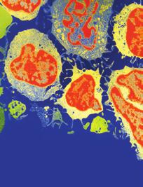
 Resolution of intrahepatic duct dilation and CBD narrowing after steroid therapy. (c) 2.")

 revealed an area of enhancement concerning for neoplasm at the biliary confluence, narrowing of the central right and left intrahepatic bile ducts and intrahepatic")

2 2 Case Reports in Gastrointestinal Medicine (a) (b) (c) (d) Figure 1: MRCP findings: Case 1. (a) Narrowing and stricture of the common bile duct with dilated intrahepatic ducts. (b) Resolution of intrahepatic duct dilation and CBD narrowing after steroid therapy. (c) 2.7 cm hypointense lesion in the pancreatic head (black arrow) near a region of normal pancreatic parenchyma (white arrow). (d) Resolution after steroid therapy. The pancreatic parenchyma is homogeneous and isointense with the adjacent liver. of which were negative. A computed tomography (CT) of the abdomen demonstrated diffuse intrahepatic biliary ductal dilatation. Magnetic resonance imaging (MRI) revealed an area of enhancement concerning for neoplasm at the biliary confluence, narrowing of the central right and left intrahepatic bile ducts and intrahepatic biliary duct dilation (Figure 1(a)). Diffuse pancreatic enhancement was noted. An ERCP was performed and demonstrated a complex narrow biliary stricture involving both right and left hepatic ducts and extending to the common hepatic duct, consistent with type 4 pattern cholangiography [5]. Biliary brushings and FISH were negative for malignancy. IgG4 immunostaining of biliary brushing specimen was not pursued. Subsequently our patient underwent three ERCPs with biliary dilation/stent exchange and biliary brushings for cytology and FISH, all of which revealed no evidence of malignancy. Six months after initial presentation, a repeat MRI was obtained which demonstrated a new 2.7 cm mass at the posterior aspect of the head of the pancreas extending into the uncinate process (Figure 1(c)). An endoscopic ultrasound with fine needle aspiration was performed and revealed benign pancreatic ductal cells and acini in addition to fragments of chronically inflamed stromal material. IgG4 immunostaining showed rare IgG4-positive inflammatory cells and was felt to be inconclusive. Periampullary mucosal biopsies showed small bowel mucosa without diagnostic abnormality. Incidentally, a CT of the chest revealed bilateral hilar, paratracheal and mediastinal enlarged lymph nodes, small pulmonary nodules, and ground glass opacities. After a thorough negative workup for malignancy, based on the available clinical, radiological, and extrapancreatic presentation, a diagnosis of type 1 AIP and IgG4-SC was suspected. The patient was started on 40 mg of prednisone and continued for 4 weeks and gradually tapered. His symptoms abated and a follow-up MRI performed demonstrated complete resolution of the pancreatic mass and biliary strictures (Figures 1(b) and 1(d)). With this clinical and radiologic response, the patient was diagnosed with IgG4-related SC and thesymptomshavenotrecurredafter3yearsoffollow-up. 3. Case 2 A 45-year-old gentleman with prior history of small duct primary sclerosing cholangitis and Crohn s disease presented with symptoms of abdominal pain, pruritus, dark urine, and jaundice. Laboratory analysis showed an AST of 152, ALT of 158, alkaline phosphatase of 531, total bilirubin of 5.6, direct bilirubin of 3.8, total protein of 7.4, albumin of 4.0, lipase of
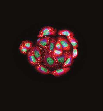
 Diffusion restriction shows hyperintensity and edema in the pancreatic tail. (d) Resolution after steroid therapy. 157, amylase of 82, CA of 19-9 194, CEA of 2.")
 and 2(c)).")


3 Case Reports in Gastrointestinal Medicine 3 (a) (b) (c) (d) Figure 2: MRCP findings: Case 2. (a) Extrahepatic bile duct narrowing. (b) Resolution after steroid therapy. (c) Diffusion restriction shows hyperintensity and edema in the pancreatic tail. (d) Resolution after steroid therapy. 157, amylase of 82, CA of , CEA of 2.9, total IgG of 1620, and IgG4 of MRI of the abdomen demonstrated diffuse enlargement of the body and the tail of the pancreas. No focal enhancing pancreatic mass was identified. MRCP revealed new irregular narrowing of the common hepatic duct, upper common bile duct beaded dilatation, and narrowing of the intrahepatic bile ducts (Figures 2(a) and 2(c)). Several prominent porta hepatis lymph nodes were noted. AnERCPwasperformedandrevealedmildintrahepatic biliary strictures and moderately severe common bile duct stricture, most consistent with a type 2 cholangiogram [5]. Sphincterotomy and dilation were performed and brushings were obtained. Biliary brushings revealed reactive epithelial cells and FISH was negative for malignancy; IgG4 immunostaining was not performed. Based on the clinical findings and parenchymal and ductal imaging, a diagnosis of autoimmune pancreatitis and IgG4-SC was made and patient was initiated on prednisone 60 mg daily. Within 2 weeks of initiating steroid therapy, his laboratory tests show significant improvement with resolution of cholestasis and MRCP showed significant improvement in intrahepatic and extrahepatic bile duct changes with resolution of pancreatic enlargement (Figures 2(b) and 2(d)). A decision was made to continue immunosuppressive therapy and he was started on mycophenolate 500 mg twice daily (prior history of Imuraninduced pancreatitis). His steroids were tapered gradually and stopped with no subsequent relapse. 4. Case 3 A 52-year-old gentleman presented to the gastroenterology clinic with a history of acute pancreatitis and complaints of abdominal pain. He denied abnormal weight loss or alcohol use. Laboratory analysis showed an amylase of 495, lipase of 1305, bilirubin of 2.8, AST of 658, ALT of 533, and alkaline phosphatase of 65. He was diagnosed with acute pancreatitis and treated conservatively. Abdominal MR imaging revealed segmental biliary dilatation from the bifurcation proximally totherightandleftsystemwithcbdstricture.thecommon hepatic duct was 10 mm and the CBD distally was 2.5 mm. The pancreatic duct was unremarkable. Additionally, there was significant retroperitoneal adenopathy and diffuse pulmonary nodules on the imaging. An ERCP was performed and revealed a 2.5 cm highgradestrictureinthedistalcommonbileduct,consistentwith type 1 cholangiogram [5]. Brushing and biopsies were taken and a biliary stent was placed. A CA 19-9 at the time was 37. The biliary biopsies and FISH were negative for malignancy. IgG4 immunostaining of biliary brushings demonstrated occasional, scattered positive cells of indeterminate clinical significance. An EUS performed revealed a hypoechoic mass in the head of the pancreas (measuring mm) and the body (measuring mm). Pancreatic biopsies showed mild atypical epithelial cells. Serum total IgG was elevated at 2,430, but IgG4 level was normal at On follow-up, he
4 4 Case Reports in Gastrointestinal Medicine was noted to have enlarged submandibular salivary glands. Given a negative diagnostic workup, a CT guided biopsy of submandibular glands was pursued which confirmed chronic inflammatory infiltrate with numerous IgG4 positive plasma cells. A diagnosis of type 1 AIP and IgG4-SC was made and the patient was treated with prednisone 40 mg daily. He was alsostartedonimuran150mgdailyandhisprednisonewas slowly tapered and stopped. The patient improved clinically, his laboratory parameters normalized, and repeat imaging showed dramatic improvement in biliary strictures with a normal-appearing pancreas. 5. Discussion IgG4-related disease is a multiorgan fibroinflammatory disorder characterized by organ infiltration with lymphoplasmacytic cells rich in IgG4, storiform fibrosis, obliterative phlebitis, elevated serum IgG4 levels, and response to steroids. Two types of AIP have been described in the literature. Type 1 AIP is related to IgG4-related disease, while type 2 (idiopathic duct centric pancreatitis) is associated with a distinct characteristic histological pattern and is usually not accompanied by extrapancreatic manifestations [6]. Type 1 AIPisprevalentworldwideandtypicallyaffectsmiddle-aged and elderly men. The most common presentation of type 1 AIP is obstructive jaundice. Other symptoms can include abdominal pain, weight loss, and anorexia [7]. Additionally, AIP can present as acute and chronic pancreatitis and could closely mimic pancreatic cancer. The commonest extrapancreatic manifestation of type 1 AIP is IgG4-SC, which can mimic cholangiocarcinoma. Other extrapancreatic manifestations include salivary gland enlargement, lymphadenopathy, pulmonary disease with nodules, hydronephrosis, and retroorbital disease [8]. In our current series, we describe three patients with type 1 AIP and IgG4-SC with normal serum IgG4 levels in whom steroid treatment was successful and surgery was entirely avoided. Each of these cases provides lessons in the diagnosis of IgG4-SC in patients with normal IgG4 levels. In Case 1, cholangiocarcinoma was suspected on the basis of clinical findings and compatible radiologic findings on CT, MR,andERCP.However,athoroughandmeasuredapproach to diagnosis led to four ERCPs with biliary brushings and FISH, which were negative for malignancy. This allowed the patient to defer needless surgery. Follow-up MRI after six months revealed a new pancreatic mass which would not be expected with cholangiocarcinoma, and pulmonary nodules and mediastinal lymphadenopathy were seen on CT imaging, suggesting extrapancreatic involvement. When an EUS with FNA of the pancreatic mass did not reveal malignancy, but rather a chronic stromal infiltrate, the combination of pathologic findings and extrapancreatic involvement led to a trial of prednisone. With dramatic clinical and radiologic response to steroids, the diagnosis of type 1 AIP was made and surgery was avoided. In Case 2, the patient presented with abdominal pain and jaundice. Imaging showed diffuse enlargement of the pancreas, as well as biliary strictures and pulmonary nodules. Serological tests were negative for IgG4, and histology and FISH were negative from ERCP brushings. Based on clinical and radiological presentation, a diagnosis of AIP was considered and empiric treatment was initiated with steroids. The patient responded well, with resolution of pancreatic enlargement and biliary strictures on imaging. In our 3rd case, the patient presented with a diagnosis of acute pancreatitis of unknown etiology. His abdominal imaging, however, revealed biliary strictures which raised the question of an uncommon cause of pancreatitis. By carefully pursuing this clinical clue, misdiagnosis was avoided. Two ERCPS with biliary brushings and FISH were negative for malignancy, and EUS demonstrated pancreatic masses in theheadandthebodyofthepancreas,biopsiesofwhich were also negative for malignancy. The subsequent detection of an enlarged submandibular gland allowed a diagnostic biopsy that demonstrated chronic inflammatory infiltrate with numerous IgG4 positive plasma cells. This patient also was treated successfully with steroids, and continues on Imuran with no relapse. The diagnosis of AIP is challenging and a number of diagnostic criteria (Japan, Italy, the United States, and Korea) have been proposed in the recent years. The HISORt criteria proposed by the Mayo Clinic to diagnose AIP include histology, imaging findings, elevated serum IgG4 levels, other organs involvement, and response to steroids [1]. In order to unify the disparate diagnostic criteria, a multinational group convened in 2011 and developed International Consensus Diagnostic Criteria (ICDC) for AIP [3]. The ICDC criteria are based on the following parameters: pancreatic parenchymal imaging, pancreatic ductal imaging (ERCP), serum IgG4 level, other organs involvement, histology of the pancreas, and response to steroid treatment. These parameters may provide level 1 (highly suggestive) or level 2 (supportive) evidence that will aid in definitive diagnosis and are associated with well-defined diagnostic algorithms. The ICDC criteria can still be successfully applied in cases of seronegative IgG4-associated AIP and IgG4-SC. In Case 1, MRI pancreatography findings were indeterminate for AIP. A thorough evaluation for possible malignancy was negative. Pancreatic ductal imaging was normal. ERCP revealed level 1 findings, but serology was negative and no additional organ involvement (OOI) was noted. Two level 1 criteria were not met, so EUS-guided pancreatic biopsy was performed and was inconclusive. Because of the presence of a single level 1 OOI, steroid trial was pursued and resulted in radiographic and symptomatic resolution, supporting a diagnosis of type 1 AIP. In Case 2,initialMRpancreatography was typical for type 1 AIP. There was a single level 1 OOI criterion (multiple proximal and distal CBD strictures); thus, a steroid trial was pursued and resulted in substantial improvement, confirming the diagnosis. In Case 3, neither pancreatic parenchymal imaging nor ductal imaging was typical for AIP. However, level 1 OOI findings were seen on ERCP. A pancreatic biopsy was thus performed and yielded inconclusive findings. At this point, a steroid trial would have been reasonable; however, the interval development of submandibular gland enlargement allowed for establishment of definitive histological diagnosis.
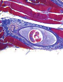
5 Case Reports in Gastrointestinal Medicine 5 The typical histological findings in type 1 AIP are comprised of abundant infiltration of lymphocytes, IgG4 positive plasma cells (greater than 10 IgG4 positive cells per high power field), fibrosis in periductal and interlobular areas, and obliterative phlebitis [9]. Tissue samples obtained with EUS-guided pancreatic trucut biopsies provide accurate histological diagnosis as compared to FNA alone [10]. However, if tissue samples are not available, the diagnosis can be made with the aid of imaging studies, extrapancreatic involvement, and response to steroids [2, 11]. In our series, two patients did not have definitive histopathological findings of lymphoplasmacytic infiltrate with IgG4 cells. Yet, based on the collateral evidence, all patients were treated with steroids and immunosuppressive medications and thus surgery was avoided. Increased serum IgG4 levels (>135mg/dL) have been frequently described in patients with type 1 AIP and have beenincludedasapartofdiagnosticcriteria.hamanoand colleagues first described an association between elevated IgG4 and AIP in 2001 and reported the sensitivity and specificity of elevated IgG4 for AIP as 95 and 97%, respectively [12]. However, subsequent studies of larger AIP patient cohorts demonstrated IgG4 seropositivity in as few as 76% [13]. Thus, further estimates of sensitivity and specificity for AIP diagnosis have been shown to vary widely and arereportedaslowas62and59%,respectively[14].this heterogeneity is largely attributable to the application of disparate diagnostic criteria in study design, a fact which prompted the ICDC s creation. Ultimately, the sole elevation of serum IgG4 is inadequate to establish a definitive diagnosis of AIP. Some have suggested that it may play a clearer role in the differentiation of AIP from malignancy, particularly at higher cutoff levels where specificity is enhanced [14]. It has also been suggested that IgG4-seronegative AIP may behave as a different clinicopathologic entity. When compared to IgG4-seropositive patients, those with negative serology are more likely to be female, present with abdominal pain or acute pancreatitis, and demonstrate segmental pancreatic body and/or tail enlargement [15, 16]. They are less likely to present with obstructive jaundice and less frequently require maintenance immunosuppression, owing to a lower relapserate[17].inthisway,igg4levelmayserveasa prognostic biomarker. Patients with IgG4 seropositivity are also noted to have a higher prevalence of extrapancreatic manifestations of IgG4-related disease, particularly lacrymal and salivary gland involvement and hilar lymphadenopathy [17]. In contrast to this data, all of our IgG4-seronegative patients were male and presented with extrapancreatic disease (i.e., IgG4-SC and submandibular involvement), with 2 of 3 presenting with obstructive jaundice and requiring maintenance immunosuppression. This divergence from previously documented patterns is likely a manifestation of the observational quality of existing data and the heterogeneous clinical course of this relatively novel disease. In Cases 1 and 3, pancreatic imaging was not suggestive of typical features for AIP, while cholangiography suggested the presence of IgG4-SC. While AIP and IgG4-SC often coexist, it is important to recognize that they are likely distinct, if closely related, manifestations of the single overarching clinical entity of IgG4-related disease. Several studies have reported histologically proven IAC (i.e., lymphoplasmacytic infiltrate, storiform fibrosis, and obliterative phlebitis on resection specimen) in the absence of any clinical or radiographic evidence of AIP [18 20]. As was seen in our cases and is corroborated by the ICDC algorithms, biliary findings suspicious for IgG4-SC without characteristic evidence of AIP should not lead one away from a meticulous evaluation for IgG4-related disease. The diagnosis of isolated IgG4-SC is challenging and IgG4 serology alone is inadequate to establish the diagnosis, as has been previously discussed. The sensitivity of IgG4 for IgG4-SC diagnosis has been estimated as 74%, a level comparable to estimates in AIP [18]. Several of the reported cases of isolated IgG4-SC demonstrate borderline elevations in IgG4 [20] and elevation of IgG4 in other stricturing biliary disorders, such as PSC, has been observed [21]. The addition of IgG4 immunostaining is often critical to accurate identification. Ampullary, hepatic, or biliary duct biopsies have been demonstrated as useful modalities, with relatively variable sensitivities (24 80%), but high specificities (91 100%) for the diagnosis of IAC [22]. Although our goal is early identification and treatment of benign IgG4-related disease and avoidance of surgery, it should be emphasized that the misdiagnosis of AIP/IgG4- SC in the setting of pancreatic cancer or cholangiocarcinoma must be strictly avoided [23]. In a recent UK study evaluating long-term outcomes in patients with AIP/IgG4-SC, 13 of 115 patients (11%) were diagnosed with a malignancy before or after the diagnosis of AIP [8]. Hence, caution should be exercised to rule out malignancy prior to embarking on investigations aimed at diagnosing AIP/IgG4-SC. On the other hand, the reported incidence of benign disease after pancreatoduodenectomy for a presumed malignancy is 5 13% and the incidence of AIP in the benign resected specimens is 30 43% [24]. Hence, diagnosis of AIP should be made cautiously taking into account all other collateral information such as characteristic imaging findings, serologic parameters, extrapancreatic involvement, and perhaps treatment with a short course of steroids prior to considering surgery. In our case series, we engaged in a thorough evaluation to exclude malignancy and include other findings within the spectrum of IgG4-related disease before diagnosis and initiation of steroid therapy. Type 1 AIP/IgG4-SC typically responds dramatically to steroids. In our series we treated all patients with prednisone 40 mg daily for 4 weeks and tapered by 5 mg/week with close clinical follow-up, liver function tests, and imaging studies. In addition, all patients required biliary drainage procedures. Even though disease relapse is common with type 1 AIP, (30% 50%) none of our patients had clinical or biochemical relapse[25,26].however,inourseriestwooutof3patients are maintained on immunosuppressive therapy (Imuran or mycophenolate). In summary, our case series highlights the benefit of early recognition and medical treatment of IgG4-negative type 1 AIP and IgG4-SC. Despite normal IgG4 levels, a diagnosis of IgG4-related AIP and IgG4-SC should be considered when there is a typical clinical presentation,
6 6 Case Reports in Gastrointestinal Medicine thepresenceofcharacteristicpancreatobiliaryimagingfindings, and extrapancreatic involvement. When IgG4-related AIP is recognized without elevation of IgG4, the patient can be spared morbidity associated with unnecessary surgical treatment by initiating a steroid trial and monitoring patients carefully. Conflict of Interests The authors declare that there is no conflict of interests regarding the publication of this paper. References [1] S. T. Chari, T. C. Smyrk, M. J. Levy et al., Diagnosis of autoimmune pancreatitis: the Mayo clinic experience, Clinical Gastroenterology and Hepatology, vol.4,no.8,pp , [2] M. Otsuki, J. B. Chung, K. Okazaki et al., Asian diagnostic criteria for autoimmune pancreatitis: consensus of the Japan- Korea symposium on autoimmune pancreatitis, Gastroenterology,vol.43,no.6,pp ,2008. [3] T. Shimosegawa, S. T. Chari, L. Frulloni et al., International consensus diagnostic criteria for autoimmune pancreatitis: guidelines of the international association of pancreatology, Pancreas,vol.40,no.3,pp ,2011. [4] J. K. Ryu, J. B. Chung, S. W. Park et al., Review of 67 patients with autoimmune pancreatitis in Korea: a multicenter nationwide study, Pancreas,vol.37,no.4,pp ,2008. [5] T. Nakazawa, I. Naitoh, K. Hayashi et al., Diagnostic criteria for IgG4-related sclerosing cholangitis based on cholangiographic classification, Gastroenterology, vol. 47, no. 1, pp , [6] G. Klöppel, S. Detlefsen, S. T. Chari, D. S. Longnecker, and G. Zamboni, Autoimmune pancreatitis: the clinicopathological characteristics of the subtype with granulocytic epithelial lesions, Gastroenterology,vol.45,no.8,pp , [7]A.Raina,D.Yadav,A.M.Krasinskasetal., Evaluationand management of autoimmune pancreatitis: experience at a large US center, The American Gastroenterology, vol. 104, no. 9, pp , [8] M. T. Huggett, E. L. Culver, M. Kumar et al., Type 1 autoimmune pancreatitis and IgG4-related sclerosing cholangitis is associated with extrapancreatic organ failure, malignancy, and mortality in a prospective UK cohort, American Gastroenterology,vol.109,pp ,2014. [9] V.Deshpande,Y.Zen,J.K.Chanetal., Consensusstatementon the pathology of IgG4-related disease, Modern Pathology, vol. 25,no.9,pp ,2012. [10] N. Mizuno, V. Bhatia, W. Hosoda et al., Histological diagnosis of autoimmune pancreatitis using EUS-guided trucut biopsy: a comparison study with EUS-FNA, Gastroenterology, vol.44,no.7,pp ,2009. [11] S. T. Chari, N. Takahashi, M. J. Levy et al., A diagnostic strategy to distinguish autoimmune pancreatitis from pancreatic cancer, Clinical Gastroenterology and Hepatology, vol. 7, no.10, pp , [12] H. Hamano, S. Kawa, A. Horiuchi et al., High serum IgG4 concentrations in patients with sclerosing pancreatitis, The New England Medicine, vol. 344, no. 10, pp , [13] R. P. Sah, S. T. Chari, R. Pannala et al., Differences in clinical profile and relapse rate of type 1 versus type 2 autoimmune pancreatitis, Gastroenterology,vol.139,no.1,pp ,2010. [14] R.Sadler,R.W.Chapman,D.Simpsonetal., Thediagnostic significance of serum IgG4 levels in patients with autoimmune pancreatitis: a UK study, European Gastroenterology and Hepatology, vol. 23, no. 2, pp , [15] T. Kamisawa, K. Takuma, T. Tabata et al., Serum IgG4-negative autoimmune pancreatitis, Gastroenterology, vol. 46, no. 1, pp , [16] A. Ghazale, S. T. Chari, T. C. Smyrk et al., Value of serum IgG4 in the diagnosis of autoimmune pancreatitis and in distinguishing it from pancreatic cancer, The American Journal of Gastroenterology,vol.102,no.8,pp ,2007. [17] S. Kawa, T. Ito, T. Watanabe et al., The utility of serum IgG4 concentrations as a biomarker, International Rheumatology, vol. 2012, Article ID , 4 pages, [18] A. Ghazale, S. T. Chari, L. Zhang et al., Immunoglobulin G4- associated cholangitis: clinical profile and response to therapy, Gastroenterology,vol.134,no.3,pp ,2008. [19]Y.Zen,K.Harada,M.Sasakietal., IgG4-relatedsclerosing cholangitis with and without hepatic inflammatory pseudotumor, and sclerosing pancreatitis-associated sclerosing cholangitis: do they belong to a spectrum of sclerosing pancreatitis? The American Surgical Pathology, vol.28,no.9,pp , [20] H. Hamano, S. Kawa, T. Uehara et al., Immunoglobulin G4- related lymphoplasmacytic sclerosing cholangitis that mimics infiltrating hilar cholangiocarcinoma: part of a spectrum of autoimmune pancreatitis? Gastrointestinal Endoscopy, vol.62, no. 1, pp , [21] F. D. Mendes, R. Jorgensen, J. Keach et al., Elevated serum IgG4 concentration in patients with primary sclerosing cholangitis, American Gastroenterology, vol.101,no.9,pp , [22] Y. Zen and Y. Nakanuma, IgG4 cholangiopathy, International Hepatology,vol.2012,ArticleID472376,6pages,2012. [23] T.B.Gardner,M.J.Levy,N.Takahashi,T.C.Smyrk,andS.T. Chari, Misdiagnosis of autoimmune pancreatitis: a caution to clinicians, AmericanJournalofGastroenterology,vol.104,no.7, pp ,2009. [24] H. J. Asbun, K. Conlon, L. Fernandez-Cruz et al., When to perform a pancreatoduodenectomy in the absence of positive histology? A consensus statement by the International Study Group of Pancreatic Surgery, Surgery, vol. 155, no. 5, pp , [25] T. Kamisawa, T. Shimosegawa, K. Okazaki et al., Standard steroid treatment for autoimmune pancreatitis, Gut, vol. 58, no. 11, pp , [26] S. T. Chari and J. A. Murray, Autoimmune pancreatitis, part II: the relapse, Gastroenterology,vol.134,no.2,pp ,2008.


7 MEDIATORS of INFLAMMATION The Scientific World Journal Gastroenterology Research and Practice Diabetes Research International Endocrinology Immunology Research Disease Markers Submit your manuscripts at BioMed Research International PPAR Research Obesity Ophthalmology Evidence-Based Complementary and Alternative Medicine Stem Cells International Oncology Parkinson s Disease Computational and Mathematical Methods in Medicine AIDS Behavioural Neurology Research and Treatment Oxidative Medicine and Cellular Longevity
Diagnostic Algorithm for Autoimmune Pancreatitis in Korea
 Review Article The Korean Journal of Pancreas and Biliary Tract 2014;19(1):7-12 pissn 1976-3573 eissn 2288-0941 한국에서자가면역췌장염의진단전략 성균관대학교의과대학삼성서울병원내과학교실 이종균 Diagnostic Algorithm for Autoimmune Pancreatitis
Autoimmune Pancreatitis: A Great Imitator
 Massachusetts General Hospital Harvard Medical School Autoimmune Pancreatitis: A Great Imitator Dushyant V Sahani MD dsahani@partners.org Autoimmune Pancreatitis: Learning Objectives Clinical manifestations
CASE 01 LA Path Slide Seminar 13 March, 08. Deepti Dhall, MD Department of Pathology and Laboratory Medicine Cedars-Sinai Medical Center
 CASE 01 LA Path Slide Seminar 13 March, 08 Deepti Dhall, MD Department of Pathology and Laboratory Medicine Cedars-Sinai Medical Center Clinical History 60 year old male presented with obstructive jaundice
Comparison of multidetector-row computed tomography findings of IgG4-related sclerosing cholangitis and cholangiocarcinoma
 Comparison of multidetector-row computed tomography findings of IgG4-related sclerosing cholangitis and cholangiocarcinoma Poster No.: C-0245 Congress: ECR 2014 Type: Scientific Exhibit Authors: M. Yata,
CASE REPORT. Abstract. Introduction. Case Report
 CASE REPORT Branch Duct Intraductal Papillary Mucinous Neoplasms of the Pancreas Involving Type 1 Localized Autoimmune Pancreatitis with Normal Serum IgG4 Levels Successfully Diagnosed by Endoscopic Ultrasound-guided
The most common presentation of autoimmune pancreatitis. A Diagnostic Strategy to Distinguish Autoimmune Pancreatitis From Pancreatic Cancer
 CLINICAL GASTROENTEROLOGY AND HEPATOLOGY 2009;7:1097 1103 A Diagnostic Strategy to Distinguish Autoimmune Pancreatitis From Pancreatic Cancer SURESH T. CHARI,* NAOKI TAKAHASHI, MICHAEL J. LEVY,* THOMAS
Immunoglobulin G4-Related Disease with Several Inflammatory Foci
 CASE REPORT Immunoglobulin G4-Related Disease with Several Inflammatory Foci Akira Sakamaki 1, Kenya Kamimura 1, Kazuhiko Shioji 1, Junko Sakurada 2, Takeshi Nakatsue 3, Yoko Wada 3, Michitaka Imai 1,
IgG4-Negative Autoimmune Pancreatitis with Sclerosing Cholangitis and Colitis: Possible Association with Primary Sclerosing Cholangitis?
 CASE REPORT IgG4-Negative Autoimmune Pancreatitis with Sclerosing Cholangitis and Colitis: Possible Association with Primary Sclerosing Cholangitis? Keita Saeki 1, Shigenari Hozawa 1, Naoteru Miyata 1,
Case Scenario 1. Discharge Summary
 Case Scenario 1 Discharge Summary A 69-year-old woman was on vacation and noted that she was becoming jaundiced. Two months prior to leaving on that trip, she had had a workup that included an abdominal
How 5 Diseases Became One. Moez Tajdin R3 McGill University
 How 5 Diseases Became One Moez Tajdin R3 McGill University Conflicts of Interest None! Mr. M. ID: 65 M PMH Benign prostatic hyperplasia Prostate cancer Awaiting biopsy Skin rash Dyslipidemia Hypertension
Value of Serum IgG4 in the Diagnosis of Autoimmune Pancreatitis and in Distinguishing it from Acute and Chronic Pancreatitis of Other Etiology
 94 Jul 2017 Vol 10 No.3 North American Journal of Medicine and Science Original Research Value of Serum IgG4 in the Diagnosis of Autoimmune Pancreatitis and in Distinguishing it from Acute and Chronic
Review Article The Utility of Serum IgG4 Concentrations as a Biomarker
 International Rheumatology Volume 2012, Article ID 198314, 4 pages doi:10.1155/2012/198314 Review Article The Utility of Serum IgG4 Concentrations as a Biomarker Shigeyuki Kawa, 1 Tetsuya Ito, 2 Takayuki
Isolated Mass-Forming IgG4-Related Cholangitis as an Initial Clinical Presentation of Systemic IgG4-Related Disease
 Journal of Pathology and Translational Medicine 2016; 50: 300-305 CASE STUDY Isolated Mass-Forming IgG4-Related Cholangitis as an Initial Clinical Presentation of Systemic IgG4-Related Disease Seokhwi
Autoimmune Pancreatitis & Cholangiopathy. Goal and Objectives
 Autoimmune Pancreatitis & Cholangiopathy Kaveh Sharzehi, MD, MS Assistant Professor of Medicine Medical Director of Endoscopy Section of Gastroenterology Lewis Katz School of Medicine at Temple University
Pictorial review of Benign Biliary tract abnormality on MRCP/MRI Liver with Endoscopic (including splyglass) and Endoscopic Ultrasound correlation
 and Endoscopic Ultrasound correlation") Pictorial review of Benign Biliary tract abnormality on MRCP/MRI Liver with Endoscopic (including splyglass) and Endoscopic Ultrasound correlation Poster No.: C-2617 Congress: ECR 2015 Type: Educational
Pictorial review of Benign Biliary tract abnormality on MRCP/MRI Liver with Endoscopic (including splyglass) and Endoscopic Ultrasound correlation Poster No.: C-2617 Congress: ECR 2015 Type: Educational
Differentiating Immunoglobulin G4-Related Sclerosing Cholangitis from Hilar Cholangiocarcinoma
 Gut and Liver, Vol. 7, No. 2, March 2013, pp. 234-238 ORiginal Article Differentiating Immunoglobulin G4-Related Sclerosing Cholangitis from Hilar Cholangiocarcinoma Taku Tabata*, Terumi Kamisawa*, Seiichi
Case Report The Challenging Diagnosis of Pancreatic Masses: Not All Tumors Are Cancers
 Case Reports in Medicine Volume 2015, Article ID 832463, 4 pages http://dx.doi.org/10.1155/2015/832463 Case Report The Challenging Diagnosis of Pancreatic Masses: Not All Tumors Are Cancers Alessandro
Type 2 Autoimmune Pancreatitis with Crohn s Disease
 doi: 10.2169/internalmedicine.0213-17 Intern Med 57: 2957-2962, 2018 http://internmed.jp CASE REPORT Type 2 Autoimmune Pancreatitis with Crohn s Disease Yoon Suk Lee, Nam-Hoon Kim, Jun Hyuk Son, Jung Wook
IgG4-related sclerosing disease
 IgG4-related sclerosing disease TERUMI KAMISAWA, KENSUKE TAKUMA, NAOTO EGAWA Department of Internal Medicine Tokyo Metropolitan Komagome Hospital 3-18-22 Honkomagome, Bunkyo-ku, Tokyo 113-8677, Japan JAPAN
Pancreas Case Scenario #1
 Pancreas Case Scenario #1 An 85 year old white female presented to her primary care physician with increasing abdominal pain. On 8/19 she had a CT scan of the abdomen and pelvis. This showed a 4.6 cm mass
Tratamiento endoscópico de la CEP. En quien como y cuando?
 Tratamiento endoscópico de la CEP. En quien como y cuando? Andrés Cárdenas, MD, MMSc, PhD, AGAF, FAASLD GI / Liver Unit, Hospital Clinic Institut de Malalties Digestives i Metaboliques University of Barcelona
Personal Profile. Name: 劉 XX Gender: Female Age: 53-y/o Past history. Hepatitis B carrier
 Personal Profile Name: 劉 XX Gender: Female Age: 53-y/o Past history Hepatitis B carrier Chief complaint Fever on and off for 2 days Present illness 94.10.14 Sudden onset of epigastric pain 94.10.15 Fever
Primary Sclerosing Cholangitis and Cholestatic liver diseases. Ahsan M Bhatti MD, FACP Bhatti Gastroenterology Consultants
 Primary Sclerosing Cholangitis and Cholestatic liver diseases Ahsan M Bhatti MD, FACP Bhatti Gastroenterology Consultants I have nothing to disclose Educational Objectives What is PSC? Understand the cholestatic
IgG4-Related Sclerosing Cholangitis
 REVIEW IgG4-Related Sclerosing Cholangitis Emma L. Culver, B.Sc., M.B.Ch.B., D.Phil., M.R.C.P.,* and Eleanor Barnes, M.B.B.S., D.Phil., M.R.C.P.*,, BACKGROUND IgG4-related sclerosing cholangitis (IgG4-SC)
Autoimmune pancreatitis (AIP) can be defined as a
 can be defined as a") CLINICAL GASTROENTEROLOGY AND HEPATOLOGY 2006;4:1010 1016 Diagnosis of Autoimmune Pancreatitis: The Mayo Clinic Experience SURESH T. CHARI,* THOMAS C. SMYRK, MICHAEL J. LEVY,* MARK D. TOPAZIAN,* NAOKI
CLINICAL GASTROENTEROLOGY AND HEPATOLOGY 2006;4:1010 1016 Diagnosis of Autoimmune Pancreatitis: The Mayo Clinic Experience SURESH T. CHARI,* THOMAS C. SMYRK, MICHAEL J. LEVY,* MARK D. TOPAZIAN,* NAOKI
Noncalculous Biliary Disease Dean Abramson, M.D. Gastroenterologists, P.C. Cedar Rapids. Cholestasis
 Noncalculous Biliary Disease Dean Abramson, M.D. Gastroenterologists, P.C. Cedar Rapids Cholestasis Biochemical hallmark Impaired bile flow from liver to small intestine Alkaline phosphatase is primary
Navigating the Biliary Tract with CT & MR: An Imaging Approach to Bile Duct Obstruction
 Navigating the Biliary Tract with CT & MR: An Imaging Approach to Bile Duct Obstruction Ann S. Fulcher, MD Medical College of Virginia Virginia Commonwealth University Richmond, Virginia Objectives To
Case Report Uncommon Mixed Type I and II Choledochal Cyst: An Indonesian Experience
 Case Reports in Surgery Volume 2013, Article ID 821032, 4 pages http://dx.doi.org/10.1155/2013/821032 Case Report Uncommon Mixed Type I and II Choledochal Cyst: An Indonesian Experience Fransisca J. Siahaya,
Colangitis Esclerosante Primaria: Manejo Clínico y Endoscópico
 Colangitis Esclerosante Primaria: Manejo Clínico y Endoscópico Andrés Cárdenas, MD, MMSc, PhD, AGAF, FAASLD GI / Liver Unit, Hospital Clinic Institut de Malalties Digestives i Metaboliques Associate Professor
IgG4 Related disease a retrospective descriptive study highlighting Canadian experiences in diagnosis and management
 Patel et al. BMC Gastroenterology 2013, 13:168 RESEARCH ARTICLE Open Access IgG4 Related disease a retrospective descriptive study highlighting Canadian experiences in diagnosis and management Harshna
Autoimmune pancreatitis (AIP), a clinical entity originally
, a clinical entity originally") Autoimmune Pancreatitis: A Multiorgan Disease Presenting a Conundrum for Clinicians in the West Eileen Kim, MD, Rebecca Voaklander, MD, Franklin E. Kasmin, MD, William H. Brown, MD, Rifat Mannan, MD, and
Autoimmune Pancreatitis: A Multiorgan Disease Presenting a Conundrum for Clinicians in the West Eileen Kim, MD, Rebecca Voaklander, MD, Franklin E. Kasmin, MD, William H. Brown, MD, Rifat Mannan, MD, and
Endoscopic Ultrasonography Assessment for Ampullary and Bile Duct Malignancy
 Diagnostic and Therapeutic Endoscopy, Vol. 3, pp. 35-40 Reprints available directly from the publisher Photocopying permitted by license only (C) 1996 OPA (Overseas Publishers Association) Amsterdam B.V.
Key Points: Autoimmune Liver Disease: Update for Pathologists from the Hepatologist s Perspective. Jenny Heathcote, MD. University of Toronto
 Autoimmune Liver Disease: Update for Pathologists from the Hepatologist s Perspective Jenny Heathcote, MD University of Toronto Key Points: AILD comprise autoimmune hepatitis, primary biliary cirrhosis
Lymphoplasmacytic sclerosing pancreatitis without IgG4 tissue infiltration or serum IgG4 elevation: IgG4-related disease without IgG4
 238 & 2015 USCAP, Inc All rights reserved 0893-3952/15 $32.00 Lymphoplasmacytic sclerosing pancreatitis without IgG4 tissue infiltration or serum IgG4 elevation: IgG4-related disease without IgG4 Phil
Autoimmune Pancreatitis: A Succinct Overview
 REVIEW ARTICLE Autoimmune Pancreatitis: A Succinct Overview Juan Putra, Xiaoying Liu Department of Pathology, Dartmouth-Hitchcock Medical Center, One Medical Center Drive Lebanon, NH 03756, USA ABSTRACT
A Review of Liver Function Tests. James Gray Gastroenterology Vancouver
 A Review of Liver Function Tests James Gray Gastroenterology Vancouver Copyright 2017 by Sea Courses Inc. All rights reserved. No part of this document may be reproduced, copied, stored, or transmitted
Case Report IgG4-Related Nasal Pseudotumor
 Case Reports in Otolaryngology Volume 2015, Article ID 749890, 4 pages http://dx.doi.org/10.1155/2015/749890 Case Report IgG4-Related Nasal Pseudotumor L. K. Døsen, 1 P. Jebsen, 2 B. Dingsør, 3 and R.
IgG4 cholangitis Case Report: Joanne Verheij, MD, PhD Department of Pathology Academic Medical Center Amsterdam.
 IgG4 cholangitis New Perspectives on Biliary Tract Disease Case Report: male, 64 yrs, truck driver and car industry worker Joanne Verheij, MD, PhD Department of Pathology Academic Medical Center Amsterdam
Autoimmune Hepatobiliary Diseases PROF. DR. SABEHA ALBAYATI CABM,FRCP
 Autoimmune Hepatobiliary Diseases PROF. DR. SABEHA ALBAYATI CABM,FRCP Autoimmune hepatobiliary diseases The liver is an important target for immunemediated injury. Three disease phenotypes are recognized:
Case Report Five-Year Survival after Surgery for Invasive Micropapillary Carcinoma of the Stomach
 Case Reports in Surgery Volume 2013, Article ID 560712, 4 pages http://dx.doi.org/10.1155/2013/560712 Case Report Five-Year Survival after Surgery for Invasive Micropapillary Carcinoma of the Stomach Shigeo
ACG Clinical Guideline: Primary Sclerosing Cholangitis
 ACG Clinical Guideline: Primary Sclerosing Cholangitis Keith D. Lindor, MD, FACG 1, Kris V. Kowdley, MD, FACG 2, and M. Edwyn Harrison, MD 3 1 College of Health Solutions, Arizona State University, Phoenix,
Autoimmune pancreatitis (AIP) was described more than a
 was described more than a") CLINICAL GASTROENTEROLOGY AND HEPATOLOGY 2007;5:1229 1234 The Use of Immunoglobulin G4 Immunostaining in Diagnosing Pancreatic and Extrapancreatic Involvement in Autoimmune Pancreatitis MAESHA G. DEHERAGODA,*
CLINICAL GASTROENTEROLOGY AND HEPATOLOGY 2007;5:1229 1234 The Use of Immunoglobulin G4 Immunostaining in Diagnosing Pancreatic and Extrapancreatic Involvement in Autoimmune Pancreatitis MAESHA G. DEHERAGODA,*
LIVER SPECIALTY CONFERENCE USCAP Maha Guindi, M.D. Clinical Professor of Pathology Cedars-Sinai Medical Center Los Angeles, CA
 LIVER SPECIALTY CONFERENCE USCAP 2016 Maha Guindi, M.D. Clinical Professor of Pathology Cedars-Sinai Medical Center Los Angeles, CA Nothing to disclose Case History 47-year-old male, long standing ileal
Case Report An IgG4-Related Salivary Gland Disorder: A Case Series Presenting with a Different Clinical Setting
 Case Reports in Immunology Volume 2011, Article ID 236079, 4 pages doi:10.1155/2011/236079 Case Report An IgG4-Related Salivary Gland Disorder: A Case Series Presenting with a Different Clinical Setting
Case Report Thoracic Paravertebral Mass as an Infrequent Manifestation of IgG4-Related Disease
 Hindawi Case Reports in Rheumatology Volume 2017, Article ID 4716245, 4 pages https://doi.org/10.1155/2017/4716245 Case Report Thoracic Paravertebral Mass as an Infrequent Manifestation of IgG4-Related
An Autopsy Case of Autoimmune Pancreatitis
 MULTIMEDIA ARTICLE - Clinical Imaging An Autopsy Case of Autoimmune Pancreatitis Yohei Kitano 1, Kakuya Matsumoto 1, Kenji Chisaka 1, Masako Imazawa 1, Kenji Takahashi 1, Yukiomi Nakade 1, Mituyoshi Okada
Overview of Diagnostic Criteria for Autoimmune Pancreatitis
 2007 년도대한췌담도학회추계학술대회 Session II: Comparison of Diagnostic Criteria for AIP: Japan, USA & Korea Overview of Diagnostic Criteria for Autoimmune Pancreatitis Department of Internal Medicine, Seoul National
Overview of PSC Jayant A. Talwalkar, MD, MPH Associate Professor of Medicine Mayo Clinic Rochester, MN
 Overview of PSC Jayant A. Talwalkar, MD, MPH Associate Professor of Medicine Mayo Clinic Rochester, MN 2012 Annual Conference PSC Partners Seeking a Cure May 5, 2012 Primary Sclerosing Cholangitis Multifocal
IgG4-related Sclerosing Disease: Autoimmune Pancreatitis and Extrapancreatic
 Note: This copy is for your personal non-commercial use only. To order presentation-ready copies for distribution to your colleagues or clients, contact us at www.rsna.org/rsnarights. GASTROINTESTINAL
Sarah Landes October 23, 2014
 Sarah Landes October 23, 2014 A T-cell mediated inflammatory destruction of intralobular bile ducts progressively leading to cholestasis and cirrhosis 9:1 F to M ratio Mostly diagnosed between 30-60 years
Autoimmune Pancreatitis, Pancreatic and Extrapancreatic Imaging Findings
 Autoimmune Pancreatitis, Pancreatic and Extrapancreatic Imaging Findings Poster No.: R-0074 Congress: RANZCR-AOCR 2012 Type: Educational Exhibit Authors: J. Stegeman, A. Borsaru; Clayton/AU Keywords: Education
State of the Art Imaging for Hepatic Malignancy: My Assignment
 State of the Art Imaging for Hepatic Malignancy: My Assignment CT vs MR vs MRCP Which one to choose for HCC vs Cholangiocarcinoma What special protocols to use for liver tumors Role of PET and Duplex US
Autoimmune pancreatitis
 Review Article Autoimmune pancreatitis Ayodeji Oluwarotimi Omiyale Department of Cellular Pathology, Maidstone Hospital, Maidstone, Kent, UK Correspondence to: Ayodeji Oluwarotimi Omiyale. Department of
What to do and not do before seeking surgical consultation for a patient with suspected pancreatic cancer
 What to do and not do before seeking surgical consultation for a patient with suspected pancreatic cancer 9 Th Annual Symposium on Gastrointestinal Cancers, St. Louis University School of Medicine Carlos
Case Report Intrabiliary Hepatic Metastasis of Colorectal Carcinoma Mimicking Primary Cholangiocarcinoma: A Case Report and Review of the Literature
 Case Reports in Pathology Volume 2016, Article ID 4704781, 5 pages http://dx.doi.org/10.1155/2016/4704781 Case Report Intrabiliary Hepatic Metastasis of Colorectal Carcinoma Mimicking Primary Cholangiocarcinoma:
Case Report Heterotopic Pancreas within the Proximal Hepatic Duct, Containing Intraductal Papillary Mucinous Neoplasm
 Case Reports in Surgery Volume 2015, Article ID 816960, 4 pages http://dx.doi.org/10.1155/2015/816960 Case Report Heterotopic Pancreas within the Proximal Hepatic Duct, Containing Intraductal Papillary
The role of endoscopy in the diagnosis and treatment of cystic pancreatic neoplasms
 The role of endoscopy in the diagnosis and treatment of cystic pancreatic neoplasms CYSTIC LESIONS AND FLUID COLLECTIONS OF THE PANCREAS Their pathology ranges from pseudocysts and pancreatic necrosis
Clinical profile and treatment outcomes in autoimmune pancreatitis: a report from North India
 ORIGINAL ARTICLE Annals of Gastroenterology (2018) 31, 1-7 Clinical profile and treatment outcomes in autoimmune pancreatitis: a report from North India Surinder S. Rana a, Rajesh Gupta b, Ritambhra Nada
Case Report Features of the Atrophic Corpus Mucosa in Three Cases of Autoimmune Gastritis Revealed by Magnifying Endoscopy
 Volume 2012, Article ID 368160, 4 pages doi:10.1155/2012/368160 Case Report Features of the Atrophic Corpus Mucosa in Three Cases of Autoimmune Gastritis Revealed by Magnifying Endoscopy Kazuyoshi Yagi,
Characteristic feautures of cholangitis with serum IgG4 elevation compared with primary sclerosing cholangitis
 Characteristic feautures of cholangitis with serum IgG4 elevation compared with primary sclerosing cholangitis Poster No.: C-2005 Congress: ECR 2011 Type: Scientific Paper Authors: T. Takeda, T. Ueda,
Chronic Cholestatic Liver Diseases
 Chronic Cholestatic Liver Diseases - EASL Clinical Practice Guidelines - Rome, 8 October 2010 Ulrich Beuers Department of Gastroenterology and Hepatology Tytgat Institute of Liver and Intestinal Research
Overview of PSC Making the Diagnosis
 Overview of PSC Making the Diagnosis Tamar Taddei, MD Assistant Professor of Medicine Yale University School of Medicine Overview Definition Epidemiology Diagnosis Modes of presentation Associated diseases
Correspondence should be addressed to Justin Cochrane;
 Case Reports in Gastrointestinal Medicine Volume 2015, Article ID 794282, 4 pages http://dx.doi.org/10.1155/2015/794282 Case Report Acute on Chronic Pancreatitis Causing a Highway to the Colon with Subsequent
AN UNUSUAL CASE OF OBSTRUCTIVE JAUNDICE- SURGICAL DILEMMA. Dr. Tejaswi Sindhiya Ragni
 AN UNUSUAL CASE OF OBSTRUCTIVE JAUNDICE- SURGICAL DILEMMA Dr. Tejaswi Sindhiya Ragni A 65 year old male from Bangalore, farmer Presented with: Fever - 1 month Yellow discolouration of eyes and urine- 1month
Localized autoimmune pancreatitis mimicking pancreatic cancer: Case report and literature review
 Case Report Localized autoimmune pancreatitis mimicking pancreatic cancer: Case report and literature review Journal of International Medical Research 2018, Vol. 46(4) 1657 1665! The Author(s) 2018 Reprints
Case Report A Case of Primary Submandibular Gland Oncocytic Carcinoma
 Case Reports in Otolaryngology Volume 2013, Article ID 384238, 4 pages http://dx.doi.org/10.1155/2013/384238 Case Report A Case of Primary Submandibular Gland Oncocytic Carcinoma Kunihiko Tokashiki, Kiyoaki
Resident, PGY1 David Geffen School of Medicine at UCLA. Los Angeles Society of Pathology Resident and Fellow Symposium 2013
 Resident, PGY1 David Geffen School of Medicine at UCLA Los Angeles Society of Pathology Resident and Fellow Symposium 2013 85 year old female with past medical history including paroxysmal atrial fibrillation,
Fat, ballooning, plasma cells and a +ANA. Yikes! USCAP 2016 Evening Specialty Conference Cynthia Guy
 Fat, ballooning, plasma cells and a +ANA. Yikes! USCAP 2016 Evening Specialty Conference Cynthia Guy Goals Share an interesting case Important because it highlights a common problem that we re likely to
Tata Memorial Centre s opinion is summarized as follows: 1. Given the type 1 stricture (as mentioned in the structured summary), assessment
, assessment") March 5 th 2016 Dear Ms. Malti Sinha, Thank you for reaching out to Tata Memorial Centre for an expert opinion in regard to assessing your treatment options. Navya Network is pleased to offer this online
March 5 th 2016 Dear Ms. Malti Sinha, Thank you for reaching out to Tata Memorial Centre for an expert opinion in regard to assessing your treatment options. Navya Network is pleased to offer this online
Biliary tree dilation - and now what?
 Biliary tree dilation - and now what? Poster No.: C-1767 Congress: ECR 2012 Type: Educational Exhibit Authors: I. Ferreira, A. B. Ramos, S. Magalhães, M. Certo; Porto/PT Keywords: Pathology, Diagnostic
Pediatric Primary Sclerosing Cholangitis and Potential Therapies
 Pediatric Primary Sclerosing Cholangitis and Potential Therapies Philip Rosenthal, M.D. Professor of Pediatrics & Surgery University of California, San Francisco DISCLOSURE I have the following financial
Endoscopic Management of Biliary Strictures. Sammy Ho, MD Director of Pancreaticobiliary Services and Endoscopic Ultrasound Montefiore Medical Center
 Endoscopic Management of Biliary Strictures Sammy Ho, MD Director of Pancreaticobiliary Services and Endoscopic Ultrasound Montefiore Medical Center Malignant Biliary Strictures Etiologies: Pancreatic
Long-term Outcome of Autoimmune Pancreatitis after Oral Prednisolone Therapy
 ORIGINAL ARTICLE Long-term Outcome of Autoimmune Pancreatitis after Oral Prednisolone Therapy Takayoshi Nishino 1, Fumitake Toki 2,HiroyasuOyama 3, Kyoko Shimizu 1 and Keiko Shiratori 1 Abstract Objective
Multiple Primary Quiz
 Multiple Primary Quiz Case 1 A 72 year old man was found to have a 12 mm solid lesion in the pancreatic tail by computed tomography carried out during a routine follow up study of this patient with adult
Use of Samples From Endoscopic Ultrasound Guided 19-Gauge Fine- Needle Aspiration in Diagnosis of Autoimmune Pancreatitis
 CLINICAL GASTROENTEROLOGY AND HEPATOLOGY 2012;10:316 322 Use of Samples From Endoscopic Ultrasound Guided 19-Gauge Fine- Needle Aspiration in Diagnosis of Autoimmune Pancreatitis TAKUJI IWASHITA,* ICHIRO
Approach to the Biliary Stricture
 Approach to the Biliary Stricture ACG Eastern Postgraduate Course Washington DC June 8, 2014 Steven A. Edmundowicz MD FASGE Chief of Endoscopy Division of Gastroenterology Professor of Medicine Disclosures
IgG4 Disease. General Principles of IgG4-related disease. EL Cluvar, AC Bateman
 IgG4 Disease General Principles of IgG4-related disease. EL Cluvar, AC Bateman Diagnostic Guidelines for IgG4-related disease with a focus on histopathological criteria. V Deshpande, A Khosroshahi Diagnostic
'I GO FOR' (IG4) Autoimmune pancreatitis (AIP) and extrapancreatic imaging features.
 Autoimmune pancreatitis (AIP) and extrapancreatic imaging features.") 'I GO FOR' (IG4) Autoimmune pancreatitis (AIP) and extrapancreatic imaging features. Poster No.: C-2649 Congress: ECR 2013 Type: Educational Exhibit Authors: R. P. Patel, T. M. Chandler, S. Barrett, J.
'I GO FOR' (IG4) Autoimmune pancreatitis (AIP) and extrapancreatic imaging features. Poster No.: C-2649 Congress: ECR 2013 Type: Educational Exhibit Authors: R. P. Patel, T. M. Chandler, S. Barrett, J.
ACG Clinical Guideline: Diagnosis and Management of Pancreatic Cysts
 ACG Clinical Guideline: Diagnosis and Management of Pancreatic Cysts Grace H. Elta, MD, FACG 1, Brintha K. Enestvedt, MD, MBA 2, Bryan G. Sauer, MD, MSc, FACG (GRADE Methodologist) 3 and Anne Marie Lennon,
Chronic pancreatitis mimicking intraductal papillary mucinous neoplasm of the pancreas; Report of tow cases
 Jichi Medical University Journal Chronic pancreatitis mimicking intraductal papillary mucinous neoplasm of the pancreas; Report of tow cases Noritoshi Mizuta, Hiroshi Noda, Nao Kakizawa, Nobuyuki Toyama,
University of Texas-MD Anderson Cancer Center, Houston, Texas, USA. University of Medicine and Pharmacy Craiova, Romania
 Case Report A Diagnostic Challenge: Pancreatic Cancer or Autoimmune Pancreatitis? IRINA MIHAELA CAZACU 1,2, ADRIANA ALEXANDRA LUZURIAGA CHAVEZ 1, ADRIAN SAFTOIU 2, TONYA G. WHITLOW 1, PRIYA BHOSALE 3,
Case Report Spontaneous Intramural Duodenal Hematoma: Pancreatitis, Obstructive Jaundice, and Upper Intestinal Obstruction
 Case Reports in Surgery Volume 2016, Article ID 5321081, 4 pages http://dx.doi.org/10.1155/2016/5321081 Case Report Spontaneous Intramural Duodenal Hematoma: Pancreatitis, Obstructive Jaundice, and Upper
Management of Patients with Suspected Cholangiocarcinoma CLINICAL GUIDELINES
 London Cancer Hepatic Pancreatic and Biliary (HPB) Faculty Management of Patients with Suspected Cholangiocarcinoma CLINICAL GUIDELINES JULY 2014 This operational policy is agreed and accepted by: Designated
Frank Burton Memorial Update on Pancreato-biliary Cancers
 Frank Burton Memorial Update on Pancreato-biliary Cancers Diagnosis and management of pancreatic cancer: common dilemmas Moderators: Banke Agarwal, MD Paul Buse, MD Evaluation of patients with obstructive
Evaluation and Management of Refractory Biliary Stricture. J. David Horwhat, MD, FACG Director of Endoscopy Lancaster Gastroenterology, Inc.
 Evaluation and Management of Refractory Biliary Stricture J. David Horwhat, MD, FACG Director of Endoscopy Lancaster Gastroenterology, Inc Outline What defines a refractory biliary stricture Endoscopic
Pancreatic Cancer Masquerading as Pancreatitis
 Pancreatic Cancer Masquerading as Pancreatitis Poster No.: C-2553 Congress: ECR 2015 Type: Educational Exhibit Authors: A. Cahalane, Y. M. Purcell, L. Lavelle, E. R. Ryan, S. Skehan ; 1 1 2 2 2 2 2 Dublin,
Mandana Moosavi 1 and Stuart Kreisman Background
 Case Reports in Endocrinology Volume 2016, Article ID 6471081, 4 pages http://dx.doi.org/10.1155/2016/6471081 Case Report A Case Report of Dramatically Increased Thyroglobulin after Lymph Node Biopsy in
Case Report Overlap of Acute Cholecystitis with Gallstones and Squamous Cell Carcinoma of the Gallbladder in an Elderly Patient
 Case Reports in Surgery Volume 2015, Article ID 767196, 4 pages http://dx.doi.org/10.1155/2015/767196 Case Report Overlap of Acute Cholecystitis with Gallstones and Squamous Cell Carcinoma of the Gallbladder
EDUCATION PRACTICE. Primary Sclerosing Cholangitis: Patients With a Rising Alkaline Phosphatase at Annual Follow-up.
 CLINICAL GASTROENTEROLOGY AND HEPATOLOGY 2007;5:32 36 EDUCATION PRACTICE Primary Sclerosing Cholangitis: Patients With a Rising Alkaline Phosphatase at Annual Follow-up PHUNCHAI CHARATCHAROENWITTHAYA and
Case Report PET/CT Imaging in Oncology: Exceptions That Prove the Rule
 Case Reports in Oncological Medicine Volume 2013, Article ID 865032, 4 pages http://dx.doi.org/10.1155/2013/865032 Case Report PET/CT Imaging in Oncology: Exceptions That Prove the Rule M. Casali, 1 A.
Intraductal papillary mucinous neoplasm of the bile ducts: a rare form of premalignant lesion of invasive cholangiocarcinoma
 Intraductal papillary mucinous neoplasm of the bile ducts: a rare form of premalignant lesion of invasive cholangiocarcinoma Authors: R. Revert Espí, Y. Fernandez Nuñez, I. Carbonell, D. P. Gómez valencia,
Chronic Pancreatitis
 Falk Symposium 161 October 12, 2007 Chronic Pancreatitis David C Whitcomb MD PhD Giant Eagle Foundation Professor of Cancer Genetics. Professor of Medicine, Cell biology & Physiology, and Human Genetics
Autoimmune pancreatitis associated to renal and aortic involvement: 3.0-TESLA magnetic resonance imaging in diagnosis and follow-up
 CASE REPORT Autoimmune pancreatitis associated to renal and aortic involvement: 3.0-TESLA magnetic resonance imaging in diagnosis and follow-up Rossella Graziani 1, Paoletta Preatoni 2, Silvia Carrara
NEW-ONSET JAUNDICE IN A PATIENT WITH HISTORY OF RETROPERITONEAL FIBROSIS, A CASE REPORT
 Acta Medica Mediterranea, 2015, 31: 1099 NEW-ONSET JAUNDICE IN A PATIENT WITH HISTORY OF RETROPERITONEAL FIBROSIS, A CASE REPORT PIETRO VALERIO FOTI 1, GIANCARLO ATTINÀ 1, GIUSEPPINA CAPPELLO 1, STEFANO
Approach to the Patient with Liver Disease
 Approach to the Patient with Liver Disease Diagnosis of liver disease Careful history taking Physical examination Laboratory tests Radiologic examination and imaging studies Liver biopsy Liver diseases
Autoimmune Liver Diseases
 2nd Pannonia Congress of pathology Hepato-biliary pathology Autoimmune Liver Diseases Vera Ferlan Marolt Institute of pathology, Medical faculty, University of Ljubljana Slovenia Siofok, Hungary, May 2012
Title. CitationHepato-Gastroenterology, 61(135): Issue Date Doc URL. Type. File Information. IgG4-Related Sclerosing Cholangitis
: Issue Date Doc URL. Type. File Information. IgG4-Related Sclerosing Cholangitis") Title Difference from Bile Duct Cancer and Relationship be IgG4-Related Sclerosing Cholangitis Kuwatani, Masaki; Kawakami, Hiroshi; Zen, Yoh; Kawak Author(s) Sakamoto, Naoya CitationHepato-Gastroenterology,
Title Difference from Bile Duct Cancer and Relationship be IgG4-Related Sclerosing Cholangitis Kuwatani, Masaki; Kawakami, Hiroshi; Zen, Yoh; Kawak Author(s) Sakamoto, Naoya CitationHepato-Gastroenterology,
A case of retroperitoneal fibrosis responding to steroid therapy
 Challenging Clinical Cases Vol. 43 (6): 1185-1189, November - December, 2017 doi: 10.1590/S1677-5538.IBJU.2016.0520 A case of retroperitoneal fibrosis responding to steroid therapy Ryuta Watanabe 1, Akira
ROLE OF RADIOLOGY IN INVESTIGATION OF JAUNDICE
 ROLE OF RADIOLOGY IN INVESTIGATION OF JAUNDICE Dr. Sohan kumar sah *, Dr. Liu Sibin, Dr. sumendra raj pandey, Dr. Prakashmaan shah, Dr. Gaurishankar pandit, Dr. Suraj kurmi and Dr. Sanjay kumar jaiswal
Hilar cholangiocarcinoma. Frank Wessels, Maarten van Leeuwen, UMCU utrecht
 Hilar cholangiocarcinoma Frank Wessels, Maarten van Leeuwen, UMCU utrecht Content Anatomy Biliary strictures (Hilar) Cholangiocarcinoom Staging Biliary tract 1 st order Ductus hepatica dextra Ductus hepaticus