Non epileptiform abnormality J U LY 2 7,
|
|
|
- Chad Lindsey
- 5 years ago
- Views:
Transcription
1 Non epileptiform abnormality S U D A J I R A S A K U L D E J, M D. C H U L A L O N G KO R N C O M P R E H E N S I V E E P I L E P S Y C E N T E R J U LY 2 7,
2 Outline Slow pattern Focal slowing Generalized or regional slowing Triphasic waves Fast frequency pattern Abnormal beta activity Breach rhythm Patterns in coma Alpha coma Alpha-theta coma Beta coma Spindle coma Amplitude asymmetry Focal attenuation Generalized change of amplitude Low voltage and suppressed patterns Generalized suppression Burst suppression Electro-cerebral inactivity High voltage pattern Abnormal frequency or reactivity of alpha Abnormal sleep potentials
3 Abnormal EEG pattern Interictal epileptiform activity Ictal patterns Non-epileptiform abnormality
4 Slow pattern Focal slowing Generalized slowing Triphasic waves
5 Focal slowing Frequency less than 8 Hz and limited distribution Delta Hz, theta Hz Indicate focal cerebral dysfunction Result of cortex deafferentation from subcortical structure Suggests underlying abnormality but nonspecific etiology Structural (tumor, infarction, trauma, abscess) Functional (postictal, migraine) Rarely: toxic-metabolic: hypoglycemia, hyperglycemia
6 Focal slowing: assessment Arrhythmic polymorphic or rhythmic monomorphic Persistence: intermittent or continuous Reactivity Amplitude Frequency
7 Focal slowing: clinical correlation Continuous focal arrhythmic polymorphic delta activity Highly correlated with localized structural lesion in subcortical white matter Tumor, stroke, abscess, intra parenchymal hematoma or contusion Continuous focal rhythmic monomorphic slowing More commonly associated with grey matter lesions Persistence Reactivity Indicators of damage degree Frequency Slow growing tumor usually associated with focal slow more frequently in theta range +/- epileptiform discharges Fast-growing tumor focal slow in delta range
8 Generalized or regional slow
9 Generalized or regional slow Intermittent or continuous Arrhythmic polymorphic or rhythmic monomorphic Synchronous or asynchronous Synchronous disordered circuits between cortex and thalamus Asynchronous interfere with structural or function of both hemispheres, and involve subcortical white matter or thalamocortical or corticothalamic pathway
10 Intermittent rhythmic delta activity (IRDA) Runs of high voltage rhythmic 2-3 Hz recurring at irregular interval Bisynchronous, monomorphic, may be asymmetric or unilateral, reactive/attenuate to eye opening or alerting Considered a projected rhythm from deep structure, may reflect diffuse grey dysfunction either cortical or subcortical Possible sources in reticular activating system or dorsomedial nucleus of thalamus
11 Rhythmic delta activity Frontal intermittent rhythmic delta activity (FIRDA) Occipital intermittent rhythmic delta activity (OIRDA) Temporal intermittent delta activity (TIRDA)
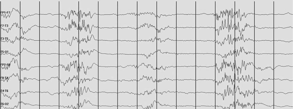
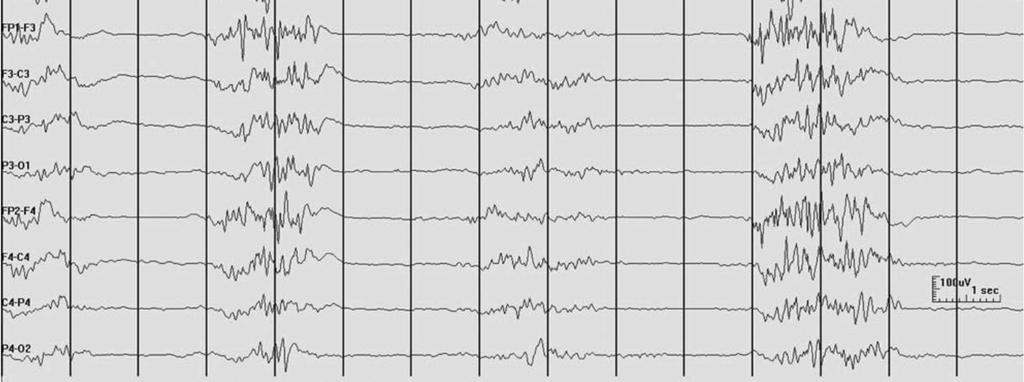
12 FIRDA Bilateral synchronous 2-3 Hz delta activity with frontal predominance Early study Tumor of posterior fossa and third ventricle, deep midline lesion, hydrocephalus More recent study Focal structural lesion: asymmetric FIRDA Diffuse brain injury in combination with metabolic disturbances Prospective controlled study Identified FIRDA in 6% of EEG Should initiate work up for toxic, metabolic or structural lesion Must be differentiate from vertical eye blinks, normal HV, hypnagogic hypersynchrony
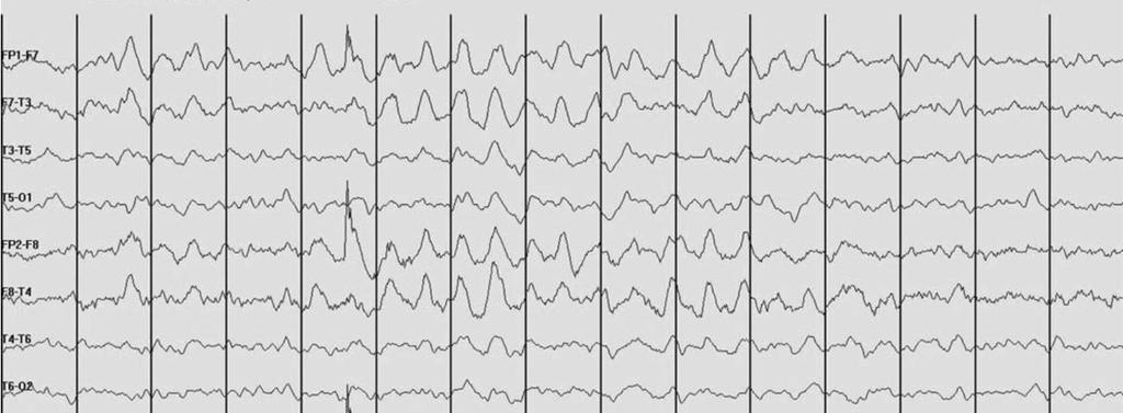
13 FIRDA
14 Temporal intermittent delta activity (TIRDA) Bursts or train of 3 seconds or more of repetitive, rhythmic, sawtoothed or sinusoidal 1-4 Hz, microvolts Predominant over anterior temporal region Associated with temporal lobe epilepsy
15 Occipital intermittent rhythmic delta activity Bi-posterior predominant rhythmic delta activity in children Occurred primarily in children with absence epilepsy Lesion in occipital lobe with involvement of posterior lateral ventricle: unilateral OIRDA
16 Rhythmic delta slow over posterior head region in absence epilepsy
17 Generalized theta activity Metabolic or drug induced encephalopathy Reactive in mild encephalopathy Non reactive in theta coma Similar prognosis with alpha coma Majority die or remain vegetative, some recover
18 Continuous generalized slow Normal feature or drowsy or sleep EEG Earliest sign of mild encephalopathy slowing of posterior waking background Encephalopathy deepens slow of anterior rhythm predominant polymorphic delta slowing less reactivity or organization Deep coma burst suppression, generalized suppression isoelectric

19 Drowsy burst in older patient

20 Mild diffuse slowing
21 Marked diffuse slowing and attenuation
22 Suppression burst pattern
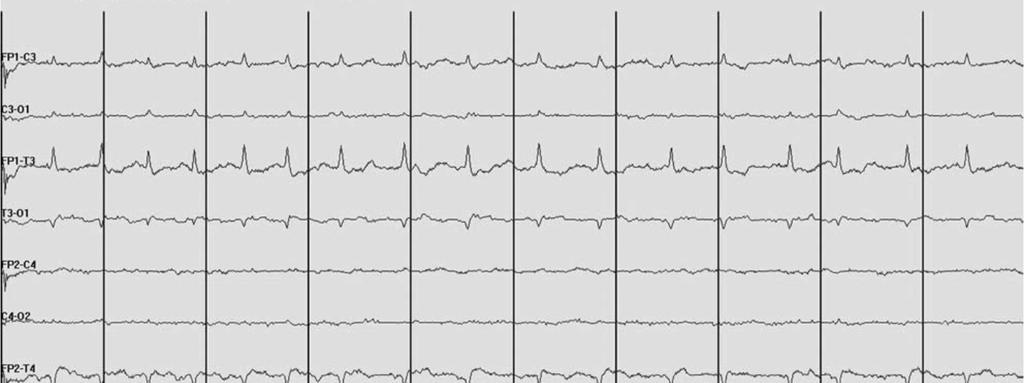
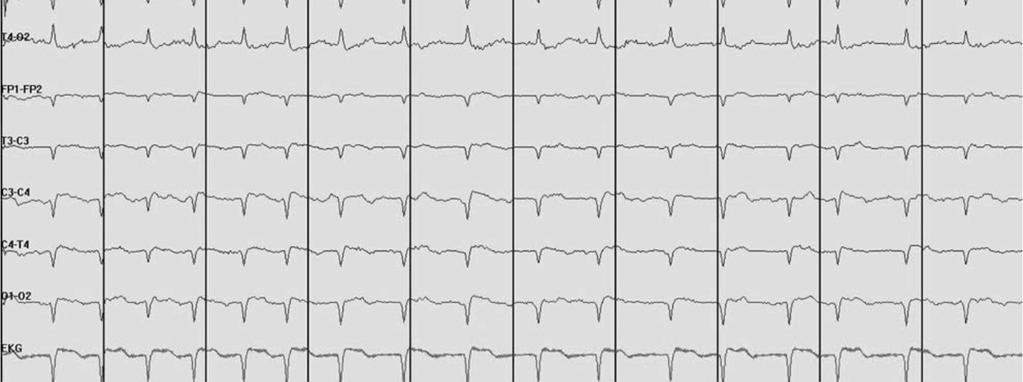
23 Triphasic wave Special type of generalized slow waves
 surrounding a prominent downward deflection on bipolar montage Typically bi-frontal predominant, 1.5-2.")
24 Triphasic waves First description: Blunt spike and slow waves pattern in hepatic coma Bilaterally synchronous and symmetrical waves Three phases: initiate and terminate with small upward deflection (negative wave) surrounding a prominent downward deflection on bipolar montage Typically bi-frontal predominant, Hz, µv May have ms anterior posterior or posterior-anterior lags May increase or attenuate with stimulation Occur intermittently in brief runs or continuous pattern
25 Triphasic waves: clinical correlation Etiology: wide variety Renal failure, hyponatremia, metabolic encephalopathy Hepatic encephalopathy Hypothyroidism Encephalitis Post-anoxic encephalopathy Sepsis Dementia: in rapidly progressive dementia CJD Drug intoxication Structural lesion: subdural hematoma, stroke Correlate closely with severity of encephalopathy Longer in duration and more widespread when patient became less responsive Usually absent in alert or deeply comatose patient

26 Triphasic wave GNCSE Kaplan PW and Schlattman DK. J Clin Neurophysiol. 2012;29(5):
27 NCSE Triphasic wave Response to benzodiazepine Higher frequency 2.4 Hz average Morphology Shorter duration > 0.3 sec Extra spike component Maximum fronto-polar First phase: absent Second phase: sharply downgoing Third phase: acutely rising, sharply descending, then return to baseline Less severe generalized BG slowing Response to benzodiazepine Slower frequency Hz Morphology Longer duration <0.3 sec Prominent amplitude of phase 2 Maximum fronto-central Phase lag First phase: blunt upward spike Second phase: blunted descent Third phase: large amplitude, blunted ascend, gradually fall to baseline Increase with painful or auditory stimulation
28 Fast frequency pattern Abnormal beta activity Breach rhythm
29 Generalized increased beta rhythm: causes Normal subjects, but persistence of increased beta activity throughout recording is abnormal Un-medicated children with chronic encephalopathy may have prominent beta especially when a/w dysgenetic lesion of the brain Barbiturates, benzodiazepines, sedative, transquilizers Hyperthyroidism: central or wide distribution Acute and chronic anxiety
30 2 year-old boy with lissencephaly Continuous generalized moderate amplitude beta

31 Generalized beta due to drugs
32 Asymmetric beta activity Asymmetrical beta activity should be considered abnormal if persistent voltage difference of 35% or more Beta activity is attenuated by subdural, epidural or subgaleal fluid collections Barbiturates bring out a beta asymmetry
33 Breach rhythm Consequence of intracranial surgery requiring craniotomy or burr hole, less often from skull fracture Attenuate high frequency filter function of the skull Enhanced voltage of fast frequency on the side of the defect Sharply contoured morphology

34 Breach rhythm
35 Patterns in coma
36 Patterns in coma Alpha coma Theta coma Beta coma Spindle coma
37 Alpha, theta, beta coma Patterns of rhythmical waves which have theta, alpha, beta frequency but occurs in isolation without other accompanying waves form in comatose patients No spontaneous variability or reactivity to sensory stimulation
38 Alpha coma Generalized or predominantly frontal alpha activty, 8-12 Hz, unreactive to stimuli in comatose patient Widespread cerebral damage, HIE, prolonged hypoglycemia, bilateral destruction of thalamic nuclei Posterior dominant alpha coma shows no reaction or variable attenuation or increase in amplitude following alerting maneuvers Brainstem lesion, pontine infarction Imply very poor prognosis

39 Alpha coma
40 Theta coma Generalized monorhythmic activity in theta frequency range Little or no spontaneous variability or reactivity to noxious stimuli Theta coma or mixed alpha-theta coma are not as reliable to predict a poor outcome as absence of reactivity and spontaneous variability
41 Beta coma Generalized, sometimes frontal dominant, pattern of mainly rhythmic beta waveforms Barbiturate or benzodiazepine intoxication Usually indicate likelihood of recovery from coma
42 Spindle coma Sleep spindles in unconscious or comatose patients Sleep spindle in 9-14 Hz diffuse distribution, often with vertex and K complex No cycles of wakefulness and sleep Preservation of intact pontine raphe nucleus and thalamocortical circuits, impairment of ARAS at midbrain level that maintains consciousness Indicate better prognosis in coma patient than its absence
43 Amplitude asymmetry
44 Amplitude asymmetry Characterized by differences in amplitude from two sides of the head Should be evaluated on referential montage Considered abnormal Consistent asymmetry of beta > 35% Consistent asymmetry of alpha > 50% Causes of asymmetry Unilateral lesion Change in conducting medium between cortex and recording electrode (SDH, skull defect, local scalp edema)
45 Clinical significance of asymmetry Normal asymmetry Asymmetry of photic driving, POSTs, mu rhythm in the absence of other abnormalities Abnormal asymmetry Usually due to decrease of amplitude on the side of cerebral abnormality
46 Abnormal amplitude asymmetry Asymmetry of all background activity Decrease amplitude of all type of activities Reduction of cortical EEG production Long lasting: Structural lesion Transient: focal seizure, TIA, migraine Increased of all media separating cortex from recording electrodes Increased amplitude of all type of activities: local skull defect
47 Focal attenuation
48 Focal attenuation Focal cortical lesion, tumor, cerebral ischemia Reversible cortical dysfunction (e.g. post ictal) Collection between cortex and recording electrode: swelling of scalp, subdural collection
49 Generalized change of amplitude
50 Patterns of generalized change of amplitude Bilateral symmetrical decrease or increase of amplitude of all types of normal frequency Abnormal low amplitude Cerebral activity < 20 microvolts during relaxed wakefulness with eye closed Low amplitude must be sustained Attenuation, suppression, burst suppression Abnormal high amplitude Not well defined, > 200 microvolts Amplitude > 100 microvolts is uncommon in wakeful adults, only considered abnormal on basis of frequency, morphology, or distribution rather than amplitude alone
51 Clinical significant Normal high amplitude occurs in waking record of children and deep stage of sleep at any age Normal low amplitude < 20 microvolts occurs transiently as a result of eye opening, mental effort, anxiety, alerting or drowsiness Abnormal low amplitude of all activity Low amplitude in normal subjects can be seen in about 5-10% of adults but is not acceptable as normal in younger persons Moderate or marked reduction if overall amplitude <10 microvolts abnormal at any age Transients and due to acute anoxic, toxic and metabolic, head injury, postictal Permanent: lasting damage or from other disease significant involve cortex
52 Burst suppression Presence of brain activity bursts of variable amplitude, duration and form, usually with sharp and spike waves lasted at least 0.5 sec, followed by marked depression of activity, occur on cyclical basis Severe cerebral damage in post anoxic encephalopathy Effect of anesthetic drug or drug induced coma
53 Burst suppression
54 Electro-cerebral inactivity Isoelectric, nonreactive EEG, flat EEG or electro-cerebral silence Expression of severe and widespread cerebral dysfunction in which EEG activity is undetectable (amplitudes of < 2 µv) Conventional scalp electrodes placed at double routine international electrode distances Body core temperature above 34 degrees centigrade At least 30 minutes of continuous recording
55 Electrocerebral activity
56 Abnormal alpha rhythms
57 Posterior/alpha rhythm Normal attenuation of alpha rhythm occurs with eye opening, alerting, mental effort, anxiety, decreased alerting to level of drowsiness Abnormal frequency of alpha rhythm Voltage asymmetry >50% in amplitude Rarely an indication of focal abnormality Skull defect result in higher voltage alpha rhythm Symmetry of frequency and reactivity are abnormal and reliably indicate the side of lesion
58 Abnormal frequency of alpha rhythm Unilateral decrease in alpha frequency is abnormal Abnormal <8-8.5 Hz Mild asymmetry (consistent asymmetry in frequency of Hz) Marked asymmetry (>1 Hz frequency asymmetry) Bilateral decrease of alpha frequency Generalized disturbance of cerebral function Bilateral structural damage to occipital cortex or its thalamic input Unilateral decrease of alpha frequency Unilateral disturbance of function of occipital cortex or its thalamic input Unilateral structural damage to occipital cortex or its thalamic input Bilateral increase of alpha frequency Metabolic disorder: fever, hyperthyroid
59 Abnormal reactivity of alpha rhythm Unilateral failure of alpha blocking (attenuation with eye opening): Bancaud s phenomenon Posterior subcortical lesion Parietal or temporal lesions Bilateral failure of alpha blocking in monocular input Disorder of one eye or optic nerve Absence of alpha rhythm, presence of occipital spikes Long standing diseases of both eyes or central visual pathway Alpha frequency coma Central pontine lesion Widespread cerebral damage
60 Abnormal alpha rhythm Focal cerebral lesion can alter alpha rhythm even though they do not involve occipital lobe or adjacent brain regions Unilateral slowing of frequency Loss of reactivity Loss of modulation Voltage attenuation
61 Asymmetry response to photic stimulation May result from lesions on the side of lower voltage Can consistently lateralized in some normal individual Should not be interpreted as abnormal in the absence of corroborative findings such as focal slow or localized attenuation of background rhythms on the same side Rarely epileptogenic lesions result in responses to photic stimulation that are of higher voltage on the side of lesion
62 Abnormal sleep potential
63 Abnormal sleep potentials Normal sleep patterns, sleep spindle and vertex can be affected by cerebral lesion Decrease potential, spindles, especially if involved cortex or thalamus Lesion of parietal lobe or thalamus can attenuate sleep spindles Thalamic lesion can result in interhemispheric asynchrony of spindles, affect regulation spindle frequency Skull defects enhance scalp-recorded voltage of sleep spindles and vertex

64 Extreme spindles Wide spread and persistent than normal spindles Diffuse encephalopathy of childhood 6 y/o with MR and seizure, persistent high voltage spindle activity during sleep
Common EEG pattern in critical care
 Common EEG pattern in critical care พ.ญ.ส ธ ดา เย นจ นทร Causes Direct neuronal injury Cerebral dysfunction : encephalopathy Psychic problems EEG in critical care 1 October 2009, Pramongkutklao Hospital
Scope. EEG patterns in Encephalopathy. Diffuse encephalopathy. EEG in adult patients with. EEG in diffuse encephalopathy
 Scope EEG patterns in Encephalopathy Dr.Pasiri Sithinamsuwan Division of Neurology Department of Medicine Phramongkutklao Hospital Diffuse encephalopathy EEG in specific encephalopathies Encephalitides
Normal EEG of wakeful resting adults of years of age. Alpha rhythm. Alpha rhythm. Alpha rhythm. Normal EEG of the wakeful adult at rest
 Normal EEG of wakeful resting adults of 20-60 years of age Suthida Yenjun, M.D. Normal EEG of the wakeful adult at rest Alpha rhythm Beta rhythm Mu rhythm Vertex sharp transients Intermittent posterior
EEG WORKSHOP Nonepileptiform Abnormalities
 EEG WORKSHOP Nonepileptiform Abnormalities Kamornwan Katanyuwong MD Chiangmai University Hospital EST: 20th July 2010 EEG reading Age Background Epileptiform Non epileptiform Activation procedure normal
EEG IN FOCAL ENCEPHALOPATHIES: CEREBROVASCULAR DISEASE, NEOPLASMS, AND INFECTIONS
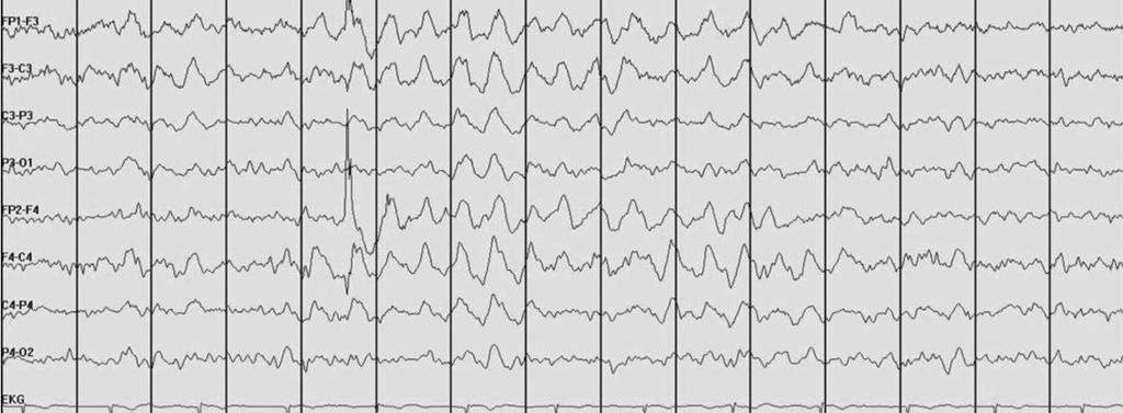
 246 Figure 8.7: FIRDA. The patient has a history of nonspecific cognitive decline and multiple small WM changes on imaging. oligodendrocytic tumors of the cerebral hemispheres (11,12). Electroencephalogram
EEG workshop. Epileptiform abnormalities. Definitions. Dr. Suthida Yenjun
 EEG workshop Epileptiform abnormalities Paroxysmal EEG activities ( focal or generalized) are often termed epileptiform activities EEG hallmark of epilepsy Dr. Suthida Yenjun Epileptiform abnormalities
Beyond the Basics in EEG Interpretation: Throughout the Life Stages
 Beyond the Basics in EEG Interpretation: Throughout the Life Stages Steve S. Chung, MD, FAAN Chairman, Neuroscience Institute Director, Epilepsy Program Banner University Medical Center University of Arizona
The secrets of conventional EEG
 The secrets of conventional EEG The spike/sharp wave activity o Electro-clinical characteristics of Spike/Sharp wave The polymorphic delta activity o Electro-clinical characteristics of Polymorphic delta
Introduction to EEG del Campo. Introduction to EEG. J.C. Martin del Campo, MD, FRCP University Health Network Toronto, Canada
 Introduction to EEG J.C. Martin, MD, FRCP University Health Network Toronto, Canada What is EEG? A graphic representation of the difference in voltage between two different cerebral locations plotted over
Subhairline EEG Part II - Encephalopathy
 Subhairline EEG Part II - Encephalopathy Teneille Gofton September 2013 Objectives To review the subhairline EEG changes seen with encephalopathy To discuss specific EEG findings in encephalopathy To outline
True Epileptiform Patterns (and some others)
") True Epileptiform Patterns (and some others) a) What is epileptiform b) Some possible surprises c) Classification of generalized epileptiform patterns An epileptiform pattern Interpretative term based
True Epileptiform Patterns (and some others) a) What is epileptiform b) Some possible surprises c) Classification of generalized epileptiform patterns An epileptiform pattern Interpretative term based
The Normal EEG, Normal Variants, Artifacts. Bassel Abou-Khalil, M.D.
 The Normal EEG, Normal Variants, Artifacts Bassel Abou-Khalil, M.D. I have no financial relationships to disclose that are relative to the content of my presentation Learning Objectives recognize normal
9 The Abnormal EEG. An EEG is considered abnormal if it has findings
 C h a p t e r 9 The Abnormal EEG An EEG is considered abnormal if it has findings known to be associated with a pathologic or disease state. As discussed in Chapter 8, The Structure and Philosophy of the
ENCEPHALOPATHY RECOGNIZING METABOLIC AND ANOXIC CHANGES
 ENCEPHALOPATHY RECOGNIZING METABOLIC AND ANOXIC CHANGES ENCEPHALOPATHY Encephalopathy is a general term that means brain disease, damage, or malfunction. The major symptom of encephalopathy is an altered
THE ACTIVITY RECORDED IN THE EEG
 Version 4. A Monthly Publication presented by Professor Yasser Metwally April 2008 THE ACTIVITY RECORDED IN THE EEG here is now considerable evidence from studies in experimental animals to suggest that
EEG in the ICU. Quiz. March Teneille E. Gofton
 EEG in the ICU Quiz March 2012 Teneille E. Gofton Quiz The next several slides will show 15 subhairline EEGs. Choose the best possible answer in each scenario. Your score and solutions will be provided
EEG in Medical Practice
 EEG in Medical Practice Dr. Md. Mahmudur Rahman Siddiqui MBBS, FCPS, FACP, FCCP Associate Professor, Dept. of Medicine Anwer Khan Modern Medical College What is the EEG? The brain normally produces tiny
states of brain activity sleep, brain waves DR. S. GOLABI PH.D. IN MEDICAL PHYSIOLOGY
 states of brain activity sleep, brain waves DR. S. GOLABI PH.D. IN MEDICAL PHYSIOLOGY introduction all of us are aware of the many different states of brain activity, including sleep, wakefulness, extreme
Neurophysiology & EEG
 Neurophysiology & EEG PG4 Core Curriculum Ian A. Cook, M.D. Associate Director, Laboratory of Brain, Behavior, & Pharmacology UCLA Department of Psychiatry & Biobehavioral Sciences Semel Institute for
Periodic and Rhythmic Patterns. Suzette M LaRoche, MD Mission Health Epilepsy Center Asheville, North Carolina
 Periodic and Rhythmic Patterns Suzette M LaRoche, MD Mission Health Epilepsy Center Asheville, North Carolina Continuum of EEG Activity Neuronal Injury LRDA GPDs SIRPIDs LPDs + NCS Burst-Suppression LPDs
The EEG in focal epilepsy. Bassel Abou-Khalil, M.D. Vanderbilt University Medical Center
 The EEG in focal epilepsy Bassel Abou-Khalil, M.D. Vanderbilt University Medical Center I have no financial relationships to disclose that are relative to the content of my presentation Learning Objectives
EEG in the ICU: Part I
 EEG in the ICU: Part I Teneille E. Gofton July 2012 Objectives To outline the importance of EEG monitoring in the ICU To briefly review the neurophysiological basis of EEG To introduce formal EEG and subhairline
Localization a quick look
 Localization a quick look Covering the basics Differential amplifiers Polarity convention 10-20 electrode system Basic montages: bipolar and referential Other aspects of displaying the EEG Localization
SLEEP STAGING AND AROUSAL. Dr. Tripat Deep Singh (MBBS, MD, RPSGT, RST) International Sleep Specialist (World Sleep Federation program)
 International Sleep Specialist (World Sleep Federation program)") SLEEP STAGING AND AROUSAL Dr. Tripat Deep Singh (MBBS, MD, RPSGT, RST) International Sleep Specialist (World Sleep Federation program) Scoring of Sleep Stages in Adults A. Stages of Sleep Stage W Stage
SLEEP STAGING AND AROUSAL Dr. Tripat Deep Singh (MBBS, MD, RPSGT, RST) International Sleep Specialist (World Sleep Federation program) Scoring of Sleep Stages in Adults A. Stages of Sleep Stage W Stage
Practical 3 Nervous System Physiology 2 nd year English Module. Dept. of Physiology, Carol Davila University of Medicine and Pharmacy
 Electroencephalography l h (EEG) Practical 3 Nervous System Physiology 2 nd year English Module Dept. of Physiology, Carol Davila University of Medicine and Pharmacy What is EEG EEG noninvasively records
Neonatal EEG Maturation
 Neonatal EEG Maturation Cindy Jenkinson, R. EEG T., CLTM October 7, 2017 Fissure Development 3 http://www.hhmi.org/biointeractive/develop ment-human-embryonic-brain 4 WHAT IS IMPORTANT TO KNOW BEFORE I
Asian Epilepsy Academy (ASEPA) EEG Certification Examination
 EEG Certification Examination") Asian Epilepsy Academy (ASEPA) EEG Certification Examination EEG Certification Examination Aims To set and improve the standard of practice of Electroencephalography (EEG) in the Asian Oceanian region
Asian Epilepsy Academy (ASEPA) EEG Certification Examination EEG Certification Examination Aims To set and improve the standard of practice of Electroencephalography (EEG) in the Asian Oceanian region
The Sonification of Human EEG and other Biomedical Data. Part 3
 The Sonification of Human EEG and other Biomedical Data Part 3 The Human EEG A data source for the sonification of cerebral dynamics The Human EEG - Outline Electric brain signals Continuous recording
Developmental Changes Including Neonatal EEG. Gregory L. Holmes, MD
 Developmental Changes Including Neonatal EEG Gregory L. Holmes, MD A A + B =: B + A.Dravet Syndrome B.Menkes syndrome C.West syndrome D.Ohtahara shyndrome The Difficult Delivery 1 day old male transferred
Generalized seizures, generalized spike-waves and other things. Charles Deacon MD FRCPC Centre Hospitalier Universitaire de Sherbrooke
 Generalized seizures, generalized spike-waves and other things Charles Deacon MD FRCPC Centre Hospitalier Universitaire de Sherbrooke Objectives Give an overview of generalized EEG discharges and seizures
Generalised Epileptiform Patterns
 Generalised Epileptiform Patterns Manori Wijayath Westmead Hospital, Sydney, Australia With slides from Elizabeth Walker and Andrew Bleasel Generalised Epilep-form Discharges: Outline 1. Generalised epilep.form
NONSPECIFIC ABNORMAL EEG PATTERNS
 Version 17 A Monthly Publication presented by Professor Yasser Metwally January 2010 NONSPECIFIC ABNORMAL EEG PATTERNS Electroencephalogram (EEG) abnormalities can be divided into three descriptive categories:
Sleep stages. Awake Stage 1 Stage 2 Stage 3 Stage 4 Rapid eye movement sleep (REM) Slow wave sleep (NREM)
 Slow wave sleep (NREM)") Sleep stages Awake Stage 1 Stage 2 Stage 3 Stage 4 Rapid eye movement sleep (REM) Slow wave sleep (NREM) EEG waves EEG Electrode Placement Classifying EEG brain waves Frequency: the number of oscillations/waves
Sleep stages Awake Stage 1 Stage 2 Stage 3 Stage 4 Rapid eye movement sleep (REM) Slow wave sleep (NREM) EEG waves EEG Electrode Placement Classifying EEG brain waves Frequency: the number of oscillations/waves
Continuous EEG Monitoring is becoming a commonly used tool
 INVITED REVIEW American Clinical Neurophysiology Society s Standardized Critical Care EEG Terminology: 2012 version L. J. Hirsch, S. M. LaRoche, N. Gaspard, E. Gerard, A. Svoronos, S. T. Herman, R. Mani,
Asian Epilepsy Academy (ASEPA) & ASEAN Neurological Association (ASNA) EEG Certification Examination
 & ASEAN Neurological Association (ASNA) EEG Certification Examination") Asian Epilepsy Academy (ASEPA) & ASEAN Neurological Association (ASNA) EEG Certification Examination EEG Certification Examination Aims To set and improve the standard of practice of Electroencephalography
Asian Epilepsy Academy (ASEPA) & ASEAN Neurological Association (ASNA) EEG Certification Examination EEG Certification Examination Aims To set and improve the standard of practice of Electroencephalography
Physiology Unit 2 CONSCIOUSNESS, THE BRAIN AND BEHAVIOR
 Physiology Unit 2 CONSCIOUSNESS, THE BRAIN AND BEHAVIOR What the Brain Does The nervous system determines states of consciousness and produces complex behaviors Any given neuron may have as many as 200,000
Physiology Unit 2 CONSCIOUSNESS, THE BRAIN AND BEHAVIOR
 Physiology Unit 2 CONSCIOUSNESS, THE BRAIN AND BEHAVIOR In Physiology Today What the Brain Does The nervous system determines states of consciousness and produces complex behaviors Any given neuron may
Neonatal EEG, Seizures and Epilepsy Syndromes
 Neonatal EEG, Seizures and Epilepsy Syndromes Introduction Over the past several decades great progress has been made in neonatal-perinatal medicine Survival of premature infants < 1 Kg is common Neonatal
Idiopathic epilepsy syndromes
 1 Idiopathic epilepsy syndromes PANISRA SUDACHAN, M.D. Pe diatric Neuro lo gis t Pediatric Neurology Department Pras at Neuro lo gic al Institute Epilepsy course 20 August 2016 Classification 2 1964 1970
EEG History. Where and why is EEG used? 8/2/2010
 EEG History Hans Berger 1873-1941 Edgar Douglas Adrian, an English physician, was one of the first scientists to record a single nerve fiber potential Although Adrian is credited with the discovery of
Electroencephalography. Role of EEG in NCSE. Continuous EEG in ICU 25/05/59. EEG pattern in status epilepticus
 EEG: ICU monitoring & 2 interesting cases Electroencephalography Techniques Paper EEG digital video electroencephalography Dr. Pasiri Sithinamsuwan PMK Hospital Routine EEG long term monitoring Continuous
EEG and some applications (seizures and sleep)
") EEG and some applications (seizures and sleep) EEG: stands for electroencephalography and is a graphed representation of the electrical activity of the brain. EEG is the recording of electrical activity
EEG and some applications (seizures and sleep) EEG: stands for electroencephalography and is a graphed representation of the electrical activity of the brain. EEG is the recording of electrical activity
Elsevier's Encyclopedia of euroscience
 REPRINTED FROM Elsevier's Encyclopedia of euroscience e Edited by George Adelman Barry H. Smith Editorial Manager Jennifer De Pasquale 1999 Elsevier Science B.V. All rights reserved. Visit the Encyclopedia's
Organic Mental Disorders. Organic Mental Disorders. Axes. Damrongsak Bulyalert Department of Internal Medicine
 Organic Mental Disorders Damrongsak Bulyalert Department of Internal Medicine www.metadon.net 1 Organic Mental Disorders In DSM (Diagnostic and Statistical Manual of Mental Disorders), OMD includes Delirium,
13 The Electroencephalogram of the Newborn
 C h a p t e r 13 The Electroencephalogram of the Newborn Newborn EEG interpretation is considered a particularly challenging area. An understanding of the appearance of the normal newborn EEG was achieved
EEG Electrode Placement
 EEG Electrode Placement Classifying EEG brain waves Frequency: the number of oscillations/waves per second, measured in Hertz (Hz) reflects the firing rate of neurons alpha, beta, theta, delta Amplitude:
Electroencephalography
 The electroencephalogram (EEG) is a measure of brain waves. It is a readily available test that provides evidence of how the brain functions over time. The EEG is used in the evaluation of brain disorders.
Drug exposure and EEG/qEEG findings
 Drug exposure and EEG/qEEG findings Dec 23rd, 2009 by Jay Gunkelman. A technical guide by Jay Gunkelman, QEEG-D General comments: There is a generally reciprocal effect between alpha and beta, as brain
Diagnosing Complicated Epilepsy: Mapping of the Epileptic Circuitry. Michael R. Sperling, M.D. Thomas Jefferson University Philadelphia, PA
 Diagnosing Complicated Epilepsy: Mapping of the Epileptic Circuitry Michael R. Sperling, M.D. Thomas Jefferson University Philadelphia, PA Overview Definition of epileptic circuitry Methods of mapping
PD233: Design of Biomedical Devices and Systems
 PD233: Design of Biomedical Devices and Systems (Lecture-7 Biopotentials- 2) Dr. Manish Arora CPDM, IISc Course Website: http://cpdm.iisc.ac.in/utsaah/courses/ Electromyogram (EMG) Skeletal muscles are
Myoclonic status epilepticus in hypoxic ischemic encephalopathy which recurred after somatosensory evoked potential testing
 ANNALS OF CLINICAL NEUROPHYSIOLOGY CASE REPORT Ann Clin Neurophysiol 2017;19(2):136-140 Myoclonic status epilepticus in hypoxic ischemic encephalopathy which recurred after somatosensory evoked potential
Multiple Choice Questions for Part I
 Multiple Choice Questions for Part I 1. Neurons in the cerebral cortex are organized in: A. Three horizontal layers B. Four horizontal layers C. Six horizontal layers with layer IV receiving inputs from
Epilepsy and EEG in Clinical Practice
 Mayo School of Professional Development Epilepsy and EEG in Clinical Practice November 10-12, 2016 Hard Rock Hotel at Universal Orlando Orlando, FL Course Directors Jeffrey Britton, MD and William Tatum,
Post-anoxic status epilepticus and EEG patterns
 Post-anoxic status epilepticus and EEG patterns Nicolas Gaspard, MD, PhD Université Libre de Bruxelles Hôpital Erasme, Bruxelles, Belgique Yale University School of Medicine, New Haven, CT, USA DISCLOSURES
ALERTNESS, SLEEP, EEG D27 (1)
") ALERTNESS, SLEEP, EEG D27 (1) Alertness, Sleep, Electroencephalography (EEG) Last updated: September 5, 2017 Introduction... 1 ELECTROENCEPHALOGRAPHY (EEG)... 2 MONTAGES... 2 RHYTHMS... 3 Alpha rhythm...
ALERTNESS, SLEEP, EEG D27 (1) Alertness, Sleep, Electroencephalography (EEG) Last updated: September 5, 2017 Introduction... 1 ELECTROENCEPHALOGRAPHY (EEG)... 2 MONTAGES... 2 RHYTHMS... 3 Alpha rhythm...
CEREBRAL FUNCTION MONITORING
 CEREBRAL FUNCTION MONITORING Introduction and Definitions The term amplitude integrated electroencephalography (aeeg) is used to denote a method for electro-cortical monitoring whereas cerebral function
Continuous EEG monitoring of the premature infant in the NICU
 Continuous EEG monitoring of the premature infant in the NICU Tom Stiris Oslo University Hospital, NICU CIP, Paris 2011 Background A method that at a very early stage diagnose those babies which would
NEURAL MECHANISMS OF SLEEP (p.1) (Rev. 3/21/07)
 (Rev. 3/21/07)") NEURAL MECHANISMS OF SLEEP (p.1) (Rev. 3/21/07) 1. Revisitation of Bremer s 1936 Isolated Brain Studies Transected the brain: a. Cut between the medulla and the spinal cord ( encephale isole ) Note: recall
NEURAL MECHANISMS OF SLEEP (p.1) (Rev. 3/21/07) 1. Revisitation of Bremer s 1936 Isolated Brain Studies Transected the brain: a. Cut between the medulla and the spinal cord ( encephale isole ) Note: recall
ROLE OF EEG IN EPILEPTIC SYNDROMES ASSOCIATED WITH MYOCLONUS
 Version 18 A Monthly Publication presented by Professor Yasser Metwally February 2010 ROLE OF EEG IN EPILEPTIC SYNDROMES ASSOCIATED WITH MYOCLONUS EEG is an essential component in the evaluation of epilepsy.
Babak Tamizi Far MD. Assistant professor of internal medicine Al-zahra hospital, Isfahan university of medical sciences
 Babak Tamizi Far MD. Assistant professor of internal medicine Al-zahra hospital, Isfahan university of medical sciences ٢ Level of consciousness is depressed Stuporous patients respond only to repeated
COMA BIOLOGY. Assist. Prof. Mehmet Akif KARAMERCAN Gazi University Faculty of Medicine Department of Emergency Medicine
 COMA BIOLOGY Assist. Prof. Mehmet Akif KARAMERCAN Gazi University Faculty of Medicine Department of Emergency Medicine Outlines Definitions Classification and Major Causes Arousal Systems (Reticular Activating
EEG ANALYSIS: ANN APPROACH
 EEG ANALYSIS: ANN APPROACH CHAPTER 5 EEG ANALYSIS: ANN APPROACH 5.1 INTRODUCTION The analysis of EEG signals using ANN deals with developing a network in order to establish a relation between input and
CHAPTER 6 INTERFERENCE CANCELLATION IN EEG SIGNAL
 116 CHAPTER 6 INTERFERENCE CANCELLATION IN EEG SIGNAL 6.1 INTRODUCTION Electrical impulses generated by nerve firings in the brain pass through the head and represent the electroencephalogram (EEG). Electrical
Cerebro-vascular stroke
 Cerebro-vascular stroke CT Terminology Hypodense lesion = lesion of lower density than the normal brain tissue Hyperdense lesion = lesion of higher density than normal brain tissue Isodense lesion = lesion
Seizures and Sleep, Sorting out the Spikes and Waves, a Polysomnographic and Clinical Review
 Seizures and Sleep, Sorting out the Spikes and Waves, a Polysomnographic and Clinical Review DR. MARK GARWOOD CLINICAL ASSISTANT PROFESSOR, DEPARTMENT OF NEUROLOGY MEDICAL DIRECTOR SLEEP DISORDERS CLINICS
Can EEG Test Helps in Identifying Brain Tumor? M. Sharanreddy, P. K. Kulkarni
 Can EEG Test Helps in Identifying Brain Tumor? M. Sharanreddy, P. K. Kulkarni Abstract Brain tumor is inherently serious and life-threatening disease. Brain tumor builds the intracranial pressure in the
Dysfunctional FINDINGS IN EEG. About 18 Cases were Seen in Consultation in our Hospital
 Cronicon OPEN ACCESS NEUROLOGY Clinical Images Angel Molina Leon* Dysfunctional FINDINGS IN EEG. About 18 Cases were Seen in Consultation in our Hospital Clinical Neurophysiology Service. Centro medico
Patient-Specific Seizure Onset Detection
 Patient-Specific Seizure Onset Detection by Ali Hossam Shoeb Submitted to the Department of Electrical Engineering and Computer Science in partial fulfillment of the requirements for the degree of Master
STRUCTURAL ORGANIZATION OF THE NERVOUS SYSTEM
 STRUCTURAL ORGANIZATION OF THE NERVOUS SYSTEM STRUCTURAL ORGANIZATION OF THE BRAIN The central nervous system (CNS), consisting of the brain and spinal cord, receives input from sensory neurons and directs
Case report. Epileptic Disord 2005; 7 (1): 37-41
: 37-41") Case report Epileptic Disord 2005; 7 (1): 37-41 Periodic lateralized epileptiform discharges (PLEDs) as the sole electrographic correlate of a complex partial seizure Gagandeep Singh, Mary-Anne Wright,
Case report Epileptic Disord 2005; 7 (1): 37-41 Periodic lateralized epileptiform discharges (PLEDs) as the sole electrographic correlate of a complex partial seizure Gagandeep Singh, Mary-Anne Wright,
The AASM Manual for the Scoring of Sleep and Associated Events
 The AASM Manual for the Scoring of Sleep and Associated Events Summary of Updates in Version 2.1 July 1, 2014 The American Academy of Sleep Medicine (AASM) is committed to ensuring that The AASM Manual
Idiopathic epilepsy syndromes
 Idiopathic epilepsy syndromes PANISRA SUDACHAN, M.D. Pediatric Neurologist Pediatric Neurology Department Prasat Neurological Institue Epilepsy course 26 August 2017 Classification 1964 1970 1981 1989
EEG Instrumentation, Montage, Polarity, and Localization
 EEG Instrumentation, Montage, Polarity, and Localization 2 Krikor Tufenkjian The Source of EEG The source of the EEG potentials recorded from the scalp is the excitatory and inhibitory postsynaptic potentials
*Pathophysiology of. Epilepsy
 *Pathophysiology of Epilepsy *Objectives * At the end of this lecture the students should be able to:- 1.Define Epilepsy 2.Etio-pathology of Epilepsy 3.Types of Epilepsy 4.Role of Genetic in Epilepsy 5.Clinical
SWI including phase and magnitude images
 On-line Table: MRI imaging recommendation and summary of key features Sequence Pathologies Visible Key Features T1 volumetric high-resolution whole-brain reformatted in axial, coronal, and sagittal planes
Classification of Seizures. Generalized Epilepsies. Classification of Seizures. Classification of Seizures. Bassel F. Shneker
 Classification of Seizures Generalized Epilepsies Bassel F. Shneker Traditionally divided into grand mal and petit mal seizures ILAE classification of epileptic seizures in 1981 based on clinical observation
Electroencephalography & Neurofeedback
 Electroencephalography & Neurofeedback A Brief Introduction to the Science of Brainwaves Glyn Blackett YORK biofeedback CENTRE Introduction This article is a brief introduction to electroencephalography
AMERICAN BOARD OF CLINICAL NEUROPHYSIOLOGY
 AMERICAN BOARD OF CLINICAL NEUROPHYSIOLOGY Part I Content Outline I. Physiology and Instrumentation 30% A. Physiology 1. Anatomy of neural generation 2. Mechanisms of EEG and evoked potential generation
1/31/2009. Paroxysmal, uncontrolled electrical discharge of neurons in brain interrupting normal function
 Paroxysmal, uncontrolled electrical discharge of neurons in brain interrupting normal function In epilepsy abnormal neurons undergo spontaneous firing Cause of abnormal firing is unclear Firing spreads
Surgery for Medically Refractory Focal Epilepsy
 Surgery for Medically Refractory Focal Epilepsy Seth F Oliveria, MD PhD The Oregon Clinic Neurosurgery Director of Functional Neurosurgery: Providence Brain and Spine Institute Portland, OR Providence
Seizure onset can be difficult to asses in scalp EEG. However, some tools can be used to increase the seizure onset activity over the EEG background:
 This presentation was given during the Dianalund Summer School on EEG and Epilepsy, July 24, 2012. The main purpose of this introductory talk is to show the possibilities of improved seizure onset analysis
Sleep-Wake Cycle I Brain Rhythms. Reading: BCP Chapter 19
 Sleep-Wake Cycle I Brain Rhythms Reading: BCP Chapter 19 Brain Rhythms and Sleep Earth has a rhythmic environment. For example, day and night cycle back and forth, tides ebb and flow and temperature varies
EEG Sleep Circadian rhythms Learning Objectives: 121, 122
 EEG Sleep Circadian rhythms Learning Objectives: 121, 122 Zoltán Lelkes Electroencenphalography Hans Berger pen time amplifier electrodes 1 The waves of the EEG gamma > 30 Hz beta: 13-30 Hz Mental activity:
This is a repository copy of An introduction to neonatal EEG. White Rose Research Online URL for this paper:
 This is a repository copy of An introduction to neonatal EEG. White Rose Research Online URL for this paper: http://eprints.whiterose.ac.uk/124766/ Version: Accepted Version Article: Alix, J.J.P. orcid.org/0000-0001-8391-9749,
Cesarean section for breech presentation. Jonathan H. Waters, M.D.
 Cesarean section for breech presentation Jonathan H. Waters, M.D. 1 26 y.o. G1P0 presented to triage in labor at 38 weeks. Patient was a known breech with a failed version 5 days before presentation. PMH
2. Subarachnoid Hemorrhage
 Causes: 2. Subarachnoid Hemorrhage A. Saccular (berry) aneurysm - Is the most frequent cause of clinically significant subarachnoid hemorrhage is rupture of a saccular (berry) aneurysm. B. Vascular malformation
Electroencephalographic Study of Essential Oils for Stress Relief
 Applied Mechanics and Materials Online: 2013-10-11 ISSN: 1662-7482, Vol. 437, pp 1085-1088 doi:10.4028/www.scientific.net/amm.437.1085 2013 Trans Tech Publications, Switzerland Electroencephalographic
V. CENTRAL NERVOUS SYSTEM TRAUMA
 V. CENTRAL NERVOUS SYSTEM TRAUMA I. Concussion - Is a clinical syndrome of altered consiousness secondary to head injury - Brought by a change in the momentum of the head when a moving head suddenly arrested
MRI OF THE THALAMUS. Mohammed J. Zafar, MD, FAAN Kalamazoo, MI
 1 MRI OF THE THALAMUS Mohammed J. Zafar, MD, FAAN Kalamazoo, MI Objectives: The thalamic nuclei can be involved in a wide variety of conditions. A systematic imaging approach would be useful for narrowing
PEDIATRIC BRAIN CARE
 PEDIATRIC BRAIN CARE The brain matters most! OVERVIEW OF NEURO ASSESSMENT 1. Overall responsiveness/activity 2. The eyes 3.? Increased ICP 4. Movements 5.? Seizures 6. Other OVERALL RESPONSIVENESS/ ACTIVITY
TOBY Cerebral Function Monitoring Addition to CFM handbook for users of the Olympic CFM 6000
 ISRCTN 89547571 TOBY Cerebral Function Monitoring Addition to CFM handbook for users of the Olympic CFM 6000 2 The contents of this booklet were originally produced for the website http://www.azzopardi.freeserve.co.uk/cfm
EEG in Benign and Malignant Epileptic Syndromes of Childhood
 Epilepsia, 43(Suppl. 3):17 26, 2002 Blackwell Publishing, Inc. International League Against Epilepsy EEG in Benign and Malignant Epileptic Syndromes of Childhood Ivo Drury Department of Neurology, Henry
NEURO IMAGING 2. Dr. Said Huwaijah Chairman of radiology Dep, Damascus Univercity
 NEURO IMAGING 2 Dr. Said Huwaijah Chairman of radiology Dep, Damascus Univercity I. EPIDURAL HEMATOMA (EDH) LOCATION Seventy to seventy-five percent occur in temporoparietal region. CAUSE Most likely caused
Circadian rhythm and Sleep. Radwan Banimustafa MD
 Circadian rhythm and Sleep Radwan Banimustafa MD Homeostasis Maintenance of equilibrium by active regulation of internal states: Cardiovascular function (blood pressure, heart rate) Body temperature Food
The EEG Analysis of Auditory Emotional Stimuli Perception in TBI Patients with Different SCG Score
 Open Journal of Modern Neurosurgery, 2014, 4, 81-96 Published Online April 2014 in SciRes. http://www.scirp.org/journal/ojmn http://dx.doi.org/10.4236/ojmn.2014.42017 The EEG Analysis of Auditory Emotional
Matrix Energetics Research Brainwaves and Heart waves Research on Matrix Energetics in Action
 Matrix Energetics Research Brainwaves and Heart waves Research on Matrix Energetics in Action QEEG (quantitative electroencephalography) and HRV (heart rate variability analysis) tests revealed Dr. Richard
PART I EXAMINATION INFOMATION. Part I Content Outline
 PART I EXAMINATION INFOMATION The three-hour examination will be administered during an established two-week testing period at PSI Computer Testing, Inc. The examination consists of 120 objective, multiple-choice
EE 4BD4 Lecture 11. The Brain and EEG
 EE 4BD4 Lecture 11 The Brain and EEG 1 Brain Wave Recordings Recorded extra-cellularly from scalp (EEG) Recorded from extra-cellularly from surface of cortex (ECOG) Recorded extra-cellularly from deep
A reappraisal of secondary bilateral synchrony
 Neurology Asia 2007; 12 : 29 35 A reappraisal of secondary bilateral synchrony Liri JIN MD, PhD Department of Neurology, Peking Union Medical College Hospital, Chinese Academy of Medical Sciences, Beijing,
Idiopathic epilepsy syndromes
 Idiopathic epilepsy syndromes Kamornwan Katanyuwong MD. Chiangmai University Hospital EST, July 2009 Diagram Sylvie Nyugen The Tich, Yann Pereon Childhood absence epilepsy (CAE) Age : onset between 4-10