The EEG in focal epilepsy. Bassel Abou-Khalil, M.D. Vanderbilt University Medical Center
|
|
|
- Logan Johnston
- 6 years ago
- Views:
Transcription
1 The EEG in focal epilepsy Bassel Abou-Khalil, M.D. Vanderbilt University Medical Center
2 I have no financial relationships to disclose that are relative to the content of my presentation
3 Learning Objectives to identify focal epileptiform abnormalities to recognize the clinical significance of epileptiform discharges in the diagnosis of epileptic syndromes to localize interictal epileptiform and ictal discharges
4 Self assessment questions
5 Q1- The difference between epileptiform sharp waves and nonepileptiform sharp transients is A. Epileptiform transients are more likely to be symmetrical and monophasic B. Epileptiform transients are more likely to be asymmetrical and polyphasic C. Epileptiform discharges are less likely to have an aftergoing slow wave D. Epileptiform discharges have a larger first segment than second segment
6 Q2- The following are most specific for epilepsy A. Occipital sharp waves B. Parietal and central sharp waves C. Anterior temporal and frontal sharp waves D. Generalized sharp waves
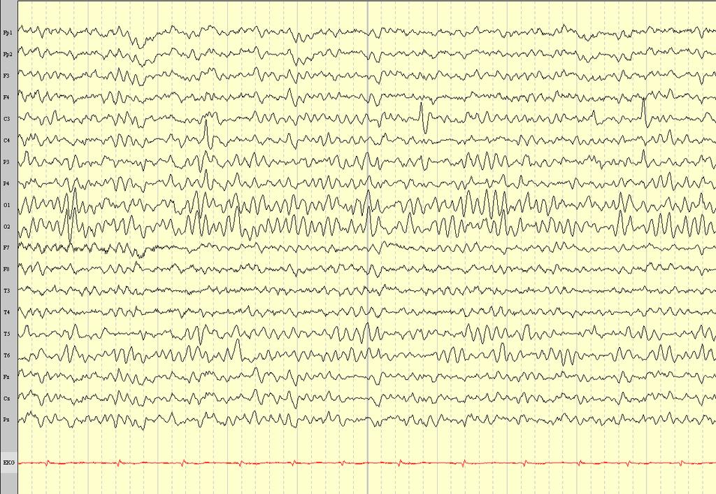
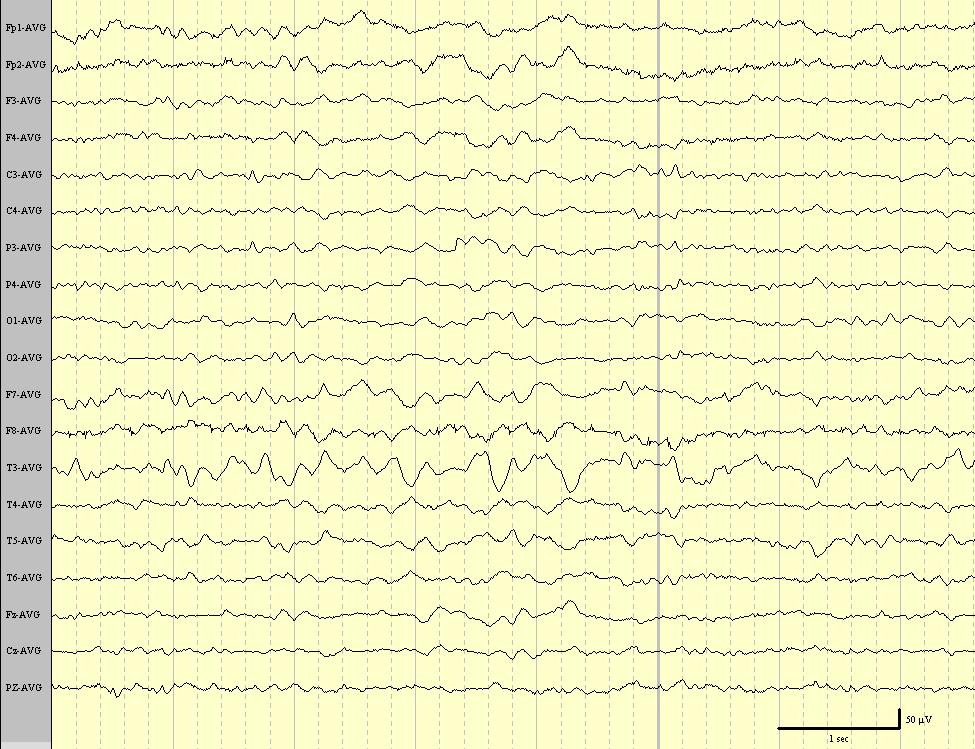
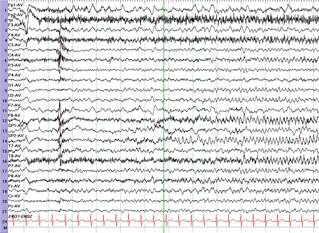
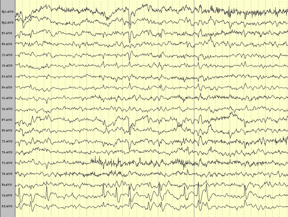
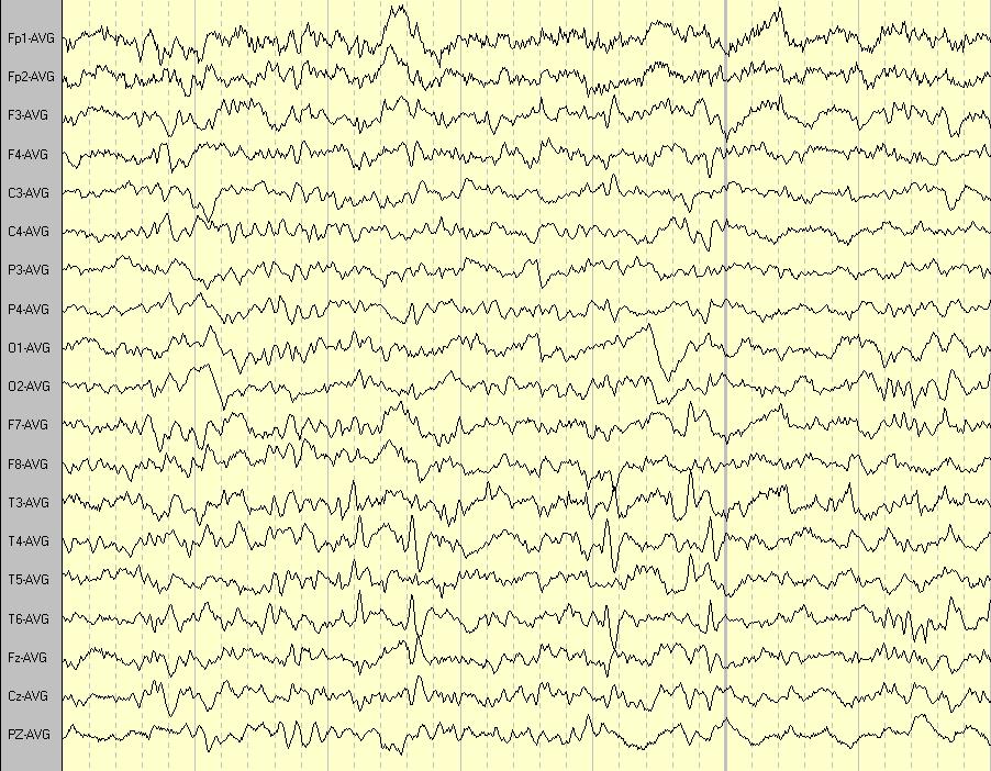
7
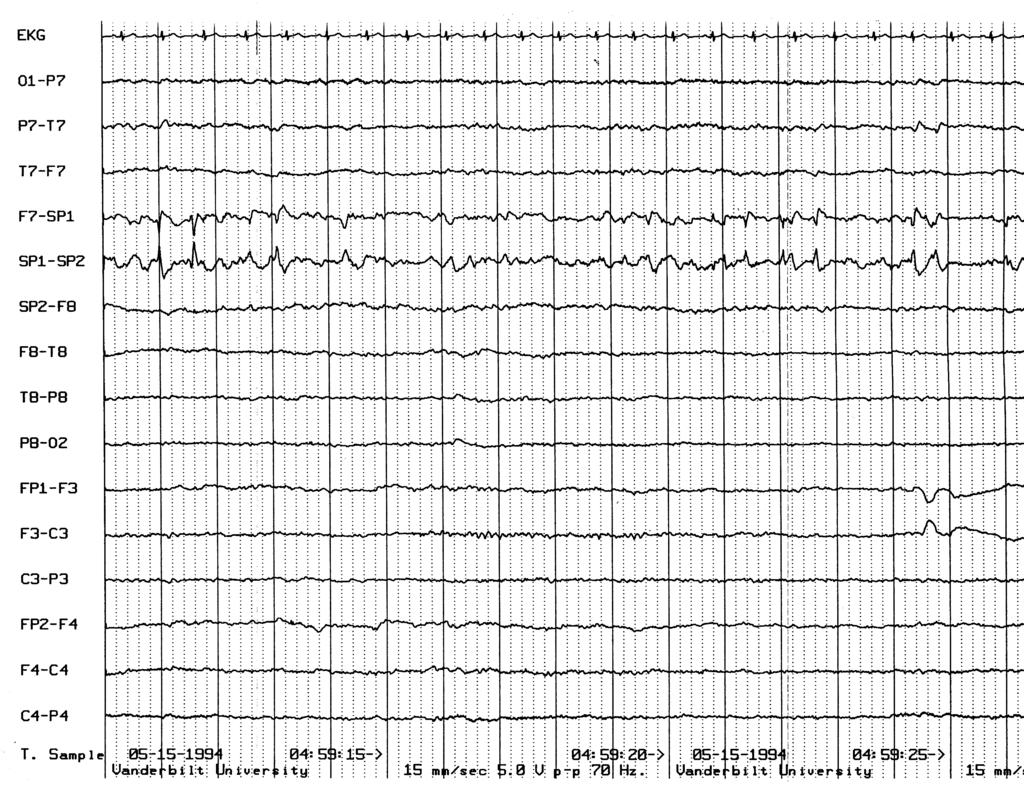
8 Q3- What is not true for the EEG above A. Findings may be seen in the absence of epilepsy B. Is expected to remit at puberty C. Seizures are likely to be hemifacial sensorimotor D. Seizures likely manifest with oroalimentary automatisms

9 Fp1-Av Fp2-Av F3-Av F4-Av C3-Av C4-Av P3-Av P4-Av O1-Av O2-Av F7-Av F8-Av T1-Av T2-Av T7-Av T8-Av P7-Av P8-Av Fz-Av Cz-Av Pz-Av ECG
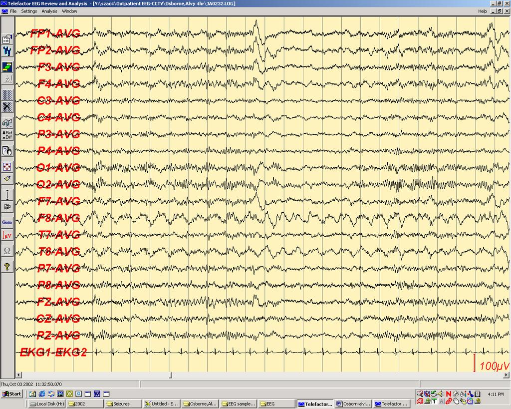
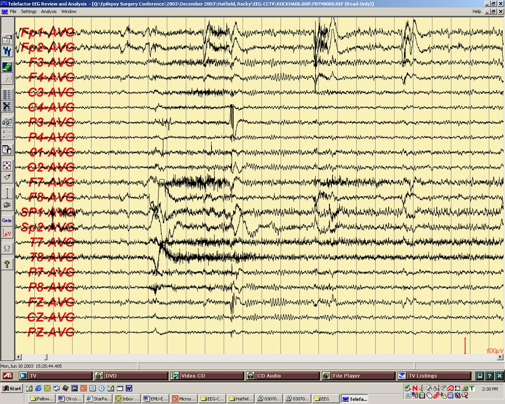
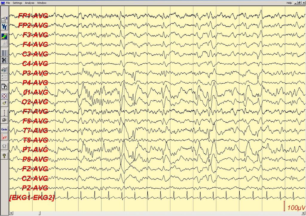
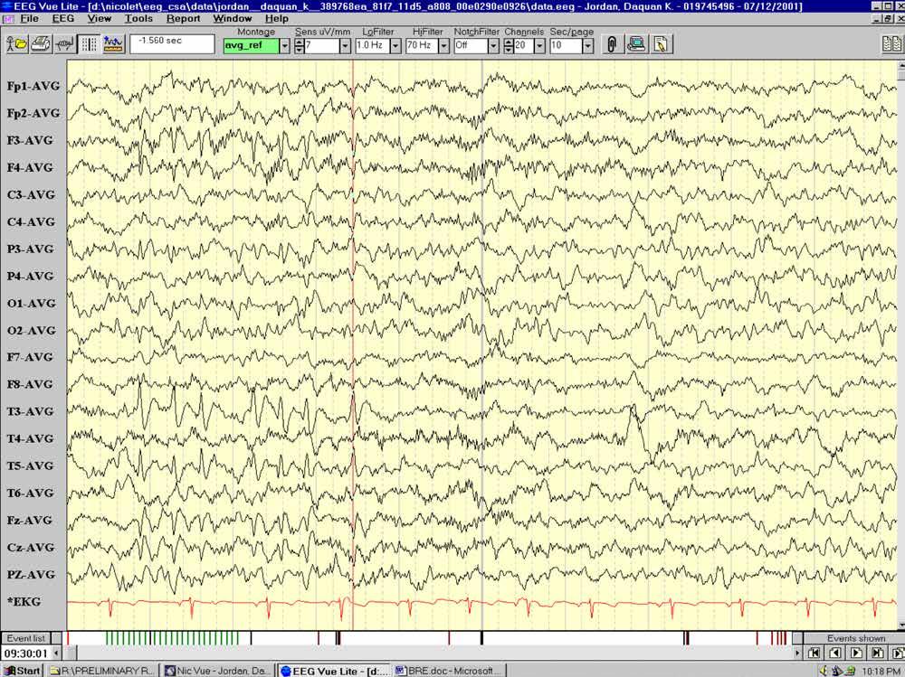
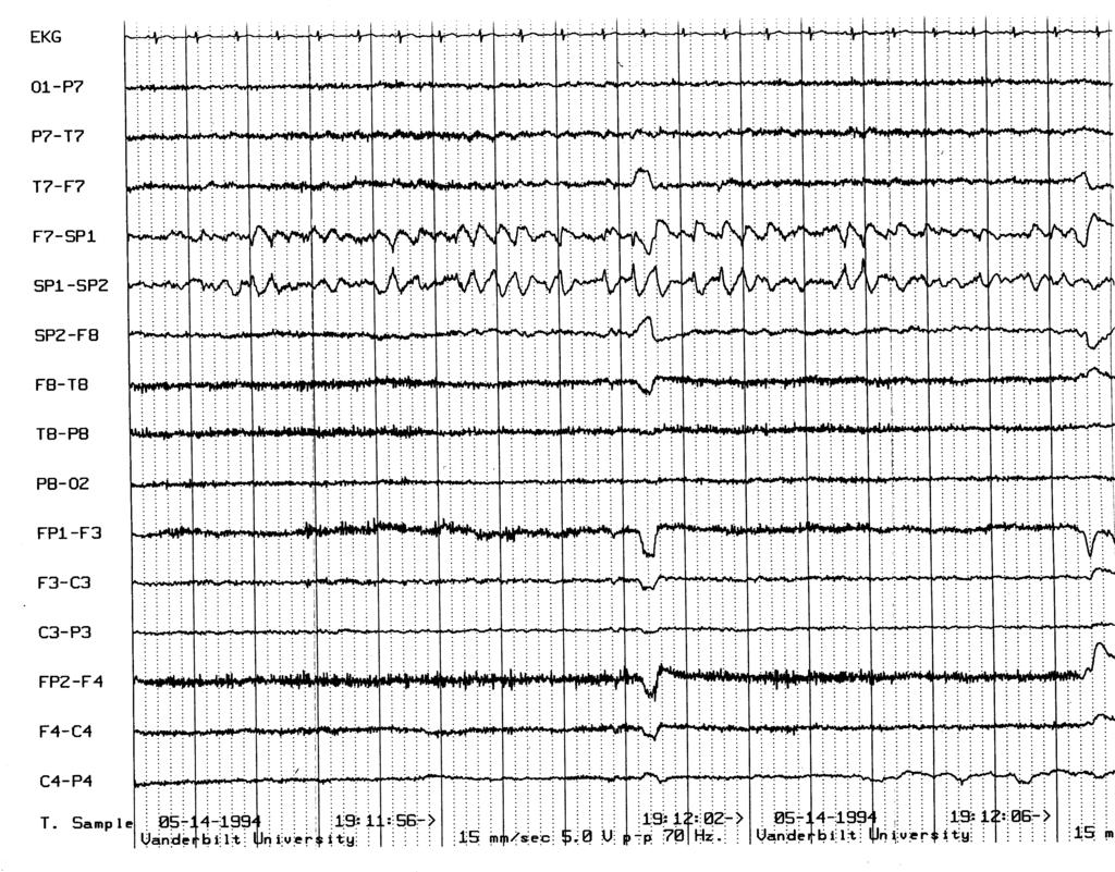
10 Q4- What is not true for the EEG above A. Seizures originate from both temporal regions B. Findings could be seen in parietal and insular lobe epilepsy C. Seizures are more difficult to lateralize than for patients with unilateral discharges D. Waking and REM recordings may help resolve lateralization
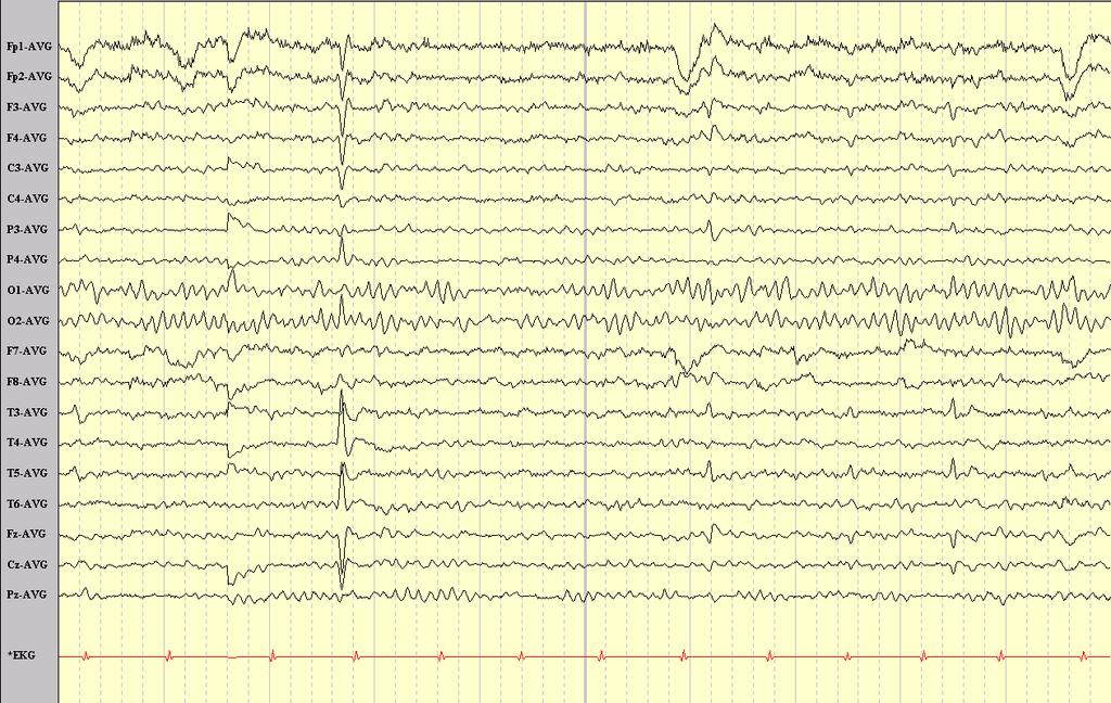
11
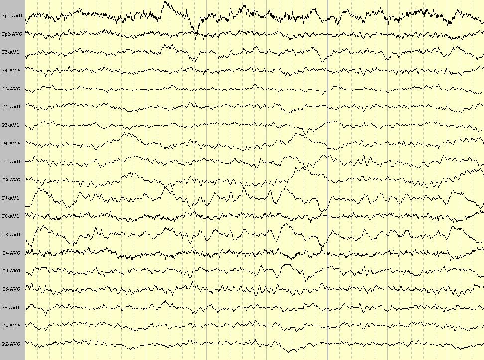
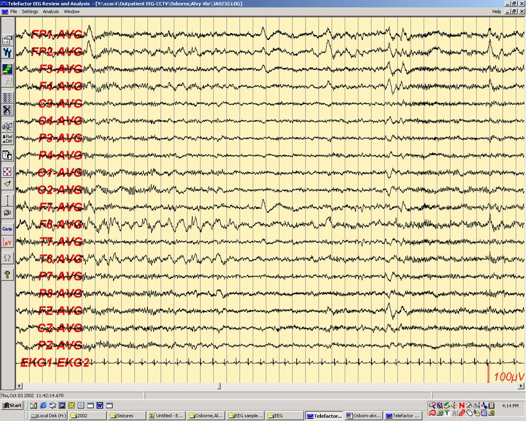
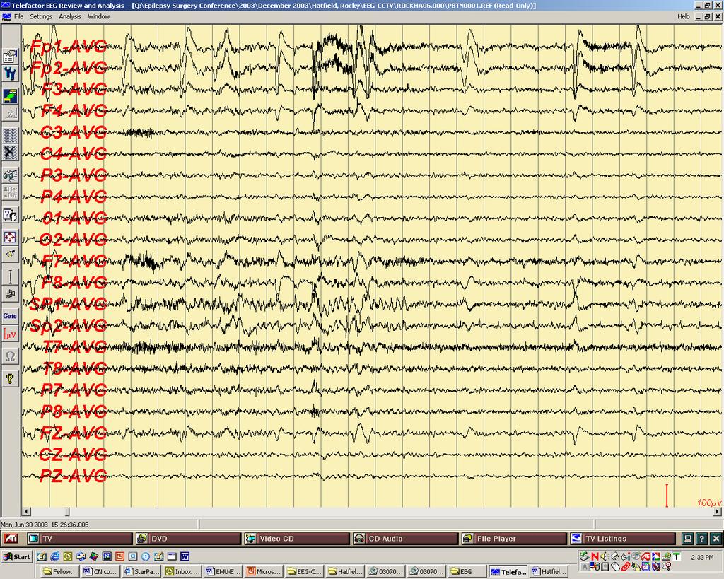
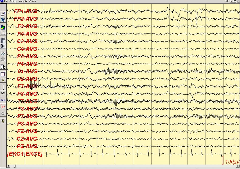
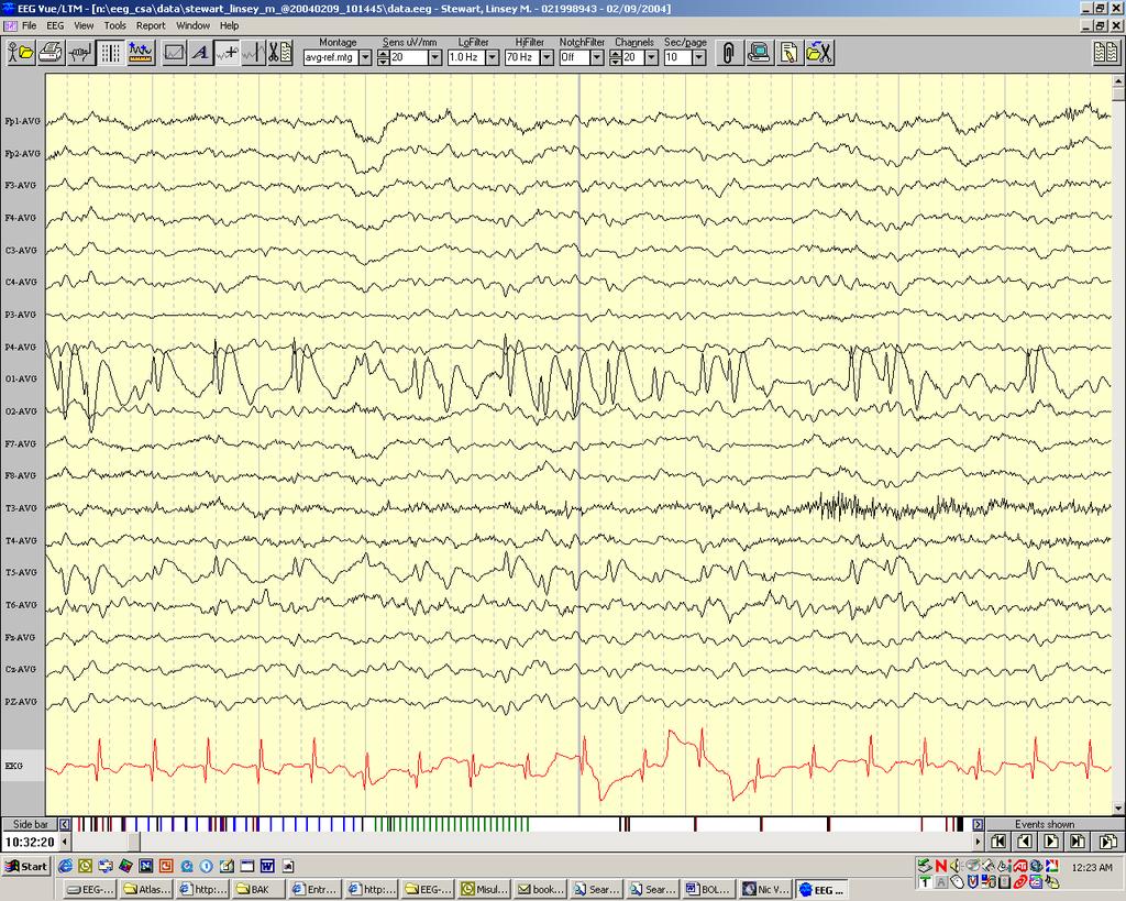
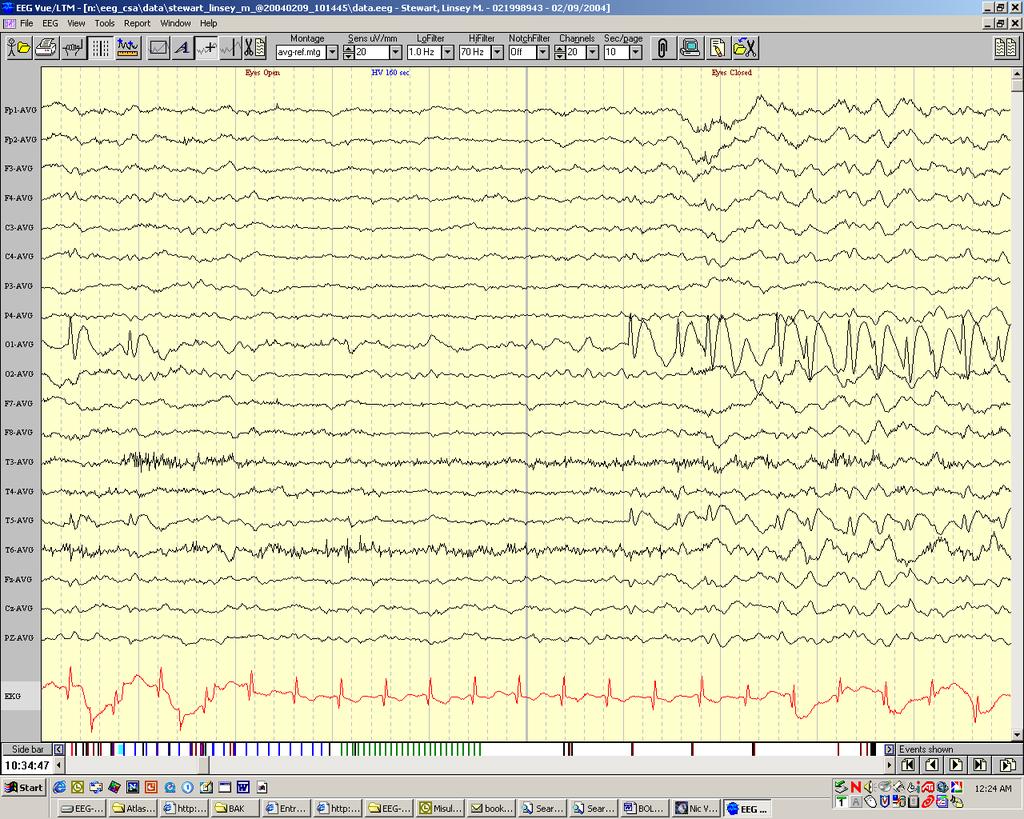
12 Q5- What is most likely true for the EEG above A. The pattern is indicative of a contaminated reference B. Seizures involve jerking of the right face C. Seizures are most likely nocturnal D. Seizures originate in the midline frontal region
13 The EEG in epilepsy Role in diagnosis and management Focal interictal nonepileptiform and epileptiform abnormalities Focal ictal patterns Specificity, sensitivity, pitfalls
14 Role in diagnosis and management of epilepsy Provide support for the clinical diagnosis (including status epilepticus) Help classify the epilepsy/ epileptic syndrome and localize the epileptogenic zone Unlikely to provide evidence for the etiology of epilepsy Occasionally help follow response to therapy (ex: absence) Help predict seizure recurrence after first unprovoked seizure or after AED withdrawal
15 Steps in the analysis of suspected abnormal transients Is the transient cerebral or artifactual? If cerebral, is it normal or abnormal? If abnormal, is it specific for epilepsy (epileptiform)? If epileptiform, is it focal (or regional, or lateralized) or generalized? If focal or regional, what is the field of the discharge?
16 Discharges Associated with Epilepsy Interictal Epileptiform Discharges Seizure Patterns or Ictal Patterns
17 Criteria for epileptiform VOLTAGE: high MORPHOLOGY: discharges shorter, lower amplitude first phase longer, higher amplitude second phase polyphasic aftergoing slow wave different from background Polarity: great majority are predominantly surface negative
18 Criteria for epileptiform discharges DURATION: short (but not too short) Spike: <70 msec Sharp wave: msec BACKGROUND: abnormal Must have a physiologic field LOCATION & STATE: unlike normal physiologic transients
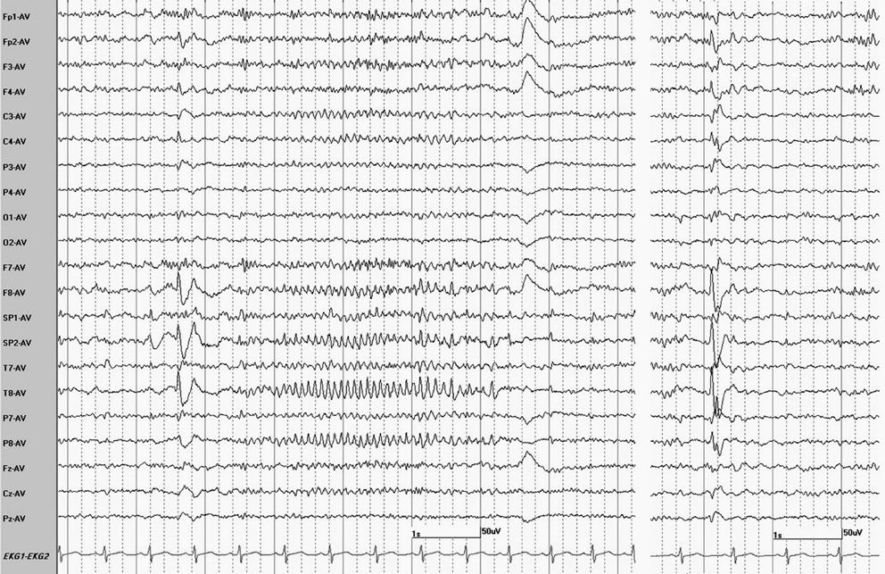
19 Epileptiform sharp wave
20 Epileptiform Discharges Spikes (20-70 msec) Sharp waves ( msec) Spike-and-wave, Slow-spike-and-wave, Sharp-and-slow-wave, Multiple spike (Polyspike), Multiple-spike-and-slowwave (Polyspike-and-slow-wave) Complexes

21 A- spike; B- sharp wave; C- spike-andwave complexes; D- sharp-and-slowwave complexes; E- slow-spike-andslow-wave complex; F- polyspike-andwave complex; G- multiple-sharp-andslow-wave complex; H- polyspike complex; I&Jmultiple sharp wave complexes. Even though spikes and sharp waves usually have aftergoing slow waves, the term spike-andwave complex is usually reserved for the situation where the slow wave is very prominent, higher in voltage than the spike.
22 Ictal vs interictal discharges Ictal discharge usually not a repetition of interictal discharges, except in generalized absence seizures Occasional patterns can be either ictal or interictal (spike-and-wave discharges, paroxysmal fast activity)
23 EEG findings in selected epileptic syndromes TLE Other cryptogenic/symptomatic partial epilepsies Benign Partial Epilepsies
24 TLE Temporal irregular slow activity- nonspecific Temporal intermittent rhythmic delta activity (TIRDA)- has a strong association with epileptiform discharges Temporal sharp waves/spikes- fairly specific for epilepsy May be bilateral independent even when seizures unilateral Sleep activates independent interictal epileptiform discharges Waking and REM restrict IED to primary focus
25
26
27
28
29
30
31 HOW CAN WE DISTINGUISH MESIAL FROM LATERAL TEMPORAL FOCI?
32 Interictal EEG features distinguishing mesial and lateral temporal lobe epilepsy Mesial IEDs predominantly over the ipsilateral mesial temporal regions (SP1/2 or FT9/10) TIRDA more likely Lateral IEDs with lateral neocortical predominance (T7/T8 ie T3/T4; P7/P8 ie T5/T6) Patient groups do not differ in the presence of mesial versus lateral discharges; only in the predominance
33 Ictal EEG features distinguishing mesial and lateral temporal lobe seizures Mesial Frequency of ictal discharge >5 Hz (frequency with inverse relationship to HS) Ictal discharge better localized, with inferomesial predominance Bilateral spread slower Lateral Lower mean frequency of ictal discharge Frequent bihemispheric distribution at onset May have transitional sharp wave at seizure onset Bilateral spread more rapid
34 Scalp EEG characteristics of temporal lobe seizures Ebersole &Pacia, Epilepsia Description of earliest ictal rhythms Most likely origin 1A- Inferotemporal rhythm of 5-9 Hz that is regular for at least 5 s 1B- Vertex rhythm of 5-9 Hz that is regular for at least 5 s 1C- Type 1B followed by type 1A 2A- Temporal and/or frontocentral rhythm of 2-5 Hz that is irregular or regular for only brief periods 2B- Type 2A followed by type 1A 2C- Type 2A or 2B preceded by irregular or repetitive sharp or slow waves 3- Unlateralized or diffuse arrythmic change in background Hippocampus Hippocampus Hippocampus Temporal neocortex Temporal neocortex Temporal neocortex Temporal neocortex
35
36
37
38 Transitional Sharp Waves at Ictal Onset- a Neocortical Ictal Pattern Azar et al, JCN ictal discharges in 13 patients started with a TShW The center of TShW field was always concordant with the presumed final localization/lateralization, while that of the subsequent ictal discharge was concordant in only six patients. None of 61 ictal discharges in 15 patients with mesial temporal lobe epilepsy studied in the same time period started with a TShW. TShW are a marker of neocortical seizure onset. The TShW field provided more accurate localization or lateralization of the ictal focus than the following rhythmic ictal discharge. TShW at seizure onset should suggest a neocortical rather than hippocampal seizure onset.
39
40 Frontal lobe epilepsy Epileptiform discharges often poorly localized Multifocal, generalized (secondary bilateral synchrony) More localized if dorsolateral (over convexity) Focal slow activity, attenuation may be helpful Interictal EEG abnormalities may be absent Midline theta may favor FLE
41 Frontal lobe epilepsy Ictal EEG on scalp may show no definite change when the epileptogenic zone is mesial frontal or orbitofrontal. Dorsolateral seizures more likely to have focal onset Focal beta activity most valuable
42
43 Perry interictal

44 Perry ictal
45 Parietal and occipital lobe seizures Interictal discharges may be bitemporal, multifocal or generalized Correctly localized in 1/3 of instances May be absent Frequent false ictal localization through rapid temporal or frontal propagation
46
47
48 Benign epilepsy with centrotemporal spikes or Benign Rolandic Epilepsy Discharges have a characteristic morphology: they are broad, blunt, followed by a slow wave Discharges commonly have a central-midtemporal field, but variations exist (ex: parieto-temporal or parieto-occipital) Markedly activated in drowsiness and sleep May be bilateral independent or multifocal Horizontal dipole is common, with bifrontal positivity Generalized spike-and-wave discharges may co-exist
49
50
51
52 Benign epilepsy with occipital paroxysms High voltage paroxysms, mostly spike-and-wave discharges, less often sharp waves, over the occipital + posterior temporal regions, unilaterally or bilaterally, synchronously or independently Repetitive (1-3/ sec) in trains Blocked by eye opening; enhanced by eye closure Occipital discharges may co-exist with centrotemporal sharp waves or generalized spikeand-wave discharges
53
54
55 EEG abnormalities with 3 EEG protocols among 85 patients (DI-EEG= EEG during drug-induced sleep, r-eeg= routine EEG; SD-EEG= EEG after sleep deprivation). Leach et, JNNP 2006 n (%) Sensitivity Generalized spike-and-wave 36 (43%) Yield SD-EEG Yield DI-EEG Yield r-eeg Focal discharges 15 (18%) Yield SD-EEG Yield DI-EEG Yield r-eeg normal EEGs 33 (39%)
56 Effectiveness of Multiple EEGs in Supporting the Diagnosis of Epilepsy Salinsky et al, Epilepsia 1987 Interictal epileptiform activity (IIEA) detected on first EEG in 50% by 3rd EEG in 84% by 4th EEG in 92% There is relatively little yield beyond the 4th EEG
57 Limitations of routine EEG Indirect assessment (seizures being an intermittent phenomenon) Relies on interictal discharges; rare opportunity to record seizures 50% of patients with epilepsy will have a normal first EEG; 10% will always have a normal interictal EEG Interictal EEG abnormalities may be unreliable for diagnosis and classification Non-epileptic seizures coexistent with epilepsy Generalized SW discharges in patients with partial epilepsy, focal D/Cs in patients with generalized seizures
58 Methods of Activation Sleep Sleep deprivation Hyperventilation Photic stimulation Specific modes of precipitation in patients with reflex seizures Prolonged recordings
59 Additional Electrodes 10/10 system T1/T2, zygomatic, cheek electrodes Nasopharyngeal electrodes Sphenoidal electrodes Minisphenoidal electrodes Supraorbital
60
61
62 EEG- predictive value after 1 st seizure (Berg & Shinnar, Neurology 1991) Recurrence following 1st seizure Higher risk with abnormal EEG GSW more predictive (Hauser et al) Pooled relative risk: 2.0 Epi abn. vs Nl / 1.3 Epi vs Non-epi abn. Pooled risk of recurrence 27% with Nl EEG 58% with epileptiform abnormalities 37% with non-epileptiform abnormalities
63 Value of the early EEG after a first unprovoked seizure- Schreiner & Pohlmann-Eden. Clin Electroencephalogr 2003;34: Prospective- standard EEG and EEG with sleep deprivation in adult patients with a first unprovoked seizure. EEGs were performed on 157 adult patients within the first 48 hours of the first seizure. Additional sleep deprived EEGs in 60. Standard EEG was abnormal in 70.7% and significantly associated with an increased risk of seizure recurrence [risk ratio 4.5, 95% confidence interval (CI) 1.8; 11.3, p=0.001].
64 Value of the early EEG after a first unprovoked seizure- Schreiner & Pohlmann-Eden. Clin Electroencephalogr 2003;34: Subgroup analysis revealed the highest recurrence rates for patients with focal epileptiform activity (risk ratio 2.2, CI 1.2; 4.2, p=0.01). EEGs with sleep deprivation were abnormal in 48.3% of all cases and revealed epileptiform discharges in 13.3% of the patients who had no epileptiform activity in the standard EEG.
65 EEG predictive value for medication withdrawal Most studies report an increased risk for relapse in patients whose EEG pattern is abnormal just before discontinuing drugs compared with those having normal EEG findings. Relative risk typical estimate MRC trial: only generalized IEDs with GTC seizures increased risk of recurrence (relative risk 1.37) Diagnosis of epileptic syndrome may be a better predictor of relapse
66 False Positive EEGs Normal nonepileptic population: 0.4% Nonepileptic patients: 1.7% Central, midtemporal and occipital IIEA may occur in children without epilepsy Errors in practice most often due to: Misinterpretation of normal EEG (particularly in drowsiness) Misinterpretation of incidental or irrelevant EEG abnormalities
67 Epileptiform discharges with weaker association with epilepsy Multifocal sporadic spikes in premature newborns- no association with epilepsy Centrotemporal spikes in children 20-40% of children with centrotemporal spikes do not have epilepsy Occipital spikes in children epilepsy in only 54%, only 33% with early onset visual deprivation (vs 91% with temporal foci)
68 Epileptiform discharges with weaker association with epilepsy Generalized spike-wave discharges 35% of asymptomatic siblings of children with generalized absence seizures have generalized spike-waves Photoparoxysmal response Up to 14% of normal children have photoparoxysmal responses with prolonged photic stimulation
69 Incidence of spikes and paroxysmal rhythmic events in normal subjects Multicenter study, J Clin Neurophys 1998 Computer-assisted ambulatory EEG 16 channels, overnight recording 135 normal adult volunteers at 11 sites Only one subject had spikes 16 subjects with migraine 2 had spikes (12.5%) 15 subjects with family history of epilepsy 2 had spikes (13.3%)
70 EEG in Epilepsy Summary The EEG remains the most sensitive and specific test in support of epilepsy There is great potential for over-reading epileptic abnormalities on EEG More damage is done by over-reading than under-reading The EEG should never be a substitute for a detailed clinical history
Beyond the Basics in EEG Interpretation: Throughout the Life Stages
 Beyond the Basics in EEG Interpretation: Throughout the Life Stages Steve S. Chung, MD, FAAN Chairman, Neuroscience Institute Director, Epilepsy Program Banner University Medical Center University of Arizona
The Normal EEG, Normal Variants, Artifacts. Bassel Abou-Khalil, M.D.
 The Normal EEG, Normal Variants, Artifacts Bassel Abou-Khalil, M.D. I have no financial relationships to disclose that are relative to the content of my presentation Learning Objectives recognize normal
Idiopathic epilepsy syndromes
 1 Idiopathic epilepsy syndromes PANISRA SUDACHAN, M.D. Pe diatric Neuro lo gis t Pediatric Neurology Department Pras at Neuro lo gic al Institute Epilepsy course 20 August 2016 Classification 2 1964 1970
True Epileptiform Patterns (and some others)
") True Epileptiform Patterns (and some others) a) What is epileptiform b) Some possible surprises c) Classification of generalized epileptiform patterns An epileptiform pattern Interpretative term based
True Epileptiform Patterns (and some others) a) What is epileptiform b) Some possible surprises c) Classification of generalized epileptiform patterns An epileptiform pattern Interpretative term based
EEG workshop. Epileptiform abnormalities. Definitions. Dr. Suthida Yenjun
 EEG workshop Epileptiform abnormalities Paroxysmal EEG activities ( focal or generalized) are often termed epileptiform activities EEG hallmark of epilepsy Dr. Suthida Yenjun Epileptiform abnormalities
Idiopathic epilepsy syndromes
 Idiopathic epilepsy syndromes PANISRA SUDACHAN, M.D. Pediatric Neurologist Pediatric Neurology Department Prasat Neurological Institue Epilepsy course 26 August 2017 Classification 1964 1970 1981 1989
The secrets of conventional EEG
 The secrets of conventional EEG The spike/sharp wave activity o Electro-clinical characteristics of Spike/Sharp wave The polymorphic delta activity o Electro-clinical characteristics of Polymorphic delta
Generalised Epileptiform Patterns
 Generalised Epileptiform Patterns Manori Wijayath Westmead Hospital, Sydney, Australia With slides from Elizabeth Walker and Andrew Bleasel Generalised Epilep-form Discharges: Outline 1. Generalised epilep.form
Non epileptiform abnormality J U LY 2 7,
 Non epileptiform abnormality S U D A J I R A S A K U L D E J, M D. C H U L A L O N G KO R N C O M P R E H E N S I V E E P I L E P S Y C E N T E R J U LY 2 7, 2 0 1 6 Outline Slow pattern Focal slowing
Normal EEG of wakeful resting adults of years of age. Alpha rhythm. Alpha rhythm. Alpha rhythm. Normal EEG of the wakeful adult at rest
 Normal EEG of wakeful resting adults of 20-60 years of age Suthida Yenjun, M.D. Normal EEG of the wakeful adult at rest Alpha rhythm Beta rhythm Mu rhythm Vertex sharp transients Intermittent posterior
Neurophysiology & EEG
 Neurophysiology & EEG PG4 Core Curriculum Ian A. Cook, M.D. Associate Director, Laboratory of Brain, Behavior, & Pharmacology UCLA Department of Psychiatry & Biobehavioral Sciences Semel Institute for
EEG IN FOCAL ENCEPHALOPATHIES: CEREBROVASCULAR DISEASE, NEOPLASMS, AND INFECTIONS
 246 Figure 8.7: FIRDA. The patient has a history of nonspecific cognitive decline and multiple small WM changes on imaging. oligodendrocytic tumors of the cerebral hemispheres (11,12). Electroencephalogram
Seizure Semiology and Neuroimaging Findings in Patients with Midline Spikes
 Epilepsia, 42(12):1563 1568, 2001 Blackwell Science, Inc. International League Against Epilepsy Seizure Semiology and Neuroimaging Findings in Patients with Midline Spikes *Ekrem Kutluay, *Erasmo A. Passaro,
ROLE OF EEG IN EPILEPTIC SYNDROMES ASSOCIATED WITH MYOCLONUS
 Version 18 A Monthly Publication presented by Professor Yasser Metwally February 2010 ROLE OF EEG IN EPILEPTIC SYNDROMES ASSOCIATED WITH MYOCLONUS EEG is an essential component in the evaluation of epilepsy.
Introduction to EEG del Campo. Introduction to EEG. J.C. Martin del Campo, MD, FRCP University Health Network Toronto, Canada
 Introduction to EEG J.C. Martin, MD, FRCP University Health Network Toronto, Canada What is EEG? A graphic representation of the difference in voltage between two different cerebral locations plotted over
Idiopathic epilepsy syndromes
 Idiopathic epilepsy syndromes PANISRA SUDACHAN, M.D. Pediatric Neurologist Pediatric Neurology Department Prasat Neurological Institue Epilepsy course 8 September 2018 Outline of topic Definition Idiopathic
EEG in Benign and Malignant Epileptic Syndromes of Childhood
 Epilepsia, 43(Suppl. 3):17 26, 2002 Blackwell Publishing, Inc. International League Against Epilepsy EEG in Benign and Malignant Epileptic Syndromes of Childhood Ivo Drury Department of Neurology, Henry
EEG in the Evaluation of Epilepsy. Douglas R. Nordli, Jr., MD
 EEG in the Evaluation of Epilepsy Douglas R. Nordli, Jr., MD Contents Epidemiology First seizure Positive predictive value Risk of recurrence Identifying epilepsy Type of epilepsy (background and IEDs)
EEG WORKSHOP Nonepileptiform Abnormalities
 EEG WORKSHOP Nonepileptiform Abnormalities Kamornwan Katanyuwong MD Chiangmai University Hospital EST: 20th July 2010 EEG reading Age Background Epileptiform Non epileptiform Activation procedure normal
Diagnosing Complicated Epilepsy: Mapping of the Epileptic Circuitry. Michael R. Sperling, M.D. Thomas Jefferson University Philadelphia, PA
 Diagnosing Complicated Epilepsy: Mapping of the Epileptic Circuitry Michael R. Sperling, M.D. Thomas Jefferson University Philadelphia, PA Overview Definition of epileptic circuitry Methods of mapping
EEG in Medical Practice
 EEG in Medical Practice Dr. Md. Mahmudur Rahman Siddiqui MBBS, FCPS, FACP, FCCP Associate Professor, Dept. of Medicine Anwer Khan Modern Medical College What is the EEG? The brain normally produces tiny
Localization of Temporal Lobe Foci by Ictal EEG Patterns
 Epilepsia, 37(4):386-399, 1996 Lippincott-Raven Publishers, Philadelphia 0 International League Against Epilepsy Localization of Temporal Lobe Foci by Ictal EEG Patterns John S. Ebersole and *Steven V.
Idiopathic epilepsy syndromes
 Idiopathic epilepsy syndromes Kamornwan Katanyuwong MD. Chiangmai University Hospital EST, July 2009 Diagram Sylvie Nyugen The Tich, Yann Pereon Childhood absence epilepsy (CAE) Age : onset between 4-10
Common EEG pattern in critical care
 Common EEG pattern in critical care พ.ญ.ส ธ ดา เย นจ นทร Causes Direct neuronal injury Cerebral dysfunction : encephalopathy Psychic problems EEG in critical care 1 October 2009, Pramongkutklao Hospital
Common Ictal Patterns in Patients with Documented Epileptic Seizures
 THE ICTAL IRAQI PATTERNS POSTGRADUATE IN EPILEPTIC MEDICAL JOURNAL PATIENTS Common Ictal Patterns in Documented Epileptic Seizures Ghaieb Bashar ALJandeel, Gonzalo Alarcon ABSTRACT: BACKGROUND: The ictal
Asian Epilepsy Academy (ASEPA) EEG Certification Examination
 EEG Certification Examination") Asian Epilepsy Academy (ASEPA) EEG Certification Examination EEG Certification Examination Aims To set and improve the standard of practice of Electroencephalography (EEG) in the Asian Oceanian region
Asian Epilepsy Academy (ASEPA) EEG Certification Examination EEG Certification Examination Aims To set and improve the standard of practice of Electroencephalography (EEG) in the Asian Oceanian region
Review Article Electroencephalography in Mesial Temporal Lobe Epilepsy: A Review
 Epilepsy Research and Treatment Volume 2012, Article ID 637430, 17 pages doi:10.1155/2012/637430 Review Article Electroencephalography in Mesial Temporal Lobe Epilepsy: A Review Manouchehr Javidan1, 2,
Practical 3 Nervous System Physiology 2 nd year English Module. Dept. of Physiology, Carol Davila University of Medicine and Pharmacy
 Electroencephalography l h (EEG) Practical 3 Nervous System Physiology 2 nd year English Module Dept. of Physiology, Carol Davila University of Medicine and Pharmacy What is EEG EEG noninvasively records
Asian Epilepsy Academy (ASEPA) & ASEAN Neurological Association (ASNA) EEG Certification Examination
 & ASEAN Neurological Association (ASNA) EEG Certification Examination") Asian Epilepsy Academy (ASEPA) & ASEAN Neurological Association (ASNA) EEG Certification Examination EEG Certification Examination Aims To set and improve the standard of practice of Electroencephalography
Asian Epilepsy Academy (ASEPA) & ASEAN Neurological Association (ASNA) EEG Certification Examination EEG Certification Examination Aims To set and improve the standard of practice of Electroencephalography
9 The Abnormal EEG. An EEG is considered abnormal if it has findings
 C h a p t e r 9 The Abnormal EEG An EEG is considered abnormal if it has findings known to be associated with a pathologic or disease state. As discussed in Chapter 8, The Structure and Philosophy of the
Scalp EEG Findings in Temporal Lobe Epilepsy
 Scalp EEG Findings in Temporal Lobe Epilepsy Seyed M Mirsattari M.D., Ph.D., F.R.C.P.(C) Assistant Professor Depts. of CNS, Medical Biophysics, Medical Imaging, and Psychology University of Western Ontario
Seizures and Sleep, Sorting out the Spikes and Waves, a Polysomnographic and Clinical Review
 Seizures and Sleep, Sorting out the Spikes and Waves, a Polysomnographic and Clinical Review DR. MARK GARWOOD CLINICAL ASSISTANT PROFESSOR, DEPARTMENT OF NEUROLOGY MEDICAL DIRECTOR SLEEP DISORDERS CLINICS
EEG source Localization (ESL): What do we know now?
: What do we know now?") EEG source Localization (ESL): What do we know now? Talk overview Theoretical background Fundamental of ESL (forward and inverse problems) Voltage topography of temporal spikes Improving source localization
EEG source Localization (ESL): What do we know now? Talk overview Theoretical background Fundamental of ESL (forward and inverse problems) Voltage topography of temporal spikes Improving source localization
Idiopathic Epileptic Syndromes
 Idiopathic Epileptic Syndromes Greek words idios = self, own and personal pathic = suffer Kamornwan Katanuwong MD Chiangmai University Hospital 1 st Epilepsy Camp, Hua Hin 20 th August 2010 Is a syndrome
Focal epilepsy recruiting a generalised network of juvenile myoclonic epilepsy: a case report
 Clinical commentary Epileptic Disord 2014; 16 (3): 370-4 Focal epilepsy recruiting a generalised network of juvenile myoclonic epilepsy: a case report Myo Khaing 1,2, Kheng-Seang Lim 1, Chong-Tin Tan 1
Neonatal EEG Maturation
 Neonatal EEG Maturation Cindy Jenkinson, R. EEG T., CLTM October 7, 2017 Fissure Development 3 http://www.hhmi.org/biointeractive/develop ment-human-embryonic-brain 4 WHAT IS IMPORTANT TO KNOW BEFORE I
EEG in the ICU. Quiz. March Teneille E. Gofton
 EEG in the ICU Quiz March 2012 Teneille E. Gofton Quiz The next several slides will show 15 subhairline EEGs. Choose the best possible answer in each scenario. Your score and solutions will be provided
Effects of Sleep and Circadian Rhythms on Epilepsy
 Effects of Sleep and Circadian Rhythms on Epilepsy Milena Pavlova, M.D. Medical Director, Faulkner Neurophysiology Laboratory Department of Neurology, Brigham and Women s Hospital Harvard Medical School
Identifying Montages that Best Detect Electrographic Seizure Activity During Polysomnography
 ELECTROGRAPHIC SEIZURE ACTIVITY DURING POLYSOMNOGRAPHY Identifying Montages that Best Detect Electrographic Seizure Activity During Polysomnography Nancy Foldvary DO, 1 A.Cosmo Caruso MD, 1 Edward Mascha
Latero-Orbital and Anterior-Temporal Electrodes "Their Usefulness in Diagnosing Complex Partial Seizures"
 Mona T. ElGhoneimy et al. LateroOrbital and AnteriorTemporal Electrodes "Their Usefulness in Diagnosing Complex Partial Seizures" Mona T. ElGhoneimy 1, Hanan Hosny 2, Faisal Abdel Wahab 3, Abdel Naser
Classification of Seizures. Generalized Epilepsies. Classification of Seizures. Classification of Seizures. Bassel F. Shneker
 Classification of Seizures Generalized Epilepsies Bassel F. Shneker Traditionally divided into grand mal and petit mal seizures ILAE classification of epileptic seizures in 1981 based on clinical observation
A reappraisal of secondary bilateral synchrony
 Neurology Asia 2007; 12 : 29 35 A reappraisal of secondary bilateral synchrony Liri JIN MD, PhD Department of Neurology, Peking Union Medical College Hospital, Chinese Academy of Medical Sciences, Beijing,
The Sonification of Human EEG and other Biomedical Data. Part 3
 The Sonification of Human EEG and other Biomedical Data Part 3 The Human EEG A data source for the sonification of cerebral dynamics The Human EEG - Outline Electric brain signals Continuous recording
Epilepsy: diagnosis and treatment. Sergiusz Jóźwiak Klinika Neurologii Dziecięcej WUM
 Epilepsy: diagnosis and treatment Sergiusz Jóźwiak Klinika Neurologii Dziecięcej WUM Definition: the clinical manifestation of an excessive excitation of a population of cortical neurons Neurotransmitters:
Localization a quick look
 Localization a quick look Covering the basics Differential amplifiers Polarity convention 10-20 electrode system Basic montages: bipolar and referential Other aspects of displaying the EEG Localization
Idiopathic Photosensitive Occipital Lobe Epilepsy
 Idiopathic Photosensitive Occipital Lobe Epilepsy 2 Idiopathic photosensitive occipital lobe epilepsy (IPOE) 5, 12, 73, 75, 109, 110 manifests with focal seizures of occipital lobe origin, which are elicited
Scope. EEG patterns in Encephalopathy. Diffuse encephalopathy. EEG in adult patients with. EEG in diffuse encephalopathy
 Scope EEG patterns in Encephalopathy Dr.Pasiri Sithinamsuwan Division of Neurology Department of Medicine Phramongkutklao Hospital Diffuse encephalopathy EEG in specific encephalopathies Encephalitides
Case report. Epileptic Disord 2005; 7 (1): 37-41
: 37-41") Case report Epileptic Disord 2005; 7 (1): 37-41 Periodic lateralized epileptiform discharges (PLEDs) as the sole electrographic correlate of a complex partial seizure Gagandeep Singh, Mary-Anne Wright,
Case report Epileptic Disord 2005; 7 (1): 37-41 Periodic lateralized epileptiform discharges (PLEDs) as the sole electrographic correlate of a complex partial seizure Gagandeep Singh, Mary-Anne Wright,
Epilepsy and EEG in Clinical Practice
 Mayo School of Professional Development Epilepsy and EEG in Clinical Practice November 10-12, 2016 Hard Rock Hotel at Universal Orlando Orlando, FL Course Directors Jeffrey Britton, MD and William Tatum,
EEG in Epileptic Syndrome
 EEG in Epileptic Syndrome Surachai Likasitwattanakul, M.D. Division of Neurology, Department of Pediatrics Faculty of Medicine, Siriraj Hospital Mahidol University Epileptic syndrome Electroclinical syndrome
Electroencephalography. Role of EEG in NCSE. Continuous EEG in ICU 25/05/59. EEG pattern in status epilepticus
 EEG: ICU monitoring & 2 interesting cases Electroencephalography Techniques Paper EEG digital video electroencephalography Dr. Pasiri Sithinamsuwan PMK Hospital Routine EEG long term monitoring Continuous
Developmental Changes Including Neonatal EEG. Gregory L. Holmes, MD
 Developmental Changes Including Neonatal EEG Gregory L. Holmes, MD A A + B =: B + A.Dravet Syndrome B.Menkes syndrome C.West syndrome D.Ohtahara shyndrome The Difficult Delivery 1 day old male transferred
EPILEPSY SURGERY EVALUATION IN ADULTS WITH SCALP VIDEO-EEG MONITORING. Meriem Bensalem-Owen, MD University of Kentucky
 EPILEPSY SURGERY EVALUATION IN ADULTS WITH SCALP VIDEO-EEG MONITORING Meriem Bensalem-Owen, MD University of Kentucky DISCLOSURES Received grants for sponsored research as investigator from: UCB Eisai
Spike voltage topography in temporal lobe epilepsy
 Thomas Jefferson University Jefferson Digital Commons Department of Neurology Faculty Papers Department of Neurology 5-17-2016 Spike voltage topography in temporal lobe epilepsy Ali Akbar Asadi-Pooya Thomas
Seizure onset can be difficult to asses in scalp EEG. However, some tools can be used to increase the seizure onset activity over the EEG background:
 This presentation was given during the Dianalund Summer School on EEG and Epilepsy, July 24, 2012. The main purpose of this introductory talk is to show the possibilities of improved seizure onset analysis
Sleep in Epilepsy. Kurupath Radhakrishnan,
 Sleep in Epilepsy Kurupath Radhakrishnan, Retired Senior Professor (Emeritus), R. Madavan Nayar Center for Comprehensive Epilepsy Care, Retired Director, Sree Chitra Tirunal Institute for Medical Sciences
Objectives. Amanda Diamond, MD
 Amanda Diamond, MD Objectives Recognize symptoms suggestive of seizure and what those clinical symptoms represent Understand classification of epilepsy and why this is important Identify the appropriate
Intracranial Studies Of Human Epilepsy In A Surgical Setting
 Intracranial Studies Of Human Epilepsy In A Surgical Setting Department of Neurology David Geffen School of Medicine at UCLA Presentation Goals Epilepsy and seizures Basics of the electroencephalogram
SLEEP STAGING AND AROUSAL. Dr. Tripat Deep Singh (MBBS, MD, RPSGT, RST) International Sleep Specialist (World Sleep Federation program)
 International Sleep Specialist (World Sleep Federation program)") SLEEP STAGING AND AROUSAL Dr. Tripat Deep Singh (MBBS, MD, RPSGT, RST) International Sleep Specialist (World Sleep Federation program) Scoring of Sleep Stages in Adults A. Stages of Sleep Stage W Stage
SLEEP STAGING AND AROUSAL Dr. Tripat Deep Singh (MBBS, MD, RPSGT, RST) International Sleep Specialist (World Sleep Federation program) Scoring of Sleep Stages in Adults A. Stages of Sleep Stage W Stage
Myoclonic status epilepticus in hypoxic ischemic encephalopathy which recurred after somatosensory evoked potential testing
 ANNALS OF CLINICAL NEUROPHYSIOLOGY CASE REPORT Ann Clin Neurophysiol 2017;19(2):136-140 Myoclonic status epilepticus in hypoxic ischemic encephalopathy which recurred after somatosensory evoked potential
EEG IN THE DIAGNOSIS, CLASSIFICATION, AND MANAGEMENT OF PATIENTS WITH EPILEPSY
 ii2 EEG IN THE DIAGNOSIS, CLASSIFICATION, AND MANAGEMENT OF PATIENTS WITH EPILEPSY Correspondence to: Dr Shelagh Smith, National Society for Epilepsy, Chalfont St Peter, Bucks SL9 0RJ, UK; shelaghs@epilepsynse.org.uk
All that blacks out is not syncope: a neurological view of transient loss of consciousness
 All that blacks out is not syncope: a neurological view of transient loss of consciousness Dr Simon Taggart Consultant Clinical Neurophysiologist. JCUH, Middlesbrough. Misdiagnosis of Blackouts Sutula
Late-onset temporal lobe epilepsy in a patient with juvenile myoclonic epilepsy
 Clinical commentary Epileptic Disord 2012; 14 (2): 190-4 Late-onset temporal lobe epilepsy in a patient with juvenile myoclonic epilepsy Octavian V Lie 1,2, Mark D Holmes 1 1 Department of Neurology, University
The AASM Manual for the Scoring of Sleep and Associated Events
 The AASM Manual for the Scoring of Sleep and Associated Events Summary of Updates in Version 2.1 July 1, 2014 The American Academy of Sleep Medicine (AASM) is committed to ensuring that The AASM Manual
Coexistence of focal and idiopathic generalized epilepsy in the same patient population
 Seizure (2006) 15, 28 34 www.elsevier.com/locate/yseiz Coexistence of focal and idiopathic generalized epilepsy in the same patient population Lara E. Jeha a, *, Harold H. Morris b, Richard C. Burgess
ENCEPHALOPATHY RECOGNIZING METABOLIC AND ANOXIC CHANGES
 ENCEPHALOPATHY RECOGNIZING METABOLIC AND ANOXIC CHANGES ENCEPHALOPATHY Encephalopathy is a general term that means brain disease, damage, or malfunction. The major symptom of encephalopathy is an altered
The EEG Findings in Extratemporal Seizures
 Epilepsa, 39(Suppl. 4):Sl-W 1998 Lippincott-Raven Publishers, Philadelphia 0 International League Against Epilepsy The EEG Findings in Extratemporal Seizures Barbara F. Westmoreland Mayo Clinic, Rochester,
Successful Treatment of Mesial Temporal Lobe Epilepsy with Bilateral Hippocampal Atrophy and False Temporal Scalp Ictal Onset: A case report
 Hiroshima J. Med. Sci. Vol. 61, No. 2, 37~41, June, 2012 HIJM 61 7 37 Successful Treatment of Mesial Temporal Lobe Epilepsy with Bilateral Hippocampal Atrophy and False Temporal Scalp Ictal Onset: A case
EEG in Children with Early-onset Benign Occipital Seizure Susceptibility Syndrome: Panayiotopoulos Syndrome
 Epilepsia, 44(3):435 442, 2003 Blackwell Publishing, Inc. 2003 International League Against Epilepsy EEG in Children with Early-onset Benign Occipital Seizure Susceptibility Syndrome: Panayiotopoulos Syndrome
La sindrome di Angelman: il problema delle crisi
 La sindrome di Angelman: il problema delle crisi Crisi o non crisi? Questo è il problema. Pedagogia dell anamnesi: ovvero come raccontare una crisi al medico, sperando che non vada fuori strada L inizio
Continuous EEG Monitoring is becoming a commonly used tool
 INVITED REVIEW American Clinical Neurophysiology Society s Standardized Critical Care EEG Terminology: 2012 version L. J. Hirsch, S. M. LaRoche, N. Gaspard, E. Gerard, A. Svoronos, S. T. Herman, R. Mani,
FRONTAL & TEMPORAL. A. Shah, MD. Director, Comprehensive Epilepsy Program Wayne State University/ Detroit Medical Center
 FRONTAL & TEMPORAL LOBE EPILEPSY A. Shah, MD Professor of Neurology Director, Comprehensive Epilepsy Program Wayne State University/ Detroit Medical Center Pretest 1. A complex partial seizure (CPS) may
*Pathophysiology of. Epilepsy
 *Pathophysiology of Epilepsy *Objectives * At the end of this lecture the students should be able to:- 1.Define Epilepsy 2.Etio-pathology of Epilepsy 3.Types of Epilepsy 4.Role of Genetic in Epilepsy 5.Clinical
Epileptic Seizures, Syndromes, and Classifications. Heidi Currier, MD Minnesota Epilepsy Group, PA St. Paul, MN
 Epileptic Seizures, Syndromes, and Classifications Heidi Currier, MD Minnesota Epilepsy Group, PA St. Paul, MN Definitions Diagnosis of Seizures A seizure is a sudden surge of electrical activity in the
A retrospective study of Electroencephalographic (EEG) findings and its interpretation in Adults and children
 findings and its interpretation in Adults and children") Original Research Article DOI: 10.18231/2455-8451.2017.0023 A retrospective study of Electroencephalographic (EEG) findings and its interpretation in Adults and children Dwajani S 1,*, Nirmala K.S. 2,
Original Research Article DOI: 10.18231/2455-8451.2017.0023 A retrospective study of Electroencephalographic (EEG) findings and its interpretation in Adults and children Dwajani S 1,*, Nirmala K.S. 2,
Early seizure propagation from the occipital lobe to medial temporal structures and its surgical implication
 Original article Epileptic Disord 2008; 10 (4): 260-5 Early seizure propagation from the occipital lobe to medial temporal structures and its surgical implication Naotaka Usui, Tadahiro Mihara, Koichi
Latency to first spike in the EEG of epilepsy patients
 Seizure (2008) 17, 34 41 www.elsevier.com/locate/yseiz Latency to first spike in the EEG of epilepsy patients Jaishree T. Narayanan a,b, *, Douglas R. Labar a, Neil Schaul a a Comprehensive Epilepsy Center,
Early marker of CLN2: Is neurophysiology helpful? A. Kaminska, Department of Neurophysiology, Hôpital Necker Enfants Malades, Paris, France
 Early marker of CLN2: Is neurophysiology helpful? A. Kaminska, Department of Neurophysiology, Hôpital Necker Enfants Malades, Paris, France U/CLN2/0142 October 2017 Lectures: Biomarin Disclosures 14 patients
Periodic and Rhythmic Patterns. Suzette M LaRoche, MD Mission Health Epilepsy Center Asheville, North Carolina
 Periodic and Rhythmic Patterns Suzette M LaRoche, MD Mission Health Epilepsy Center Asheville, North Carolina Continuum of EEG Activity Neuronal Injury LRDA GPDs SIRPIDs LPDs + NCS Burst-Suppression LPDs
Epilepsy. Hyunmi Choi, M.D., M.S. Columbia Comprehensive Epilepsy Center The Neurological Institute. Seizure
 Epilepsy Hyunmi Choi, M.D., M.S. Columbia Comprehensive Epilepsy Center The Neurological Institute Seizure Symptom Transient event Paroxysmal Temporary physiologic dysfunction Caused by self-limited, abnormal,
Temporal lobe dysembryoplastic neuroepithelial tumour: significance of discordant interictal spikes
 Original article Epileptic Disord 2004; 6: 10-14 Temporal lobe dysembryoplastic neuroepithelial tumour: significance of discordant interictal spikes Angelo Labate 1, Regula S. Briellmann 1,6, Anthony S.
Assessment of EEG as a Diagnostic and Prognostic Indicator Tool in the Febrile Seizures
 Indian J Physiol Pharmacol 2015; 59(3) : 251 260 Febrile Seizure, Diagnostic and Prognostic Indicator, EEG 251 Original Article Assessment of EEG as a Diagnostic and Prognostic Indicator Tool in the Febrile
Early predictors of refractory epileptic seizures
 Mohamed M.Mostafa et al. Early predictors of refractory epileptic seizures Mohamed M.Mostafa 1, Ali Akram 2, Mohamed Osman 3, Mamdooh Eissa 4 Depatments of Neurology, Ain Shams University 1, Psychiatry
Chronic PLEDs with transitional rhythmic discharges (PLEDs-plus) in remote stroke
 in remote stroke") Original article Epileptic Disord 2007; 9 (2): 164-9 Chronic PLEDs with transitional rhythmic discharges (PLEDs-plus) in remote stroke José F. Téllez-Zenteno 1, Sylaja N. Pillai 2, Michael D. Hill 2, Neelan
Original article Epileptic Disord 2007; 9 (2): 164-9 Chronic PLEDs with transitional rhythmic discharges (PLEDs-plus) in remote stroke José F. Téllez-Zenteno 1, Sylaja N. Pillai 2, Michael D. Hill 2, Neelan
Source localisation in the clinical practice: spontaneous EEG examinations with LORETA. Ph.D. thesis. Márton Tamás Tóth M.D.
 Source localisation in the clinical practice: spontaneous EEG examinations with LORETA Ph.D. thesis Márton Tamás Tóth M.D. Department of Neurology, University of Pécs Leader of project:: Prof. István Kondákor,
BESA Research Quick Guide
 BESA Research Quick Guide BESA 3D Maps Quick Guide An introduction how to interpret 3D voltage and phase maps in the scalp EEG Copyright and Trademarks The BESA products and their documentation are copyrighted
Epilepsy, a common chronic neurological disorder, is a
 10 SUPPLEMENT TO Journal of the association of physicians of india august 2013 VOL. 61 Epilepsy: Diagnostic Evaluation JMK Murthy* Epilepsy, a common chronic neurological disorder, is a potentially treatable
SEIZURE OUTCOME AFTER EPILEPSY SURGERY
 SEIZURE OUTCOME AFTER EPILEPSY SURGERY Prakash Kotagal, M.D. Head, Pediatric Epilepsy Cleveland Clinic Epilepsy Center LEFT TEMPORAL LOBE ASTROCYTOMA SEIZURE OUTCOME 1 YEAR AFTER EPILEPSY SURGERY IN ADULTS
Epilepsy DOJ Lecture Masud Seyal, M.D., Ph.D. Department of Neurology University of California, Davis
 Epilepsy DOJ Lecture - 2005 Masud Seyal, M.D., Ph.D. Department of Neurology University of California, Davis Epilepsy SEIZURE: A temporary dysfunction of the brain resulting from a self-limited abnormal
EEG in the ICU: Part I
 EEG in the ICU: Part I Teneille E. Gofton July 2012 Objectives To outline the importance of EEG monitoring in the ICU To briefly review the neurophysiological basis of EEG To introduce formal EEG and subhairline
Focal fast rhythmic epileptiform discharges on scalp EEG in a patient with cortical dysplasia
 Seizure 2002; 11: 330 334 doi:10.1053/seiz.2001.0610, available online at http://www.idealibrary.com on CASE REPORT Focal fast rhythmic epileptiform discharges on scalp EEG in a patient with cortical dysplasia
13 The Electroencephalogram of the Newborn
 C h a p t e r 13 The Electroencephalogram of the Newborn Newborn EEG interpretation is considered a particularly challenging area. An understanding of the appearance of the normal newborn EEG was achieved
Est-ce que l'eeg a toujours sa place en 2019?
 Est-ce que l'eeg a toujours sa place en 2019? Thomas Bast Epilepsy Center Kork, Germany Does EEG still play a role in 2019? What a question 7T-MRI, fmri, DTI, MEG, SISCOM, Of ieeg course! /HFO, Genetics
EEG History. Where and why is EEG used? 8/2/2010
 EEG History Hans Berger 1873-1941 Edgar Douglas Adrian, an English physician, was one of the first scientists to record a single nerve fiber potential Although Adrian is credited with the discovery of
TEMPORAL LOBE EPILEPSY: A CLINICAL VIEW POINT
 Version 9 A Monthly Publication presented by Professor Yasser Metwally January 2009 TEMPORAL LOBE EPILEPSY: A CLINICAL VIEW POINT Background: Temporal lobe epilepsy (TLE) was defined in 1985 by the International
Epileptic syndrome in Neonates and Infants. Piradee Suwanpakdee, MD. Division of Neurology Department of Pediatrics Phramongkutklao Hospital
 Epileptic syndrome in Neonates and Infants Piradee Suwanpakdee, MD. Division of Neurology Department of Pediatrics Phramongkutklao Hospital AGE SPECIFIC INCIDENCE OF EPILEPSY Hauser WA, et al. Epilepsia.
Interictal High Frequency Oscillations as Neurophysiologic Biomarkers of Epileptogenicity
 Interictal High Frequency Oscillations as Neurophysiologic Biomarkers of Epileptogenicity December 10, 2013 Joyce Y. Wu, MD Associate Professor Division of Pediatric Neurology David Geffen School of Medicine
MRI-negative frontal lobe epilepsy with ipsilateral akinesia and reflex activation
 Anatomo-electro-clinical correlations with video sequences Epileptic Disord 2008; 10 (4): 349-55 Anatomo-electro-clinical correlations: the Miami Children s Hospital, USA Case Report - Case 04-2008 MRI-negative
Subhairline EEG Part II - Encephalopathy
 Subhairline EEG Part II - Encephalopathy Teneille Gofton September 2013 Objectives To review the subhairline EEG changes seen with encephalopathy To discuss specific EEG findings in encephalopathy To outline
Fixation-off sensitivity in epilepsies other than the idiopathic epilepsies of childhood with occipital paroxysms: a 12-year clinical-video EEG study
 Original article Epileptic Disord 2009; 11 (1): 20-36 Fixation-off sensitivity in epilepsies other than the idiopathic epilepsies of childhood with occipital paroxysms: a 12-year clinical-video EEG study