Muscular System. SKELETAL CARDIAC SMOOTH Location Attached to bones or skin Forms walls of heart In walls of hollow visceral organs stomach, bladder,
|
|
|
- Frederica Francis
- 6 years ago
- Views:
Transcription

1 Muscular System 3 Types of Muscle Tissue Cells are called muscle fibers. SKELETAL CARDIAC SMOOTH Location Attached to bones or skin Forms walls of heart In walls of hollow visceral organs stomach, bladder, Cell shape & appearance Regulation of contraction Speed of contraction Single, long cylindrical cells arranged in bundles Multinucleated cells Striated due to arrangement of protein filaments Voluntary subject to conscious control via somatic nervous system Slow to fast -- Contracts rapidly, tires easily, powerful Function(s) Produces movement Maintains posture and provides support Stabilizes joints Body heat production Branching chains of cylindrical cells Uninucleated cells Striated Fibers arranged in spiral bundles joined by intercalated discs Involuntary controlled by intrinsic nodal system (pacemaker), autonomic nervous system and endocrine system Slow rhythmic contractions Changes size & shape of organ space to TRANSPORT MATERIALS through the body pushing blood through vessels respiratory passages, etc Spindle shaped cells muscle arranged in longitudinal and circular layers Uninucleated cells No striations Involuntary controlled by autonomic nervous system and endocrine system Very slow rhythmic contractions Changes size & shape of organ space to TRANSPORT MATERIALS through the body SPECIAL CHARACTERISTICS of muscular tissue: Excitability ability to receive and respond to electrical stimuli Contractility ability to shorten forcibly when stimulated Extensibility ability to be stretched Elasticity ability to recoil to resting length

2 Gross Anatomy of Skeletal Muscle Each muscle served by one artery, one nerve, and one or more veins that enter or exit near the central part of the muscle and branch through the connective tissue sheaths Every skeletal muscle fiber supplied by nerve ending that controls its activity. Contracting muscle fibers use huge amount of energy needs lots of oxygen and nutrients. Muscles generate lots of metabolic waste that is removed through veins. Individual muscle fibers are wrapped and help together by several different connective tissue sheaths that support each cell and reinforce the entire muscle. o Epimysium dense irregular connective tissue surrounding entire muscle; may blend with fascia o Perimysium fibrous connective tissue surrounding fascicles (groups of muscle fibers) o Endomysium fine areolar connective tissue surrounding each muscle fiber All the connective tissue sheaths are continuous with each other and with the tendons that join muscle to bone. When muscle fibers contract, they pull on these sheaths which transmit the pulling force to the bone to be moved. These sheaths contribute to the natural elasticity of muscle tissue and provide routes for blood vessels and nerves that serve the muscle. Most skeletal muscles span joints and are attached to bones at least two places. When a muscle contracts, the moveable bone, the muscle insertion, moves toward the immovable or less movable bone, the muscles origin. Muscle attachment may be direct or indirect. In direct attachment epimysium is fused to the periosteum of bone or perichondrium of cartilage. In indirect attachment the epimysium extends beyond the muscle either as a tendon or an aponeurosis which anchor the muscle to the bone, cartilage or to fascia of other muscles. Indirect attachments are more common because of durability and small size.


 proteins on midline of I-band that anchors thin filament and connects myofibrils to each other o H zone lighter region in midsection of dark A-band where filaments do not overlap.")
3 Microscopic Anatomy of Skeletal Muscle Fiber Muscle fibers are long, cylindrical cells (10 to 100 µm in diameter; up to 30 cm long) --- have multiple peripheral nuclei. Fiber components: o Sarcolemma cell membrane o Sarcoplasm cytoplasm; contains glycosomes for glycogen storage, myoglobin for O 2 storage o Sarcoplasmic reticulum contains interconnecting tubules (terminal cisternae) surrounding each myofibril; regulates intracellular levels of calcium ions o Myofibrils densely packed rodlike organelle that contains contracting units called sarcomeres Muscle Fiber Myofibril Sarcomeres are made up of an A band and I band. Striations are caused by repeating series of dark A bands and light I bands. Sarcomeres are regions on the myofibrils between two Z discs. o Z disc (line) proteins on midline of I-band that anchors thin filament and connects myofibrils to each other o H zone lighter region in midsection of dark A-band where filaments do not overlap. Each H zone is bisected vertically by dark line called the M line. o Myofilaments contractile protein filaments. Myosin thick myofilament runs length of an A band and is connected at M line. Actin thin myofilament runs length of I band and part of A band and anchored to Z line. Titin - elastic filaments maintain the organization of the A band and provide elastic recoil when muscle contraction ends.


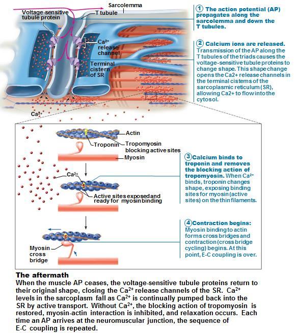
4 Myofilaments: Thick filaments composed of protein myosin. Each myosin molecule has a rodlike tail terminating via a flexible hinge in two globular heads. Myosin tails contain 2 interwoven, heavy polypeptide chains. Myosin heads contain two smaller, light polypeptide chains that act as cross bridges during contraction. Myosin heads contain binding sites for actin (thin filament), binding sites for ATP and ATPase enzymes that split ATP to generate energy for muscle contraction. Thin filament composed of protein actin. The polypeptide subunits of actin called globular actin or G actin which contains active sites for myosin head attachment during contraction. Thin filaments contain regulatory proteins tropomyosin and troponin. Tropomyosin molecules are arranged along the actin filaments and block myosin binding sites on actin so myosin heads cannot bind to the thin filaments during a relaxed muscle fiber. Troponin binds to actin, binds to tropomyosin to help position it on actin and binds calcium ions. Elastic filament titin holds the thick filaments in place and assists the muscle cell to spring back after being stretched or shortened. Dystropin links the thin filaments to the integral proteins of the sarcolemma. Nebulin, myomesin and C proteins bind filaments or sarcomeres together and maintain alignment. T tubules Elongated tubes formed by the sarcolemma extending into cells interior; at each A band-i band junction. As each T tubule protrudes deep into the cell, it runs between paired terminal cisternae of SR so that triads (terminal cisterna, T tubule, terminal cisterna) are formed encircle each sarcomere. T tubules conduct impulses deep into muscle fiber and to every sarcomere. Integral proteins protrude into intermembrane space from T tubule and SR cistern membranes and connect with each other. T tubule integral proteins act as voltage sensors and change shape in response to voltage changes. SR integral proteins are channels that release Ca 2+ from SR cisterns when voltage sensors change shape.


 Physiology of a Skeletal Muscle Fiber For a skeletal muscle fiber to contract, it must be stimulated by a nerve ending and must")
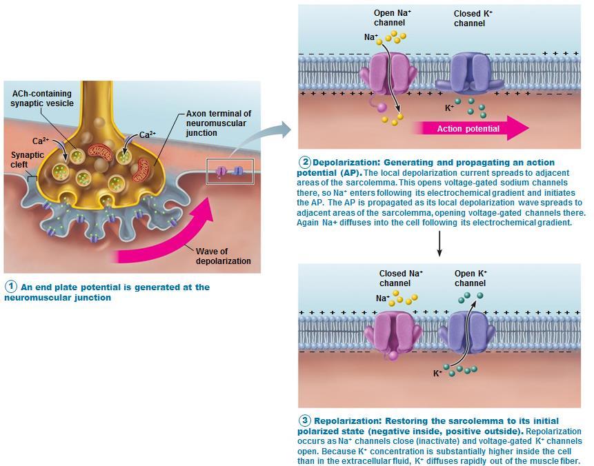
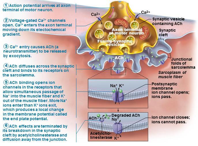
5 Sliding Filament Model of Contraction Shortening occurs when tension generated by cross bridges on thin filaments exceeds forces opposing shortening. Contraction ends when cross bridges become inactive and tension generated declines relaxation of the muscle fiber. During contraction, the thin filaments slide past the thick filaments so the actin and myosin filaments overlap to a greater degree. Occurs when myosin heads bind to actin, forming cross bridges the sliding begins. Cross bridges form and break several times during a contraction pulling the thin filaments toward the center of the sarcomere causing the muscle cell to shorten. (Z disc moves toward the M line ; I bands shorten; H Zone disappears) Physiology of a Skeletal Muscle Fiber For a skeletal muscle fiber to contract, it must be stimulated by a nerve ending and must propagate an electric current or action potential along its sarcolemma. This propagation causes a rise in intracellular Ca 2+ levels. These events linking the electrical signaling to contraction is called excitation-contraction coupling. Neuromuscular Junction - Skeletal muscles stimulated by somatic motor neurons. Axons of motor neurons travel from central nervous system via nerves to skeletal muscle. Each axon forms several branches as it enters muscle. Each axon ending forms neuromuscular junction with single muscle fiber - usually only one per muscle fiber and situated midway along length of muscle fiber. Axon terminal and muscle fiber separated by gel-filled space called synaptic cleft. Synaptic vesicles of axon terminal contain neurotransmitter acetylcholine (ACh) Junctional folds of sarcolemma contain ACh receptors.

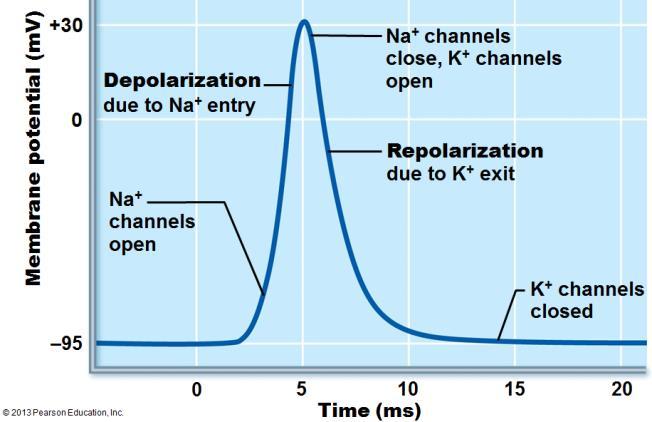
6 Events at the neuromuscular junction: 1. The nerve impulse arrives at axon terminal ACh released into synaptic cleft 2. ACh diffuses across cleft and binds with receptors on sarcolemma ACh effects quickly terminated by enzyme acetylcholinesterase in synaptic cleft. Enzyme breaks down ACh to acetic acid and choline to prevent continued muscle fiber contraction in absence of additional stimulation. 3. ACh binding opens chemically gated ion channels -- simultaneous diffusion of Na+ (inward) and K + (outward). More Na + diffuses in, so interior of sarcolemma becomes less negative. (Resting membrane is polarized more negative on the inside.) 4. Depolarization (end plate potential) generation and propagation of an action potential (AP). End plate potentials spread to adjacent membrane areas opening voltage gated Na + channels. Na + influx decreases membrane voltage toward critical voltage called threshold. If threshold is reached, AP initiated. Once initiated, AP is unstoppable muscle fiber contraction 5. Action Potential spread across sarcolemma voltage gated Na + channels open in adjacent patch, causing it to depolarize to threshold. 6. Repolarization restoring electrical conditions resting membrane potential. Na + channels close and voltage-gated K + channels open. K + efflux rapidly restores resting polarity. Fibers cannot be stimulated in refractory period until repolarization is complete. Ionic conditions of resting state restored by Na + -K + pump 7. Action potential propagated along sarcomere to T tubules causing voltage-sensitive proteins to stimulate Ca 2+ release from SR. Ca 2+ binds to troponin troponin moves tropomyosin away from myosin-binding sites. Myosin heads bind to actin, causing sarcomere shortening and muscle contraction. Cross-bridge cycling continues as Ca 2+ signal and adequate ATP is present. Cross bridge formation high energy myosin head attaches to thin filament. Power stroke myosin head pivots and pulls thin filament toward the M line. Cross bridge detachment ATP attaches to myosin head and cross bridge detaches. Cocking of myosin head energy from hydrolysis of ATP cocks myosin head into high-energy state. When nervous stimulation ceases, Ca 2+ pumped back into SR and contraction ends At low intracellular Ca 2+ concentration tropomyosin blocks active sites on actin, myosin heads cannot attach to actin and muscle fiber is relaxed. Although the action potential is very brief (1-2 milliseconds), the contraction phase of the muscle fiber may persist for 100 ms and outlast the electrical event that triggers. The action potential ends before any signs of contraction are obvious. The events of excitation-coupling occur during the latent phase between action potential initiation and beginning of mechanical activity (shortening). The electrical signal does not act directly on the myofilaments -- it causes the rise of intracellular calcium ions concentration that allows the filaments to slide.
7 Neuromuscular Junction Activity: Depolarization & Repolarization: :
8 Excitation-Contraction Coupling:

9 Cross Bridge Action: Homeostatic Imbalance: Rigor mortis - cross bridge detachment requires ATP; 3 4 hours after death muscles begin to stiffen with weak rigidity at 12 hours post mortem. Dying cells take in calcium cross bridge formation No ATP generated to break cross bridges. Contraction of a Skeletal Muscle Same principles apply to contraction of single fiber and whole muscle. Contraction produces muscle tension, force exerted on load or object to be moved. Contraction may or may not shorten muscle. Isometric contraction - no shortening; muscle tension increases but does not exceed load. Isotonic contraction - muscle shortens because muscle tension exceeds load. Force and duration of contraction vary in response to different frequencies and intensities of stimuli. Motor Unit A motor neuron and all the muscle fibers it supplies. Each muscle is served by at least one motor neuron. Motor nerves contain axons of up to hundreds of motor neurons axons branch into terminals, each of which forms a neuromuscular junction with a single muscle fiber. When a motor neuron fires, all the muscle fibers it innervates contract. Motor units in muscle usually contract asynchronously; helps prevent fatigue.


10 Muscle Twitch single contractile response to a single stimulus. Three phases of muscle twitch: o Latent period: events of excitationcontraction coupling; no muscle tension o Period of contraction: cross bridge formation; tension increases o Period of relaxation: Ca 2+ reentry into SR; tension declines to zero Muscle contracts fast then relaxes. Different strength and duration of twitches due to variations in metabolic properties and enzymes between muscles. Graded Muscle Response varying strength of contraction for different demands. Required for proper control of skeletal movement. Responses graded by changing frequency of stimulation and changing strength of stimulation. Response to change in frequency Wave (temporal) summation - Increased stimulus frequency (muscle does not completely relax between stimuli) second contraction of greater force Additional Ca 2+ release with second stimulus stimulates more shortening Produces smooth, continuous contractions Further increase in stimulus frequency unfused (incomplete) tetanus
 controls force of contraction Subthreshold stimuli no observable contractions")

11 If stimuli are given quickly enough, muscle reaches maximal tension fused (complete) tetany results. Smooth, sustained contraction; No muscle relaxation muscle fatigue. Muscle cannot contract; zero tension Response to change in Stimulus Strength Recruitment (multiple motor unit summation) controls force of contraction Subthreshold stimuli no observable contractions Threshold stimulus stimulus strength causing first observable muscle contraction Maximal stimulus strongest stimulus that increases contractile force Muscle contracts more vigorously as stimulus strength increases above threshold Contraction force precisely controlled by recruitment activates more and more muscle fibers Beyond maximal stimulus no increase in force of contraction Recruitment works on size principle o Motor units with smallest muscle fibers recruited first o Motor units with larger fibers recruited as stimulus intensity increases o Largest motor units activated only for most powerful contractions Treppe the staircase effect: When a muscle begins to contract after a long period of rest, its initial contractions may be only half as strong as those that occur later in response to stimuli of the same strength. Probably reflects an increase in availability of Ca ions; as muscle begins tow work it liberates heat; enzymes become more efficient This is the basis of the warm-up period. Isotonic and Isometric Contractions Muscle changes in length and moves load - thin filaments slide Isotonic contractions either concentric (muscle shortens and does work) or eccentric (muscles generate force as it lengthens) Isometric contractions - load is greater than tension muscle can develop; tension increases to muscle's capacity, but muscle neither shortens nor lengthens. Cross bridges generate force but do not move actin filaments.


12 Muscle Tone Constant, slightly contracted state of all muscles Due to spinal reflexes -- groups of motor units alternately activated in response to input from stretch receptors in muscles Keeps muscles firm, healthy, and ready to respond Muscle Metabolism As a muscle contracts, ATP provides energy for cross bridge movements and detachment and for the operation of the calcium pump. Muscles store very limited reserves of ATP 4 to 6 seconds worth. ATP is the only energy source used for contractile activities it must be regenerated as fast as it is broken down if contraction is to continue. ATP is regenerated by these 3 pathways: Creatine Phosphate Once the stored ATP is used (4-6 sec worth), creatine phosphate (high-energy molecule stored in muscles) is tapped to regenerate ATP while the metabolic pathways are adjusting to higher demands of ATP. Muscle cells store more CP as ATP the CP/ADP reaction is catalyzed by the enzyme, creatine kinase. Stored ATP and CP provide for maximum muscle power for secs (enough energy to run a 100-meter dash). CP reserves are replenished during periods of inactivity. Anaerobic Respiration As stored ATP and CP are used, more ATP is generated by the catabolism of glucose. Glucose is stored in the muscles as glycogen or in the blood. Glucose is broken down during anaerobic respiration to produce 2 ATP molecules and 2 pyruvic acid molecules. Normally, the 2 pyruvic acid enter the mitochondria and undergo aerobic respiration. But when muscles contract vigorously and contractile activity reaches about 70% of the maximum possible, the bulging muscles compress the blood vessels within them, impairing blood flow and oxygen delivery. Under these anaerobic conditions, most of the pyruvic acid is converted to lactic acid --- most of the lactic acid diffuses out of the muscles into the blood and used as fuel for the liver, heart or kidney cells. When large amounts of ATP are needed for moderate activity (30-40 sec), glycolysis can provide most of the ATP needed. Anaerobic glycolysis fuels spurts of vigorous exercise as glycolysis produces small amounts of ATP, you accumulate lactic acid which contributes to muscle fatigue and soreness. Aerobic Respiration During rest and light-moderate exercise, 95% of ATP is generated by aerobic respiration. The 2 pyruvic acid formed during glycolysis enter the mitochondria and continue to be broken down yielding water, carbon dioxide and large amounts of ATP. As exercise begins, muscle glycogen provides most of the fuel. Then pyruvic acid from glycolysis and free fatty acids are major sources of fuel for oxidation. Aerobic respiration provides about 38 ATPs per glucose, but is relatively sluggish because it requires oxygen and nutrients to keep going. When oxygen is plentiful, muscles cells will form ATP by aerobic respiration --- moderate muscle activity can continue for several hours.

13 When exercise demands begin to exceed the ability of muscle cells to carry out aerobic respiration, glycolysis beings to contribute more of the ATP generated. The length of time a muscle can continue to contract using aerobic pathways is aerobic endurance and the points at which muscle metabolism converts to anaerobic glycolysis is anaerobic threshold. Muscle Fatigue The state of physiological inability to contract even though the muscle may be receiving stimuli. Occurs when ionic imbalances (K +, Ca 2+, P i ) interfere with Excitation-Contraction coupling or prolonged exercise may damage SR and interferes with Ca 2+ regulation and release. ATP availability declines during contraction and total lack of ATP results in contractures continuous contraction because the cross brides are unable to detach. (Cramps are temporary contractures.) For muscles to return to a resting state, oxygen reserves must be replenished, the accumulated lactic acid must be converted to pyruvic acid, glycogen stores must be replaced and ATP/CP reserves must be resynthesized. Oxygen debt is the extra amounts of oxygen that the body must take in for these restoration processes. About 40% of energy released during muscle contraction is useful as work and the remaining energy (60%) is given off as heat. Heat buildup is prevented from reaching dangerous levels by several homeostatic mechanisms sweat and radiation of heat from skin surfaces. Shivering produces more heat by contracting muscles when cold. Force of Muscle Contraction The force of muscle contraction depends on number of cross bridges attached, which is affected by: 1. As more muscle fibers are recruited (as more are stimulated) more force 2. Relative size of fibers bulkier muscles & hypertrophy of cells more force 3. Frequency of stimulation - frequency contractions are summed up, becoming stronger, more vigorous and ultimately producing tetanus more time for transfer of tension to noncontractile components more force 4. Degree of muscle stretch: Length-tension relationship muscle fibers at % normal resting length more force


14 Velocity and Duration of Contraction Muscles vary in how fast they can contract and in how long they can continue to contract before they fatigue. These characteristics are influenced by muscle fiber type, load and recruitment. Muscle fiber types are classified according to two characteristics: 1. Speed of contraction: slow or fast fibers according to speed at which myosin ATPases split ATP and pattern of electrical activity of motor neurons. 2. Metabolic pathways for ATP synthesis: Oxidative fibers use aerobic pathways; Glycolytic fibers use anaerobic glycolysis Three types of muscle fibers Slow oxidative fibers; Fast oxidative fibers; Fast glycolytic fibers Most muscles contain mixture of fiber types this provides a range of contractile speed and fatigue resistance. All muscle fibers in a particular motor unit are the same type. Some people have relatively more of one variety of fibers initially due to genetics, but are modified by exercise and determine endurance vs. strength. Load Muscles contract faster when there is no added load on them. The greater the load the longer the latent phase, the slower the contraction and the shorter the duration of the contraction. If the load exceeds the muscles maximum tension, the speed of shortening is zero and the contraction is isometric. Recruitment the more motor units that are contracting, the faster and more prolonged the contraction.


15 Effects of Exercise on Muscles Aerobic (endurance) exercise leads to increased muscle capillaries, number of mitochondria, myoglobin synthesis; results in greater endurance, strength and resistance to fatigue; may convert fast glycolytic fibers into fast oxidative fibers. Resistance exercise (typically anaerobic) results in muscle hypertrophy due to increase in fiber size; increased mitochondria, myofilaments, glycogen stores and connective tissue Increased muscle strength and size Balanced exercise program Exercise gains adhere to the overload principle. Forcing muscle to work hard promotes increased muscle strength and endurance Muscles adapt to increased demands and must be overloaded to produce further gains. Overuse injuries may result from lack of rest. Best programs alternate aerobic and anaerobic activities. Homeostatic Imbalances Disuse atrophy results from immobilization; muscle strength declines 5% per day. Without neural stimulation muscles atrophy to ¼ initial size. Fibrous connective tissue replaces lost muscle tissue rehabilitation impossible. Smooth Muscle Found in walls of most hollow visceral organs; usually in two layers (longitudinal and circular). Spindle-shaped fibers - thin and short compared with skeletal muscle fibers; only one nucleus; no striations Lacks connective tissue sheaths; endomysium only Pouchlike infoldings (caveolae) of sarcolemma sequester Ca 2+ - most calcium influx from outside cell; rapid No sarcomeres, myofibrils, or T tubules Longitudinal layer - Fibers parallel to long axis of organ; contraction dilates and shortened Circular layer - Fibers in circumference of organ; contraction constricts lumen, elongates organ Longitudinal & Circular layers allows peristalsis - Alternating contractions and relaxations of smooth muscle layers that mix and squeeze substances through lumen of hollow organs No neuromuscular junction, instead autonomic nerve fibers innervate smooth muscle at diffuse junctions. Varicosities (bulbous swellings) of nerve fibers store and release neurotransmitters into diffuse junctions.


16 Myofilaments in Smooth Muscle Ratio of thick to thin filaments (1:13) is much lower than in skeletal muscle (1:2) Thick filaments have heads along entire length. No troponin complex; protein calmodulin binds Ca 2+ Myofilaments are spirally arranged, causing smooth muscle to contract in corkscrew manner Dense bodies - Proteins that anchor noncontractile intermediate filaments to sarcolemma at regular intervals; correspond to Z discs of skeletal muscle Smooth Muscle Contraction Slow, synchronized contractions. Cells are electrically coupled by gap junctions. Action potentials transmitted from fiber to fiber Some cells self-excitatory (depolarize without external stimuli); act as pacemakers for sheets of muscle -- Rate and intensity of contraction may be modified by neural and chemical stimuli. Ca 2+ binds to and activates calmodulin; activated calmodulin activates myosin (light chain) kinase Phosphorylates and activates myosin, cross bridges interact with actin. When intracellular Ca 2+ levels drop relaxation. Slow to contract and relax but maintains for prolonged periods with little energy cost -- slow ATPases and myofilaments may latch together to save energy. Relaxation requires Ca 2+ detachment from calmodulin; active transport of Ca 2+ into SR and ECF; dephosphorylation of myosin to reduce myosin ATPase activity Regulation of Contraction Neural regulation Neurotransmitter binding [Ca 2+ ] in sarcoplasm; either graded (local) potential or action potential; Response depends on neurotransmitter released and type of receptor molecules Hormones and local chemicals Some smooth muscle cells have no nerve supply, some respond to both neural & chemical stimuli (hormones, ph, CO 2 ) o Depolarize spontaneously or in response to chemical stimuli that bind to G protein linked receptors
17 Special Features of Smooth Muscle Contraction Stress-relaxation response Responds to stretch only briefly, then adapts to new length; retains ability to contract on demand; enables organs such as stomach and bladder to temporarily store contents. Length and tension changes can contract when between half and twice its resting length. Hyperplasia - Smooth muscle cells can divide and increase numbers. Types of Smooth Muscle Smooth muscle varies in different organs fiber arrangement/organization, innervation, responsiveness to various stimuli. Categorized as unitary and multi-unit. Unitary (visceral) smooth muscle in all hollow organs except heart; arranged in opposing sheets; innervated by varicosities; often exhibit spontaneous action potentials; electrically coupled by gap junctions; respond to various chemical stimuli Multiunit smooth muscle located in large airways, large arteries, arrector pili muscles, and iris of eye; gap junctions; spontaneous depolarization rare; independent muscle fibers; innervated by autonomic NS; graded contractions occur in response to neural stimuli; has motor units; responds to hormones Developmental Aspects of Muscle All muscle tissues develop from embryonic myoblasts Multinucleated skeletal muscle cells form by fusion Growth factor agrin stimulates clustering of ACh receptors at neuromuscular junctions Cardiac and smooth muscle myoblasts develop gap junctions Cardiac and skeletal muscle become amitotic, but can lengthen and thicken in growing child Myoblast-like skeletal muscle satellite cells have limited regenerative ability Cardiomyocytes can divide at modest rate, but injured heart muscle mostly replaced by connective tissue Smooth muscle regenerates throughout life Muscular development reflects neuromuscular coordination Peak natural neural control occurs by mid-adolescence; athletics can improve neuromuscular control Female skeletal muscle makes up 36% of body mass Male skeletal muscle makes up 42% of body mass, primarily due to testosterone With age, connective tissue increases and muscle fibers decrease; by age 30, loss of muscle mass (sarcopenia) begins --- regular exercise reverses sarcopenia Muscular Dystrophy Group of inherited muscle-destroying diseases; generally appear in childhood Muscles enlarge due to fat and connective tissue deposits. Muscle fibers atrophy and degenerate. Duchenne muscular dystrophy (DMD): most common and severe type; inherited, sex-linked, carried by females and expressed in males (1/3500) as lack of dystrophin (cytoplasmic protein that stabilizes sarcolemma)fragile sarcolemma tears Ca 2+ entry damaged contractile fibers inflammatory cells muscle mass drops Victims become clumsy and fall frequently; usually die of respiratory failure in 20s; no cure; Prednisone improves muscle strength and function
Muscle and Muscle Tissue
 Muscle and Muscle Tissue Make up about half of total body mass Exerts force by converting chemical energy, ATP, to mechanical energy Muscle tissue is classified based on Shape Number and position of nuclei
Muscles and Muscle Tissue
 1 Muscles and Muscle Tissue Chapter 9 2 Overview of Muscle Tissues Compare and Contrast the three basic types of muscle tissue List four important functions of muscle tissue 3 Muscle Terminology Muscle
Chapter 10 Muscle Tissue Lecture Outline
 Chapter 10 Muscle Tissue Lecture Outline Muscle tissue types 1. Skeletal muscle = voluntary striated 2. Cardiac muscle = involuntary striated 3. Smooth muscle = involuntary nonstriated Characteristics
2/19/2018. Learn and Understand:
 Muscular System with Special Emphasis on Skeletal Muscle Anatomy and Physiology Learn and Understand: The definition of cell changes again The contractile unit of muscle is the sarcomere. ATP and Ca 2+
Warm Up! Test review (already! ;))
)") Warm Up! Test review (already! ;)) Write a question you might find on the Unit 5 test next week! (Multiple choice, matching, fill in, or short answer!) - challenge yourself and be ready to share!!! PowerPoint
Warm Up! Test review (already! ;)) Write a question you might find on the Unit 5 test next week! (Multiple choice, matching, fill in, or short answer!) - challenge yourself and be ready to share!!! PowerPoint
MUSCULAR TISSUE. Dr. Gary Mumaugh
 MUSCULAR TISSUE Dr. Gary Mumaugh MUSCLE OVERVIEW The three types of muscle tissue are skeletal, cardiac, and smooth These types differ in structure, location, function, and means of activation FUNCTIONAL
Skeletal Muscle Tissue
 Functions of Skeletal Muscle Skeletal Muscle Tissue Keri Muma Bio 6 Movement muscles attach directly or indirectly to bone, pull on bone or tissue when they contract Maintain posture / body position muscles
Skeletal Muscle. Connective tissue: Binding, support and insulation. Blood vessels
 Chapter 12 Muscle Physiology Outline o Skeletal Muscle Structure o The mechanism of Force Generation in Muscle o The mechanics of Skeletal Muscle Contraction o Skeletal Muscle Metabolism o Control of Skeletal
MUSCLE TISSUE (MUSCLE PHYSIOLOGY) PART I: MUSCLE STRUCTURE
 PART I: MUSCLE STRUCTURE") PART I: MUSCLE STRUCTURE Muscle Tissue A primary tissue type, divided into: skeletal muscle cardiac muscle smooth muscle Functions of Skeletal Muscles Produce skeletal movement Maintain body position Support
PART I: MUSCLE STRUCTURE Muscle Tissue A primary tissue type, divided into: skeletal muscle cardiac muscle smooth muscle Functions of Skeletal Muscles Produce skeletal movement Maintain body position Support
MODULE 6 MUSCLE PHYSIOLOGY
 MODULE 6 MUSCLE PHYSIOLOGY III SEMESTER BOTANY Syllabi: Striated, Non striated and Cardiac muscle, Ultra structure of striated muscle fibre, Mechanism of muscle contraction, Threshold and spike potential,
Chapter 9 - Muscle and Muscle Tissue
 Chapter 9 - Muscle and Muscle Tissue I. Overview of muscle tissue A. Three muscle types in the body: B. Special characteristics 1. Excitability: able to receive and respond to a stimulus 2. Contractility:
Lecture Overview. Muscular System. Marieb s Human Anatomy and Physiology. Chapter 9 Muscles and Muscle Tissue Lecture 16
 Marieb s Human Anatomy and Physiology Marieb Hoehn Chapter 9 Muscles and Muscle Tissue Lecture 16 1 Lecture Overview Types, characteristics, functions of muscle Structure of skeletal muscle Mechanism of
1/4/2017. Introduction. Connective Tissue Coverings. 9.1: Structure of a Skeletal Muscle. Skeletal Muscle Fibers. Connective Tissue Coverings
 Introduction Chapter 09 Lecture Outline See separate PowerPoint slides for all figures and tables preinserted into PowerPoint without notes. Copyright McGraw-Hill Education. Permission required for reproduction
Microanatomy of Muscles. Anatomy & Physiology Class
 Microanatomy of Muscles Anatomy & Physiology Class Three Main Muscle Types Objectives: By the end of this presentation you will have the information to: 1. 2. 3. 4. 5. 6. Describe the 3 main types of muscles.
Muscle Tissue- 3 Types
 AN INTRODUCTION TO MUSCLE TISSUE Muscle Tissue- 3 Types Skeletal muscle (focus on these) Cardiac muscle Smooth muscle FUNCTIONS OF SKELETAL MUSCLES Produce movement of the skeleton Maintain posture and
Chapter 10 Muscle Tissue and Physiology Chapter Outline
 Chapter 10 Muscle Tissue and Physiology Chapter Outline Module 10.1 Overview of muscle tissue (Figures 10.1 10.2) A. Types of Muscle Tissue (Figure 10.1) 1. The three types of cells in muscle tissue are,,
Anatomy and Physiology 1 Chapter 10 self quiz Pro, Dima Darwish,MD.
 Anatomy and Physiology 1 Chapter 10 self quiz Pro, Dima Darwish,MD. 1) Which of the following is a recognized function of skeletal muscle? A) produce movement B) maintain posture C) maintain body temperature
MUSCULAR SYSTEM CHAPTER 09 BIO 211: ANATOMY & PHYSIOLOGY I
 1 BIO 211: ANATOMY & PHYSIOLOGY I 1 CHAPTER 09 MUSCULAR SYSTEM Part 2 of 2 Dr. Dr. Lawrence G. G. Altman www.lawrencegaltman.com Some illustrations are courtesy of McGraw-Hill. Some illustrations are courtesy
Hole s Human Anatomy and Physiology Eleventh Edition. Mrs. Hummer. Chapter 9 Muscular System
 Hole s Human Anatomy and Physiology Eleventh Edition Mrs. Hummer Chapter 9 Muscular System 1 Chapter 9 Muscular System Skeletal Muscle usually attached to bones under conscious control striated Three Types
Muscle Tissue. Dr. Heba Kalbouneh Associate Professor of Anatomy and Histology
 Muscle Tissue Dr. Heba Kalbouneh Associate Professor of Anatomy and Histology Functions of muscle tissue Movement Maintenance of posture Joint stabilization Heat generation Tendon Belly Tendon Types of
Page 1. Chapter 9: Muscle Tissue. Types of Muscle Tissue: Skeletal Muscle Cardiac Muscle Smooth Muscle. Gross Anatomy of Muscle:
 1 Chapter 9: Muscle Tissue Types of Muscle Tissue: Skeletal Muscle Cardiac Muscle Smooth Muscle Characteristics: Attaches to skeleton Voluntary control Striated / multi-nucleated Characteristics: Composes
Chapter 10: Muscle Tissue
 Chapter 10: Muscle Tissue Muscle is one of the 4 primary types of tissue. It is subdivided into skeletal, cardiac and smooth muscle. I. Skeletal Muscle Tissue and the Muscular System, p. 284 Objective
I. Overview of Muscle Tissues
 I. Overview of Muscle Tissues A. Types of Muscle Tissue 1. Terminology 1. Muscle fibers = muscle cells are greatly elongated therefore known as fibers; true for skeletal and smooth muscles only 2. Myo
Page 1. Chapter 9: Muscle Tissue. Types of Muscle Tissue: Skeletal Muscle Cardiac Muscle Smooth Muscle. Characteristics of Muscle:
 1 Chapter 9: Muscle Tissue Muscle little mouse Types of Muscle Tissue: Skeletal Muscle Cardiac Muscle Smooth Muscle Characteristics: Attaches to skeleton Voluntary control Striated / multi-nucleated Characteristics:
Principles of Anatomy and Physiology
 Principles of Anatomy and Physiology 14 th Edition CHAPTER 10 Muscular Tissue Introduction The purpose of the chapter is to: 1. Learn about the structure and function of the 3 types of muscular tissue
Essentials of Human Anatomy & Physiology. The Muscular System
 Essentials of Human Anatomy & Physiology The Muscular System The Muscular System Muscles are responsible for all types of body movement they contract or shorten and are the machine of the body Three basic
Chapter 10: Muscles. Vocabulary: aponeurosis, fatigue
 Chapter 10: Muscles 37. Describe the structural components of skeletal muscle tissue from the molecular to the organ level. 38. Describe the structure, function, and importance of sarcomeres. 39. Identify
Human Anatomy. Muscle Tissue and Organization. DR.SADIQ ALI (K.E Medalist) 10-1
 10-1") Human Anatomy Muscle Tissue and Organization DR.SADIQ ALI (K.E Medalist) 10-1 Tissue and Organization Over 700 skeletal muscles have been named. Form the muscular system. Muscle tissue is distributed almost
Human Anatomy Muscle Tissue and Organization DR.SADIQ ALI (K.E Medalist) 10-1 Tissue and Organization Over 700 skeletal muscles have been named. Form the muscular system. Muscle tissue is distributed almost
Muscle Tissue. C h a p t e r. PowerPoint Lecture Slides prepared by Jason LaPres Lone Star College - North Harris
 C h a p t e r 10 Muscle Tissue PowerPoint Lecture Slides prepared by Jason LaPres Lone Star College - North Harris Copyright 2009 Pearson Education, Inc., publishing as Pearson Benjamin Cummings An Introduction
Chapter 10! Chapter 10, Part 2 Muscle. Muscle Tissue - Part 2! Pages !
 ! Chapter 10, Part 2 Muscle Chapter 10! Muscle Tissue - Part 2! Pages 308-324! SECTION 10-5! Sarcomere shortening and muscle fiber stimulation produce tension! 2! Tension Production - Muscle FIBER! All-or-none
Muscle Physiology. Dr. Ebneshahidi Ebneshahidi
 Muscle Physiology Dr. Ebneshahidi Skeletal Muscle Figure 9.2 (a) Functions of the muscular system 1. Locomotion body movements are due to skeletal muscle contraction. 2. Vasoconstriction and vasodilatation
Chapter 8 Notes. Muscles
 Chapter 8 Notes Muscles 8.1 Intro Three muscle types Skeletal Smooth cardiac 8.2 Structure of Skeletal Muscle Composition Skeletal muscle tissue Nervous tissue Blood Connective tissue Connective tissue
Chapter 10 -Muscle Tissue
 Chapter 10 -Muscle Tissue Muscles: 1. Overview of Muscle Tissue A. Review 5 functions of muscle tissue. B. Review the 5 properties of muscle tissue. WHICH do they share with nervous tissue? (2, plus the
CHAPTER 6 2/9/2016. Learning Objectives List the four traits that all muscle types have in common.
 Learning Objectives List the four traits that all muscle types have in common. CHAPTER 6 The Muscular System Demonstrate and explain the use of antagonistic muscle pairs. Describe the attachment of muscle
Ch.10 Muscle Tissue. Copyright 2009, John Wiley & Sons, Inc.
 Ch.10 Muscle Tissue Preview Chapter 10 In groups we will define the following terms 1. Skeletal muscle 2. Smooth muscle 3. Cardiac muscle 4. Sarcomere 5. Myofibril 6. Myofilament 7. Sarcoplasmic reticulum
Chapter 9: Muscles and Muscle Tissue
 Chapter 9: Muscles and Muscle Tissue Objectives: 1. Compare and contrast the basic types of muscle tissue. 2. List four important functions of muscle tissue. 3. Describe the gross structure of a skeletal
Muscle Tissue. Muscle Tissue Outline. General Function of Muscle Tissue
 Muscle Tissue Muscle Tissue Outline General Functions of Muscle Tissue Characteristics of Muscle Tissue Classification of Muscle Tissue Skeletal Muscle Structure and Function Muscle Energetics Muscle Mechanics
Ch 12: Muscles sarcolemma, t-tubules, sarcoplasmic reticulum, myofibrils, myofilaments, sarcomere...
 Ch 12: Muscles Review micro-anatomy of muscle tissue Terminology examples: sarcolemma, t-tubules, sarcoplasmic reticulum, myofibrils, myofilaments, sarcomere... SLOs Differentiate levels of muscle structure:
Smooth Cardiac Skeletal Location Around tubes Heart tissue attached to skeleton Moves stuff thru Heart beat pumps Moves body parts
 Biology 067 - Muscular system A. Type of muscles: Smooth Cardiac Skeletal Location Around tubes Heart tissue attached to skeleton Function Moves stuff thru Heart beat pumps Moves body parts tubes blood
Muscle Tissue. PowerPoint Lecture Presentations prepared by Jason LaPres. Lone Star College North Harris Pearson Education, Inc.
 10 Muscle Tissue PowerPoint Lecture Presentations prepared by Jason LaPres Lone Star College North Harris An Introduction to Muscle Tissue Muscle Tissue A primary tissue type, divided into: Skeletal muscle
Muscles and Metabolism
 Muscles and Metabolism How does the body provide the energy needed for contraction? -as muscles contract, ATP supplies the energy fro cross bridge movement and detachment and for operation of the calcium
Lecture 9A. Muscle structure. Outline
 Lecture 9A Muscle structure Outline Smooth, skeletal, and cardiac muscle tissues Structure and function of skeletal muscle cells. Sarcomeres structure and contraction Actin-myosin interaction and sliding
Muscle Histology. Dr. Heba Kalbouneh Assistant Professor of Anatomy and Histology
 Muscle Histology Dr. Heba Kalbouneh Assistant Professor of Anatomy and Histology Functions of muscle tissue Movement Maintenance of posture Joint stabilization Heat generation Types of Muscle Tissue Skeletal
Skeletal Muscle and the Molecular Basis of Contraction. Lanny Shulman, O.D., Ph.D. University of Houston College of Optometry
 Skeletal Muscle and the Molecular Basis of Contraction Lanny Shulman, O.D., Ph.D. University of Houston College of Optometry Like neurons, all muscle cells can be excited chemically, electrically, and
BIOH111. o Cell Module o Tissue Module o Integumentary system o Skeletal system o Muscle system o Nervous system o Endocrine system
 BIOH111 o Cell Module o Tissue Module o Integumentary system o Skeletal system o Muscle system o Nervous system o Endocrine system Endeavour College of Natural Health endeavour.edu.au 1 TEXTBOOK AND REQUIRED/RECOMMENDED
Muscle Tissue. General concepts. Classification of muscle. I. Functional classification is based on the type of neural control.
 Muscle Tissue LEARNING OBJECTIVES 1. Identify the three types of muscle tissue at the light microscopic level. 2. List and compare the structural and functional features of each of the three muscle fiber
PSK4U THE NEUROMUSCULAR SYSTEM
 PSK4U THE NEUROMUSCULAR SYSTEM REVIEW Review of muscle so we can see how the neuromuscular system works This is not on today's note Skeletal Muscle Cell: Cellular System A) Excitation System Electrical
Fig Copyright McGraw-Hill Education. Permission required for reproduction or display. Nucleus. Muscle fiber. Endomysium. Striations.
 Fig. 11.1 Nucleus Muscle fiber Endomysium Striations Ed Reschke 1 Fig. 11.2 Muscle fiber Nucleus I band A band Z disc Mitochondria Openings into transverse tubules Sarcoplasmic reticulum Triad: Terminal
CLASS SET Unit 4: The Muscular System STUDY GUIDE
 NPHS Anatomy & Physiology Questions to answer: 1) List three functions of the muscular system. 1) movement 2) thermogenesis (generates heat) 3) posture & body/joint support CLASS SET Unit 4: The Muscular
Muscles and Muscle Tissue
 Chapter 9 Part A Muscles and Muscle Tissue Annie Leibovitz/Contact Press Images PowerPoint Lecture Slides prepared by Karen Dunbar Kareiva Ivy Tech Community College Why This Matters Understanding skeletal
Muscle Cell Anatomy & Function (mainly striated muscle tissue)
") Muscle Cell Anatomy & Function (mainly striated muscle tissue) General Structure of Muscle Cells (skeletal) several nuclei (skeletal muscle) skeletal muscles are formed when embryonic cells fuse together
Muscle Cell Anatomy & Function (mainly striated muscle tissue) General Structure of Muscle Cells (skeletal) several nuclei (skeletal muscle) skeletal muscles are formed when embryonic cells fuse together
Muscle Tissue. Alternating contraction and relaxation of cells. Chemical energy changed into mechanical energy
 Know these muscles Muscle Tissue Alternating contraction and relaxation of cells Chemical energy changed into mechanical energy 3 Types of Muscle Tissue Skeletal muscle attaches to bone, skin or fascia
Muscle tissues. Dr. Hersh Abdul Ham-Karim BVM&S, PG Dip, MSc and PhD
 Muscle tissues Dr. Hersh Abdul Ham-Karim BVM&S, PG Dip, MSc and PhD Muscle tissue is a soft tissue that composes muscles in animal bodies, and gives rise to muscles' ability to contract. Muscle tissue
Chapter 9 Muscle. Types of muscle Skeletal muscle Cardiac muscle Smooth muscle. Striated muscle
 Chapter 9 Muscle Types of muscle Skeletal muscle Cardiac muscle Smooth muscle Striated muscle Chapter 9 Muscle (cont.) The sliding filament mechanism, in which myosin filaments bind to and move actin
Chapter 10 Muscle Tissue
 Chapter 10 Muscle Tissue Skeletal muscle, cardiac muscle and smooth muscle. Differ in their microscopic anatomy, location and how they are controlled by the endocrine and nervous system. 3 Types of Muscle
About This Chapter. Skeletal muscle Mechanics of body movement Smooth muscle Cardiac muscle Pearson Education, Inc.
 About This Chapter Skeletal muscle Mechanics of body movement Smooth muscle Cardiac muscle Skeletal Muscle Usually attached to bones by tendons Origin: closest to the trunk or to more stationary bone Insertion:
LECTURE NOTES BY: PROFESSOR RODRIGUEZ
 LECTURE NOTES BY: PROFESSOR RODRIGUEZ Muscle Overview The three types of muscle tissue: 1. 2. 3. These types differ in structure, location,, and means of activation Muscle Similarities Skeletal and smooth
Skeletal Muscle Contraction 4/11/2018 Dr. Hiwa Shafiq
 Skeletal Muscle Contraction 4/11/2018 Dr. Hiwa Shafiq Skeletal Muscle Fiber About 40 per cent of the body is skeletal muscle, and 10 per cent is smooth and cardiac muscle. Skeletal muscles are composed
Muscular System - Part III. Tension, Contractions, & Metabolism
 Do Now: What is the neurotransmitter that is released from the neuron at the NMJ? When it binds to sarcolemma receptors, what occurs? To what does calcium bind? What occurs when this bond forms? Muscular
Muscular System. SKELETAL CARDIAC SMOOTH Location Attached to bones or skin Forms walls of heart In walls of hollow visceral organs stomach, bladder,
 Muscular System 3 Types of Muscle Tissue Cells are called muscle fibers. SKELETAL CARDIAC SMOOTH Location Attached to bones or skin Forms walls of heart In walls of hollow visceral organs stomach, bladder,
BCH 450 Biochemistry of Specialized Tissues. V. Muscle Tissues
 BCH 450 Biochemistry of Specialized Tissues V. Muscle Tissues Nomenclature Sarcolemma = plasma membrane Sarcoplasmic reticulum = endoplasmic reticulum Muscle fiber = cell Myofibril = subcellular fibers
Session 3-Part 2: Skeletal Muscle
 Session 3-Part 2: Skeletal Muscle Course: Introduction to Exercise Science-Level 2 (Exercise Physiology) Presentation Created by Ken Baldwin, M.ED, ACSM-H/FI Copyright EFS Inc. All Rights Reserved. Skeletal
Muscles & Muscle Tissue
 Muscles & Muscle Tissue Chapter 6 I. Overview of Muscle 1 A. MUSCLE TYPES SKELETAL: striated, voluntary CARDIAC: only in heart involuntary striated SMOOTH: walls of organs involuntary nonstriated All Muscle
Nerve Cell (aka neuron)
") Nerve Cell (aka neuron) Neuromuscular Junction Nerve cell Muscle fiber (cell) The Nerve Stimulus and Action Potential The Nerve Stimulus and Action Potential Skeletal muscles must be stimulated by a motor
Nerve Cell (aka neuron) Neuromuscular Junction Nerve cell Muscle fiber (cell) The Nerve Stimulus and Action Potential The Nerve Stimulus and Action Potential Skeletal muscles must be stimulated by a motor
Chapter 10! Muscle Tissue - Part 2! Pages ! SECTION 10-5! Sarcomere shortening and muscle fiber stimulation produce tension!
 ! Chapter 10, Part 2 Muscle Chapter 10! Muscle Tissue - Part 2! Pages 308-324! SECTION 10-5! Sarcomere shortening and muscle fiber stimulation produce tension! 2! 1 Tension Production - MUSCLE FIBER! All-or-none
Warm-Up. 2. What structure connects muscle to bone?
 Warm-Up 1. Based on what you know about Latin root words, what do you think these terms refer to? Sarcomere Sarcoplasm Myofibril Epimysium Perimysium Endomysium 2. What structure connects muscle to bone?
Skeletal Muscle. Skeletal Muscle
 Skeletal Muscle Skeletal Muscle Types of muscle Skeletal muscle-moves the skeleton by pulling on the tendons that are connected to the bones Cardiac muscle-pumps blood through the heart and blood vessels
Muscle Tissue. Alternating contraction and relaxation of cells Chemical energy changed into mechanical energy 10:32
 Muscle Tissue Alternating contraction and relaxation of cells Chemical energy changed into mechanical energy 1 Properties of Muscle Tissue Excitability responds to chemical messengers (neurotransmitters)
Nerve regeneration. Somatic nervous system
 Somatic nervous system Signals from CNS are sent to skeletal muscles. Final result is a muscle contraction. Motor neuron starts in CNS and its axon ends at a muscle cell. Alpha motor neuron Alpha motor
Nerve meets muscle. Nerve regeneration. Somatic nervous system
 Somatic nervous system Signals from CNS are sent to skeletal muscles. Final result is a muscle contraction. Alpha motor neurons branch into several terminals (can be over 1000), each contacting a separate
Biology 201-Worksheet on Muscle System (Answers are in your power point outlines-there is no key!)
") Bio 201 Tissues and Skin 1 February 23, 2011 Biology 201-Worksheet on Muscle System (Answers are in your power point outlines-there is no key!) 1. Name and define 5 characteristics of the muscle system.
Bio 201 Tissues and Skin 1 February 23, 2011 Biology 201-Worksheet on Muscle System (Answers are in your power point outlines-there is no key!) 1. Name and define 5 characteristics of the muscle system.
Musculoskeletal Systems. Anatomy: Arrangement of Cells Physiology: Contractions
 Musculoskeletal Systems Anatomy: Arrangement of Cells Physiology: Contractions Characteristics of all muscle Contractile: it shortens Excitable: receives & responds to electrical signals Extensible: stretches
Types of Muscle. Skeletal striated & voluntary Smooth involuntary Cardiac - heart
 Muscular System Types of Muscle Skeletal striated & voluntary Smooth involuntary Cardiac - heart The word striated means striped. Skeletal muscle appears striped under a microscope. Muscles and Muscle
1. Locomotion. 2. Repositioning. 3. Internal movement
 MUSCLE and MOVEMENT Chapters 20, 8, 21 1. Locomotion A. Movement B. 2. Repositioning A. 3. Internal movement A. 1 Muscle Cells 1. Contractile 2. Myocytes 3. Striated A. Skeletal B. Cardiac 4. Smooth 5.
1. Locomotion. 2. Repositioning. 3. Internal movement
 MUSCLE and MOVEMENT Chapters 20, 8, 21 1. Locomotion A. Movement B. 2. Repositioning A. 3. Internal movement A. Muscle Cells 1. Contractile 2. Myocytes 3. Striated A. Skeletal B. Cardiac 4. Smooth 5. Striated
Chapter 8: Skeletal Muscle: Structure and Function
 Chapter 8: Skeletal Muscle: Structure and Function Objectives Draw & label the microstructure of skeletal muscle Outline the steps leading to muscle shortening Define the concentric and isometric Discuss:
Skeletal Muscle Contraction 5/11/2017 Dr. Hiwa Shafiq
 Skeletal Muscle Contraction 5/11/2017 Dr. Hiwa Shafiq Skeletal Muscle Fiber About 40 per cent of the body is skeletal muscle, and 10 per cent is smooth and cardiac muscle. Skeletal muscles are composed
BIOH111. o Cell Module o Tissue Module o Integumentary system o Skeletal system o Muscle system o Nervous system o Endocrine system
 BIOH111 o Cell Module o Tissue Module o Integumentary system o Skeletal system o Muscle system o Nervous system o Endocrine system Endeavour College of Natural Health endeavour.edu.au 1 Textbook and required/recommended
Muscle Tissue. PowerPoint Lecture Presentations prepared by Jason LaPres. Lone Star College North Harris Pearson Education, Inc.
 10 Muscle Tissue PowerPoint Lecture Presentations prepared by Jason LaPres Lone Star College North Harris 10-1 An Introduction to Muscle Tissue Learning Outcomes 10-1 Specify the functions of skeletal
Muscular System. Human A & P
 Muscular System Human A & P There are 3 types of muscle tissue: A. Skeletal B. Smooth C. Cardiac The essential function of a muscle is contraction, or shortening, and are responsible for essentially all
Skeletal Muscle Qiang XIA (
 Skeletal Muscle Qiang XIA ( 夏强 ), PhD Department of Physiology Rm C518, Block C, Research Building, School of Medicine Tel: 88208252 Email: xiaqiang@zju.edu.cn Course website: http://10.71.121.151/physiology
Muscular Tissue. Functions of Muscular Tissue. Types of Muscular Tissue. Skeletal Muscular Tissue. Properties of Muscular Tissue
 Muscular Tissue Functions of Muscular Tissue Muscle makes up a large percentage of the body s weight (40-50%) Their main functions are to: Create motion muscles work with nerves, bones, and joints to produce
Muscular System. This chapter will focus on muscle cells and tissues. Muscle tissue has several functions:
 Muscular System Slide 2 This chapter will focus on muscle cells and tissues. Muscle tissue has several functions: Movement: Muscles work as pulleys on bones to help create changes in body position. Muscles
10 - Muscular Contraction. Taft College Human Physiology
 10 - Muscular Contraction Taft College Human Physiology Muscular Contraction Sliding filament theory (Hanson and Huxley, 1954) These 2 investigators proposed that skeletal muscle shortens during contraction
Bio 103 Muscular System 61
 61 Lecture Outline: MUSCULAR SYSTEM [Chapter 9] A. Functions of Skeletal Muscle 1. Movement 2. Maintain posture 3. Support 4. Guard openings 5. Maintain body temperature (thermogenesis) B. Muscle Tissue
Ch 10: Skeletal Muscle Tissue (Myology)
") Ch 10: Skeletal Muscle Tissue (Myology) main objectives: Describe the distinguishing characteristics of the different muscle tissues Discuss the organization of skeletal muscle Explain the micro-anatomy
Ch 10: Skeletal Muscle Tissue (Myology) main objectives: Describe the distinguishing characteristics of the different muscle tissues Discuss the organization of skeletal muscle Explain the micro-anatomy
Concept 50.5: The physical interaction of protein filaments is required for muscle function
 Concept 50.5: The physical interaction of protein filaments is required for muscle function Muscle activity is a response to input from the nervous system The action of a muscle is always to contract Vertebrate
A and P CH 8 Lecture Notes.notebook. February 10, Table of Contents # Date Title Page # /30/17 Ch 8: Muscular System
 Table of Contents # Date Title Page # 1. 01/30/17 Ch 8: Muscular System 2. 1 3. 4. 5. 6. 7. i 1 Turnitin.com Class #: 13551662 Password: GoViks 2 01/30/17 Ch 8: Muscular System Objective: Students will
The All-or-None Principle Motor units also comply to a rule known as the all-ornone principle (or law).
.") The All-or-None Principle Motor units also comply to a rule known as the all-ornone principle (or law). This principle stipulates that, when a motor unit is stimulated to contract, it will do so to its
The All-or-None Principle Motor units also comply to a rule known as the all-ornone principle (or law). This principle stipulates that, when a motor unit is stimulated to contract, it will do so to its
The Muscular System PART A
 6 The Muscular System PART A PowerPoint Lecture Slide Presentation by Jerry L. Cook, Sam Houston University ESSENTIALS OF HUMAN ANATOMY & PHYSIOLOGY EIGHTH EDITION ELAINE N. MARIEB The Muscular System
Chapter Skeletal Muscle Structure and Function
 Chapter 10.2 Skeletal Muscle Structure and Function Introduction to Muscle Physiology Movement is a fundamental characteristic of all living things All muscle cells (skeletal, cardiac, and smooth) are
Skeletal Muscle. Bởi: OpenStaxCollege
 Bởi: OpenStaxCollege The best-known feature of skeletal muscle is its ability to contract and cause movement. Skeletal muscles act not only to produce movement but also to stop movement, such as resisting
SKELETAL MUSCLE CHARACTERISTICS
 THE MUSCULAR SYSTEM SKELETAL MUSCLE CHARACTERISTICS Most are attached by tendons to bones Cells are multinucleate Striated have visible banding Voluntary subject to conscious control Cells are surrounded
Outline. Bio 105: Muscular System. Muscular System. Types of Muscles. Smooth Muscle. Cardiac Muscle 4/6/2016
 Outline Bio 105: Muscular System Lecture 11 Chapter 6 Characteristics of muscles 3 types of muscles Functions of muscles Structure of skeletal muscles Mechanics of muscle contraction Energy sources for
The Musculoskeletal System. Chapter 46
 The Musculoskeletal System Chapter 46 Types of Skeletal Systems Changes in movement occur because muscles pull against a support structure Zoologists recognize three types: 1. Hydrostatic skeletons a fluid
Anatomy & Physiology. Unit Two. Muscular System URLs Frog Dissection
 Anatomy & Physiology 9 Muscular System URLs Frog Dissection http://curry.edschool.virginia.edu/go/frog/home.html Cat Dissection http://www.mhhe.com/biosci/ap/cat_dissect/index.htm List of Muscles http://www.meddean.luc.edu/lumen/meded/
Anatomy & Physiology Muscular System Worksheet
 Anatomy & Physiology Muscular System Worksheet 1. What are the three categories of muscle tissue? a) b) c) 2. The smallest functional unit of a muscle fiber is called a. 3. What are the four characteristics
Human Anatomy and Physiology - Problem Drill 09: The Muscular System
 Human Anatomy and Physiology - Problem Drill 09: The Muscular System Question No. 1 of 10 The muscular system of the human body fulfills many different roles. Which of the following statements about the
Muscle and Neuromuscular Junction. Peter Takizawa Department of Cell Biology
 Muscle and Neuromuscular Junction Peter Takizawa Department of Cell Biology Types and structure of muscle cells Structural basis of contraction Triggering muscle contraction Skeletal muscle consists of
Smooth Muscle. OpenStax College
 OpenStax-CNX module: m46478 1 Smooth Muscle OpenStax College This work is produced by OpenStax-CNX and licensed under the Creative Commons Attribution License 3.0 By the end of this section, you will be