Walsh University. A Thesis by. Matthew D. Thomas. Mathematics and Sciences. Submitted in partial fulfillment of the requirements for a
|
|
|
- Trevor Goodman
- 6 years ago
- Views:
Transcription
1 Walsh University Effects of Reduced Uterine Perfusion Pressure on Cerebral Artery Tone in Pregnant Rats Treated with Nanoparticles Containing VEGF Receptor A Thesis by Matthew D. Thomas Mathematics and Sciences Submitted in partial fulfillment of the requirements for a Bachelor of Science Degree with University Honors May 2016 Accepted by the Honors Program Jacqueline Novak, Ph.D., Advisor Date Adam Underwood, Ph.D., Reader Date Ty Hawkins, Ph.D., Honors Director Date
2
3 Acknowledgments I would like to offer a special thanks to Dr. Jackie Novak and Dr. J.J. Ramirez for graciously sharing their time and knowledge and their persistent patience. Additionally, I would like to thank Dr. Adam Underwood for being the reader of this thesis and his assistance in the lab. I would like to thank Dr. Ty Hawkins, Dr. Koop Berry, and Walsh University s Honors program for the wonderful opportunity to grow intellectually and personally. Lastly, I would like to thank my family for their continued support throughout this entire process.
4 iv Table of Contents Figures, Tables, and Graphs v Abstract and Introduction... 1 Literature Review Purpose Methodology Results Discussion Limitations Conclusion References Appendix A-1
5 v Figures, Graphs, and Tables Figure 1: Spiral Artery Remodeling Figure 2: VEGF s 3D Structure Figure 3: Non-Viral Gene Transfection Figure 4: Rupp Model Clip Placement Figure 5: Polyacrylamide Gel Graph 1: Percent Tone for SD Graph 2: Percent Tone Comparison: Rupp/Sham Graph 3: Percent Tone Comparison: Sham-Trt/Sham...30 Graph 4: Percent Tone Comparison: Rupp-Trt/Rupp...31 Graph 5: Strain vs. Pressure Graph 6: Myogenic Reactivity Uterine Arteries Table 1: Sample Strain Calculation: SD Table 2: Raw Data Diameter Measurements Table 3: Average Percent Tone and Standard Error: Sham Table 4: Average Percent Tone and Standard Error: Rupp Table 5: Average Percent Tone and Standard Error: Rupp-Trt Table 6: Average Percent Tone and Standard Error: Sham-Trt Table 7: Average Strain: Sham Table 8: Average Strain: Rupp Table 9: Average Strain: Sham-Trt Table 10: Average Strain: Rupp-Trt
6 1 Abstract Preeclampsia is a potentially fatal condition that affects maternal and fetal health at the utero-placental barrier. Researchers propose that preeclampsia is a result of an improper balance of angiogenic factors and their receptors, specifically a decreased amount of free vascular endothelial growth factor (VEGF) available to bind to membrane bound VEGF receptor. 10 This imbalance is believed to impair spiral artery remodeling in the decidua, which results in maternal hypertension. The primary purpose of this study was to evaluate the effects of L-Tyrosine Polyphosphate (LTP) nanoparticles injections containing exogenous DNA which result in an up regulation of membrane bound VEGF receptor (VEGF-R 2 ) had on the maternal and fetal health, specifically the cerebral arteries of the mother. We used the reduced uterine perfusion pressure (RUPP) model to induce preeclamptic-like symptoms in pregnant Sprauge-Dawley rats. The RUPP model reduced blood flow to the uterus by partially occluding the descending abdominal aorta and uterine-ovarian arcades, and had been proven to illicit physiologic changes similar to preeclampsia including altered vessel tone. 18 The control group (Sham) underwent surgery, but did not receive the occluding clips, thus did not develop preeclamptic-like symptoms. Additionally, at the time of surgery the rats were either injected with LTPnanoparticles or not, which resulted in four groups Rupp, Rupp-Treated, Sham, and Sham-Treated. The rats were euthanized at day 20 of pregnancy, and their posterior cerebral arteries (PCA) were removed. The vessels were hung on an isobaric arteriograph and subjected to an array of pressures to assess the vessels myogenic tones. We found that VEGF-R 2 injections via nanoparticles significantly reduced maternal hypertension (p < 0.5) 32 significantly improved uterine artery myogenic responsiveness (p < 0.5) 32, significantly increased fetal weight (p < 0.5) 32, and most likely reduces myogenic tone of the posterior cerebral arteries. Western Blotting was also performed to compare the amount of phosphorylated VEGF-R 2 present in the in the various groups. The results of the Western Blotting were inconclusive. The results suggest that VEGF-R 2 injections via nanoparticles did improve spiral artery angiogenesis improving maternal health. Introduction Preeclampsia is a serious medical condition that is one of the leading causes of preterm births and maternal fatalities worldwide, and affects between 2-9% of all pregnancies. 1,2,3 Clinically, preeclampsia is defined as new onset hypertension associated with pregnancy after 20 weeks gestation with the presence of endothelial dysfunction, hyperuricemia, and proteinuria- abnormally high amounts of protein found in the urine. 2 Severe preeclampsia can lead to either stroke or a condition called eclampsia, which is
7 2 the occurrence of seizures resulting from preeclamptic hypertension. 4,5 The pathology of preeclampsia is unknown, which makes the condition difficult to treat. Researchers believe that the hypertension that occurs in preeclampsia results from improper remodeling of spiral arteries in the uterine lining. The spiral arteries are important for supplying blood to the feto-placental unit since, they supply blood to the intervillous space. 3,6,7,10 In preeclampsia, cells located in the placenta called cytotrophoblasts (CTB) do not adequately invade and untwist the spiral arteries during a process called placentation, thus hindering angiogenesis, which the process of making new blood vessels 3,6,10 Due to lack of remodeling, the spiral arteries are smaller in diameter, therefore requiring maternal blood pressure to increase in order to sufficiently supply blood to the placenta and fetus. 6 Increasing maternal mean arterial pressure (MAP) causes systemic hypertension and many of the symptoms associated with preeclampsia, including potentially fatal strokes and seizures. 4,5 Vascular endothelial growth factor (VEGF), an angiogenic factor, is thought to have a positive impact on CTB s during placentation, enhancing spiral artery remodeling. 6 Norris et al. hypothesize that CTB s are activated by the binding of VEGF to VEGF receptors such as Fms-like tyrosine kinase 1 (Flt-1) during a healthy pregnancy, and a decrease in VEFG binding to membrane bound receptors could lead to preeclampsia via the improper remodeling of spiral arteries. 10 Increasing the amount of VEGF bound to the membrane bound Flt-1 may lead to proper placentation, thus treating preeclampsia. One option would be to directly administer exogenous VEGF; however, VEGF has been proven to be carcinogenic. 11 A safer option to treat preeclampsia, is to increase the amount VEGF receptor present on
8 3 cells of the uterus, therefore increasing the amount of VEGF bound to Flt-1 without the risk of cancer. This study focused on the effect that VEGF-R 2 injections had on the vasculature of pregnant rats. Several laboratories had shown that the reduced uterine perfusion pressure (RUPP) model alters vasculature, specifically angiogenic factors and myogenic tone, similar to what is observed in preeclamptic patients making RUPP the most accurate animal model for preeclampsia. 12,13,14 The RUPP model was utilized to induce preeclamptic like symptoms, then VEGF-R 2 was administered via microscopic packages called nanoparticles. The effectiveness of the treatment was determined by comparing the myogenic tone of the posterior cerebral arteries (PCA) between rats injected with VEGF- R 2 and the non-injected control. Vessels in the brain were studied to evaluate the potential that VEGF-R 2 injections for reducing the incidence of strokes. The PCA was chosen because it was a representation of the other cerebral arteries, it had relatively low arterial branching, and was consistent with other previous studies. Western Blot Analysis was performed to determine if VEGF-R 2 s effects occur systemically or only near the injection site in the uterus. Fellow researchers in our lab used the same animals to assess how these injections affect uterine artery reactivity and mean arterial pressure (MAP). We hypothesized by increasing the amount of VEGF receptor via nanoparticle injections, the balance between VEGF and the membrane bound VEGF-R 2 receptor would be restored, allowing for proper spiral artery angiogenesis to occur. We believed the proper angiogenesis will decrease MAP and improve the health of the maternal posterior cerebral arteries by reducing tone which could reduce risk of stroke. Additionally, we expected to find increased amounts of phosphorylated VEGF-R 2 in the RUPP group that
9 4 received the nanoparticle injections compared to the other groups via Western Blot analysis on the placental tissue, but not in the thoracic aorta. Literature Preeclampsia. Hypertensive pregnancy disorders account for 62,000-77,000 worldwide maternal deaths each year, which is roughly 18% of all maternal fatalities. 1 Preeclampsia is a serious, sometimes fatal, hypertensive condition that affects pregnant women and developing fetuses. The disease is one of the leading causes of morbidity and mortality in pregnant mothers and neonates worldwide, and presents itself in approximately 2-9% of pregnancies. 3 Many serious maternal complications surround preeclampsia, including liver and kidney malfunctions, strokes, increased risk of cardiovascular disease, and eclampsia. 3 Eclampsia is defined as potentially fatal seizures or convulsions associated with preeclamptic women that are unrelated to any other medical condition. 5 Eclampsia reportedly occurs between % of all deliveries in the Western world, and researchers expect a much higher rate in unindustrialized nations. 4 Scientists have conducted vast amounts of research on preeclampsia in an attempt to understand, properly diagnose, effectively treat, and ultimately cure this detrimental disease. The pathophysiology and etiology of this disease are unknown, however, researchers have some understanding of certain aspects of the condition. 15 Preeclampsia has negative effects on variety of maternal tissues, resulting in definite symptoms, which physicians use to diagnose. The National High Blood Pressure Education Program defines preeclampsia as hypertension, hyperuricemia, proteinuria
10 5 beyond 20 weeks of gestation in a patient who had previously been normotensive and did not have protein in her urine. 10 Typically, a preeclamptic patient manifests a blood pressure greater than 140/90 mmhg and proteinuria of 300mg/day or more, as opposed to normal values of 120/80 mmhg and less than 150mg/day, respectively. Additionally, mothers with the disease can exhibit swelling, kidney and respiratory system complications, and a condition called HELLP syndrome, which includes hemolysis, elevated liver enzymes, and a low platelet count. 3 Patients presenting with a combination of proteinuria and hypertension with some or many of the above signs most likely have preeclampsia. These factors can lead to potentially fatal strokes and seizures, which contribute to high morbidity and mortality rates of the disease. 6 The placenta is formed during pregnancy and connects the embryo/fetus to the uterus, and is thought to be the place where preeclampsia originates. 6 The only current treatment option for preeclamptic women is the removal of the placenta. 3 If the fetus is not viable yet then removal of the placenta is not an option, so bed rest is the way to treat for preeclampsia until the placenta and a healthy baby can be delivered. Researchers believe that abnormal remodeling of spiral arteries in the placenta contributes to the pathophysiology of preeclampsia. 7 One of the easiest ways to understand preeclampsia was to compare the diseased state with a healthy pregnancy. During a healthy pregnancy, angiogenesis- the formation of new blood vessels from existing vasculature- occurs in the spiral arteries of the placenta via a process called placentation as displayed Figure 1. The spiral arteries undergo remodeling; changing from high-resistant, narrow blood vessels to low-resistance, compliant vessels. 8 This is essential because it allows the mother to effectively supply the fetus with oxygen and

11 6 nutrients. In a preeclamptic pregnancy, proper placentation does not occur, shown at the center of Figure 1. Angiogenesis of spiral arteries of the uterine dedicua and myometrium do not occur to the same extent as a healthy pregnancy, and the spiral arteries remain narrow. 3 Thus, resistance is higher in the preeclamptic spiral arteries. Since resistance is higher, maternal blood pressure must increase to adequately supply Figure 1: Displays spiral artery remodeling in a non-pregnant, preeclamptic-pregnant, and healthy pregnancy. 34 the fetus with vital nutrients and oxygen. 6 Consequently, these maternal adjustments result in many of the complications associated with preeclampsia, such as hypertension, proteinuria and hyperuricemia. Research suggests that cytotrophoblasts (CTB), trophoblastic stem cells, may play a pivotal role in the pathogenesis of preeclampsia. CTB contribute largely to the angiogenesis that occurs during placentation. 6 Therefore, it is currently hypothesized that CTBs dysfunction has a role in the pathology of preeclampsia. During a healthy pregnancy, CTBs invade maternal tissue and allow for the placenta to attach to the uterus. 9 These CTBs contribute to angiogenesis by replacing some of the endothelial and smooth muscle cells that surround the uterine arterioles and spiral arteries. CTB s are able to replace endothelial cells by mimicking the phenotype of the existing cells, making them more invasive. Ultimately, active CTBs promote the remodeling of spiral arteries, allowing an increased blood supply, which allows the fetus to grow and are necessary for

12 7 proper placentation. 6 In the case of preeclampsia, the most affected area is the fetalmaternal junction, which is the site of spiral arteries. In patients with preeclampsia, CTBs do not adequately invade the endothelial and smooth muscle cells of the spiral arteries, thus resulting in improper remodeling and lack of blood flow. 9 The diameter of the preeclamptic spiral vessels is less than half of the fully remodeled arteries in a healthy pregnancy. In addition, fewer number of spiral arteries are remodeled in a preeclamptic pregnancy. The combination of fewer remodeled vessels and inadequate invasion lead to an increase in maternal blood pressure. 6 Increasing maternal blood pressure is the only way to counterbalance the lack of remodeling. Along with improper CTB invasion during placentation, angiogenic factors, factors that contribute to angiogenesis, are believed to have a significant part to the pathology of preeclampsia. Natural killer (NK) cells work in sync with CTB to remodel the highly resistant spiral arteries. They do this by releasing cytokines that promote angiogenesis, such as vascular endothelial growth factor (VEGF) and placental growth factor (P1GF). VEGF stimulates Figure 2: The 3D protein structure of VEGF protein. 35 endothelial cells to express α v β 3 integrin. Noris et. al hypothesized that endothelial cells presenting α v β 3 integrin are likely to be invaded by CTBs, resulting in angiogenesis. 10 Furthermore, CTB have VEGF receptors such as Fms-like tyrosine kinase 1 (Flt1) receptor, and P1GF receptors. The researchers proposed that interactions between these
13 8 receptors and VEGF and P1GF contribute to appropriate CTB invasion of endothelial cells of the spiral arteries. Another form of Flt1 receptor exists, known as soluble Flt1 (sflt1). sflt1 is not membrane bound, but circulates freely in blood. CTBs of preeclamptic patients have been isolated and shown to produce more sflt1 than healthier CTBs, and unregulated sflt1 mrna has been found in preeclamptic patients. 10 This soluble VEGF receptor competes with bound Flt1 receptors for the endogenous VEGF. It is believed that increased sflt1-vegf complexes result in a decreased amount of VEGF available to bind to Flt1. Thus, the VEGF does not bind to the Flt-1 receptor and the invasiveness of CTBs is altered. 10 Zhou et al. suggested that autoantibodies lead to increased sflt-1 receptors via activation of AT 1 receptors. 28 The AT 1 receptor is a binding site for angiotensin II (Ang II), which is found to be up-regulated in preeclamptic patients. 30 Dechend et al. found that autoantibodies do indeed bind to AT 1 receptors and initiate a response. 29 This response results in an increase in tissue factor (TF), which may participate in preeclamptic like symptoms by promoting local coagulation and ischemia, yet the exact effects remain unclear. 29 Additionally, AT 1 stimulation by autoantibodies results in a cascade of events that lead to an increase in sflt-1, which may lead to preeclampsia. 28 Nanoparticles. Viral gene transfection is a rather common method of introducing DNA into an organism. However, viral gene transfection has many complications including high cost, limited DNA capacity, potential for viral replication, and it can elicit an immune response. 24 Thus, there was a need for creating a non-viral gene transfection method.
 with polyethylene glycol grafted to chitosan (PEGg-CHN) and linear polyethylenimine")
14 9 Ditto et al. engineered a method of non-viral gene transfection via nanospheres created from L-tyrosine polyphosphate (LTP) with polyethylene glycol grafted to chitosan (PEGg-CHN) and linear polyethylenimine (LEPI) conjugated to plasmid DNA (pdna) (Ditto 2009). PEG-g-CHN stabilizes emulsion, while LEPI protects pdna from degradation during nanosphere formation, enhances gene expression, and Figure 3: A depiction of non-viral gene transfection via LTP-nanoparticles prevents endosomal escape. 25 The developed nanoparticles were shown to be nontoxic, degrade hydrolytically in 7 days, and are adequately sized for endocytosis by human cells. 26 In the in vitro model, electrophoresis and PicoGreen assay confirmed that LTP nanoparticles released their contents and were successful in non-viral gene transfection. 25 Ditto et al. tested LTP nanoparticles in vivo on the uterine tissue of rats, and found after 9 days successful transfection had occurred. 27 Furthermore, the empty LTP nanoparticles used as a control showed results similar to the untreated groups, thus indicating the empty nanoparticles themselves caused no physiologic change. 27 Lastly, no significant immune response was activated by the injection of LTP nanoparticles, thus making LTP nanoparticles and effective gene transfection method. 27
15 10 The RUPP Model. Although the exact origin of preeclampsia is unknown reduced uteroplacental perfusion is generally accepted as key factor in its etiology. Over the years, investigators have used various animals to model preeclampsia by reducing uterine perfusion. 14 In 1914, Young hypothesized that uteroplacental ischemia can induce hypertension in pregnant animals. 31 In the first attempt to model the pregnancy specific hypertension, researchers occluded a portion of a pregnant dog s abdominal aorta and observed an increase in maternal blood pressure. 12,16,31 Subsequent researchers refined the procedure by occluding various parts of the descending aorta and utero-ovarian arteries. 12 These surgeries were performed on a wide variety of animals including rabbits, dogs, rhesus monkeys, baboons, and sheep. 16 The reduction of uteroplacental blood flow appears to illicit symptoms, such as hypertension, similar to preeclampsia, thus it was and currently is the most effective model of preeclampsia to date. 12 Recently, the uteroplacental ischemia model on baboons appears to resemble the human conditions presenting symptoms of hypertension, proteinuria, and renal endothelial changes, and an increase in sflt-1 receptor. Though primate models appear to be an accurate option when studying preeclampsia, they are not the most popular research model because of the high cost of maintaining primates, legal restrictions, and ethical concerns. 16 Therefore, researchers developed a more practical model utilizing rodents. 16 In 2006, Granger et al. published an effective animal model of preeclampsia called the Reduced Uterine Perfusion Pressure (RUPP) model, which was applied to pregnant rats. 12 This model involves reducing uterine perfusion pressure by nearly forty percent by placing silver clips below the renal arteries on the descending aorta and on

16 11 each of the uterine arcades at the ovarian ends before the segmental arteries at the start of the third trimester, day 14 of gestation. 17 Figure 4 shows the clips placed on the aorta and utero-ovarian arcades. The ovarian artery clips were required due to the compensatory increases in blood flow to the uterus when only the descending aorta was partially occluded. 17 The RUPP rat model has proven to show an increase in systemic arterial pressure and proteinuria consistent with preeclampsia. 14,21 Additionally, the RUPP model altered Figure 4: This shows were the clips are placed in the RUPP model. The clips are represented by the yellow circles with a line through them. angiogenic factors in a similar manner as preeclampsia, including increased sflt-1 and decreased in VEGF and PIGF. 16 McCarthy et al. have proven that the RUPP model has damaged the endothelium of the mother s vessels a phenomenon which occurs in human preeclampsia. 16 The RUPP model closely mirrors changes of a patient with preeclampsia and serves as a viable model to test potential treatments for preeclampsia. 17 Myogenic reactivity is a measurement of a vessel s response to range of varying conditions and considers any external factors that may contribute to myogenic changes over a period of time. 18 Myogenic tone accounts for a vessel s state of contraction at a single point in time, and serves as an indication to myogenic reactive at a specific instant. 19 Percent tone is a way to measure myogenic tone that involves comparing changes in diameter allowing for normalization across many different vessels. In a
17 12 disease state, myogenic reactivity can be altered uncontrollably and can produce ischemia, which could lead to a stroke. 20 Recent studies have found that placental ischemia caused by the RUPP model increased the myogenic reactivity in the mesenteric and uterine arteries of the pregnant rats. 13,18 Ryan et al. performed a study that found the RUPP model did not alter myogenic tone of the middle cerebral arteries. 21 The effect on myogenic tone of posterior cerebral arteries by the RUPP model had not been previously studied. Gilbert et al. utilized the RUPP model to test VEGF by administering VEGF to rats via osmotic mini pumps. 22 They found that the VEGF treatment had resolved the hypertension associated with preeclampsia, and alleviated many of the other symptoms associated with preeclampsia. 22 However, administering VEGF is not a plausible treatment for preeclampsia because it has been proven to cause cancer. 11 Kroll et al. discovered that VEGF receptor 2 (VEGF-R 2 ) is directly related to the up-regulation of endothelial nitric oxide synthase (enos), which is essential for proper angiogenesis. 23 The literature has no studies reporting the treatment of preeclampsia with VEGF-R 2 that utilize the RUPP model. Preliminary studies performed by colleagues at the University of Akron had tested VEGF-R 2 as a potential treatment for preeclampsia the RUPP model, and yielded promising results. However, these studies focused on the effect of VEGF-R 2 on the uterine arteries and mean arterial blood pressure. The effects of VEGF-R 2 on the myogenic tone of cerebral arteries, which is the location of the potentially fatal strokes, were tested in this study.
18 13 Purpose As discussed in the literature, preeclampsia is the leading cause of mortality in maternal pregnancies and the vast majorities of these deaths are associated with strokes. 3 Therefore, successful preeclamptic treatment must decrease the risk of strokes indirectly by altering uterine perfusion or directly by modifying cerebral endothelial cells. The purpose of this study is to determine the effects of vascular endothelial growth factor receptor 2 (VEGF-R 2 ), a potential treatment for preeclampsia, reduces percent (myogenic) tone the posterior cerebral artery (PCA), therefore reducing the risk of maternal strokes. The study consisted of creating preeclamptic rat models by the RUPP surgery, injecting VEGF-R 2 via nanoparticles to the uterus, and quantitatively determining the effect VEGF-R 2 has on the PCA by comparing myogenic tone. It included a comparison of two independent variables by using four different test groups consisting of the Rupp-Treated, Rupp-non-treated, Sham-Treated, and Sham-non-treated. Comparisons of percent tone and protein concentrations between these models provided valuable insight concerning the effectiveness of VEGF-R 2 as a possible treatment for preeclampsia.
19 14 Methodology (All procedures were IACUC approved by the University of Akron.) Animal Procedures. Before testing the effectiveness of VEGF receptor as a treatment for preeclampsia in pregnant rats, healthy pregnant rats were modified to exhibit the signs and symptoms associated with preeclampsia. The reduced uterine perfusion pressure (RUPP) surgery was chosen as the optimal model for testing potential treatments for preeclampsia, because it creates preeclamptic-like symptoms in the rats including, hypertension, proteinuria, altered angiogenic factors, elevated myogenic tone, and decreased pup size. 17 The RUPP surgery was performed at the end of the second trimester, day 14, of a Sprague Dawley rat s gestational period. Surgical clips were cut from a sheet of silver with surgical scissors, cleaned, and prepared for insertion on the descending aorta and ovarian arteries. The surgical procedure began with sedation of the rat by administering 2-5% isoflurane gas as an anesthesia (Phoenix brand. Webster, Sterling MA). Next, the rat s abdominal fur was shaved and cleaned with alcohol and iodine solutions. Then, a pair of surgical scissors were used to make a inch incision through the skin. Likewise, a similar cut was made through the muscle tissue of the abdomen wall. The uterus with the developing fetuses were temporarily placed outside the abdominal cavity and kept most in gauze pads soaked in a normal saline solution. Next, the descending aorta was separated from the inferior vena cava and one of the silver clips was placed around the vessel. Forceps were used to partially close the clip, which resulted in partial occlusion of the artery. Similarly, the ovarian arteries were isolated and the remaining
20 15 two silver clips were inserted in a similar fashion. After the clips were positioned on the vessels, the uterus was placed back into the rat s abdominal cavity, and irrigated with saline solution. Lastly, the abdominal muscles were sutured and the skin was stapled closed. 17 The control (Sham) group had the same surgical procedure and arteries were isolated, but did not receive the occluding silver clips. It was necessary to perform surgery on the Sham model for consistency when comparing the two groups. Thus, both the RUPP and Sham models underwent the same surgical procedure- with the exception of the insertion of the silver clips- to ensure the trauma related to surgery was the same for all of the animals in the study. Nanoparticles were used to deliver genes via gene transfection that will allow the organism to synthesize specific receptors. 25 In this study, nanoparticles delivered a specific gene that codes VEFG-R 2 to the uterine horns of the rats to up-regulate VEGF-R 2 production in the the arteries of the uterus. 27 The nanoparticle injection was administered to the myometrium directly after the RUPP surgery had been performed, before the abdominal cavity was closed. The injection solution, which contains 100 microliters of a 25 milligram per milliliter solution, was injected with a 0.5 milliliter syringe equipped with a 28 gauge needle. After the injection, the incision was sutured and the animal was revived. The 2-5% isoflurane gas was continually administered throughout the completion of the RUPP surgery and nanoparticle injection. The rats that did not receive the nanoparticle injection underwent the same protocol, but did not receive the injection of VEGF-R 2 containing LTP nanoparticles is administered. Again, this was to promote consistency between the control and RUPP model.
21 16 The rats were sacrificed approximately five days after the RUPP surgery and injection of the VEGF-R 2 containing nanoparticles, day 19 of gestation. The anesthetized rat was weighed and a catheter was placed into the carotid artery to record the mean arterial pressure. The animal was then sacrificed by the removal of the heart. Organs and tissues of interest for this study were harvested from the rat. Specifically, thoracic aorta, uterus, and placenta were removed and stored for Western Blot analyses. The uterine arteries were harvested in order to measure myogenic reactivity. Additionally, the brain was removed and placed into a cold 1X HEPES active buffer solution. HEPES is an organic buffer that maintains homeostasis by accommodating for any changes in ph. The use of the HEPES buffer solution allows for the cells of the brain tissue to live for up to a day after removal. Vessel Studies. Myogenic tone is the amount of contraction the smooth muscle of a vessel is exerting at specific instant and pressure. 19 Myogenic tone plays an intricate role in the regulation of blood pressure, especially in the brain. In certain diseased states, myogenic tone is altered uncontrollably, vasospasm, which can produce ischemia leading to strokes. 20 Therefore, myogenic tone was a good indicator of VEGF-R 2 s ability to decrease the likelihood of strokes. A lower myogenic tone in the VEGF-R 2 treated RUPP group would suggest VEGF-R 2 pathway has potential as a treatment for preeclamptic patients. Thus, the myogenic tone of the posterior cerebral arteries (PCA) was selected as the dependent variable for this study. The PCA was selected for this study due to its optimal size, relative low level of branching, and superficial location.
22 17 Once the brain was removed from the rat, it was placed under a dissecting microscope, and the posterior cerebral arteries (PCA) were located. Connective tissue surrounding the PCA was carefully removed, and the vessel was harvested using forceps and surgical scissors. After removing the vessel, any excess connective tissue was detached from the vessel, and the artery was transferred to the Living Systems Instrumentation Isobaric Arteriograph (Burlington, VT). The vessel was mounted to two, hollow glass cannulea of the arteriograph with four nylon ties. The blood was then pumped flushed out of the vessel by pumping active buffer through the cannulae and vessel. Then the distal cannula was occluded, and the vessel was pressurized to 75 mmhg. If the pressure did not hold due to a branch in the vessel, then vessel is adjusted so that the branch was moved beyond the nylon tie and out of the region withstanding pressure. Once the vessel maintains a 75 mmhg pressure, the temperature controller is attached gradually raising the bath temperature to 37 degrees Celsius and the vessel equilibrates for 30 minutes. After the initial equilibration, the intraluminal pressure was increased to 140 mmhg and held for 10 seconds to ensure that the vessel will withstand the high pressures of the experiment. Fresh 1X HEPES active buffer was added to the bath, and the vessel equilibrates for an additional 15 minutes. The 1X HEPES active buffer resembles an in vivo-like environment, with the ph being 7.4 and the temperature kept at 37 C. This was necessary because it allows cells to contract and relax as they would in a living organism. A camera, dimension analyzer, and video monitor were utilized to measure the inner diameter of the vessel. A pressure servo and transducer allowed for precise measurement and control of the intraluminal space. After equilibration, the pressure was dropped to 25 mmhg for 10 minutes. Next, the pressure
23 18 was raised in a stepwise manner to 50, 75, 100, 125, 150, 175 and 200 mmhg. At each step, the pressure was equilibrated for four minutes to allow the cells to respond to the changing environment, and intraluminal diameter was measured and recorded at each pressure. The 1X HEPES active buffer was then replaced with 1X HEPES passive buffer. Passive buffer was used to inhibit contraction of the smooth muscle in the vessel, which provides insight to the compliance of the vessel. The buffer solution was composed of sodium hydroxide, papaverine, calcium ion (Ca 2+ ) free HEPES solution, and ethylene glycol tetraacetic acid (EGTA). Papaverine was utilized to inhibit receptors necessary for cellular contraction. The EGTA served as a calcium chelating agent by bounding to any free Ca 2+ ions, thus preventing smooth muscle contraction that require Ca 2+ ions. Inhibiting smooth muscle contraction with these compounds was required to collect data about the vessel without no tone. The vessel was subject the same series of pressure changes as in the active buffer (25, 50, 75, 100, 125, 150, 175, and 200 mmhg) and the intraluminal diameter and vessel wall thicknesses were recorded. Comparisons between active (responsive) and passive (unresponsive) intraluminal diameters were utilized to calculate percent tone. After the passive buffer data was collected the vessel was discarded. Comparisons In order to appropriately analyze the results of the study the rats were divided into four groups. The rats either received the RUPP surgery or did not, which placed them in the RUPP (RUPP-surgery) or SHAM (non RUPP-surgery) groups. The next distinction
24 19 made was whether they received the nanoparticle containing VEGF-R 2 injections or were a control. The injection groups were identified with Trt for example, Rupp-Trt and Sham-Trt were the groups that received VEGF-R 2 injections. The untreated groups were simply labeled Rupp or Sham. The four groups in the study were as follows, Rupp-Trt, Rupp, Sham-Trt, and Sham. Comparisons of the the four groups were used determine the effectiveness of VEGF-R 2 on myogenic tone. Percent Tone Calculations utilizing both the active and passive data were used to determine the percent tone of the PCA. Percent tone, an indication of myogenic tone, provides valuable insight regarding the vascular effects of VEGF receptor treatment. Percent tone was calculated by the following calculation: Percent Tone for Pressure X = [(Passive Diameter-Active Diameter)/Passive Diameter] x 100 The active and passive data collected at each pressure allows the percent tone of each pressure to be calculated and compared amongst various groups. Figure 1 displays a sample of how percent tone was calculated for a specific rat at every recorded pressure. The percent tone of each animal at each pressure was calculated. The percent tone of each group was averaged for each pressure step (25, 50, 75, 100, 125, 150 mmhg).
25 20 Graph 1: Percent Tone for SD 102 at Various Pressure Steps In Graph 1, the passive diameters, the orange points, have a greater diameter than the active measurements, the blue points. This is logical because the vascular smooth muscle was contracting during the active diameter measurements, but not during the passive. The percent tone of each pressure step was calculated by taking the difference of the passive and active diameter and dividing by the passive diameter. This calculation normalizes percent tone, and allowed comparisons to be made between various animals. Strain The ability of a vessel to stretch or change in compliance with varying pressures was labeled strain. The passive diameters were utilized to calculate vessel strain. The strain for a specific pressure is equal to the passive diameter at a given pressure
26 21 subtracted by the passive diameter at 25mmHg then divided by the passive diameter at 25 mmhg. The equation is shown below. Strain for Pressure X = (Passive D. at Pressure X - Passive D. at Pressure 25)/ Passive 25) A sample calculation for an individual rat is shown in Table 2 below. Average strain was calculated for each group at the recorded pressures (25, 50, 75, 100, 125, 150 mmhg). Table 1: Sample Calculation of Strain for SD 100 SD 100 Date: 07/09/2015 Weight : 429g Int. Diameter: 117 Group: Rupp-Trt Intraluminal Pressure (mmhg) Passive Diameter Strain Exclusion Criteria Any vessels that displayed negative percent tone were removed from the study. Negative percent tone occurs when the passive diameter was recorded as smaller than the active diameter. Under no circumstances should the active diameter, which the vessel was
27 22 able to contract in response to the changing pressure be larger than the passive diameter, which is inhibited from contracting by removal of calcium. Thus, negative percent tone was an apparent indication of experimental error and that vessel was removed from the study. Additionally, data from the 175 and 200 mmhg pressure steps appeared sporadic and inconsistent within each group. We believed some of the vessels were leaking at these pressures because they could not handle the intense pressures and as a result no longer had a myogenic response. Therefore, they were removed from the study. Western Blot Analysis. Western Blot Analysis was utilized to compare the abundance of phosphorylated VEGF-R 2 in the thoracic aorta and placenta of each of the four test groups. Western Blot Analysis required a relatively significant amount of tissue in order to obtain an adequate amount of protein to yield accurate results, thus the thoracic aortas were used instead of the posterior cerebral arteries. If the presence of administered VEGF-R 2 was found in the thoracic aorta, it would indicate that LTP-nanoparticle injections increased the amount of VEGF-R 2 systemically, rather than only at the injection site in the uterus. In the placental tissue, we expected to find the amount of phosphorylated VEGF-R 2 increased in the Rupp-VEGF-R 2 compared to the other three groups, which would indicated increased spiral artery angiogenesis. The thoracic aortas were removed from the rats upon sacrifice and immediately snap-frozen using dry ice. The vessels were then stored in a freezer at -80 C until homogenization. During homogenization, one sample of the thoracic aorta was removed it was transferred from the freezer to a scale with pre-chilled forceps and weighed. A
28 23 portion of the vessel, approximately mg, was cut on dry ice with a pre-chilled razor and refrozen on dry ice. Once refrozen, the vessel was removed from the dry ice with pre-chilled forceps and transferred into a chilled 1000µL BeadBug tube containing a single chilled steal ball, which was used to pulverize the frozen thoracic aorta. The tube was the placed in the BeadBug homogenizer (Benchmark Scientific Inc; Edison, NJ) and ran on maximum speed for 10 seconds. The BeadBug tube is then removed and 1X Laemeli s loading buffer was added to the BeadBug container, and the sample was homogenized for another cycle. Next, the content of the BeadBug tube was transferred into a 1.5µL bullet tube using a 1000µL pipet. The bullet tube was placed in the Eppendorf Microcentrifuge and ran at 20000xg for five minutes. Upon removal of the bullet tube from the Eppendorf Microcentrifuge, the contents of the tube were separated into two distinct layers. The supernate, protein containing top layer, was removed in 25µL aliquots and frozen and stored at -80 C. The bottom layer was discarded. A Bradford Standard Assay (BSA) was run to determine the protein concentration of the aliquots for each sample. Serum albumin was serially diluted to specific concentrations that will serve as a standard for the BSA to various wells of a laboratory plate. The samples from the homogenization process were also added to the wells of the laboratory plate. BCA Protein Assay Reagent A and BCA Protein Assay Reagent B were added to each well and a reaction ensues. The plate was covered and incubated for an hour to ensure the reaction had completed. Next, absorbance of the serum albumin standards was measured and recorded via the Nanodrop 2000 spectrophotometer (Waltham, MA). The Nanodrop software created a standard curve, and absorbance of the
29 24 samples were then measured and compared to the standard curve. The exact concentration of each sample was calculated and recorded. The next step of Western Blotting involves running electrophoreses in a sodium dodecyl sulfate (SDS) gel. One aliquot of each sample was removed from the -80 C freezer and thawed for use. The specific amounts of each sample was mixed with 2x sample buffer containing bromophenol blue, which was essential for observing the proteins run through the SDS gel. The SDS gel was loaded with 15µL of protein ladder in the first and last lanes and 10µL of each sample was added to each individual lane. The gel was then electrophoresed, which resulted in the separation of proteins based on molecular weight. The negatively charged proteins move downward through the gel as electricity courses through the gel. The larger proteins move more slowly than the smaller proteins, thus the large proteins were located near the top of the gel, while the smaller proteins traveled a farther distance towards the bottom. The proteins were then horizontally transferred to a polyvinylidene difluoride (PVDF) membrane. Next, the blots were exposed to 5% TBST and a membrane blocker in SEA BLOCK Blocking Buffer (Thermoscientific), for two hours at room temperature. The PVDF membrane containing the separated proteins was added to a solution containing the primary antibodies for β- actin (Pierce Actin Beta Thermosceintfic) and phosphorylated-tyr 1214 VEGF-R 2 (SignalwayAntibody) at concentrations of 1:3000 and 1:500, respectively. The β-actin was used as a housekeeping protein to monitor the amount of protein placed in the gel. The blots were then rinsed in 300ml of TBST for 10 minutes, and exposed to an appropriate concentration (1:50,000) of Goat Anti-Rabbit Poly HRP secondary antibody (Thermoscientific) containing the enzyme horseradish peroxidase. The PVDF membrane
30 25 was removed from the secondary antibody solution and washed four times for five minutes per wash, the first two in TBST and the second two in TBS. Next, the membrane was added to a solution containing Super Signal West-Pico chemiluminscent substrate (Thermal Scientific). The role of the secondary antibody was to bind to the primary antibody and illuminate the protein band by catalyzing the reaction of the chemiluminscent substrates with the attached horseradish peroxidase. Next, a dark room was utilized to develop the film, which was placed over the membrane. Once exposed to the illuminated secondary antibody, the film was placed in a developer and fixative. Densitometry was used to compare the concentration of phosphorylated VEGF-R 2 amongst the various samples.
31 26 Results All vessels were placed into four groups based on the type of treatment the rats received. The groups consisted of Rupp-Trt (surgery and VEGF-R 2 injections), Rupp (surgery but no VEGF-R 2 injection), Sham-Trt (no surgery but VEGF-R 2 injections), and Sham (no surgery or VEGF-R 2 injections). The weights, active diameters at each pressure step, and passive diameters at each pressure step were collected and recorded for every vessel. Diameters were found to the nearest micron and the pressure steps were measured in millimeters of mercury. The raw data was averaged based on treatment group and displayed in Table 2. Table 2: Raw Data Average Diameters by Group Avg. Weight SHAM SHAM-TrT RUPP RUPP-TrT Number of animals Avg. Active Diameters Avg. Passive Diameters

32 27 In order to asses the effectiveness of the VEGF-R 2 nanoparticle injections on myogenic tone of the posterior cerebral arteries (PCAs), percent tone was calculated for each group by utilizing active and passive diameters. The standard error for the Rupp and Sham groups was also calculated; however, the Rupp-Trt and Sham-Trt groups did not have a large enough sample size to calculate standard error. The results of the averages and standard error are presented in tables on the next pages. Table 3: Average Percent Tone and Standard Error- Sham Group Group: SHAM (n = 9) (n = 1) Pressure (mmhg) Avg. % Tone Avg. Std. Err Table 4: Average Percent Tone and Standard Error- Rupp Group
 Pressure (mmhg) Avg. % Tone Avg. Std. Err 25.00 14.03 n/a 50.00 14.34 n/a 75.00 15.42 n/a 100.00 16.05 n/a 125.00 16.78 n/a 150.00 15.69 n/a")
33 28 Table 5: Average Percent Tone and Standard Error- Rupp-Trt Group Table 6: Average Percent Tone and Standard Error- SHAM-Trt Group Group: SHAM-Trt (n = 1) Pressure (mmhg) Avg. % Tone Avg. Std. Err n/a n/a n/a n/a n/a n/a

34 29 Graphs were constructed in order to appropriately compare the percent tone of the various groups at each pressure step. Graph 2 assessed the effect of the RUPP surgery by comparing the percent tone of the Rupp group with the Sham, the the only variable being tested was the RUPP surgery. Graph 2: Percent Tone Comparison: Rupp vs Sham 25 Percent Tone: Rupp vs Sham 20 % TONE Rupp (n=7) Sham (n=9) PRESSURE Figure 2 displays a trend indicating that there was a higher myogenic tone in the Rupp group than in the Sham group. This would indicate that the RUPP surgery does alter the myogenic tone in the PCA and is consistent with our hypothesis. A statistical analysis was performed to assess the significance of this trend (see Statistical Analysis of Percent Tone for result of the test).

35 30 Graph 3 compares the Sham group with the Sham-Trt group. This comparison demonstrated the effect that VEGF-R 2 injections have on the non-surgical Sham animals. Graph 3: Percent Tone Comparison: Sham vs Sham-Trt 25 Percent Tone: Sham vs Sham-VEGFR2 20 % TONE PRESSURE Sham (n=9) Sham-VEGFR2 (n=1) Graph 3 shows that the Sham-Trt group had a higher percent tone throughout all of the pressure steps. In order to properly assess this trend, a statistical analysis was run (see Statistical Analysis of Percent Tone for result of the test). The Rupp and Rupp-Trt group were compared in Graph 4. This relationship was used to compare the percent tone of the Rupp-Trt group when compared to the Rupp group.
36 31 Graph 4: Percent Tone Comparison: Rupp vs. Rupp-Trt 25 Percent Tone: Rupp vs Rupp-VEGFR2 20 % TONE PRESSURE Rupp (n=7) Rupp-VEGFR2 (n=1) This graph shows a trend that was consistent with our hypothesis and it appears that VEGF-R 2 has a reduced percent tone in the Rupp-Trt. However, further statistical analysis is needed to determine if the trend is statistically significant (see Statistical Analysis of Percent Tone for test results). Statistical Analysis of Percent Tone Sigma Plot 13.0 software was utilized for all statistical analyses performed. Two separate two-way repeated measures ANOVA with Post Hoc testing and Bonferroni correction were completed to compare Sham to Rupp groups and Sham-Trt to Rupp-Trt. The Sham to Rupp comparison for percent tone was not statistically significant


37 32 (p=0.154). Additionally, the Sham-Trt to Rupp-Trt analysis could not be performed do to lack of sample size, thus not statistical significance could be determined. Strain The compliance of each vessel was measured and averaged in the form of strain. The strain at each pressure step (25, 50, 75, 100, 125, 150 mmhg) was recorded and averaged by group. The findings were reported in Tables 7, 8, 9, and 10 located below, and Graph 5 displays graphically how strain and pressure were related. Table 7: Sham Avg. Strain Table 8: Rupp Avg. Strain Table 9: Sham-Trt Avg. Strain Table 10: Rupp-Trt Avg. Strain

38 33 Graph 5: Strain-Pressure Relationship at Each Pressure Step Graph 5 and Tables 7-10 do not indicate any trends that could potentially have had statistical significance when average strain of each of the groups was compared to one another, so there was not a need to perform a statistical comparing the average strain of each group.
39 34 Systemic Results Research partners at the University of Akron used the same animals to explore the effect that VEGF-R 2 injections had elsewhere in the body. The mean arterial pressure was measured by inserting a carotid catheter and compared. The Rupp group (n=8) showed a higher MAP than the Sham group (n=7) with a p-value of less than 0.05, indicating statistical significance. The VEGF-R 2 treatment lowered the MAP similar to the Sham group. 32 Thus, the Rupp group displayed high MAP 99.0 ± 4.6 mmhg, while the Rupp- Trt group exhibited an average MAP of 71.8 ± 4.5 mmhg. 32 Additionally, fellow researchers found a trend that suggests VEGF-R 2 injection reduce myogenic tone in the Rupp-Trt group compared to the Rupp group. 32 The myogenic reactivity of the uterine arteries was also tested and compared for the four groups. The study found a positive trend suggesting that VEGF-R 2 injections have a reduce myogenic tone, which could have a positive maternal effect. No statistical significance was found, but a trend that VEGF-R 2 reduces uterine myogenic reactivity is presented in Figure 6.
40 35 Graph 6: Myogenic Reactivity of Uterine Arteries Graph 6 was retrieved from a poster by Balser et al.

41 36 Western Blot Results Western Blotting data remains inconclusive. Preliminary data suggests there was phosphorylated VEGF-R 2 found in placenta and thoracic aorta. However, the images were not clear enough to run densitometry measurements to compare the bands of the various groups. Thus, further testing is required to adequately quantify and compare the amount of phosphorylated VEGF-R 2 between the four test groups. Figure 5 displays the polyacrylamide gels with stained proteins. This was an indication that the proteins were successfully separated by molecular weight, and the error must have occurred at some point through the transferring or developing steps. Figure 5: Polyacrylamide Gel from Western Blotting
42 37 Discussion Systemic Effects of VEGF-R 2 Injections. The RUPP model has previously been shown to significantly increase the mean arterial pressure (MAP) of pregnant rats compared to the control animals (Li, Ramirez, Ryan). Preliminary studies performed by members of our research team at the University of Akron have found the Rupp group s MAP to be significantly higher (p > 0.05) than the Sham group, which coincides with the data previously presented in the literature. The effects of VEGF-R 2 injections via nanoparticles on MAP were previously untested. This study found that the VEGF-R 2 injections in the Rupp-Trt group had a significantly lowered MAP (71.8 ± 4.5) than the Rupp group (99.0 ± 4.6). 32 This finding suggests that VEGF-R 2 injections do in fact alleviate some of the negative effects associated with high MAP caused by RUPP surgery and preeclampsia. We believe that the VEGF-R 2 injections are resulting in an up-regulation of VEGF-R 2 receptors, thus promoting proper angiogenesis of the spiral arteries and sufficient blood flow to the placenta. Further testing is required to provide the evidence necessary to prove these claims. The myogenic reactivity of the uterine arteries, which is displayed in Figure 5, was reduced in the Rupp VEGF-R 2 group. Though not statistically significant, a discernible trend was found, which would likely be statistically significant with a larger sample size. Decreased myogenic reactivity in the VEGF-R 2 treated group indicates improved blood flow to the placenta. Consequently, MAP was not increased, which could alleviate many of the symptoms associated with preeclampsia. This finding suggests
43 38 VEGF-R 2 nanoparticle injections would improve maternal and fetal health due to increased utereoplacental perfusion. Myogenic Tone We anticipated that myogenic tone would be significantly higher in the posterior cerebral arteries (PCA) of the Rupp group than the Sham group. According to Ryan et al. the myogenic tone of the middle cerebral arteries did not increase in the RUPP experimental model when compared to a control group. 21 However, we felt the PCA s myogenic tone would change do to the increase mean arterial pressure (MAP) associated with the RUPP model. The average percent tones for each group were calculated in Tables 3-6 and displayed in Figure 1. The graph (Figure 1) shows a trend indicating that the Rupp group had higher myogenic tone than the control at each pressure. Though the numbers were higher, the statistical analysis found the difference to not be significant (p > 0.05, p = 0.154). Indicating that the RUPP model does not change myogenic tone in the PCA. We assessed the effect of the VEGF-R 2 injections via nanoparticles had on percent tone of the PCA of the Sham group. Figure 2 displayed the findings of average percent tone for each pressure step of the Sham and Sham-Trt groups. It appears that there was a slight trend indicating the Sham-Trt group had higher tone than the Sham group. A statistical analysis was not performed due to small sample size, thus the slight change was most likely due to random error. This finding was expected because the uterine perfusion was not hindered in the Sham model, therefore a raise in MAP was not
44 39 needed to adequately supply the fetus with nutrients. Enhanced remodeling of the spiral arteries by VEGF-R 2 injections did have any effect on the percent tone of the PCAs. In the RUPP model uterine perfusion was decreased, thus an increase in MAP was needed to supply the developing fetus with an appropriate amount of nutrients. We hypothesized that the Rupp-Trt group would have a lower percent tone than the Rupp group, because the VEGF-R 2 injections would improve spiral artery remodeling, reducing the need for the increase in MAP, and decrease the tone found in the PCA. A statistical analysis was not performed due to lack of sample size; however, average percent tones were presented in Figure 3. The graph reveals a trend suggesting that VEGF-R 2 injections lower percent tone in the RUPP model, and a drastic difference between percent tone of the Rupp-Trt and Rupp groups was observed at 50 and 75 mmhg pressure steps. These pressures were significant because they were closest to the arterial pressure expected to be found in the PCA of Sprague Dawley rats, which estimated near 60 mmhg. 33 VEGF- R 2 injections via LTP nanoparticles appear to lower the percent tone of the PCA, which could have a positive effect on maternal health and potentially decrease the risk of strokes associated with preeclampsia. Strain. The average compliances at each pressure step, measured as strain, were found in Tables 7-10, and Figure 4 displays a graph of all four groups at the various pressures. No marked change was recorded when comparing the compliance of the PCA among all four groups. It is highly unlikely that the vessel composition, specifically collagen fibers,
45 40 would significantly change in the 5 days between injection and sacrifice. Therefore, we did not expect to find a significant change in strain of the vessels. Limitations The most significant limitation in this study was sample size for the Rupp-Trt and the Sham-Trt group, both of which only contained one sample. Many issues arose throughout the research process that interfered with our ability to achieve the intended sample size of six animals per group. Construction in the facility that housed the animals that stressed the animals and altered their breeding habits. Also, a malfunction in the temperature controller, which had to be shipped back to the company for repair, rendered us unable to perform experiments for several weeks. Lastly, we had to exclude a sample from each of the two groups (Rupp-Trt and Sham-Trt) due to human error, which occurred before techniques were mastered.
46 41 Conclusion This study proved to provide insight to objectives stated in our thesis. We found that MAP was significantly increased in the RUPP model, and VEGF-R 2 treatment lowers MAP in rats with reduced uterine perfusion. 32 Additionally, myogenic tone was decreased in RUPP rats treated with VEGF-R 2 nanoparticle injections. Furthermore, no change was found in percent tone in the PCA of the Sham and Rupp groups, but a marked trend was observed indicating that the Rupp-Trt group had a lower percent tone that the Rupp untreated group. This study unveiled the many positive effects that VEGF-R 2 injections can have on treating preeclampsia. For instance, many of the symptoms associated with preeclampsia are related to high MAP, which VEGF-R 2 injections lowered. Reducing myogenic reactivity in the uterine arteries is an indication that the VEGF-R 2 injections are remodeling spiral arteries allowing for an adequate amount of blood to nourish the developing fetus(es). The appropriate remodeling is believed to be the reason behind the decreased MAP. The decreased percent tone in the PCA suggests that VEGF-R 2 treatment reduces the risk of potentially fatal strokes. Though this study provided meaningful incite regarding the effectiveness of VEGF-R 2 treatment, future studies are needed to further assess the treatments effects on preeclampsia. An increased sample size could provide more insight into the role for VEGF-R 2 in RUPP. Also, studies that evaluate the myogenic tone on other arteries would provide information regarding the systemic effects of VEGF-R 2 treatments.
47 42 References 1. Abalos E, Cuesta C, Carroli G, et al. Pre-eclampsia, eclampsia and adverse maternal perinatal outcomes: A secondary analysis of the World Health Organization multicountry survey on maternal and newborn health. BJOG. 2013; 121: doi: / Townsend R, O Brian P, Khalil, A. Diagnosis and management of pre-eclampsia: A clinical perspective on recent advances in the field. Br J Midwifery. 2015; 23(4): doi: /bjom Pratt A, Costa F, Borg A. Placenta-derived angiogenic proteins and their contribution to the pathogenesis of preeclampsia. Angiogenesis. 2014; 18(2): doi: /s Mattar F, Sibai B. EclampsiaVIII. Risk factors for maternal morbidity. Am J of Obs and Gyn. 2000; 182(2): url: 5. Cunningham F, Leveno K, Bloom S, et al. Pregnancy hypertension. In: Williams Obstetric. 23 rd ed. New York, NY: McGraw-Hill; 2010: \ 6. McMaster M, Zhou Y, Fisher, S. Abnormal Placentation and the Syndrome of Preeclampsia. Seminars in Nephrology. 2004; 1(2): url: 7. Vadillo-Ortega F, Perichart-Perera O, Salvador E, et al. Effect of supplementation during pregnancy with L-arginine and antioxidant vitamins in medical food on pre-eclampsia in high risk population: randomized controlled trial. BMJ. 2011; 342:1-8. doi: /bmj.d Brosens I, Robertson W, Dixon H, The role of the spiral arteries in the pathogenesis of preeclampsia. Obstetrics and Gynecology. 1972; 1: Zhou Y, Damsky C, Fisher S. Preeclampsia is associated with failure of human cytotrophoblasts to mimic a vascular adhesion phenotype. One cause of defective endovascular invasion in this syndrome? J Clin Invest. 1997;99(9): doi: /JCI Noris M, Norberto P, Remuzzi G. Mechanisms of Disease: Pre-eclampsia. Nature Clinical Practice Nephrology. 2000; 1(2): doi: /ncpneph0035
48 Herbst R, Onn A, Sandler A. Angiogenesis and lung cancer: prognostic and therapeutic implications. J Clin Oncol. 2005; 23(14): doi: /JCO Granger J, Alexander B, Llinas M, et al. Pathophysiology of preeclampsia: Linking placental ischemia/hypoxia with microvascular dysfunction. Microcirulation. 2002; 9: dio: /sj.mn Ramirez R, Debrah J, Novak J. Increased myogenic responses of resistance-sized mesenteric arteries after reduced uterine perfusion pressure in pregnant rats. Hypertension in Pregnancy. 2011; 30: dio: / Li J, LaMarca B, Reckenlhoff J. A model of preeclampsia in rats: the reduced uterine perfusion pressure (RUPP) model. Am Phsiol Heart Circ Physiol. 2012; 303: H1- H8. doi: /aipheart Villar J, Abdel-Aleem H, Meriald M, et al. World Health Organization randomized trial of calcium supplementation among low calcium intake pregnant women. Am J of Obs and Gyn. 2006; 194(3): doi: /j.ajog McCarthy F, Kingdom J, Kenny L, et al. Animal models of preeclampsia; uses and limitations. Placenta. 2011; 32: dio: /j.placenta Granger J, LaMarca B, Cockrell K, et al. Reduced uterine perfusion pressure (RUPP) model for studying cardiovascular-renal dysfunction in response to placental ischemia. Methods Mol Med. 2006; 122; Reho J, Toot J, Peck J, et al. Increased myogenic reactivity of uterine arteries from pregnant rats with reduced uterine perfusion pressure. Prenancy Hypertesion: An International Journal of Women s Cardiovascular Health. 2011; 2: dio: /j.prghy Johannson B. Myogenic tone and reactivity: definitions based on muscle physiology. Journal of Hypertension Supply. 1989; 4: Cipolla, M. (2009). Regulation of Cerebrovascular Tone. The Cerebral Circulation. 1 st ed. San Rafael, CA: Morgan and Claypool Life Sciences; 2010: Ryan M, Gilbert E, Glover P, et al. Placental ischemia impairs middle cerebral artery myogenic responses in the pregnant rat. Hypertension. 2011; 58: doi: /hypertensionaha Gilbert J, Verzwyvelt J, Colson D, et al. Recombinant vascular endothelial growth factor 121 infusion lowers blood pressure and improves renal function in rats with placental ischemia induced hypertension. Hypertension. 2009; 55: dio: /hypertensionaha
49 Kroll J, Waltenberger J. VEGF-A induces experession of enos and inos in endothelial cells via VEGF receptor -2 (KDR). Biochemical and Biophysical Research Communications. 1998; 252: dio: /bbrc Aknic A, Thomas, M, Klibanov A, et al. Exploring polyethyleninimine mediated DNA transfection and the proton sponge hypothesis. J. Gene Med. 2005; 7 (5), Ditto A, Shah P, Gump L, et al. Nanospheres formulated from L-tyrosines polyphosphate exhibiting sustained release of polyplexes and in vitro controlled transfection properties. Mol Pharm. 2009; 6 (3), Ditto A, Shah P, Lopina S, et al. Nanoshperes formulated from L-tyrosine polyphosphate as a potential as a potential intracellular delivery device. Int. J. Pharm. 2008; Ditto A, Reho J, Shah K, et al. In vivo gene delivery with L-tyrosine polyphosphate nanoparticles. Mol. Pharm. 2013; Zhou C, Ahmad T, Mi T, et al. Autoantibody from women with preeclampsia induces soluble Fms-Like tyrosine kinase-1 production via angiotensin type 1 receptor and Calcineurin/Nuclear factor of activated T-cells signaling. Hypertension. 2008; Deschend R, Homuth V, Wallukat G, et al. AT 1 Receptor agonistic antibodies preeclamptic patients cause vascular cells to express tissue factor. Circulation. 2000; Wallukat G, Homuth V, Fischer T, et al. Patients with preeclampsia develop agonistic autoantibodies against the angiotesnin AT 1 receptor. J. Clin. Inv. 1999; 103(7) Podjarny E, Baylis C, Losonczy G. Animal Models of Preeclampsia. Seminars in Perinatology, (1) Balser et al. Upregulation of VEGFR2 Improves Uterine Artery Myogenic Response and Maternal Hypertension Altered by Uterine Perfusion Pressure Reductions. APS Sex and Gender. (2015). 33. Cipolla, M. The adaptation of the cerebral circulation to pregnancy: mechanisms and consequences. Journal of Cerebral Blood Flow and Metabolism. 2013; 33 (4) Moffet-King A. Natural killer cells and pregnancy. Nature Reviews Immunology. 2002; Samarasinghe B. The Hallmarks of Cancer: Growth Factors and Cell Signaling. Australian Science
50 A-1 Appendix Posterior Cerebral Artery Active Protocol Sheet... A-2 Posterior Cerebral Artery Passive Protocol Sheet... A-3 Stock Solutions- Papaverine, EGTA, NaOH, Sodium Acetate... A-4 Active and Passive Buffer... A-5 Arteriograph System... A-6 Vessel Hanging... A-7 Tissue Homogenazation... A-8 Bradford Asssay... A-9 Polyacrylamide Gel Casting... A-10 Western Blot Plot Protocol... A-11 Gels Electrophoresis... A-12 Western Blot Diagram... A-13
51 A-2
52 A-3
53 A-4
54 A-5
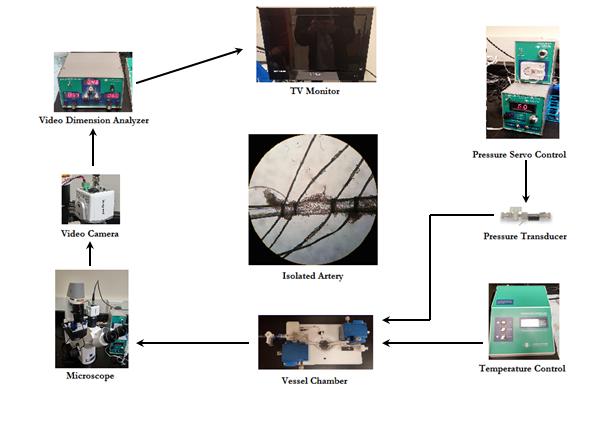
55 Arteriograph System A-6


56 A-7 Vessel Hanging This image, taken through the lens of dissecting microscope, displays a vessel that has been hung and tied between two hollow glass cannula. The image to the right shows what a vessel looks like projected on to a screen using the video dimension analyzer.
57 A-8 Tissue Homogenization 1. Cut tissue using a chilled razor and forceps, approximately 0.10g of tissue that was stored at -80 C (be sure tissue is taken from center of sample). *be sure to keep tissue frozen (on dry ice) 2. Add tissue and the steal bead pre-chilled in liquid nitrogen to the pre-chilled homogenization capsule. 3. And homogenize for 10 seconds, until tissue is a powder. 4. Add 2x buffer to the capsule. Total volume should be 1000uL (1000µL 0.10g = 900µL of buffer) 2x Lamelli s Buffer (2x Sample Buffer): 1000µL 0.5 ml 4x Stacking Gel Buffer (0.5 M Tris-Cl, ph 6.8) 0.2 ml 50% glycerol 0.2 ml 10% SDS 30µL Molecular Water 20µL Protease Inhibitor 20µL Phosphatase Inhibitor (only if looking for phosphorylated tissue) 5. Place capsule and pulse on homogenizer (2-3 seconds). Then place on regular ice. 6. Remove homogenate with 1000µL pipette and place into bullet tube. 7. Centrifuge bullet tube with homogenate. 8. Collect the supernatant in 25µL aliquots and place them in the -80 C. 9. Run Bradford assay to determine protein concentration (see A-9)
58 A-9 Bradford Assay 1. Open program (Bradford Protein) 2. Transfer 2.0 µl of 0.0 concentration standard and click blank 3. Type in standard values (125, 250, 500, 750, 1000, 1250 µg/µl) 4. Wipe lens and transfer the corresponding standard to the NanoDrop (x3 for each standard) 5. R-squared value must be above Switch screen to sample, then put 2.0 µl of 1:10 dilution Bradford reagent solution of desired sample (also 3x per sample) Bradford reagent solution: 50:1 Reagent A to Reagent B
59 A-10 Polyacrylimde Gel Casting 7. Mix 10% APS solution (0.2g + 2mL water). 8. Prepare Stacking and Resolving solutions without adding APS or TEMED. 9. Set pipettes to appropriate volumes, prepare cassettes (remove comb and leave tape on) 10. Add the appropriate amounts of TEMED and APS to the resolving and stacking solution. 11. Use the pipette gun to load Resolving Solution into cassette 1 cm below well-area. 12. Load Stacking solution (slowly) until well-area of the cassette is full. 13. Insert comb with desired well number and let sit for one hour. 14. Wrap with plastic rap and store in fridge Note: Steps 4-6 should be completely at a brisk pace, so gel does not prematurely set. Bio-Rad TGX Stain-Free FastCast Acrylamide Solutions Resolving Solution: Stacking Solution: 12 ml Resolving Solution A ml Stacking Solution A 12 ml Resolving Solution B 3.0 ml Stacking Solution B 120 µl 10% APS 30 µl 10% APS 12 µl TEMED 6 µl TEMED 4 µl Bromophenol Blue *Makes two gels
60 A-11 Western Blotting Protocol 1. Fill out protocol sheet for each sample/well. a. Calculate the amount of each sample necessary to load desired amount (30µg of Protein) to the micro plate (0.2mL). b. Calculate the amount of molecular water need to normalize each sample to the same volume. (twice the amount being loaded to the wells) 2. Add 1.0 µl Bromophenol blue to each well that will receive a sample 3. Add reducing agent DTT or ß mercaptoethanol at 100µM concentration for final volume (i.e. 30µL solution = 3.0µL reducing agent) 4. Pipet calculated amount of sample and molecular water to each well (keep on ice) 5. Denature the protein by covering the samples and placing the plate in the denaturing machine. 6. Fill gel reservoir with cold running buffer 7. Load molecular ladder and each sample into each specific lane 8. Electrophorese the gel at 250mV until the sample runs off (approx. 1hr) 9. Wet PVDF membrane in methanol then soak with blot pads in transfer buffer 10. Place blot pad, then gel, then PVDF membrane, and finally other pad and transfer with electricity (2hrs) 11. Block for 2hrs 12. Add primary antibody at desired concentration. Sit overnight 13. Perform 3 washes in TBST each for 5 minutes 14. Add secondary antibody in a 50:50 solution of blocker and TBS (1-2hrs) 15. Wash 2 times in TBST for 5 mins; 2 times TBS for 5 minutes 16. Add chemiluminscent substrates (femto) 650 µl of A and B let sit for 5 min 17. Develop in the dark room


61 A-12 Gel Electrophoresis These figures depict the processes of electrophoresis. The proteins are being separated by molecular weight using polyacrylamide gels and electricity in an electrophoresing chamber.

62 A-13 Western Blotting Diagram This diagram demonstrates how primary antibody binds to a specific protein on the PDVF membrane. Then the secondary antibody, which contains horseradish peroxidase, binds to the primary antibody. Ultimately, the horseradish peroxidase will catalyze the reaction of the two chemiluminescent substrates making them glow. The film is then places on the membrane, and the glowing part exposes the film.
Agonistic Autoantibodies to Angiotensin II Type I Receptor Contributes Partly to Placental Ischemia-Induced Cerebrovascular Abnormalities
 Agonistic Autoantibodies to Angiotensin II Type I Receptor Contributes Partly to Placental Ischemia-Induced Cerebrovascular Abnormalities Junie Paula Warrington 1, Fan Fan 1, Babbette B. LaMarca 1, Ralf
THE CIRCULATORY SYSTEM
 Biology 30S THE CIRCULATORY SYSTEM Name: This module adapted from bblearn.merlin.mb.ca 1 Introduction to Circulation The first organ to form, and the last organ to die. The heart is the pump of life. The
Procine sphingomyelin ELISA Kit
 Procine sphingomyelin ELISA Kit For the quantitative in vitro determination of Procine sphingomyelin concentrations in serum - plasma - celiac fluid - tissue homogenate - body fluid FOR LABORATORY RESEARCH
Human Apolipoprotein A1 EIA Kit
 A helping hand for your research Product Manual Human Apolipoprotein A1 EIA Kit Catalog Number: 83901 96 assays 1 Table of Content Product Description 3 Assay Principle 3 Kit Components 3 Storage 4 Reagent
TECHNICAL BULLETIN. Catalog Number RAB0447 Storage Temperature 20 C
 Phospho-Stat3 (ptyr 705 ) and pan-stat3 ELISA Kit for detection of human, mouse, or rat phospho-stat3 (ptyr 705 ) and pan-stat3 in cell and tissue lysates Catalog Number RAB0447 Storage Temperature 20
TECHNICAL BULLETIN. Phospho-Akt (pser 473 ) ELISA Kit for detection of human, mouse, or rat phospho-akt (pser 473 ) in cell and tissue lysates
 ELISA Kit for detection of human, mouse, or rat phospho-akt (pser 473 ) in cell and tissue lysates") Phospho-Akt (pser 473 ) ELISA Kit for detection of human, mouse, or rat phospho-akt (pser 473 ) in cell and tissue lysates Catalog Number RAB0011 Storage Temperature 20 C TECHNICAL BULLETIN Product Description
Phospho-Akt (pser 473 ) ELISA Kit for detection of human, mouse, or rat phospho-akt (pser 473 ) in cell and tissue lysates Catalog Number RAB0011 Storage Temperature 20 C TECHNICAL BULLETIN Product Description
CHAPTER 12 HYPERTENSION IN SPECIAL GROUPS HYPERTENSION IN PREGNANCY
 CHAPTER 12 HYPERTENSION IN SPECIAL GROUPS HYPERTENSION IN PREGNANCY v Mild preeclampsia is managed by close observation of the mother and fetus preferably in hospital. If the diastolic blood pressure remains
Mouse Cathepsin B ELISA Kit
 GenWay Biotech, Inc. 6777 Nancy Ridge Drive San Diego, CA 92121 Phone: 858.458.0866 Fax: 858.458.0833 Email: techline@genwaybio.com http://www.genwaybio.com Mouse Cathepsin B ELISA Kit Catalog No. GWB-ZZD154
Direct blood pressure monitoring was done using radiotelemetry (DataSciences
 Supplemental Methods: Blood Pressure Monitoring Direct blood pressure monitoring was done using radiotelemetry (DataSciences International; DSI). Surgical implantation of TA11PA-C1 transmitters was performed
RayBio Human Phospho-DDR2 (Tyr740) and Total DDR2 ELISA Kit
 and Total DDR2 ELISA Kit") RayBio Human Phospho-DDR2 (Tyr740) and Total DDR2 ELISA Kit Catalog #: PEL-DDR2-Y740-T User Manual Last revised March 22, 2018 Caution: Extraordinarily useful information enclosed ISO 13485 Certified 3607
RayBio Human Phospho-DDR2 (Tyr740) and Total DDR2 ELISA Kit Catalog #: PEL-DDR2-Y740-T User Manual Last revised March 22, 2018 Caution: Extraordinarily useful information enclosed ISO 13485 Certified 3607
FOCUS SubCell. For the Enrichment of Subcellular Fractions. (Cat. # ) think proteins! think G-Biosciences
 think proteins! think G-Biosciences") 169PR 01 G-Biosciences 1-800-628-7730 1-314-991-6034 technical@gbiosciences.com A Geno Technology, Inc. (USA) brand name FOCUS SubCell For the Enrichment of Subcellular Fractions (Cat. # 786 260) think
169PR 01 G-Biosciences 1-800-628-7730 1-314-991-6034 technical@gbiosciences.com A Geno Technology, Inc. (USA) brand name FOCUS SubCell For the Enrichment of Subcellular Fractions (Cat. # 786 260) think
Human IL-2. Pre-Coated ELISA Kit
 Human IL-2 (Interleukin 2) Pre-Coated ELISA Kit Catalog No: 90-2083 1 96 well Format (96 tests) Detection Range: 31.2 2000 pg/ml Sensitivity: < 18.75 pg/ml This immunoassay kit allows for the in vitro
Mouse TrkB ELISA Kit
 Mouse TrkB ELISA Kit CATALOG NO: IRKTAH5472 LOT NO: SAMPLE INTENDED USE For quantitative detection of mouse TrkB in cell culture supernates, cell lysates and tissue homogenates. BACKGROUND TrkB receptor
Human TSH ELISA Kit. User Manual
 Human TSH ELISA Kit User Manual Catalog number: GTX15585 GeneTex Table of Contents A. Product Description... 2 B. Kit Components... 3 C. Additional Required Materials (not included)... 3 D. Reagent Preparation...
EXPERIMENT 26: Detection of DNA-binding Proteins using an Electrophoretic Mobility Shift Assay Gel shift
 EXPERIMENT 26: Detection of DNA-binding Proteins using an Electrophoretic Mobility Shift Assay Gel shift Remember to use sterile conditions (tips, tubes, etc.) throughout this experiment Day 1: Biotinylation
Human Obestatin ELISA
 K-ASSAY Human Obestatin ELISA For the quantitative determination of obestatin in human serum and plasma Cat. No. KT-495 For Research Use Only. 1 Rev. 081309 K-ASSAY PRODUCT INFORMATION Human Obestatin
Human Cathepsin D ELISA Kit
 GenWay Biotech, Inc. 6777 Nancy Ridge Drive San Diego, CA 92121 Phone: 858.458.0866 Fax: 858.458.0833 Email: techline@genwaybio.com http://www.genwaybio.com Human Cathepsin D ELISA Kit Catalog No. GWB-J4JVV9
Mitochondrial Trifunctional Protein (TFP) Protein Quantity Microplate Assay Kit
 Protein Quantity Microplate Assay Kit") PROTOCOL Mitochondrial Trifunctional Protein (TFP) Protein Quantity Microplate Assay Kit DESCRIPTION Mitochondrial Trifunctional Protein (TFP) Protein Quantity Microplate Assay Kit Sufficient materials
PROTOCOL Mitochondrial Trifunctional Protein (TFP) Protein Quantity Microplate Assay Kit DESCRIPTION Mitochondrial Trifunctional Protein (TFP) Protein Quantity Microplate Assay Kit Sufficient materials
(PDGF), 9 ( -2 (FGF-2), SMO
, 9 ( -2 (FGF-2), SMO") Abstract An ethanol extract from shark muscle has been shown to have potent angiogenic activity when mixed together with olive oil in a ratio of 1part extract to 9 parts olive oil. This mixture has been
Abstract An ethanol extract from shark muscle has been shown to have potent angiogenic activity when mixed together with olive oil in a ratio of 1part extract to 9 parts olive oil. This mixture has been
In the name of GOD. Animal models of cardiovascular diseases: myocardial infarction & hypertension
 In the name of GOD Animal models of cardiovascular diseases: myocardial infarction & hypertension 44 Presentation outline: Cardiovascular diseases Acute myocardial infarction Animal models for myocardial
RayBio Human Phosphotyrosine BTK ELISA Kit
 RayBio Human Phosphotyrosine BTK ELISA Kit Catalog #: PEL-BTK-Y User Manual Last revised August 10, 2016 Caution: Extraordinarily useful information enclosed ISO 13485 Certified 3607 Parkway Lane, Suite
RayBio Human Phospho-DDR1 (Tyr792) ELISA Kit
 ELISA Kit") RayBio Human Phospho-DDR1 (Tyr792) ELISA Kit Catalog #: PEL-DDR1-Y792 User Manual Last revised March 22, 2018 Caution: Extraordinarily useful information enclosed ISO 13485 Certified 3607 Parkway Lane,
RayBio Human Phospho-DDR1 (Tyr792) ELISA Kit Catalog #: PEL-DDR1-Y792 User Manual Last revised March 22, 2018 Caution: Extraordinarily useful information enclosed ISO 13485 Certified 3607 Parkway Lane,
1. Which of the following blood vessels has a thin elastic layer? A. Aorta. B. Pulmonary artery. C. Posterior vena cava. D. Mesenteric capillary.
 CIRCULATORY SYSTEM 1. Which of the following blood vessels has a thin elastic layer? A. Aorta. B. Pulmonary artery. C. Posterior vena cava. D. Mesenteric capillary. 2. Capillary beds are equipped with
RayBio Human Phospho-DDR1 (Tyr792) and Total DDR1 ELISA Kit
 and Total DDR1 ELISA Kit") RayBio Human Phospho-DDR1 (Tyr792) and Total DDR1 ELISA Kit Catalog #: PEL-DDR1-Y792-T User Manual Last revised March 22, 2018 Caution: Extraordinarily useful information enclosed ISO 13485 Certified 3607
RayBio Human Phospho-DDR1 (Tyr792) and Total DDR1 ELISA Kit Catalog #: PEL-DDR1-Y792-T User Manual Last revised March 22, 2018 Caution: Extraordinarily useful information enclosed ISO 13485 Certified 3607
Estrone EIA kit. For the quantitative determination of estrone in dried fecal extracts, urine and tissue culture media. Cat. No.
 K-ASSAY KAMIYA BIOMEDICAL COMPANY KAMIYA BIOMEDICAL COMPANY Estrone EIA kit For the quantitative determination of estrone in dried fecal extracts, urine and tissue culture media Cat. No. KT-720 For Research
EGFR (py1045)/ Pan EGFR (Human) ELISA Kit
/ Pan EGFR (Human) ELISA Kit") EGFR (py1045)/ Pan EGFR (Human) ELISA Kit Catalog Number KA2156 96 assays Version: 01 Intended for research use only www.abnova.com I. INTRODUCTION EGFR (py1045)/pan EGFR (Human) ELISA (Enzyme-Linked Immunosorbent
EGFR (py1045)/ Pan EGFR (Human) ELISA Kit Catalog Number KA2156 96 assays Version: 01 Intended for research use only www.abnova.com I. INTRODUCTION EGFR (py1045)/pan EGFR (Human) ELISA (Enzyme-Linked Immunosorbent
LAB 12-1 HEART DISSECTION GROSS ANATOMY OF THE HEART
 LAB 12-1 HEART DISSECTION GROSS ANATOMY OF THE HEART Because mammals are warm-blooded and generally very active animals, they require high metabolic rates. One major requirement of a high metabolism is
STAT3 (py705) (Human/Mouse/Rat) ELISA Kit
 (Human/Mouse/Rat) ELISA Kit") STAT3 (py705) (Human/Mouse/Rat) ELISA Kit Catalog Number KA2175 96 assays Version: 01 Intended for research use only www.abnova.com I. INTRODUCTION STAT3 (py705) (Human/Mouse/Rat) ELISA (Enzyme-Linked
STAT3 (py705) (Human/Mouse/Rat) ELISA Kit Catalog Number KA2175 96 assays Version: 01 Intended for research use only www.abnova.com I. INTRODUCTION STAT3 (py705) (Human/Mouse/Rat) ELISA (Enzyme-Linked
RayBio Human, Mouse and Rat Phospho-NF-kB P65 (Ser536) and Total NF-kB P65 ELISA Kit
 and Total NF-kB P65 ELISA Kit") RayBio Human, Mouse and Rat Phospho-NF-kB P65 (Ser536) and Total NF-kB P65 ELISA Kit Catalog #: PEL-NFKBP65-S536-T User Manual Last revised October 10, 2017 Caution: Extraordinarily useful information
RayBio Human, Mouse and Rat Phospho-NF-kB P65 (Ser536) and Total NF-kB P65 ELISA Kit Catalog #: PEL-NFKBP65-S536-T User Manual Last revised October 10, 2017 Caution: Extraordinarily useful information
Increased risk for the development of preeclampsia in obese pregnancies: weighing in on the mechanisms
 Review Am J Physiol Regul Integr Comp Physiol 39: R1326 R1343, 215. First published October 7, 215; doi:1.1152/ajpregu.178.215. Increased risk for the development of preeclampsia in obese pregnancies:
Influenza A H1N1 (Swine Flu 2009) Hemagglutinin / HA ELISA Pair Set
 Hemagglutinin / HA ELISA Pair Set") Influenza A H1N1 (Swine Flu 2009) Hemagglutinin / HA ELISA Pair Set Catalog Number : SEK001 To achieve the best assay results, this manual must be read carefully before using this product and the assay
Influenza A H1N1 (Swine Flu 2009) Hemagglutinin / HA ELISA Pair Set Catalog Number : SEK001 To achieve the best assay results, this manual must be read carefully before using this product and the assay
Human Alpha 1 microglobulin ELISA Kit
 Human Alpha 1 microglobulin ELISA Kit Catalogue No.: EH4144 Size: 48T/96T Reactivity: Human Range:0.625-40ng/ml Sensitivity:
#6 - Cardiovascular III Heart Sounds, Pulse Rate, Hemoglobin Saturation, and Blood Pressure
 #6 - Cardiovascular III Heart Sounds, Pulse Rate, Hemoglobin Saturation, and Blood Pressure Objectives: Observe slide of artery and vein cross-section Auscultate heart sounds using a stethoscope Measure
STAT3 (py705)/ Pan STAT3 (Human/Mouse/Rat) ELISA Kit
/ Pan STAT3 (Human/Mouse/Rat) ELISA Kit") STAT3 (py705)/ Pan STAT3 (Human/Mouse/Rat) ELISA Kit Catalog Number KA2176 96 assays Version: 02 Intended for research use only www.abnova.com Table of Contents Introduction... 3 Principle of the Assay...
STAT3 (py705)/ Pan STAT3 (Human/Mouse/Rat) ELISA Kit Catalog Number KA2176 96 assays Version: 02 Intended for research use only www.abnova.com Table of Contents Introduction... 3 Principle of the Assay...
2D protein gel analysis tool for studying the protein phosphorylation changes associated with contraction and relaxation of vascular smooth muscle
 2D protein gel analysis tool for studying the protein phosphorylation changes associated with contraction and relaxation of vascular smooth muscle Internship details Where: Department of Bioengineering
Amersham ECL Prime Western blotting reagent
 GE Healthcare Life Sciences Data file 28-9857-23 AA Western blotting reagents Amersham ECL Prime Western blotting reagent Since its introduction in 199, the enhanced chemiluminescence (ECL) Western Blotting
PHYSIOEX 3.0 EXERCISE 33B: CARDIOVASCULAR DYNAMICS
 PHYSIOEX 3.0 EXERCISE 33B: CARDIOVASCULAR DYNAMICS Objectives 1. To define the following: blood flow; viscosity; peripheral resistance; systole; diastole; end diastolic volume; end systolic volume; stroke
SURGICAL PROCEDURE DESCRIPTIONS
 SURGICAL PROCEDURE DESCRIPTIONS GONADECTOMY: CASTRATION USING SCROTAL METHOD 1. The animal is anesthetized and placed in dorsal recumbency with the tail toward the surgeon. 2. The abdominal and scrotal
Osmosis and Diffusion: How biological membranes are important This page is a lab preparation guide for instructors.
 Osmosis and Diffusion: How biological membranes are important This page is a lab preparation guide for instructors. **All solutions and dialysis bags can easily be prepared prior to lab start to maximize
Saphenous Vein Autograft Replacement
 Saphenous Vein Autograft Replacement of Severe Segmental Coronary Artery Occlusion Operative Technique Rene G. Favaloro, M.D. D irect operation on the coronary artery has been performed in 180 patients
ab LDL Uptake Assay Kit (Fluorometric)
") ab204716 LDL Uptake Assay Kit (Fluorometric) Instructions for Use For rapid, sensitive and accurate measuring of LDL uptake. This product is for research use only and is not intended for diagnostic use.
ab204716 LDL Uptake Assay Kit (Fluorometric) Instructions for Use For rapid, sensitive and accurate measuring of LDL uptake. This product is for research use only and is not intended for diagnostic use.
Blood Pressure and its Regulation
 Blood Pressure and its Regulation Blood pressure in your blood vessels is closely monitored by baroreceptors; they send messages to the cardio regulatory center of your medulla oblongata to regulate your
HiPer Western Blotting Teaching Kit
 HiPer Western Blotting Teaching Kit Product Code: HTI009 Number of experiments that can be performed: 5/20 Duration of Experiment: ~ 2 days Day 1: 6-8 hours (SDS- PAGE and Electroblotting) Day 2: 3 hours
Human Immunodeficiency Virus type 1 (HIV-1) gp120 / Glycoprotein 120 ELISA Pair Set
 gp120 / Glycoprotein 120 ELISA Pair Set") Human Immunodeficiency Virus type 1 (HIV-1) gp120 / Glycoprotein 120 ELISA Pair Set Catalog Number : SEK11233 To achieve the best assay results, this manual must be read carefully before using this product
Human Immunodeficiency Virus type 1 (HIV-1) gp120 / Glycoprotein 120 ELISA Pair Set Catalog Number : SEK11233 To achieve the best assay results, this manual must be read carefully before using this product
CoQ10(Coenzyme Q10) ELISA Kit
 ELISA Kit") CoQ10(Coenzyme Q10) ELISA Kit Catalogue No.: EU0196 Size: 48T/96T Reactivity: Universal Detection Range: 0.781-50ng/ml Sensitivity:
CoQ10(Coenzyme Q10) ELISA Kit Catalogue No.: EU0196 Size: 48T/96T Reactivity: Universal Detection Range: 0.781-50ng/ml Sensitivity:
STAT1 (ps727) (Human/Mouse) ELISA Kit
 (Human/Mouse) ELISA Kit") STAT1 (ps727) (Human/Mouse) ELISA Kit Catalog Number KA2171 96 assays Version: 01 Intended for research use only www.abnova.com I. INTRODUCTION STAT1 (ps727) (Human/Mouse) ELISA (Enzyme-Linked Immunosorbent
STAT1 (ps727) (Human/Mouse) ELISA Kit Catalog Number KA2171 96 assays Version: 01 Intended for research use only www.abnova.com I. INTRODUCTION STAT1 (ps727) (Human/Mouse) ELISA (Enzyme-Linked Immunosorbent
ab ATP Synthase Enzyme Activity Microplate Assay Kit
 ab109714 ATP Synthase Enzyme Activity Microplate Assay Kit Instructions for Use For the quantitative measurement of ATP Synthase activity in samples from Human, Rat and Cow This product is for research
Biodiversity Study & Biomass Analysis
 PR072 G-Biosciences 1-800-628-7730 1-314-991-6034 technical@gbiosciences.com A Geno Technology, Inc. (USA) brand name Biodiversity Study & Biomass Analysis Teacher s Guidebook (Cat. # BE-403) think proteins!
Human LDL Receptor / LDLR ELISA Pair Set
 Human LDL Receptor / LDLR ELISA Pair Set Catalog Number : SEK10231 To achieve the best assay results, this manual must be read carefully before using this product and the assay is run as summarized in
4/19/2018. St. Cloud Hospital Perinatology Kristin Olson, RDMS, RVT
 St. Cloud Hospital Perinatology Kristin Olson, RDMS, RVT Review Fetal Circulation Provide Indications for Umbilical Artery, Middle Cerebral Artery, and Ductus Venosus Doppler studies. Demonstrate normal
Minimal Effects When Acute Nicotine Exposure Introduced to 3 Day Old Chick Embryos
 Minimal Effects When Acute Nicotine Exposure Introduced to 3 Day Old Chick Embryos Amy Langevin Independent Research Project Bio254- Developmental Biology November 28, 2007 Introduction Thousands of mothers
11/10/2014. Muscular pump Two atria Two ventricles. In mediastinum of thoracic cavity 2/3 of heart's mass lies left of midline of sternum
 It beats over 100,000 times a day to pump over 1,800 gallons of blood per day through over 60,000 miles of blood vessels. During the average lifetime, the heart pumps nearly 3 billion times, delivering
Page 1 of 2. Standard curve of Human IFN gamma ELISA Ready- SET-Go! Product Information Contents: Human IFN gamma ELISA Ready- SET-Go!
 Page 1 of 2 Human IFN gamma ELISA Ready-SET-Go! Catalog Number: 88-7316 Also known as: Interferon-gamma, IFN-g, IFNg RUO: For. Not for use in diagnostic procedures. Standard curve of Human IFN gamma ELISA
ACTG Laboratory Technologist Committee Revised Version 2.0 ACTG Lab Man Coulter HIV-1 p24 ELISA May 21, 2004
 Coulter HIV p24 1. PRINCIPLE The Human Immunodeficiency Virus Type 1 (HIV-1) is recognized as the etiologic agent of acquired immunodeficiency syndrome (AIDS). The virus is transmitted by sexual contact,
Exosome ELISA Complete Kits
 Exosome ELISA Complete Kits EXOEL-CD9A-1, EXOEL-CD63A-1, EXOEL-CD81A-1 User Manual See PAC for Storage Conditions for Individual Components Version 12 4/17/2017 A limited-use label license covers this
Virus Harvest AAV 15 cm 2
 Virus Harvest AAV 15 cm 2 1) Gently scrape cells from plate in 10 ml of media, place remaining 10 ml of media in plate lid. 2) Once cells are removed from plate, wash plate by pipetting 20 ml of media
ER REBOA Catheter. Instructions for Use
 ER REBOA Catheter Instructions for Use Prytime Medical Devices, Inc. 229 N. Main Street Boerne, TX 78006, USA feedback@prytimemedical.com www.prytimemedical.com US 1 210 340 0116 U.S. and Foreign Patents
Influenza A H1N1 HA ELISA Pair Set
 Influenza A H1N1 HA ELISA Pair Set for H1N1 ( A/Puerto Rico/8/1934 ) HA Catalog Number : SEK11684 To achieve the best assay results, this manual must be read carefully before using this product and the
TECHNICAL BULLETIN. GLP-1 EIA Kit for serum, plasma, culture supernatant, and cell lysates. Catalog Number RAB0201 Storage Temperature 20 C
 GLP-1 EIA Kit for serum, plasma, culture supernatant, and cell lysates Catalog Number RAB0201 Storage Temperature 20 C TECHNICAL BULLETIN Product Description The GLP-1 (Glucagon-like Peptide 1) Enzyme
RayBio Human ENA-78 ELISA Kit
 RayBio Human ENA-78 ELISA Kit Catalog #: ELH-ENA78 User Manual Last revised April 15, 2016 Caution: Extraordinarily useful information enclosed ISO 13485 Certified 3607 Parkway Lane, Suite 100 Norcross,
Protocols. Harvesting and Sectioning the Ascending Aorta
 DAUGHERTY LAB Saha Cardiovascular Research Center University of Kentucky Protocols Harvesting and Sectioning the Ascending Aorta Page 1 of 7 Harvesting the ascending aorta Materials: 1. Insulin syringe
3 To gain experience monitoring a titration with a ph electrode and determining the equivalence point.
 Titrations PURPOSE To determine the concentration of acetic acid in vinegar. GOALS 1 To perform an acid-base titration. 2 To gain experience titrating carefully to a visible endpoint. 3 To gain experience
Mouse Myeloperoxidase/MPO ELISA Kit
 OriGene Technologies, Inc 9620 Medical Center Dr., Suite 200, Rockville, MD 20850 Phone: 1.888.267.4436 Fax: 301-340-9254 Email: techsupport@origene.com Web: Mouse Myeloperoxidase/MPO ELISA Kit Catalog
For the quantitative measurement of ATP Synthase Specific activity in samples from Human, Rat and Cow
 ab109716 ATP Synthase Specific Activity Microplate Assay Kit Instructions for Use For the quantitative measurement of ATP Synthase Specific activity in samples from Human, Rat and Cow This product is for
2009 H1N1 Influenza ( Swine Flu ) Hemagglutinin ELISA kit
 Hemagglutinin ELISA kit") 2009 H1N1 Influenza ( Swine Flu ) Hemagglutinin ELISA kit Catalog Number : SEK001 To achieve the best assay results, this manual must be read carefully before using this product and the assay is run as
2009 H1N1 Influenza ( Swine Flu ) Hemagglutinin ELISA kit Catalog Number : SEK001 To achieve the best assay results, this manual must be read carefully before using this product and the assay is run as
Health Science 20 Circulatory System Notes
 Health Science 20 Circulatory System Notes Functions of the Circulatory System The circulatory system functions mainly as the body s transport system. It transports: o Oxygen o Nutrients o Cell waste o
BIOL 347L Laboratory Three
 Introduction BIOL 347L Laboratory Three Osmosis in potato and carrot samples Osmosis is the movement of water molecules through a selectively permeable membrane into a region of higher solute concentration,
The Living Environment
 Name: Class: 1. Asexual reproduction primarily involves the process of 1) pollination 3) spermatogenesis 2) mitosis 4) ovulation The Living Environment Ms. Fazio Base your answers to questions 4 and 5
RayBio Human Granzyme B ELISA Kit
 RayBio Human Granzyme B ELISA Kit Catalog #: ELH-GZMB User Manual Last revised April 15, 2016 Caution: Extraordinarily useful information enclosed ISO 13485 Certified 3607 Parkway Lane, Suite 100 Norcross,
Cell Lysis Buffer. Catalog number: AR0103
 Cell Lysis Buffer Catalog number: AR0103 Boster s Cell Lysis Buffer is a ready-to-use Western blot related reagent solution used for efficient extraction of total soluble protein in nondenatured state
Human Urokinase / PLAU / UPA ELISA Pair Set
 Human Urokinase / PLAU / UPA ELISA Pair Set Catalog Number : SEK10815 To achieve the best assay results, this manual must be read carefully before using this product and the assay is run as summarized
Preeclampsia is estimated to affect 7% to 10% of all
 Pathophysiology of Hypertension During Preeclampsia Linking Placental Ischemia With Endothelial Dysfunction Joey P. Granger, Barbara T. Alexander, Maria T. Llinas, William A. Bennett, Raouf A. Khalil Abstract
9( )- Hydroxyoctadecadienoic Acid ELISA
- Hydroxyoctadecadienoic Acid ELISA") Package Insert 9( )- Hydroxyoctadecadienoic Acid ELISA 96 Wells For Research Use Only v. 1.0 Eagle Biosciences, Inc. 82 Broad Street, Suite 383, Boston, MA 02110 Phone: 866-419-2019 Fax: 617-419-1110 INTRODUCTION
Package Insert 9( )- Hydroxyoctadecadienoic Acid ELISA 96 Wells For Research Use Only v. 1.0 Eagle Biosciences, Inc. 82 Broad Street, Suite 383, Boston, MA 02110 Phone: 866-419-2019 Fax: 617-419-1110 INTRODUCTION
Influenza A H7N9 (A/Anhui/1/2013) Hemagglutinin / HA ELISA Pair Set
 Hemagglutinin / HA ELISA Pair Set") Influenza A H7N9 (A/Anhui/1/2013) Hemagglutinin / HA ELISA Pair Set Catalog Number : SEK40103 To achieve the best assay results, this manual must be read carefully before using this product and the assay
Influenza A H7N9 (A/Anhui/1/2013) Hemagglutinin / HA ELISA Pair Set Catalog Number : SEK40103 To achieve the best assay results, this manual must be read carefully before using this product and the assay
Physiology Unit 3 CARDIOVASCULAR PHYSIOLOGY: THE VASCULAR SYSTEM
 Physiology Unit 3 CARDIOVASCULAR PHYSIOLOGY: THE VASCULAR SYSTEM In Physiology Today Hemodynamics F = ΔP/R Blood flow (F) High to low pressure Rate = L/min Pressure (P) Hydrostatic pressure Pressure exerted
Relaxation responses of aortic rings from salt-loaded high calcium fed rats to potassium chloride, calcium chloride and magnesium sulphate
 Pathophysiology 4 (1998) 275 280 Relaxation responses of aortic rings from salt-loaded high calcium fed rats to potassium chloride, calcium chloride and magnesium sulphate B.J. Adegunloye, O.A. Sofola
02006B 1 vial 02006B 1 vial Store at -20 C. Lyophilized recombinant IL-2
 For detection and measurement of human interleukin 2 Catalog #02006 Catalog #02007 2 Plates 10 Plates Product Description The Human Interleukin 2 (IL-2) ELISA Kit is designed for the quantitative detection
Protocol for protein SDS PAGE and Transfer
 Protocol for protein SDS PAGE and Transfer According to Laemmli, (1970) Alaa El -Din Hamid Sayed, Alaa_h254@yahoo.com Serum Selection of a protein source cell cultures (bacteria, yeast, mammalian, etc.)
Cardiovascular Physiology
 Cardiovascular Physiology Lecture 1 objectives Explain the basic anatomy of the heart and its arrangement into 4 chambers. Appreciate that blood flows in series through the systemic and pulmonary circulations.
Exosome ELISA Complete Kits
 Exosome ELISA Complete Kits EXOEL-CD9A-1, EXOEL-CD63A-1, EXOEL-CD81A-1 User Manual See PAC for Storage Conditions for Individual Components Version 12 4/17/2017 A limited-use label license covers this
Influenza B Hemagglutinin / HA ELISA Pair Set
 Influenza B Hemagglutinin / HA ELISA Pair Set Catalog Number : SEK11053 To achieve the best assay results, this manual must be read carefully before using this product and the assay is run as summarized
Instructions for use. TSH rat ELISA. Please use only the valid version of the Instructions for Use provided with the kit AR E-8600
 Instructions for use TSH rat ELISA AR E-8600 TSH rat ELISA 1. INTRODUCTION 1.1 Intended use The TSH rat ELISA is an enzyme immunoassay for the quantitative measurement of TSH in rat serum. For research
PROTEIN LAB BASED ON THE RESEARCH OF DR. RICHARD LONDRAVILLE
 PROTEIN LAB BASED ON THE RESEARCH OF DR. RICHARD LONDRAVILLE LAB OUTLINE- WEEK ONE PROCCESS WORMS FOR ANALYSIS SET UP A STANDARD CURVE WITH KNOWN CONCENTRATIONS PERFORM PROTEIN ASSAYS ON WORM SECTION INTERPRET
Evaluation of directed and random motility in microslides Assessment of leukocyte adhesion in flow chambers
 Evaluation of directed and random motility in microslides Motility experiments in IBIDI microslides, image acquisition and processing were performed as described. PMN, which ended up in an angle < 180
ab Creatinine Assay Kit (Colorimetric)
") ab204537 Creatinine Assay Kit (Colorimetric) Instructions for Use For the quantitative determination of Creatinine in urine samples. This product is for research use only and is not intended for diagnostic
ab204537 Creatinine Assay Kit (Colorimetric) Instructions for Use For the quantitative determination of Creatinine in urine samples. This product is for research use only and is not intended for diagnostic
HST 071. IN SUMMARY PREGNANCY INDUCED HYPERTENSION Classification of Hypertensive Disorders of Pregnancy
 Harvard-MIT Division of Health Sciences and Technology HST.071: Human Reproductive Biology Course Director: Professor Henry Klapholz IN SUMMARY Classification of Hypertensive Disorders of Pregnancy HST
RayBio Human Thyroglobulin ELISA Kit
 RayBio Human Thyroglobulin ELISA Kit Catalog #: ELH-Thyroglobulin User Manual Last revised July 6, 2017 Caution: Extraordinarily useful information enclosed ISO 13485 Certified 3607 Parkway Lane, Suite
PIAF study: Placental insufficiency and aortic isthmus flow Jean-Claude Fouron, MD
 Dear colleagues, I would like to thank you very sincerely for agreeing to participate in our multicentre study on the clinical significance of recording fetal aortic isthmus flow during placental circulatory
Laparoscopic Gastric Bypass Information
 1441 Constitution Boulevard, Salinas, CA 93906 (831) 783-2556 www.natividad.com/weight-loss (Roux-en-Y Gastric Bypass) What is gastric bypass surgery? Gastric bypass surgery, a type of bariatric surgery
Rat cholesterol ELISA Kit
 Rat cholesterol ELISA Kit Catalog No. CSB-E11706r (96T) This immunoassay kit allows for the in vitro quantitative determination of rat Cholesterol concentrations in serum, plasma and other biological fluids.
Influenza A IgG ELISA
 Influenza A IgG ELISA For the qualitative determination of IgG-class antibodies against Influenza A virus in human serum or plasma (citrate, heparin). For Research Use Only. Not For Use In Diagnostic Procedures.
Human Immunodeficiency Virus type 1 (HIV-1) p24 / Capsid Protein p24 ELISA Pair Set
 p24 / Capsid Protein p24 ELISA Pair Set") Human Immunodeficiency Virus type 1 (HIV-1) p24 / Capsid Protein p24 ELISA Pair Set Catalog Number : SEK11695 To achieve the best assay results, this manual must be read carefully before using this product
Human Immunodeficiency Virus type 1 (HIV-1) p24 / Capsid Protein p24 ELISA Pair Set Catalog Number : SEK11695 To achieve the best assay results, this manual must be read carefully before using this product
Blood flows away from the heart in arteries, to the capillaries and back to the heart in the veins
 Cardiovascular System Summary Notes The cardiovascular system includes: The heart, a muscular pump The blood, a fluid connective tissue The blood vessels, arteries, veins and capillaries Blood flows away
Blood Flow and Blood Pressure Regulation *
 OpenStax-CNX module: m44806 1 Blood Flow and Blood Pressure Regulation * OpenStax This work is produced by OpenStax-CNX and licensed under the Creative Commons Attribution License 4.0 By the end of this
Cardiovascular System Blood Vessels
 Cardiovascular System Blood Vessels Structure of Blood Vessels The three layers (tunics) Tunica intima composed of simple squamous epithelium Tunica media sheets of smooth muscle Contraction vasoconstriction
The blood returns from the body and enters right atrium using the vena cava. It passes through the tricuspid valve to the right ventricle.
 The blood returns from the body and enters right atrium using the vena cava. It passes through the tricuspid valve to the right ventricle. From this camber, it passes through the pulmonary semilunar valve
Rat Proinsulin ELISA
 Rat Proinsulin ELISA For the quantitative determination of proinsulin in rat serum For Research Use Only. Not For Use In Diagnostic Procedures. Catalog Number: 80-PINRT-E01 Size: 96 wells Version: June