PIAF study: Placental insufficiency and aortic isthmus flow Jean-Claude Fouron, MD
|
|
|
- Annice Simpson
- 5 years ago
- Views:
Transcription
1 Dear colleagues, I would like to thank you very sincerely for agreeing to participate in our multicentre study on the clinical significance of recording fetal aortic isthmus flow during placental circulatory insufficiency. The primary objective of this project is to identify the threshold value of the isthmic flow index (IFI) below which a fetus suffering placental insufficiency is at risk of intrauterine cerebral hypoxic injury. The project will be undertaken in three phases. The first phase is designed to ensure uniformity in all centres for Doppler recording techniques in all vascular sites included in the study. The second phase is the collection of obstetrical and neonatal data and, the final phase involves the assessment of neurodevelopment at 2 years of age. The internet site that you are currently viewing was designed primarily to facilitate phase I, which is the knowledge transfer phase. Prior to that, it seemed necessary to justify our working theory concerning the isthmus. A physiological review is first required for this purpose. 1

2 Why the fetal aortic isthmus? Two basic elements characterize fetal circulatory dynamics: first, the parallel arrangement of the two ventricles and their arterial outlet and, second, the presence of shunts. The parallel arrangement of the fetal ventricles, well demonstrated experimentally, is observed daily in any Fetal Cardiology unit where we can see the two cardiac pumps perfusing the systemic circulation in parallel, the left ventricle via the aortic arch and the right ventricle via the pulmonary arch made up of the main pulmonary trunk and arterial duct. By definition, a shunt diverts a portion of blood flow from its normal trajectory to a circuit with less resistance. In the fetus, the ductus venosus corresponds very well to the definition of a shunt since the umbilical vein normally drains into the portal system. Part of the umbilical venous 2
3 blood, which should make it to the liver, is diverted towards the inferior vena cava through the ductus venosus. This is also true for the foramen ovale which allows the passage of blood from the right to the left atrium, whereas normally it should go to the right ventricle. However, the concept of a fetal circulation with two cardiac pumps, left and right, arranged in parallel is incompatible with identification of the arterial duct as a shunt. Indeed, describing a right to left shunt between the pulmonary artery and the thoracic aorta logically means that the normal destination of blood, ejected by the right ventricle in the fetal pulmonary artery, would be the lungs and, subsequently, this blood would go to the left heart. In other words, it would be a circulation in series, as described classically in the postnatal period. Therefore, it is totally irrational to affirm that the two cardiac pumps and their respective outlet are arranged in parallel during the fetal period, and describe, in the same breath, the flow to the arterial duct as a right to left shunt. According to the next figure, we can very easily understand that the aortic arch and the pulmonary arch are perfusing the systemic circulation in parallel. Considering this figure, the right and left pulmonary arteries during fetal life are branches of the pulmonary arch in the same way as carotid and left subclavian arteries are branches of the aortic arch. We can also easily conclude from this figure that the isthmus, not the arterial duct, is the sole arterial shunt in the fetal circulation connecting the two parallel arterial arches. 3

4 In systole, the orientation of the isthmic shunt to the head or to the feet will depend both on left ventricular function which has an antegrade influence on the shunt, and on right ventricular function which influences the isthmic shunt in a retrograde manner. There are several examples of malformations that demonstrate this phenomenon very well. In diastole, while the pulmonary and aortic valves are closed, the orientation of the shunt will essentially depend on balance between the supradiaphragmatic circulation, excluding that of the lungs, and the infradiaphragmatic circulations. Since cerebrovascular resistance decreases while a plateau is observed at the level of placental resistance from the second trimester to the end of the pregnancy, we observe a progressive diminution of isthmic antegrade diastolic flow. It is important to note, on velocity Doppler curves in the isthmus, that we encounter a retrograde telesystolic incisure that gradually increases from the beginning of the third trimester to the end of the pregnancy. It is a physiological phenomenon related to the increasing preponderance of right ventricular stroke volume which at the very end of systole 4

5 influences the isthmic flow in a retrograde manner. The fact that this brief retrograde flow occurs during systole is well illustrated by simultaneous recording of flows in the isthmus and arterial duct. We can see that the left ventricular antegrade influence stops just before the end of systole and leaves room for the right ventricular influence through the arterial duct. This point is very important in the interpretation of pathological Doppler tracings. Simultaneous recording of the aortic isthmus flow and the arterial duct flow Aortic isthmus Arterial duct In the presence of increased placental resistance, as this is the case in placental insufficiency, we note the disappearance of the diastolic flow in the umbilical artery, and then very quickly, a retrograde flow arises, first telediastolic and subsequently holodiastolic in the more severe cases. Those changes in the Doppler waveforms observed at the level of the umbilical artery can also be recorded at the level of the aortic isthmus. 5

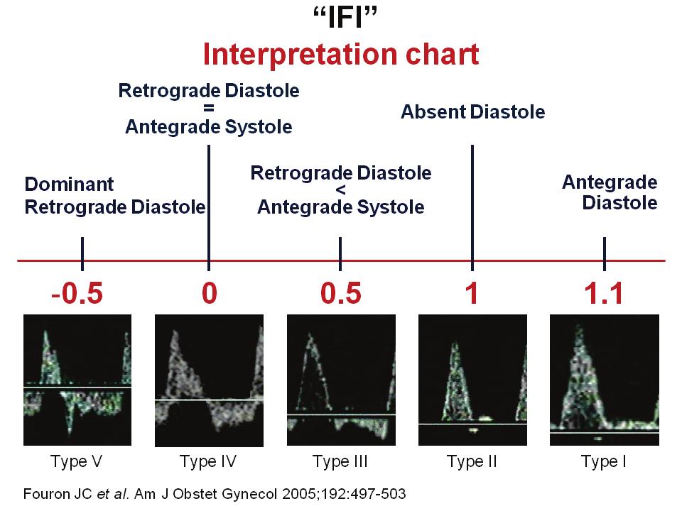
6 Aortic isthmus flow recording 1) Normal case, antegrade diastole, 2) Absent diastole, 3) Retrograde diastole, 4) Severe case, dominant retrograde diastole An isthmic flow index has been proposed, based on the ratio of integral systolic and diastolic velocities, and corrected for systolic velocities. In normal cases, the IFI is always above 1.2 regardless of gestational age. Five types of IFIs changes occur in the presence of increased placental resistance: - Type I: IFI > 1, antegrade diastolic flow is still present, but reduced; - Type II: IFI = 1, diastolic flow is absent; - Type III: IFI is a fraction of 1 but greater than 0, which reflects the appearance of a diastolic retrograde flow, but with a dominant antegrade flow; - Type IV: IFI = 0, antegrade and retrograde isthmic flows are equal, which is equivalent to an absence of flow in the isthmus; - Type V: IFI < 0, retrograde diastolic flow is predominant, the index being negative. 6
7 7
8 In closing, it is important to remember the limits of various Doppler tracings currently used to decide whether to deliver a fetus with growth restriction due to placental circulatory insufficiency. These limits are well illustrated in this figure taken from the publication of Hecher and coworkers. Pulsatility indices (umbilical artery, ductus venosus, descending aorta, inferior vena cava, middle cerebral artery), amniotic fluid index and, short-term variations Hecher K et al. Ultrasound Obstet Gynecol 2001; 18:
9 The values of standard deviations of different parameters are on the y-axis. All values between +2 and -2 standard deviations are considered to be within the limits of normal. The days that precede childbirth are on the x-axis, the date of delivery corresponding to day 0. These fetuses have been followed for more than a month before delivery. It is very easy to see, from the onset, that the umbilical artery is abnormal. It has indeed been well demonstrated that the degree of hypoxemia cannot be evaluated by the pulsatility index at the level of the umbilical artery. This index informs us only about the degree of resistance of a single vascular bed, the placenta, without telling us what is happening throughout the fetal body. It is noteworthy that the dominant retrograde flow observed at the level of the aortic isthmus in a fetus with restricted growth is never observed in the umbilical artery since it is obviously incompatible with fetal life. Doppler recording in the abdominal aorta and the amniotic fluid volume index are also abnormal very early. This is also the case for the pulsatility index of the middle cerebral artery, reflecting a compensatory phenomenon in response to hypoxemia ensuring adequate cerebral oxygenation. During a major part of the observation period, the flows in the ductus venosus and inferior vena cava as well as short-term variations in cardiac rate remain within the limits of normal. It is only five to six days before the expected date of delivery that these variables became abnormal. We know now that the appearance of changes at the level of the ductus venosus or vena cava corresponds to a state of severe hypoxia which leads to myocardial diastolic dysfunction. The myocardium is by far more resistant to hypoxia than the brain, which allows us to anticipate at this stage a central nervous system injury of hypoxic origin. It is not surprising to observe also that venous flow anomalies are associated almost simultaneously with the deterioration and loss of short-term normal variation of fetal cardiac heart beats. We know indeed that this variation is normally under the influence of the central nervous system. In other words, most of the current criteria presently used to decide the delivery of a fetus suffering from IUGR are based on signs of cardiocirculatory decompensation and central nervous system dysfunction. That is what we want to avoid in our study, indeed. Obviously, the ideal criterion is one that would allow the delivery of these fetuses just before the appearance of decompensation. 9

10 This last figure illustrates why the isthmus appears to be potentially the ideal site for evaluating the degree of secondary fetal hypoxemia. Indeed, in the presence of increased placental circulatory resistance, hypoxemia causes cerebral and coronary vasodilatation decreasing vascular impedance in the supradiaphragmatic circulation. In the infradiaphragmatic circulation of IUGR fetuses, the elevated vascular resistance is not only due to the changes in placental resistance but also to the intense hypoxic vasoconstriction observed in the mesenteric artery network. The balance between supradiaphragmatic and infradiaphragmatic resistances is then disrupted and the isthmus flow becomes abnormal. This pattern of changes which reflects the degree of hypoxemia should be reliably expressed by the wide range of possible alterations previously shown in the isthmus flow index. Noteworthy, a dominant retrograde diastolic aortic flow can be observed at the level of the aortic isthmus in a fetus with severe IUGR, of course at the level of the umbilical artery such a flow pattern would be incompatible with fetal life. On behalf of my colleagues and the entire technical staff who participate in preparing this Internet site, I want to thank you all for your attention. 10
COPYRIGHTED MATERIAL. The fetal circulation CHAPTER 1. Postnatal circulation
 1 CHAPTER 1 The fetal circulation The circulation in the fetus differs from that in the adult. Knowledge of the course and distribution of the fetal circulation is important to our understanding of the
4/19/2018. St. Cloud Hospital Perinatology Kristin Olson, RDMS, RVT
 St. Cloud Hospital Perinatology Kristin Olson, RDMS, RVT Review Fetal Circulation Provide Indications for Umbilical Artery, Middle Cerebral Artery, and Ductus Venosus Doppler studies. Demonstrate normal
Large Arteries of Heart
 Cardiovascular System (Part A-2) Module 5 -Chapter 8 Overview Arteries Capillaries Veins Heart Anatomy Conduction System Blood pressure Fetal circulation Susie Turner, M.D. 1/5/13 Large Arteries of Heart
The Physiology of the Fetal Cardiovascular System
 The Physiology of the Fetal Cardiovascular System Jeff Vergales, MD, MS Department of Pediatrics Division of Pediatric Cardiology jvergales@virginia.edu Disclosures I serve as the medical director for
The sinus venosus represent the venous end of the heart It receives 3 veins: 1- Common cardinal vein body wall 2- Umbilical vein from placenta 3-
 1 2 The sinus venosus represent the venous end of the heart It receives 3 veins: 1- Common cardinal vein body wall 2- Umbilical vein from placenta 3- Vitelline vein from yolk sac 3 However!!!!! The left
Chapter 14. The Cardiovascular System
 Chapter 14 The Cardiovascular System Introduction Cardiovascular system - heart, blood and blood vessels Cardiac muscle makes up bulk of heart provides force to pump blood Function - transports blood 2
C3, 4, 5, 6, & 7 Worksheet. C3 Describe the inter-relationships of the structures of the heart
 Name: Date: C3, 4, 5, 6, & 7 Worksheet C3 Describe the inter-relationships of the structures of the heart 1. Label and give the functions of the following: a. left and right atrium: b. left and right ventricle:
First Trimester Fetal Echocardiography: Insight Into the Fetal Circulation
 First Trimester Fetal Echocardiography: Insight Into the Fetal Circulation Lisa K. Hornberger, MD Fetal & Neonatal Cardiology Program Department of Pediatrics, Division of Cardiology Department of Obstetrics
Figure ) The specific chamber of the heart that is indicated by letter A is called the. Diff: 1 Page Ref: 364
 The specific chamber of the heart that is indicated by letter A is called the. Diff: 1 Page Ref: 364") Essentials of Anatomy and Physiology, 9e (Marieb) Chapter 11 The Cardiovascular System Short Answer Figure 11.1 Using Figure 11.1, identify the following: 1) The Purkinje fibers are indicated by label.
Essentials of Anatomy and Physiology, 9e (Marieb) Chapter 11 The Cardiovascular System Short Answer Figure 11.1 Using Figure 11.1, identify the following: 1) The Purkinje fibers are indicated by label.
A (quasi)evidence-based approach to the management of early-onset IUGR
evidence-based approach to the management of early-onset IUGR") A (quasi)evidence-based approach to the management of early-onset IUGR Eduard Gratacós Barcelona Center for Maternal-Fetal and Neonatal Medicine Hospital Clínic and Hospital Sant Joan de Deu, University
A (quasi)evidence-based approach to the management of early-onset IUGR Eduard Gratacós Barcelona Center for Maternal-Fetal and Neonatal Medicine Hospital Clínic and Hospital Sant Joan de Deu, University
THE CIRCULATORY SYSTEM
 Biology 30S THE CIRCULATORY SYSTEM Name: This module adapted from bblearn.merlin.mb.ca 1 Introduction to Circulation The first organ to form, and the last organ to die. The heart is the pump of life. The
1. Which of the following blood vessels has a thin elastic layer? A. Aorta. B. Pulmonary artery. C. Posterior vena cava. D. Mesenteric capillary.
 CIRCULATORY SYSTEM 1. Which of the following blood vessels has a thin elastic layer? A. Aorta. B. Pulmonary artery. C. Posterior vena cava. D. Mesenteric capillary. 2. Capillary beds are equipped with
Blood Vessels. Types of Blood Vessels Arteries carry blood away from the heart Capillaries smallest blood vessels. Veins carry blood toward the heart
 C H A P T E R Blood Vessels 20 Types of Blood Vessels Arteries carry blood away from the heart Capillaries smallest blood vessels The site of exchange of molecules between blood and tissue fluid Veins
DEVELOPMENT OF THE CIRCULATORY SYSTEM L E C T U R E 5
 DEVELOPMENT OF THE CIRCULATORY SYSTEM L E C T U R E 5 REVIEW OF CARDIAC ANATOMY Heart 4 chambers Base and apex Valves Pericardial sac 3 layers: epi, myo, endo cardium Major blood vessels Aorta and its
Key issues in (early and late) IUGR
 IUGR") Key issues in (early and late) IUGR Eduard Gratacós Maternal-Fetal Medicine Department, Hospital Clínic, University of Barcelona www.fetalmedicinebarcelona.org (early-onset) IUGR vs SGA: the era of UA
Key issues in (early and late) IUGR Eduard Gratacós Maternal-Fetal Medicine Department, Hospital Clínic, University of Barcelona www.fetalmedicinebarcelona.org (early-onset) IUGR vs SGA: the era of UA
REVIEW SHEET Anatomy of Blood Vessels
 REVIEW SHEET Anatomy of Blood Vessels Name LabTime/Date Microscopic Structure of the Blood Vessels 1. Cross-sectional views of an aftery of a vein are shown here. ldentify each; on the lines to the sides,
Development of Blood Vessels and Fetal Circulation *
 OpenStax-CNX module: m46610 1 Development of Blood Vessels and Fetal Circulation * OpenStax This work is produced by OpenStax-CNX and licensed under the Creative Commons Attribution License 3.0 By the
The Cardiovascular System
 PowerPoint Lecture Slide Presentation by Patty Bostwick-Taylor, Florence-Darlington Technical College The Cardiovascular System 11PART B The Heart: Cardiac Output Cardiac output (CO) Amount of blood pumped
Doppler assessment of fetal aortic isthmus blood flow in two different sonographic planes during the second half of gestation
 Ultrasound Obstet Gynecol 2005; 26: 170 174 Published online in Wiley InterScience (www.interscience.wiley.com). DOI: 10.1002/uog.1955 Doppler assessment of fetal aortic isthmus blood flow in two different
THE VESSELS OF BLOOD CIRCULATION
 THE VESSELS OF BLOOD CIRCULATION scientistcindy.com /the-vessels-of-blood-circulation.html NOTE: You should familiarize yourself with the anatomy of the heart and have a good understanding of the flow
The Fetus: Five Top Do Not Miss Diagnoses. Doppler Ultrasound
 The Fetus: Five Top Do Not Miss Diagnoses Doppler Ultrasound Giancarlo Mari, MD, MBA Professor and Chair Department of Obstetrics and Gynecology University of Tennessee Health Science Center Memphis, TN
Circulatory System Review
 Circulatory System Review 1. Know the diagrams of the heart, internal and external. a) What is the pericardium? What is myocardium? What is the septum? b) Explain the 4 valves of the heart. What is their
Spleen. Vertebrate hearts Pericardial cavity division in coelum. Vessel walls. Endocardium = endothelium of blood vessels. Artery elastic tissue
 Spleen White pulp macrophages, monocyte storage Red pulp - (RBC) storage, and prod n (in nonmammals) Vertebrate hearts Pericardial cavity division in coelum Endocardium = endothelium of blood vessels Fig.
Editorial. Color and pulsed Doppler in fetal echocardiography A. ABUHAMAD
 Ultrasound Obstet Gynecol 2004; 24: 1 9 Published online in Wiley InterScience (www.interscience.wiley.com). DOI: 10.1002/uog.1096 Editorial Color and pulsed Doppler in fetal echocardiography A. ABUHAMAD
Chapter 11. The Cardiovascular System. Clicker Questions Pearson Education, Inc.
 Chapter 11 The Cardiovascular System Clicker Questions Oxygen-poor blood is pumped through the venae cavae to the right side of the heart, and then through the pulmonary arteries to the lungs and back
2. capillaries - allow exchange of materials between blood and tissue fluid
 Chapter 19 - Vascular System A. categories and general functions: 1. arteries - carry blood away from heart 2. capillaries - allow exchange of materials between blood and tissue fluid 3. veins - return
1. Distinguish among the types of blood vessels on the basis of their structure and function.
 Blood Vessels and Circulation Objectives This chapter describes the structure and functions of the blood vessels Additional subjects contained in Chapter 13 include cardiovascular physiology, regulation,
The Cardiovascular System. The Structure of Blood Vessels. The Structure of Blood Vessels. The Blood Vessels. Blood Vessel Review
 The Cardiovascular System The Blood Vessels The Structure of Blood Vessels Blood Vessel Review Arteries carry blood away from the heart Pulmonary trunk to lungs Aorta to everything else Microcirculation
Ch.15 Cardiovascular System Pgs {15-12} {15-13}
 Ch.15 Cardiovascular System Pgs {15-12} {15-13} E. Skeleton of the Heart 1. The skeleton of the heart is composed of rings of dense connective tissue and other masses of connective tissue in the interventricular
COMPREHENSIVE EVALUATION OF FETAL HEART R. GOWDAMARAJAN MD
 COMPREHENSIVE EVALUATION OF FETAL HEART R. GOWDAMARAJAN MD Disclosure No Relevant Financial Relationships with Commercial Interests Fetal Echo: How to do it? Timing of Study -optimally between 22-24 weeks
Function: Transportation of. Oxygen Nutrients Waste Hormones gases
 Function: Transportation of Oxygen Nutrients Waste Hormones gases Pericardium: double sac of serous membrane filled with fluid (pericardial fluid to be exact) that surrounds the heart. Parietal pericardium:
Unit 11 - The Cardiovascular System 1
 Unit 11 - The Cardiovascular System 1 I. Unit 11: The Cardiovascular System A. The Cardiovascular System 1. A closed system of the heart and blood vessels a) The heart pumps blood b) Blood vessels allow
Cardiovascular System. Heart Anatomy
 Cardiovascular System Heart Anatomy 1 The Heart Location & general description: Atria vs. ventricles Pulmonary vs. systemic circulation Coverings Walls The heart is found in the mediastinum, the medial
Venous Doppler Evaluation of the Growth-Restricted Fetus
 Venous Doppler Evaluation of the Growth-Restricted Fetus Ahmet Alexander Baschat, MD KEYWORDS Fetal growth restriction Doppler Ductus venosus Venous circulation Fetal surveillance Integrated testing The
Opinion. Technical aspects of aortic isthmus Doppler velocimetry in human fetuses
 Ultrasound Obstet Gynecol 2009; 33: 628 633 Published online in Wiley InterScience (www.interscience.wiley.com). DOI: 10.1002/uog.6406 Opinion Technical aspects of aortic isthmus Doppler velocimetry in
10. Thick deposits of lipids on the walls of blood vessels, called, can lead to serious circulatory issues. A. aneurysm B. atherosclerosis C.
 Heart Student: 1. carry blood away from the heart. A. Arteries B. Veins C. Capillaries 2. What is the leading cause of heart attack and stroke in North America? A. alcohol B. smoking C. arteriosclerosis
39 th Annual Perinatal Conference Vanderbilt University December 6, 2013 IUGR. Diagnosis and Management
 39 th Annual Perinatal Conference Vanderbilt University December 6, 2013 IUGR Diagnosis and Management Giancarlo Mari, M.D., M.B.A. Professor and Chair Department of Obstetrics and Gynecology University
Paediatric Cardiology. Acyanotic CHD. Prof F F Takawira
 Paediatric Cardiology Acyanotic CHD Prof F F Takawira Aetiology Chromosomal Down syndrome, T13, T18 Genetic syndromes (gene defects) Velo-Cardio-facial (22 del) Genetic syndromes (undefined aetiology)
Failing right ventricle
 Failing right ventricle U. Herberg 1, U. Gembruch 2 1 Pediatric Cardiology, 2 Prenatal Diagnostics and Fetal Therapy, University of Bonn, Germany Prenatal Physiology Right ventricle dominant ventricle
Anatomy & Physiology
 1 Anatomy & Physiology Heart is divided into four chambers, two atrias & two ventricles. Atrioventricular valves (tricuspid & mitral) separate the atria from ventricles. they open & close to control flow
AOGS ORIGINAL RESEARCH ARTICLE
 AOGS ORIGINAL RESEARCH ARTICLE Ventricular outputs, central blood flow distribution and flow pattern through the aortic isthmus of fetuses with simple transposition of the great arteries JULIE BLANC 1,2,
Skeletal muscle. Flow increases and decreases with each muscular contraction - as a result of compression of the blood vessels by contracted muscle
 Regional blood flow Skeletal muscle Extreme increases during exercises Flow increases and decreases with each muscular contraction - as a result of compression of the blood vessels by contracted muscle
PRACTICAL GUIDE TO FETAL ECHOCARDIOGRAPHY IC Huggon and LD Allan
 PRACTICAL GUIDE TO FETAL ECHOCARDIOGRAPHY IC Huggon and LD Allan Fetal Cardiology Unit, Harris Birthright Research Centre for Fetal Medicine, King's College Hospital, London, UK IMPORTANCE OF PRENATAL
The Cardiovascular System
 PowerPoint Lecture Slide Presentation by Patty Bostwick-Taylor, Florence-Darlington Technical College The Cardiovascular System 11PART A The Cardiovascular System A closed system of the heart and blood
Principles of Biomedical Systems & Devices. Lecture 8: Cardiovascular Dynamics Dr. Maria Tahamont
 Principles of Biomedical Systems & Devices Lecture 8: Cardiovascular Dynamics Dr. Maria Tahamont Review of Cardiac Anatomy Four chambers Two atria-receive blood from the vena cave and pulmonary veins Two
(2) (1) (3) (4) BLOOD PATHWAY ASSESSMENT RUBRIC
 (1) (3) (4) BLOOD PATHWAY ASSESSMENT RUBRIC") BLOODPATHWAYASSESSMENT(4) BLOOD%PATHWAY%ASSESSMENT%(3)% BLOODPATHWAYASSESSMENT(3) (4) (3) (2) (1) Using a completely blank diagram of the heart, all valves, chambers, great vessels, and direction of blood
BLOODPATHWAYASSESSMENT(4) BLOOD%PATHWAY%ASSESSMENT%(3)% BLOODPATHWAYASSESSMENT(3) (4) (3) (2) (1) Using a completely blank diagram of the heart, all valves, chambers, great vessels, and direction of blood
The Cardiovascular System. Preview of Heart Action. The CV system provides oxygen & nutrients to tissues-removes wastes.
 The Cardiovascular System BIO 250 Human Anatomy & Physiology Preview of Heart Action http://www.youtube.com/watch?v=d3zdj gfddk0&nr=1 The CV system provides oxygen & nutrients to tissues-removes wastes.
11/10/2014. Muscular pump Two atria Two ventricles. In mediastinum of thoracic cavity 2/3 of heart's mass lies left of midline of sternum
 It beats over 100,000 times a day to pump over 1,800 gallons of blood per day through over 60,000 miles of blood vessels. During the average lifetime, the heart pumps nearly 3 billion times, delivering
Figure 10.1A Transparency Master 79
 Brain Carotid arteries Jugular vein Right front leg Lungs (inflated) Cranial Right atrium To left front leg Left subclavian Bronchus capillaries Brachiocephalic vein Left atrium Dorsal aorta Right ventricle
Fetal cardiovascular parameters for the prediction of postnatal cardiovascular risk in intrauterine growth-restriction?
 17 th International Conference on Prenatal Diagnosis and Therapy Lisbon, June 2013 Fetal cardiovascular parameters for the prediction of postnatal cardiovascular risk in intrauterine growth-restriction?
TRACE A DROP OF BLOOD FROM RIGHT EAR TO LEFT OCULOMOTOR NERVE
 TRACE A DROP OF BLOOD FROM RIGHT EAR TO LEFT OCULOMOTOR NERVE KEY: TRACE A DROP OF BLOOD FROM RIGHT EAR TO LEFT OCULOMOTOR NERVE RIGHT EAR RIGHT ATRIUM LEFT SUBCLAVIAN ARTERY RIGHT EXTERNAL JUGULAR VEIN
Unit 11: The Cardiovascular System
 Unit 11: The Cardiovascular System I. The Cardiovascular System A. A closed system of the heart and blood vessels 1. The heart pumps blood 2. Blood vessels allow blood to circulate to all parts of the
The Mammalian Circulatory System
 The Mammalian Heart The Mammalian Circulatory System Recall: What are the 3 cycles of the mammalian circulatory system? What are their functions? What are the three main vessel types in the mammalian circulatory
Diagnosis and Management of the Early Growth Restricted Fetus
 11 th Congress of Maternal Fetal Medicine and Perinatology Society of Turkey Diagnosis and Management of the Early Growth Restricted Fetus Giancarlo Mari, MD, MBA, FACOG, FAIUM Professor and Chair Department
Chapter 10 The Circulatory & Lymphatic Systems
 Biology 12 Name: Human Biology Per: Date: Chapter 10 The Circulatory & Lymphatic Systems Complete using BC Biology 12, pages 298 325 10.1 The Blood Vessels pages 298-299 1. Label the blood vessels in this
1. What kind of blood is found in the rt. atrium? (oxygenated or deoxygenated)
") Carl Christennsen, PhD Chap. 19, 20, & 21 - Circulatory System Bio. 2304 Human Anatomy HEART 1. What kind of blood is found in the rt. atrium? (oxygenated or deoxygenated) Where does this blood come from?
Carl Christennsen, PhD Chap. 19, 20, & 21 - Circulatory System Bio. 2304 Human Anatomy HEART 1. What kind of blood is found in the rt. atrium? (oxygenated or deoxygenated) Where does this blood come from?
AN ATOMY OF THE CARDIOVASCULAR SYSTEM
 Student Name CHAPTER 18 AN ATOMY OF THE CARDIOVASCULAR SYSTEM T he heart is actually two pumps one moves blood to the lungs, the other pushes it out into the body. These two functions seem rather elementary
Overview of Anatomy and Physioloy II Second Year Students
 University of Baghdad College of Nursing Department of Basic Medical Sciences Overview of Anatomy and Physioloy II Second Year Students Asaad Ismail Ahmad, Ph.D. Asaad Ismail Ahmad, Ph.D. Electrolyte and
Summary. HVRA s Cardio Vascular Genetic Detailed L2 Obstetrical Ultrasound. CPT 76811, 76825, _ 90% CHD detection. _ 90% DS detection.
 What is the role of fetal echocardiography (2D 76825, cardiovascular color flow mapping 93325) as performed in conjunction with detailed fetal anatomy scan (CPT 76811) now that AIUM requires limited outflow
Cardiovascular Physiology. Heart Physiology. Introduction. The heart. Electrophysiology of the heart
 Cardiovascular Physiology Heart Physiology Introduction The cardiovascular system consists of the heart and two vascular systems, the systemic and pulmonary circulations. The heart pumps blood through
IP: Regulation of Cardiac Output
 ANP 1105D Winter 2013 Assignment 9: The Heart, part 2: Chap... Assignment 9: The Heart, part 2: Chapter 18 Signed in as Alex Sokolowski Help Close Resources Due: 11:59pm on Monday, March 25, 2013 Note:
Assessment of fetal heart function and rhythm
 Assessment of fetal heart function and rhythm The fetal myocardium Early Gestation Myofibrils 30% of myocytes Less sarcoplasmic reticula Late Gestation Myofibrils 60% of myocytes Increased force per unit
Heart and Lungs. LUNG Coronal section demonstrates relationship of pulmonary parenchyma to heart and chest wall.
 Heart and Lungs Normal Sonographic Anatomy THORAX Axial and coronal sections demonstrate integrity of thorax, fetal breathing movements, and overall size and shape. LUNG Coronal section demonstrates relationship
TRAINING NEONATOLOGY SILVANA PARIS
 TRAINING ON NEONATOLOGY SILVANA PARIS RESUSCITATION IN DELIVERY ROOM INTRODUCTION THE GLOBAL RESUSCITATION BURDEN IN NEWBORN 136 MILL NEWBORN BABIES EACH YEAR (WHO WORLD REPORT) 5-8 MILL NEWBORN INFANTS
The Cardiovascular System (Heart)
") The Cardiovascular System The Cardiovascular System (Heart) A closed system of the heart and blood vessels The heart pumps blood Blood vessels allow blood to circulate to all parts of the body The function
The Cardiovascular System The Cardiovascular System (Heart) A closed system of the heart and blood vessels The heart pumps blood Blood vessels allow blood to circulate to all parts of the body The function
SWISS SOCIETY OF NEONATOLOGY. Prenatal closure of the ductus arteriosus
 SWISS SOCIETY OF NEONATOLOGY Prenatal closure of the ductus arteriosus March 2007 Leone A, Fasnacht M, Beinder E, Arlettaz R, Neonatal Intensive Care Unit (LA, AR), University Hospital Zurich, Cardiology
From Head to Toe Use of Advanced Dynamic Flow in prenatal ultrasound
 From Head to Toe Use of Advanced Dynamic Flow in prenatal ultrasound Without doubt, the B- Schwerdtfeger, R. tant diagnostic instrument. Furthermore, we use colour in feto- mode imaging is the most important
Contents. Page 1. Homework 11 Chapter Blood Vessels Due: Week 6 Lec 11
 Page 1 Homework 11 Chapter 18-19 Blood Vessels Due: Week 6 Lec 11 Contents When printing, make sure that you specify the page range that you want to print out! Learning objectives for Lecture 11:...pg
Pearson's Comprehensive Medical Assisting Administrative and Clinical Competencies
 Pearson's Comprehensive Medical Assisting Administrative and Clinical Competencies THIRD EDITION CHAPTER 27 The Cardiovascular System Lesson 1: Overview of the Cardiovascular System Lesson Objectives Upon
The Heart. Made up of 3 different tissue: cardiac muscle tissue, nerve tissue, and connective tissue.
 The Heart The Heart Made up of 3 different tissue: cardiac muscle tissue, nerve tissue, and connective tissue. Your heart pumps with a regular beat (Heart Rate) Your heart rate can change depending on
The Cardiovascular System
 11 PART A The Cardiovascular System PowerPoint Lecture Slide Presentation by Jerry L. Cook, Sam Houston University ESSENTIALS OF HUMAN ANATOMY & PHYSIOLOGY EIGHTH EDITION ELAINE N. MARIEB The Cardiovascular
Foetal Cardiology: How to predict perinatal problems. Prof. I.Witters Prof.M.Gewillig UZ Leuven
 Foetal Cardiology: How to predict perinatal problems Prof. I.Witters Prof.M.Gewillig UZ Leuven Cardiopathies Incidence : 8-12 / 1000 births ( 1% ) Most frequent - Ventricle Septum Defect 20% - Atrium Septum
Diagnosis of Congenital Cardiac Defects Between 11 and 14 Weeks Gestation in High-Risk Patients
 Article Diagnosis of Congenital Cardiac Defects Between 11 and 14 Weeks Gestation in High-Risk Patients Zeev Weiner, MD, Abraham Lorber, MD, Eliezer Shalev, MD Objective. To examine the feasibility of
Cardiovascular Physiology
 Cardiovascular Physiology Lecture 1 objectives Explain the basic anatomy of the heart and its arrangement into 4 chambers. Appreciate that blood flows in series through the systemic and pulmonary circulations.
The cardiovascular system is composed of a pump the heart and blood
 5 E X E R C I S E Cardiovascular Dynamics O B J E C T I V E S 1. To understand the relationships among blood flow, pressure gradient, and resistance 2. To define resistance and describe the main factors
Chapter 05 Cardiovascular System
 Chapter 05 Cardiovascular System 1 Cardiovascular System: Heart and Blood Vessels 2 Points to ponder What are the functions of the cardiovascular system? What is the anatomy of the heart? Of blood vessels,
Health Science 20 Circulatory System Notes
 Health Science 20 Circulatory System Notes Functions of the Circulatory System The circulatory system functions mainly as the body s transport system. It transports: o Oxygen o Nutrients o Cell waste o
Cardiovascular Anatomy Dr. Gary Mumaugh
 Cardiovascular Anatomy Dr. Gary Mumaugh Location of Heart Approximately the size of your fist Location o Superior surface of diaphragm o Left of the midline in mediastinum o Anterior to the vertebral column,
Approximately the size of your fist Location Superior surface of diaphragm Left of the midline in mediastinum Anterior to the vertebral column,
 Dr. Gary Mumaugh Approximately the size of your fist Location Superior surface of diaphragm Left of the midline in mediastinum Anterior to the vertebral column, posterior to the sternum Posteriorly the
Chapter 11. The Cardiovascular System
 Chapter 11 The Cardiovascular System The Cardiovascular System The Cardiovascular System A closed system of the heart and blood vessels Heart pumps blood Blood vessels circulate blood to all parts of the
Circulatory Systems. All cells need to take in nutrients and expel metabolic wastes.
 Circulatory Systems All cells need to take in nutrients and expel metabolic wastes. Single celled organisms: nutrients from the environment can diffuse (or be actively transported) directly in to the cell
Fetal cardiac function: what to use and does it make a difference?
 17 th International Conference on Prenatal Diagnosis and Therapy Lisbon, June 2013 Fetal cardiac function: what to use and does it make a difference? Fàtima Crispi Department of Maternal-Fetal Medicine,
Chapter 12 Lecture Outline
 Chapter 12 Lecture Outline See separate PowerPoint slides for all figures and tables preinserted into PowerPoint without notes. Copyright The McGraw-Hill Companies, Inc. Permission required for reproduction
ISUOG Basic Training. Obtaining & Interpreting Heart Views Correctly Alfred Abuhamad, USA. Basic training. Editable text here
 ISUOG Basic Training Obtaining & Interpreting Heart Views Correctly Alfred Abuhamad, USA Learning Objectives 6, 7 & 8 At the end of the lecture you will be able to: describe how to assess cardiac situs
Systematic approach to Fetal Echocardiography. Objectives. Introduction 11/2/2015
 Systematic approach to Fetal Echocardiography. Pediatric Echocardiography Conference, JCMCH November 7, 2015 Rajani Anand Objectives Fetal cardiology pre-test Introduction Embryology and Physiology of
Management of IUGR Prof. Dr. Acar KOÇ
 Management of IUGR Prof. Dr. Acar KOÇ Ankara University School of Medicine Department of OB&GYN Department of Perinatology Definition and Diagnosis: SGA IUGR EFW: < 10th percentile EFW: < 10th percentile
4. The two inferior chambers of the heart are known as the atria. the superior and inferior vena cava, which empty into the left atrium.
 Answer each statement true or false. If the statement is false, change the underlined word to make it true. 1. The heart is located approximately between the second and fifth ribs and posterior to the
THE HEART. Unit 3: Transportation and Respiration
 THE HEART Unit 3: Transportation and Respiration The Circulatory System Also called the Cardiovascular System Circulates blood in the body Transports nutrients, oxygen, carbon dioxide, hormones, and blood
Chapter 23. Circulation
 Chapter 23 Circulation Standards CORE: I can describe the components and function of blood. I can describe structure and function of blood vessels. I can compare and contrast systemic and pulmonary systems.
Biology 12 - Circulation - Chapter Notes
 Biology 12 - Circulation - Chapter Notes Multicellular organisms (above the level of roundworms) rely on a circulatory system to bring nutrients to, and take wastes away from, cells. In higher organisms
AP2 Lab 3 Coronary Vessels, Valves, Sounds, and Dissection
 AP2 Lab 3 Coronary Vessels, Valves, Sounds, and Dissection Project 1 - BLOOD Supply to the Myocardium (Figs. 18.5 &18.10) The myocardium is not nourished by the blood while it is being pumped through the
MODULE 2: CARDIOVASCULAR SYSTEM ANTOMY An Introduction to the Anatomy of the Heart and Blood vessels
 MODULE 2: CARDIOVASCULAR SYSTEM ANTOMY An Introduction to the Anatomy of the Heart and Blood vessels The cardiovascular system includes a pump (the heart) and the vessels that carry blood from the heart
Exam 3 Study Guide. 4) The process whereby the binding of antibodies to antigens causes RBCs to clump is called:
 The process whereby the binding of antibodies to antigens causes RBCs to clump is called:") Exam 3 Study Guide 1) Where does hematopoiesis produce new red blood cells: 2) Which of the following is a blood clotting disorder: 3) Treatment of hemophilia often involves: 4) The process whereby the
Exam 3 Study Guide 1) Where does hematopoiesis produce new red blood cells: 2) Which of the following is a blood clotting disorder: 3) Treatment of hemophilia often involves: 4) The process whereby the
CRITICAL THINKING QUESTIONS AND ANSWERS AND CYCLE 2 LAB EXAM TEMPLATE. There are two main mechanisms that work in conjunction to return the blood
 CRITICAL THINKING QUESTIONS AND ANSWERS AND CYCLE 2 LAB EXAM TEMPLATE There are two main mechanisms that work in conjunction to return the blood THE CARDIAC PUMP 1) The forward pull(vis a fronte) This
The Cardiovascular System (Part II)
") The Cardiovascular System (Part II) 黃敏銓 mchuang@ntu.edu.tw 解剖學暨細胞生物學研究所 1 Development of veins Three paired veins drain into the tubular heart of a 4-week embryo Vitelline veins: poorly oxygenated blood
The Cardiovascular System (Part II) 黃敏銓 mchuang@ntu.edu.tw 解剖學暨細胞生物學研究所 1 Development of veins Three paired veins drain into the tubular heart of a 4-week embryo Vitelline veins: poorly oxygenated blood
Volume Flow. Volume Flow
 Volume Flow Jonathan M. Rubin, M.D., Ph.D. Department of Radiology Volume Flow Technique initially described by Hottenger and Meindl in 1974 Describes method for measuring the total flux across a flow
Circulatory System Notes
 Circulatory System Notes Functions of Circulatory System A. Transports B. Transports C. Transports D. Transports E. of fluids F. G. Regulate temperature H. Blood clotting Characteristics of various blood
Chapter 9, Part 2. Cardiocirculatory Adjustments to Exercise
 Chapter 9, Part 2 Cardiocirculatory Adjustments to Exercise Electrical Activity of the Heart Contraction of the heart depends on electrical stimulation of the myocardium Impulse is initiated in the right
Project 1: Circulation
 Project 1: Circulation This project refers to the matlab files located at: http://www.math.nyu.edu/faculty/peskin/modsimprograms/ch1/. Model of the systemic arteries. The first thing to do is adjust the
Principles of Anatomy and Physiology
 Principles of Anatomy and Physiology 14 th Edition CHAPTER 20 The Cardiovascular System: The Heart Introduction The purpose of the chapter is to: 1. Learn about the components of the cardiovascular system