3-Dimensional Echocardiography in Aortic Valve Repair 13. September Saarland University Medical Center - Homburg/Saar, Germany
|
|
|
- Marjory Wells
- 5 years ago
- Views:
Transcription


1 3-Dimensional Echocardiography in Aortic Valve Repair 13. September Saarland University Medical Center - Homburg/Saar, Germany Prof. Dr. med. Andreas Hagendorff, Universitätsklinikum Leipzig Klinik für Kardiologie Liebigstraße Leipzig Andreas.Hagendorff@medizin.uni-leipzig.de 1
2 I declare for the last 3 years and the subsequent 12 month the following conflicts of interests: Section I: Support for Research Activities - grant of the DEGUM - no other financial research support Section II: Support for Educational Activities - MIFO, GE Healthcare, Astra Zeneca, Servier, Novartis, Berlin-Chemie, Pfizer, Cardiac Dimension, Abbott, Bayer, Canon, Kelcon Section III: Honorarium for Promotional Activities - none Section IV: Personal Financial Interests in Vommercial Activities - none IB1; 2A11 Member of the German Society of Cardiology, the German Society of Ultrasound DEGUM), the German Society of Internal Medicine and the European Society of Cardiology/Cardiovascular Imaging Secretary of the DEGUM Board 2
 Imaging planes and leaflet attachments from (A) shown superimposed on postmortem specimen. A-M aorto-mitral; VA ventriculo-arterial.")
3 Aortic Root Anatomy (A) Diagram of aortic root anatomy showing coronet shape and location of various annular planes and coronary ostia relative to leaflet attachments. (B) Imaging planes and leaflet attachments from (A) shown superimposed on postmortem specimen. A-M aorto-mitral; VA ventriculo-arterial. according to Piazza N, de Jaegere P, Schultz C, Becker AE, Serruys PW, Anderson RH. Anatomy of the aortic valvar complex and its implications for transcatheter implantation of the aortic valve. Circ Cardiovasc Interv 2008;1:


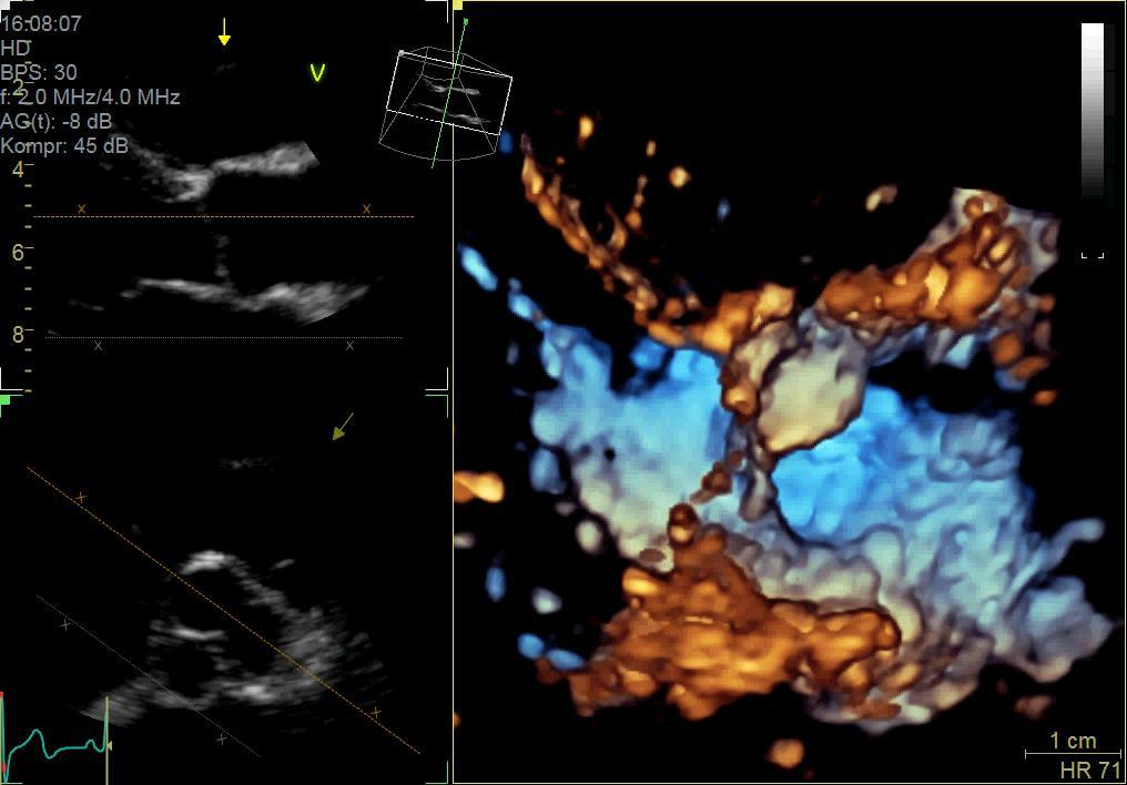
4 The anatomy of the aortic valve and the aortic root be better visualized multidimensional than in a two-dimensional images. 4

5 5


6 according to Van Dyck et al., Anest Analg 2010; 111:


7 7

8 according to Basiswissen Echokardiografie Hagendorff/ Stöbe 5/2017 Abb : Multidimensionale Schrägansichten der Aortenklappe zur Darstellung der komplexen Anatomie. In der linken Bildhälfte ist ein ZOOM-Ausschnitt eines transthorakalen 5-Kammerblicks, darunter der zugehörige perpendikuläre Schnitt sowie die multidimensionale Ansicht, die durch die jeweiligen Pfeile in den roten Kreisen gekennzeichnet ist, während der Diastole links und während der Systole rechts dargestellt. In der rechten Bildhälfte ist ein ZOOM-Ausschnitt eines transösophagealen Kurzachsenschnittes der AV, darunter der zugehörige perpendikuläre Schnitt sowie die multidimensionale Ansicht, die durch die jeweiligen Pfeile in den roten Kreisen gekennzeichnet ist, während der Diastole links und während der Systole rechts dargestellt. Links sind gut die Hinge Points der Taschen im "en-face" Blick, rechts in einem Querschnitt zu erkennen. 8

9 according to Basiswissen Echokardiografie Hagendorff/ Stöbe 5/2017 Abb : Darstellung der Lage der Koronarostien zu den Taschen der Aortenklappe in der multidimensionalen Echokardiographie. In der oberen Bildreihe sind links zwei Sequenzen während der Systole und rechts zwei Sequenzen während der Diastole abgebildet. Darunter sind die jeweiligen zweidimensionalen biplanen Schnittebenen mit den entsprechenden Blickrichtungen (siehe Pfeile) angeführt. In der unteren Bildreihe ist der Kommissur-bildende Taschenrand in schwarz, der Übergangsbereich zwischen Taschen und Aortenwand in rot sowie das Ostium der rechten Koronararterie mit einem Pfeil im multidimensionalen Bild links beschriftet. Rechts daneben ist eine Schemazeichnung der "aufgeklappten" AV mit Darstellung der Taschenränder abgebildet. 9


10 Righ coronary cusp Non coronary cusp Left coronary cusp 10


11 Sinutubular junction Anatomical ventriculoarterial junction Crown-like ring Virtual ring formed by the hinge points of the aortic cusps 11


12 * 1 3 * * * * * * * * * * * 3 is mirror inverted to 1. 12


13 Diastole Thus, how to operate correctly? Systole Intersection with the commissure Intersection with the commissure Intersection with the cusp Intersection with the cusp Intersection at the same level of the cups 13


14 1. step: adjust the central axis of AV during diastole to label the perpendicular plane through the hinge points 2. step: go to systole to measure the widest expansion of the LVOT 3. step: adjust the annulus plane in the LAX view 14

 and is defined as a virtual ring (green line)")
.")
15 aus: J Am Coll Cardiol Img. 2013;6(2): doi: /j.jcmg Standardized Imaging for Aortic Annular Sizing: Implications for Transcatheter Valve Selection Normal Anatomy of the Aortic Annulus The aortic annulus accounts for the tightest part of the aortic root (A) and is defined as a virtual ring (green line) with 3 anatomical anchor points at the nadir (green points) of each of the attachments of the 3 aortic leaflets (B). LCC = left coronary cusp; NCC = noncoronary cusp; RCC = right coronary cusp 15


16 gh = geometric height aus Swanson WM and Clark RE. Dimensions and Geometric Relationships of the Human Aortic Valve as a Function of Pressure. Circ Res 1974; 35:


17 17


18 18


19 Diastole 1 1= diameter at the level of the tpis of the crownlike ring 1 3 = effective height 2 2= diameter at the level of the hinge points 2 3 Systole = coaptationlength and 6 = geometric heights of the cusps - Really visualized only for the right coronary cusp 19

 3.")

20 Steps for measurement of geometric and effective height Acquire a ZOOM data set of the complete mitral and aortic valve 2. Adjust the central axis of the aortic root in the long axis (systole/diastole) 3. Adjust the central axis of the aortic root in the perpendicular axis 4. Adjust the short axis to the hindge points by translation during diastole 5. Rotate the short axis view to control the sectional short axis plane.. 20


21 21
22 Abstract: Reconstructive surgery of the aortic valve repair has become increasingly employed in patients with aortic regurgitation and/or aortic aneurysm. Its success depends on restoring normal aortic valve and root form. Echocardiography is the most reliable and precise imaging technique since it defines abnormal morphology and function, essential for selecting appropriate substrates and guiding the surgical strategy. This review focuses on the use of 3D-echocardiography to characterize different forms of aortic valve and root abnormalities and attempts to define echocardiographic predictors of successful valve/root complex repair. in press JACC Imaging
 and aortic root (long-axis view: a, short-axis view: b, 3D view: c).")
23 Central Illustration: The figure summarizes the advantages of applying the 3D technique to the analysis of aortic valve and aortic root complex prior to surgical correction. Sketches of the aortic valve (AV) and aortic root (long-axis view: a, short-axis view: b, 3D view: c). Parameters such as diameter of the ventricularaortic (VA)-junction, aortic root and sinotubular (ST)-junction as well as effective height (eh) and geometric height (gh) of each cusp can be more objectively and accurately assessed by 3D echocardiography versus 2D echocardiography. Labeling of the essential parameters in 3D longaxis view (d), 3D short-axis view of the AV (e) and VA junction (f). Exemplary illustration of the adjustment of the correct sectional plane of the VA junction (g-i). Exemplary illustration of the adjustment of eh (yellow) and coaptation length (CL) (blue) between non- (NCC) and right coronary cusp (RCC) and of gh (red) of the RCC. according Hagendorff et al. J Am Coll Cardiol Img 2018; in press 23

24 To analyze the aortic valve and the aortic root the main prerequisite is to acquire 3D-image data sets with optimal image quality and the best possible temporal and spatial resolution. Optimization of ultrasound settings in 2 D an 3D is extremely important 24


25 Prerquisite for excellent image quality in 2D as well as 3D echocardiography: knowledge about ultrasound physics and implementation of these aspects into the workflow by just technical knowledge about the buttons. and in case of interventions and surgery training for a fast workflow. Then, detailed information about aortic valve and aortic root morphology is possible. The spatial and temporal resolution of 3D TEE is at least comparable to cardiac-ct. The same patients: bad settings versus optimized settings in 2D and 3D-TEE. 25


26 LVOT Sinuses ST-junction TAA VA-junction 26

27 according Hagendorff et al. J Am Coll Cardiol Img 2018; in press 27
28 Final recommendations according to the authors` experience and literature (1) The results of echocardiographic measurements of the AV and aortic root strongly depend on the time point of the cardiac cycle. The maximum anterior-posterior diameter of the LVOT, AV annulus, SV, ST junction and TAA obviously vary between systole and diastole. These dimensions are larger during systole than diastole, especially in younger patients with preserved compliance of the aortic root. These dimensions are important for decision making in AV reconstruction, which is generally performed in younger patients. Given these considerations, we strongly feel that the AV complex measurements should be performed in mid-systole. according Hagendorff et al. J Am Coll Cardiol Img 2018; in press 28
29 Final recommendations according to the authors` experience and literature (2).. In addition, spatial resolution of the external aortic wall using 3D TTE and 3D TEE may be limited. Owing superior demarcation of the inner aortic wall especially using 3D TEE we believe that I-I measurements are superior to L-L measurements when using 3D echocardiography. Finally, underestimation is unavoidable in 2D TEE for the reasons stated. Thus, correct determination of these important diameters can best be achieved by I-I measurements during mid-systole using standardized sectional planes within the 3D data sets by postprocessing. This creates a contradiction regarding proposed mid-systolic measurements and current guideline recommendations. Current guidelines, however, do not address the specific aspects of AV repair. Furthermore, several studies showed that using I-I convention underestimation was compensated for by measuring aortic diameters mid-end-systole. according Hagendorff et al. J Am Coll Cardiol Img 2018; in press 29
30 Final recommendations according to the authors` experience and literature (3).. Measurements of cusp morphology and geometry especially CL, eh and gh obviously have to be performed during diastole when the cusps are stretched by diastolic pressure. All findings and parameters for the assessment of the AV root complex which are relevant and mandatory according to our experience, are highlighted in Table 1. according Hagendorff et al. J Am Coll Cardiol Img 2018; in press 30
31 Klinik-Interne Fortbildung Summary 3D Echocardiography is the best imaging technique for patient selection for surgical AV repair and AV-sparing surgery. 2D-TTE and 2D-TEE are inferior to 3D-echocardiography owing to misleading measurements in non-standardized, oblique sectional planes. 3D echocardiography should include analysis of AV morphology, aortic root dimensions and AR severity. Cusp morphology and commissures and measurements of coaptation length, eh and gh parameters should be described in a systematic approach using mainly 3D TTE and 3D TEE. Complete and concise analysis by 3D echocardiography enables correct decision-making and planning of surgical procedures in patients with AR and aortic valve/root abnormalities. It can be assumed that automatic quantification of the aortic root complex will facilitate the dynamic analysis of the aortic root complex in the future. according Hagendorff et al. J Am Coll Cardiol Img 2018; in press 31


32 Hagendorff et al., Global Cardiology Science and Practice 2018:

33 Optimal and transparent objective analysis of aortic regurgitant orifice area by 3Dechocardiography Hagendorff et al., Global Cardiology Science and Practice 2018:


34 3D- and Multidimensional Echocardiography in Aortic Valve Repair Additional information and better diagnostic impact: Quantification of an excentric regurgitation in biscuspid aortic valve ERO cm 2 34

35 3D-analysis of left and right ventricular volumes to determine total, effective and regurgitant volume by 3D echocardiography Hagendorff et al., Global Cardiology Science and Practice 2018:

36 Complex 3D-analysis of the aortic valve and aortic root by 3D echocardiography Hagendorff et al., Global Cardiology Science and Practice 2018:

37 3D- visualization of the specific cardiac structures Hagendorff et al., Global Cardiology Science and Practice 2018:

38 Dynamics of the aortic annulus and the aortic root between diastole and systole. The dimensions are larger during systole than during diastole. Hagendorff et al., Global Cardiology Science and Practice 2018:

39 Differences of spatial resolution due to different ultrasound settings in a case of vegetations at the aortic valve. Thus, settings have to be standardized to be comparable. Hagendorff et al., Global Cardiology Science and Practice 2018:


40 Documentation of the dynamics between the mitral and aortic annulus between systole and diastole Hagendorff et al., Global Cardiology Science and Practice 2018:

41 Acurate, transparent and objective measurements of all cardiac structures by 3D-echocardiography Hagendorff et al., Global Cardiology Science and Practice 2018:

42 Measurements of coaptation length and effective height by 3D-echocardiography Hagendorff et al., Global Cardiology Science and Practice 2018:
43 Measurements of geometric height by 3D-echocardiography Hagendorff et al., Global Cardiology Science and Practice 2018:

44 Acurate, transparent and objective measurements of intercommissural distances by 3D-echocardiography Hagendorff et al., Global Cardiology Science and Practice 2018:


45 3D- and Multidimensional Echocardiography in Aortic Valve Repair Example of 2D-imaging Aortic regurgitation type Ia FAA functional aortic annulus; STJ - sinotubular junction; SCA - subcommissural anuloplasty 45

46 Documentation of different types of aortic regurgitation by 3D-echocardiography Hagendorff et al., Global Cardiology Science and Practice 2018:
47 Documentation of different types of aortic regurgitation by 3D-echocardiography Hagendorff et al., Global Cardiology Science and Practice 2018:

48 Documentation of different types of aortic regurgitation by 3D-echocardiography Hagendorff et al., Global Cardiology Science and Practice 2018:

49 Documentation of different types of aortic regurgitation by 3D-echocardiography Hagendorff et al., Global Cardiology Science and Practice 2018:

50 Documentation of different types of aortic regurgitation by 3D-echocardiography Hagendorff et al., Global Cardiology Science and Practice 2018:

51 Documentation of fenestrations at the aortic cusps by 3D-echocardiography Hagendorff et al., Global Cardiology Science and Practice 2018:

52 Documentation of complications (endocarditis, aortic dissection) by 3D-echocardiography and contrast echocardiography Hagendorff et al., Global Cardiology Science and Practice 2018:
53 Documentation of calcifications and retraction by 3D-echocardiography Hagendorff et al., Global Cardiology Science and Practice 2018:
54 Documentation of cusp tethering and cusp restriction by 3D-echocardiography Hagendorff et al., Global Cardiology Science and Practice 2018:

55 Documentation of cusp prolapses by 2D-echocardiography Hagendorff et al., Global Cardiology Science and Practice 2018:

56 Visualization of blood flow turbulences by blood flow speckle tracking Hagendorff et al., Global Cardiology Science and Practice 2018:
57 SUMMARY 3D-echocardiography should be established in clinical routine to evaluate patients for AV repair and AV-sparing surgery. It is obvious that 3D-transthoracic and transesophageal echocardiography has additional value in characterizing AV and aortic root abnormalities if the image quality of the 3D data sets is sufficient. The assessment of cusp morphology and function can be best performed by CL, eh and gh, which is possible for all cusps using 3Dechocardiography. Moreover, misleading measurements due to nonstandardized, oblique sectional planes can be avoided by 3Dechocardiography. Thus, 3D-echocardiography should be mandatory for the analysis of AV and aortic root dimensions as well as the grading AR severity. However, sufficient training and experience is required before it can be applied in clinical routine. Hagendorff et al., Global Cardiology Science and Practice 2018:


58 3D- and Multidimensional Echocardiography in Aortic Valve Repair Multidimensional analysis of aortic arch: Objective measurements of aortic dimensions 58


 4. Especially for the decicion making and the planning of the surgical strategy 3D echocardiography can provide important")
59 3D- and Multidimensional Echocardiography in Aortic Valve Repair Final Summary: 1. 3D echocardiography enables a completely new modality of imaging in echocardiography the visualization of surfaces (endocardium and the cusps). 2. Biplane and triplane simultaneous sectional planes enables a better and more acurate standardization of imaging with improvement of measurements of anatomical structures. 3. Postprocessing in 3D data sets offers the possibility of new views (e.g. en-face view of the coronary ostia, etc.) 4. Especially for the decicion making and the planning of the surgical strategy 3D echocardiography can provide important informations. 5. The higher the image quality, the better the information. 6. Thus, training and expertise in 3D echocardiography is a prerequisite for a better diagnosis. 59


60 Siegel der Universität Leipzig Thank You for Your Attention 60
3D- and Multidimensional Echocardiography in Aortic Valve Repair
 Reconstruction of the Aortic Valve and Root - A Practical Approach - 3D- and Multidimensional Echocardiography Thursday 15 th September - 14.00 Practice must always be founded on sound theory. Leonardo
Aortic Regurgitation and Aortic Aneurysm - Epidemiology and Guidelines -
 Reconstruction of the Aortic Valve and Root - A Practical Approach - Aortic Regurgitation and Aortic Aneurysm Wednesday 14 th September - 9.45 Practice must always be founded on sound theory. Leonardo
Technical consideration of aquiring and analyzing 3D TEE volume data sets (EchoPac ) Phasic changes of the aortic root throughout the cardiac cycle
 Phasic changes of the aortic root throughout the cardiac cycle") Technical consideration of aquiring and analyzing 3D TEE volume data sets (EchoPac ) Phasic changes of the aortic root throughout the cardiac cycle Specific application in aortic regurgitation and aortic
Technical consideration of aquiring and analyzing 3D TEE volume data sets (EchoPac ) Phasic changes of the aortic root throughout the cardiac cycle Specific application in aortic regurgitation and aortic
Aortic Valve Repair a Modular and Geometric Approach. H.-J. Schäfers Dept. of Thoracic and Cardiovascular Surgery University Hospital of Saarland
 Aortic Valve Repair a Modular and Geometric Approach H.-J. Schäfers Dept. of Thoracic and Cardiovascular Surgery University Hospital of Saarland 12.09.2018 Limitations: Purely echocardiographic, does not
Part II: Fundamentals of 3D Echocardiography: Acquisition and Application
 Part II: Fundamentals of 3D Echocardiography: Acquisition and Application Dr. Bruce Bollen 3D matrix array TEE probes provide options for both 2D and 3D imaging. Indeed, their utility in obtaining multiple
When Does 3D Echo Make A Difference?
 When Does 3D Echo Make A Difference? Wendy Tsang, MD, SM Assistant Professor, University of Toronto Toronto General Hospital, University Health Network 1 Practical Applications of 3D Echocardiography Recommended
Image Assistance in TAVI Why CT? Won-Jang Kim, MD, PhD Clinical Assistant Professor of Medicine, Heart Institute, Asan Medical Center, Seoul, Korea
 Image Assistance in TAVI Why CT? Won-Jang Kim, MD, PhD Clinical Assistant Professor of Medicine, Heart Institute, Asan Medical Center, Seoul, Korea Major Uses of CT in TAVI Ileofemoral Patient Arterial
Outline. EuroScore II. Society of Thoracic Surgeons Score. EuroScore II
 SURGICAL RISK IN VALVULAR HEART DISEASE: WHAT 2D AND 3D ECHO CAN TELL YOU AND WHAT THEY CAN'T Ernesto E Salcedo, MD Professor of Medicine University of Colorado School of Medicine Director of Echocardiography
Pre-procedural CT angiography for Transcatheter Aortic Valve Implantation: What a Radiologist Needs to Know?
 Pre-procedural CT angiography for Transcatheter Aortic Valve Implantation: What a Radiologist Needs to Know? E O Dwyer, C O Brien, I Murphy, C Shortt, O Buckley Department of Radiology, AMNCH, Dublin,
Preprocedural evaluation for TAVR
 KEBE 30/05/15 Preprocedural evaluation for TAVR Ioannis Iakovou, MD, PhD Interventional Cardiology Onassis Cardiac Surgery Center Athens, Greece Clinical Pathway: Developing Peri- Procedural Protocols
Introduction. Aortic Valve. Outflow Tract and Aortic Valve Annulus
 Chapter 1: Surgical anatomy of the aortic and mitral valves Jordan RH Hoffman MD, David A. Fullerton MD, FACC University of Colorado School of Medicine, Department of Surgery, Division of Cardiothoracic
Questions of the webinar "Imaging in TAVI procedures" Answered by Andreas Hagendorff, Victoria Delgado and Bernard Cosyns
 Questions of the webinar "Imaging in TAVI procedures" Answered by Andreas Hagendorff, Victoria Delgado and Bernard Cosyns 1. The incidence in AR I think that this question focuses on the incidence in AR
Functional anatomy of the aortic root. ΔΡΟΣΟΣ ΓΕΩΡΓΙΟΣ Διεσθσνηής Καρδιοθωρακοτειροσργικής Κλινικής Γ.Ν. «Γ. Παπανικολάοσ» Θεζζαλονίκη
 Functional anatomy of the aortic root ΔΡΟΣΟΣ ΓΕΩΡΓΙΟΣ Διεσθσνηής Καρδιοθωρακοτειροσργικής Κλινικής Γ.Ν. «Γ. Παπανικολάοσ» Θεζζαλονίκη What is the aortic root? represents the outflow tract from the LV provides
Failed Aortic Valve Repairs Lessons Learned
 Failed Aortic Valve Repairs Lessons Learned A. Stephane Lambert, MD, FRCPC Munir Boodhwani, MD, MMSc, FRCSC University of Ottawa Heart Institute Ottawa Ontario No Disclosure Why do repairs fail? Basic
Imaging in TAVI. Jeroen J Bax Dept of Cardiology Leiden Univ Medical Center The Netherlands Davos, feb 2013
 Imaging in TAVI Jeroen J Bax Dept of Cardiology Leiden Univ Medical Center The Netherlands Davos, feb 2013 Research grants: Medtronic, Biotronik, Boston Scientific, St Jude, BMS imaging, GE Healthcare,
Regurgitant Lesions. Bicol Hospital, Legazpi City, Philippines July Gregg S. Pressman MD, FACC, FASE Einstein Medical Center Philadelphia, USA
 Regurgitant Lesions Bicol Hospital, Legazpi City, Philippines July 2016 Gregg S. Pressman MD, FACC, FASE Einstein Medical Center Philadelphia, USA Aortic Insufficiency Valve anatomy and function LVOT and
Dr Winnie Sze-Wun Chan. Cardiac Team Deputy Team Head Department of Radiology and Imaging Queen Elizabeth Hospital Hong Kong
 Dr Winnie Sze-Wun Chan Cardiac Team Deputy Team Head Department of Radiology and Imaging Queen Elizabeth Hospital Hong Kong Why? Is CT reliable? How to perform the CT study? How to interpret the CT study?
Severity of AS Degree of AV calcification (? Bicuspid AV), annulus size, & aortic root
, annulus size, & aortic root") The role of Cardiac Imaging modalities in evaluation & selection of patients for Trans-catheter Aortic Valve Implantation Dr.Saeed AL Ahmari Consultant Cardiologist Prince Sultan Cardaic Center, Riyadh
The role of Cardiac Imaging modalities in evaluation & selection of patients for Trans-catheter Aortic Valve Implantation Dr.Saeed AL Ahmari Consultant Cardiologist Prince Sultan Cardaic Center, Riyadh
The Role of Imaging in Transcatheter Aortic Valve Implantation
 The Role of Imaging in Transcatheter Aortic Valve Implantation Helmut Baumgartner Westfälische Wilhelms-Universität Münster Division of Adult Congenital and Valvular Heart Disease Department of Cardiovascular
Annular Stabilization Techniques in the Context of Aortic Valve Repair
 Annular Stabilization Techniques in the Context of Aortic Valve Repair Prashanth Vallabhajosyula, MD MS University of Pennsylvania, Philadelphia, Pennsylvania 2 nd North American Aortic Valve Repair Symposium
Aortic Regurgitation & Aorta Evaluation
 VALVULAR HEART DISEASE Regurgitation Valvular Lessions 2017 Aortic Regurgitation & Aorta Evaluation Jorge Eduardo Cossío-Aranda MD, FACC Chairman of Outpatient Care Department Instituto Nacional de Cardiología
NEW GUIDELINES. A Guideline Protocol for the Assessment of Aortic Regurgitation From the British Society of Echocardiography Education Committee
 NEW GUIDELINES A Guideline Protocol for the Assessment of Aortic Regurgitation From the British Society of Echocardiography Education Committee Gill Wharton, Prathap Kanagala (Lead Authors) Richard Steeds
Anatomy of aortic valve and root Emmanuel Lansac MD PhD
 Anatomy of aortic valve and root Emmanuel Lansac MD PhD Cardiac Surgery Institut Mutualiste Montsouris, Paris, France The aortic valve : a passive or dynamic structure? Leonardo da Vinci 1508 Quadr Anat
Indications and Late Results of Aortic Valve Repair
 Indications and Late Results of Aortic Valve Repair Prof. Gebrine El Khoury Department of Cardiovascular and Thoracic Surgery Cliniques St. Luc Brussels, Belgium Aortic Valve Repair Question # 1 Can the
Aortic Valve Repair - Alternative to Replacement
 Aortic Valve Repair - Alternative to Replacement Seite 1 Dept. of Thoracic and Cardiovascular Surgery University Hospital of Saarland Homburg/ Saar Germany Seite 2 Aortic Valve - Historic Repair Attempts
JOINT MEETING 2 Tricuspid club Chairpersons: G. Athanassopoulos, A. Avgeropoulou, M. Khoury, G. Stavridis
 JOINT MEETING 2 Tricuspid club Chairpersons: G. Athanassopoulos, A. Avgeropoulou, M. Khoury, G. Stavridis Similarities and differences in Tricuspid vs. Mitral Valve Anatomy and Imaging. Echo evaluation
Anatomy of aortic valve and root
 Anatomy of aortic valve and root Emmanuel Lansac, Isabelle Di Centa Cardiac Surgery Institut Mutualiste Montsouris, Paris, France The aortic valve : a passive or dynamic structure? Leonardo da Vinci 1508
Aortic valve repair: When and how to employ this novel approach?
 Aortic valve repair: When and how to employ this novel approach? Konstadinos A Plestis, MD System Chief of Cardiac Thoracic and Vascular Surgery Main Line Health Care System Professor Sidney Kimmel Medical
Back to Basics: Common Errors In Quantitation In Everyday Practice
 Back to Basics: Common Errors In Quantitation In Everyday Practice Deborah Agler, ACS, RDCS, FASE October 9, 2017 ASE: Echo Florida Rebecca T. Hahn, MD Director of Interventional Echocardiography Professor
Aortic Valve Repair: The Brussels Approach Laurent de Kerchove, MD, PhD Cliniques Universitaires St-Luc, IREC, UCL, Brussels, Belgium
 Reconstruction of the Aortic Valve and Root: A Practical Approach September 14 th -16 th, Homburg/Saar, Germany Aortic Valve Repair: The Brussels Approach Laurent de Kerchove, MD, PhD Cliniques Universitaires
Imaging Assessment of Aortic Stenosis/Aortic Regurgitation
 Imaging Assessment of Aortic Stenosis/Aortic Regurgitation Craig E Fleishman, MD FACC FASE The Heart Center at Arnold Palmer Hospital for Children, Orlando SCAI Fall Fellows Course 2014 Las Vegas Disclosure
Boston, MA 2 Service Chief, Cardiovascular Imaging, Department of Radiology, Massachusetts General Hospital,
 Chapter 9: Cardiac imaging for TAVR: CTA, TTE, TEE, and valve sizing Lucy M. Safi, DO 1 ; Brian Ghoshhajra, MD, MBA 2 ; Jonathan J. Passeri, MD 3 1 Clinical and Research Fellow in Medicine, Massachusetts
Organic mitral regurgitation
 The best in heart valve disease Organic mitral regurgitation Ewa Szymczyk Department of Cardiology Medical University of Lodz, Poland I have nothing to declare Organic mitral regurgitation leaflet abnormality
Jonathon Leipsic MD FRCPC FSCCT. Vice Chairman of Radiology University of British Columbia. Disclosures
 Jonathon Leipsic MD FRCPC FSCCT Vice Chairman of Radiology University of British Columbia Disclosures Speaker s bureau: GE Healthcare and Edwards LifeSciences Grant Support- CIHR, GE Healthcare Advisory
ECHOCARDIOGRAPHY DATA REPORT FORM
 Patient ID Patient Study ID AVM - - Date of form completion / / 20 Initials of person completing the form mm dd yyyy Study period Preoperative Postoperative Operative 6-month f/u 1-year f/u 2-year f/u
Revealing new insights. irotate electronic rotation and xplane adjustable biplane imaging. Ultrasound cardiology. irotate and xplane
 Ultrasound cardiology irotate and xplane Revealing new insights irotate electronic rotation and xplane adjustable biplane imaging Annemien van den Bosch and Jackie McGhie Department of Cardiology, Erasmus
Joseph E. Bavaria, M.D. Roberts Measy Professor and Vice Chief CardioVascular Surgery Director: Thoracic Aortic Surgery Program University of
 Joseph E. Bavaria, M.D. Roberts Measy Professor and Vice Chief CardioVascular Surgery Director: Thoracic Aortic Surgery Program University of Pennsylvania, USA AVRS Philadelphia Sept 2016 Pictures courtesy
Aortic valve calcium load before TAVI: Is it important?
 Research Highlight Aortic valve calcium load before TAVI: Is it important? Martin Haensig 1, Ardawan Julian Rastan 2 1 Department of Cardiac Surgery, Heart Center, University of Leipzig, Germany; 2 Department
Aortic valve repair: Techniques and Pitfalls. Allan Stewart, MD Columbia University Medical Center New York, NY
 Aortic valve repair: Techniques and Pitfalls Allan Stewart, MD Columbia University Medical Center New York, NY Take Away Points 1. Valve anatomy is essential to assess repair 2. Unique Decisions with Aneurysm/AI
Comments restricted to Sapien and Corevalve 9/12/2016. Disclosures: Core Lab contracts with Edwards Lifesciences, Middlepeak, Medtronic
 Para-ValvularRegurgitation post TAVR: Predict, Prevent, Quantitate, Manage Linda D. Gillam, MD, MPH, FACC, FASE Chair, Department of Cardiovascular Medicine Morristown Medical Center/Atlantic Health System
Rotation: Echocardiography: Transthoracic Echocardiography (TTE)
") Rotation: Echocardiography: Transthoracic Echocardiography (TTE) Rotation Format and Responsibilities: Fellows rotate in the echocardiography laboratory in each clinical year. Rotations during the first
Rotation: Echocardiography: Transthoracic Echocardiography (TTE) Rotation Format and Responsibilities: Fellows rotate in the echocardiography laboratory in each clinical year. Rotations during the first
Imaging to select patients for Transcatheter TV
 Imaging to select patients for Transcatheter TV Jeroen J Bax Dept of Cardiology Leiden Univ Medical Center The Netherlands San Diego, february 2018 Research grants: Medtronic, Biotronik, Boston Scientific,
Transoesophageal echocardiography and decision making in valve surgery
 Transoesophageal echocardiography and decision making in valve surgery Intraoperative evaluation of the surgical results in aortic valve / root surgery Catherine Szymanski Disclosures None Sino-tubular
What I Have Learned from 3D Imaging of Heart Valve Disease
 What I Have Learned from 3D Imaging of Heart Valve Disease Rebecca T. Hahn, MD Director of Interventional Echocardiography Columbia University Core Lab Director for multiple tricuspid device trials for
Results of Aortic Valve Preservation and Repair
 Results of Aortic Valve Preservation and Repair Department of Cardiothoracic and Vascular Surgery Cliniques Universitaires St. Luc Brussels, Belgium Gebrine Elkhoury Institutional experience in AV preservation
Atrioventricular valve repair: The limits of operability
 Atrioventricular valve repair: The limits of operability Francis Fynn-Thompson, MD Co-Director, Center for Airway Disorders Surgical Director, Pediatric Mechanical Support Program Surgical Director, Heart
Milind Desai Christine Jellis Teerapat Yingchoncharoen Editors. An Atlas of Mitral Valve Imaging
 Milind Desai Editors An Atlas of Mitral Valve Imaging 123 An Atlas of Mitral Valve Imaging Milind Desai Editors An Atlas of Mitral Valve Imaging Editors Milind Desai Department of Cardiovascular Medicine
Case 47 Clinical Presentation
 93 Case 47 C Clinical Presentation 45-year-old man presents with chest pain and new onset of a murmur. Echocardiography shows severe aortic insufficiency. 94 RadCases Cardiac Imaging Imaging Findings C
New Cardiovascular Devices and Interventions: Non-Contrast MRI for TAVR Abhishek Chaturvedi Assistant Professor. Cardiothoracic Radiology
 New Cardiovascular Devices and Interventions: Non-Contrast MRI for TAVR Abhishek Chaturvedi Assistant Professor Cardiothoracic Radiology Disclosure I have no disclosure pertinent to this presentation.
8/31/2016. Mitraclip in Matthew Johnson, MD
 Mitraclip in 2016 Matthew Johnson, MD 1 Abnormal Valve Function Valve Stenosis Obstruction to valve flow during that phase of the cardiac cycle when the valve is normally open. Hemodynamic hallmark - pressure
Chamber Quantitation Guidelines: What is New?
 Chamber Quantitation Guidelines: What is New? Roberto M Lang, MD J AM Soc Echocardiogr 2005; 18:1440-1463 1 Approximately 10,000 citations iase in itune Cardiac Chamber Quantification: What is New? Database
3D Printing & Echocardiography
 ASE SOTA Feb 19, 2018 3D Printing & Echocardiography Stephen H. Little, MD John S. Dunn Chair in Cardiovascular Research and Education, Associate professor, Weill Cornell Medicine Disclosures Personal
Cardioband: una chance per l insufficienza mitralica funzionale
 HEARTLINE Genova 10-11 novembre 2017 Cardioband: una chance per l insufficienza mitralica funzionale Sergio Berti Ospedale del Cuore Fondazione C.N.R. Reg Toscana Massa/Pisa Reduction of Septo Lateral
Conflict of Interests
 Introduction to Interventional Echocardiography Roberto M Lang, MD Tomtec Conflict of Interests Research Grants Philips Medical Imaging Research Grants Speakers bureau Advisory bureau 1 Structural Heart
Joseph E. Bavaria, M.D. Roberts Measy Professor and Vice Chief CardioVascular Surgery Director: Thoracic Aortic Surgery Program University of
 Joseph E. Bavaria, M.D. Roberts Measy Professor and Vice Chief CardioVascular Surgery Director: Thoracic Aortic Surgery Program University of Pennsylvania, USA North American Valve Repair, Philadelphia
Sparing aortic valve techniques
 Surgical Technique Sparing aortic valve techniques Rubén Álvarez-Cabo Cardiac Surgery Department, Heart Area, Central University Hospital of Asturias (HUCA), Oviedo, Spain Correspondence to: Rubén Álvarez-Cabo.
Reconstruction of the Aortic Valve and Root A Practical approach Failures after aortic valve repair. Diana Aicher. September 16 th -18 th 2015
 Reconstruction of the Aortic Valve and Root A Practical approach Failures after aortic valve repair Diana Aicher September 16 th -18 th 2015 Classification of failures- root repair 51/810 acute/ intraoperative
How to assess ischaemic MR?
 ESC 2012 How to assess ischaemic MR? Luc A. Pierard, MD, PhD, FESC, FACC Professor of Medicine Head, Department of Cardiology University Hospital Sart Tilman, Liège ESC 2012 No conflict of interest Luc
Anatomy determines the close vicinity of the sinuses of
 Aortic Valve Reimplantation According to the David Type I Technique Matthias Karck, MD, and Axel Haverich, MD Department of Thoracic and Cardiovascular Surgery, Hannover Medical School, Hannover, Germany.
Reconstructive surgery of the aortic root
 Reconstructive surgery of the aortic root Reconstructive surgery of the aortic root Academician d-r Zan Mitrev MDFETCS Special hospital for surgery Fillip II Skopje - Macedonia february, 2011 Reconstructive
Lessons From The Computer Model and How We Do Root Replacement
 Lessons From The Computer Model and How We Do Root Replacement Ehud Raanani, MD Cardiac Surgery Leviev Cardiothoracic and Vascular Center Sheba Medical Center Sackler School of Medicine, Tel Aviv University
A systematic approach to 3D echocardiographic assessment of the aortic root
 OPEN ACCESS 1 Dep. of Cardiology, University Hospital Leipzig, Germany 2 Dep. of Cardiology, Aalborg University Hospital, Denmark *Email: Andreas.Hagendorff@medizin.unileipzig.de Review article A systematic
The Bicuspid AV Surgical Conisiderations
 The Bicuspid AV Surgical Conisiderations Ehud Raanani, MD Cardiothoracic Surgery, Sheba Medical Center Sackler School of Medicine, Tel Aviv University MAY 15, 2014 Homburg BAV Repair Congenital variations
Reconstruction of the Aortic Valve and Root A Practical approach Why and when to repair the aortic valve. Diana Aicher. September 16 th - 18 th 2015
 Reconstruction of the Aortic Valve and Root A Practical approach Why and when to repair the aortic valve Diana Aicher September 16 th - 18 th 2015 Why repair the aortic valve? Aortic Valve Replacement
ICE: Echo Core Lab-CRF
 APPENDIX 1 ICE: Echo Core Lab-CRF Study #: - Pt Initials: 1. Date of study: / / D D M M M Y Y Y Y 2. Type of Study: TTE TEE 3. Quality of Study: Poor Moderate Excellent Ejection Fraction 4. Ejection Fraction
Optimal Imaging Technique Prior to TAVI -Echocardiography-
 2014 KSC meeting Optimal Imaging Technique Prior to TAVI -Echocardiography- Geu-Ru Hong, M.D. Ph D Associate Professor of Medicine Division of Cardiology, Severance Cardiovascular Hospital Yonsei University
The stentless bioprosthesis has many salient features that
 Aortic Valve Replacement with the Medtronic Freestyle Xenograft Using the Subcoronary Implantation Technique D. Michael Deeb, MD The stentless bioprosthesis has many salient features that make it an attractive
State of the art in reconstruction of the ascending aorta with or without valve reconstruction
 State of the art in reconstruction of the ascending aorta with or without valve reconstruction PD Dr Diana Aicher Universitätskliniken des Saarlandes Homburg/Germany ESBV Straßbourg, May 10 2013 Background
ΔΙΑΔΕΡΜΙΚΗ ΑΝΤΙΜΕΤΩΠΙΣΗ ΔΟΜΙΚΩΝ ΠΑΘΗΣΕΩΝ: Ο ΡΟΛΟΣ ΤΗΣ ΑΠΕΙΚΟΝΙΣΗΣ ΣΤΟ ΑΙΜΟΔΥΝΑΜΙΚΟ ΕΡΓΑΣΤΗΡΙΟ ΣΤΗΝ ΤΟΠΟΘΕΤΗΣΗ MITRACLIP
 ΔΙΑΔΕΡΜΙΚΗ ΑΝΤΙΜΕΤΩΠΙΣΗ ΔΟΜΙΚΩΝ ΠΑΘΗΣΕΩΝ: Ο ΡΟΛΟΣ ΤΗΣ ΑΠΕΙΚΟΝΙΣΗΣ ΣΤΟ ΑΙΜΟΔΥΝΑΜΙΚΟ ΕΡΓΑΣΤΗΡΙΟ ΣΤΗΝ ΤΟΠΟΘΕΤΗΣΗ MITRACLIP ΒΛΑΣΗΣ ΝΙΝΙΟΣ MD MRCP ΚΛΙΝΙΚΗ ΑΓΙΟΣ ΛΟΥΚΑΣ ΘΕΣΣΑΛΟΝΙΚΗ CONFLICT OF INTEREST PROCTOR
3D Printing & Echocardiography
 Echo Hawaii Jan 18, 2018 3D Printing & Echocardiography Stephen H. Little, MD John S. Dunn Chair in Cardiovascular Research and Education, Associate professor, Weill Cornell Medicine Rapid Prototyping
Aortic root reconstructive surgery - new created technique for aortic stenosis
 Aortic root reconstructive surgery - new created technique for aortic stenosis Reconstructive surgery of the aortic root Academician d-r Zan Mitrev, T.Anguseva, E.Stoicovski, E Idoski Special hospital
Percutaneous Mitral Valve Repair
 Percutaneous Mitral Valve Repair MitraClip: Procedure, Data, Patient Selection Chad Rammohan, MD FACC Director, Cardiac Cath Lab El Camino Hospital Mountain View, California Mitral Regurgitation MitraClip
Will we face a big problem with the aortic valve/root after ASO?
 Will we face a big problem with the aortic valve/root after ASO? Laurence Iserin Unité médico-chirurgicale de Cardiologie Congénitale Adulte Hôpital Universitaire Européen Georges Pompidou APHP, Université
Eva Maria Delmo Walter Takeshi Komoda Roland Hetzer
 Surgical repair of the congenitally malformed mitral valve leaflets in infants and children Eva Maria Delmo Walter Takeshi Komoda Roland Hetzer Deutsches Herzzentrum Berlin Germany Background and Objective
MAYON VOLCANO: FAST FACTS
 MAYON VOLCANO: FAST FACTS Type of Volcano: Stratovolcano Elevation: 2.46 km Base Diameter: 20 km Base Circumference: 62.8 km Area: 314.1 km 2 Reference: http://www.phivolcs.dost.gov.ph/html/update_vmepd/volcano/volcanolist/mayon.htm
SURGICAL INTERVENTION IN AORTOPATHIES ZOHAIR ALHALEES, MD RIYADH, SAUDI ARABIA
 SURGICAL INTERVENTION IN AORTOPATHIES ZOHAIR ALHALEES, MD RIYADH, SAUDI ARABIA In patients born with CHD, dilatation of the aorta is a frequent feature at presentation and during follow up after surgical
What are the best diagnostic tools to quantify aortic regurgitation?
 What are the best diagnostic tools to quantify aortic regurgitation? Agnès Pasquet, MD, PhD Pôle de Recherche Cardiovasculaire Institut de Recherche Expérimentale et Clinique Université catholique de Louvain
The aortic valve and its root: the modern Babylonian tower still stands
 Page 1 of 7 Gross Anatomy The aortic valve and its root: the modern Babylonian tower still stands WP Mistiaen* Abstract Introduction The complexity of the aortic valve and aortic root is appreciated, especially
CAS SECTION 3 PASSPORT
 CAS SECTION 3 PASSPORT Section 3 activities are awarded to CPD activities that provide assessment of knowledge or performance. Each one hour provides 3 credits under the Approved self-assessment and simulation
MITRAL REGURGITATION ECHO PARAMETERS TOOL
 Comprehensive assessment of qualitative and quantitative parameters, along with the use of standardized nomenclature when reporting echocardiographic findings, helps to better define a patient s MR and
Outcomes of Mitral Valve Repair for Mitral Regurgitation Due to Degenerative Disease
 Outcomes of Mitral Valve Repair for Mitral Regurgitation Due to Degenerative Disease TIRONE E. DAVID, MD ; SEMIN THORAC CARDIOVASC SURG 19:116-120c 2007 ELSEVIER INC. PRESENTED BY INTERN 許士盟 Mitral valve
The Bicuspid AV Surgical Considerations
 The Bicuspid AV Surgical Considerations Ehud Raanani, MD Cardiothoracic Surgery, Sheba Medical Center Sackler School of Medicine, Tel Aviv University September 12, 2014 Homburg BAV Repair Congenital variations
Echo Assessment Pre-TAVI
 Disclosure Statement of Financial Interest Within the past 12 months, I or my spouse/partner have had a financial Interest /arrangement or affiliation with the organization(s) listed below Echocardiographic
Introduction RECOMMENDATIONS
 European Journal of Echocardiography (2010) 11, 223 244 doi:10.1093/ejechocard/jeq030 RECOMMENDATIONS European Association of Echocardiography recommendations for the assessment of valvular regurgitation.
How to Perform a Valve Sparing Root Replacement Joseph S. Coselli, M.D.
 How to Perform a Valve Sparing Root Replacement Joseph S. Coselli, M.D. AATS International Cardiovascular Symposium 2017 Session 6: Technical Aspects of Open Surgery on the Aortic Valve Sao Paulo, Brazil
Tayyar Gökdeniz, Emre Ertürk, M. Ali Astarcıoğlu, Sabahattin Gündüz, A. Çağrı Aykan, A. Emrah Oğuz, Zübeyde Bayram, Mustafa Yıldız, N.
 Tayyar Gökdeniz, Emre Ertürk, M. Ali Astarcıoğlu, Sabahattin Gündüz, A. Çağrı Aykan, A. Emrah Oğuz, Zübeyde Bayram, Mustafa Yıldız, N. Ekşi Duran, Mehmet Özkan Introduction The incidence of development
912 Schultz et al. JACC Vol. 54, No. 10, 2009 CoreValve Geometry and Apposition by MSCT September 1, 2009:911 8 Abbreviations and Acronyms CRS CoreVal
 Journal of the American College of Cardiology Vol. 54, No. 10, 2009 2009 by the American College of Cardiology Foundation ISSN 0735-1097/09/$36.00 Published by Elsevier Inc. doi:10.1016/j.jacc.2009.04.075
New imaging modalities for assessment of TAVI procedure and results. R Dulgheru, MD Heart Valve Clinic CHU, Liege
 New imaging modalities for assessment of TAVI procedure and results R Dulgheru, MD Heart Valve Clinic CHU, Liege Disclosure of Interest I, Raluca Dulgheru, DO NOT HAVE a financial interest/arrangement
Cardiac MRI in ACHD What We. ACHD Patients
 Cardiac MRI in ACHD What We Have Learned to Apply to ACHD Patients Faris Al Mousily, MBChB, FAAC, FACC Consultant, Pediatric Cardiology, KFSH&RC/Jeddah Adjunct Faculty, Division of Pediatric Cardiology
MEMO 3D. The true reflection of the mitral annulus. Natural physiological 3D motion
 MEMO 3D TM The true reflection of the mitral annulus Natural physiological 3D motion Imagine... if there was one annuloplasty ring that provided an optimal solution across the entire spectrum of mitral
Prof. Dr. Thomas Walther. TAVI in ascending aorta / aortic root dilatation
 Prof. Dr. Thomas Walther TAVI in ascending aorta / aortic root dilatation nn AorticStenosis - Guidelines TAVI and aortic aneurysm? Few data published. EJCTS 2014;46:228-33 TAVI and aortic aneurysm? Few
Cite this article as:
 doi: 10.21037/acs.2018.09.05 Cite this article as: Cheung A. Early experience of TIARA transcatheter mitral valve replacement system.. doi: 10.21037/acs.2018.09.05 This is a PDF file of an edited manuscript
Echocardiographic Evaluation of Primary Mitral Regurgitation
 Echocardiographic Evaluation of Primary Mitral Regurgitation Roberto M Lang, MD 0-10 o ME 4CH Med A2 P2 50-70 o Commissural P3 P1 A2 80-100 o ME 2CH P3 A2 A1 A1 125-135 o - ME Long axis P2 A2 P3 A3 P2
Surgical Repair of the Mitral Valve Presenter: Graham McCrystal Cardiothoracic Surgeon Christchurch Public Hospital
 Mitral Valve Surgical intervention Graham McCrystal Chairs: Rajesh Nair & Gerard Wilkins Surgical Repair of the Mitral Valve Presenter: Graham McCrystal Cardiothoracic Surgeon Christchurch Public Hospital
Late secondary TR after left sided heart disease correction: is it predictibale and preventable
 Late secondary TR after left sided heart disease correction: is it predictibale and preventable Gilles D. Dreyfus Professor of Cardiothoracic surgery Nath J, et al. JACC 2004 PREDICT Incidence of secondary
School, The University of Texas Health Science Center at Houston, TX.
 Chapter 4: Basics of echocardiography for transcatheter interventions Adrian DaSilva-DeAbreu, MD 1, Catalin Loghin, MD 2 1 Resident, Department of Internal Medicine, McGovern Medical School, The University
British Society of Echocardiography
 British Society of Echocardiography Affiliated to the British Cardiac Society A Minimum Dataset for a Standard Adult Transthoracic Echocardiogram From the British Society of Echocardiography Education
Multi-imaging modality approach. Covadonga Fernández-Golfín Cardiac Imaging Unit. Cardiology Department. Ramón y Cajal Hospital.
 Multi-imaging modality approach Covadonga Fernández-Golfín Cardiac Imaging Unit. Cardiology Department. Ramón y Cajal Hospital.Madrid Faculty Disclosure Covadonga Fernández-Golfín I have no financial relationships
Valve-sparing aortic root replacement in patients with Marfan syndrome the Homburg experience
 Masters of Cardiothoracic Surgery Valve-sparing aortic root replacement in patients with Marfan syndrome the Homburg experience Ulrich Schneider, Tristan Ehrlich, Irem Karliova, Christian Giebels, Hans-Joachim