1. Department of Medical Radiation Sciences, Curtin University, Perth, Western Australia, 6845,
|
|
|
- Justina Colleen McDaniel
- 5 years ago
- Views:
Transcription
1 CT virtual intravascular endoscopy in the visualization of coronary plaques: A pictorial essay Zhonghua Sun 1, Lei Xu 2 1. Department of Medical Radiation Sciences, Curtin University, Perth, Western Australia, 6845, Australia 2. Department of Radiology, Beijing Anzhen Hospital, Capital Medical University, Beijing, China Corresponding author: Prof Lei Xu, Department of Radiology, Beijing Anzhen Hospital, Capital Medical University, No. 2, Anzhen Road, Chaoyang District, , Beijing, China Tel: Fax: leixu2001@hotmail.com 1
2 Abstract Coronary CT angiography has been increasingly used to diagnose coronary artery disease with high diagnostic value achieved. For further assessment of coronary lumen stenosis, coronary CT angiography allows characterization of coronary plaques in terms of plaque morphology and composition. Coronary plaques are commonly assessed on 2D axial and multiplanar reformatted images. However, these visualization tools are limited to viewing extraluminal changes in the coronary artery but fail to provide intralumina l appearances of the coronary wall due to presence of plaques. In this pictorial essay, we present various intraluminal appearances of coronary wall and plaques with use of 3D virtual intravascular endoscopy (VIE) visualization in relation to different types of plaques. VIE appearances of coronary stents in patients treated with coronary stenting are also briefly demonstrated to show both normal stents and in-stent restenosis. Keywords: Coronary artery disease, coronary CT angiography, plaque, virtual intravascular endoscopy, visualization 2
3 Introduction Coronary CT angiography (CCTA) is a well-established less invasive imaging modality for the diagnosis of coronary artery disease (CAD) with high diagnostic value having been reported in many studies [1-10]. In addition to the detection and assessment of coronary artery stenosis, CCTA allows characterization of coronary plaques in terms of plaque components [11-13]. It is generally agreed that plaque composition rather than the degree of luminal narrowing may provide more accurate information for prediction of the patient s risk for further cardiac events [14]. Specific plaques features such as low-attenuation area, positive remodelling, spotty calcification and napkin ring sign demonstrated by CCTA have been reported to be directly related to the development of major adverse cardiac events [15-19]. Therefore, analysis of plaque features plays an important role in the identification of high-risk patients. Assessment of coronary plaques is commonly performed with use of 2D axial and multiplanar reformatted as well as 3D volume rendering images. However, the main limitation of these visualizations is lack of direct intraluminal views of plaque appearances and corresponding coronary wall changes. This limitation is overcome by a 3D visualization technique, virtual intravascular endoscopy (VIE) which is proved to have diagnostic applications in cardiovascular disease [20-25]. These previous studies have shown that VIE offers additional value to conventional visualizations for accurate assessment of cardiovascular disease. Accurate assessment of plaque morphology with subsequent coronary wall changes helps identification of high-risk patients by detecting vulnerable plaques, thus leading to better risk stratification and patient management [19, 21, 25]. In this pictorial 3
4 essay, we further extend the applications of VIE to coronary artery disease with the aim of demonstrating a spectrum of intravascular appearances of coronary plaques in a group of patients with suspected CAD. Furthermore, VIE visualization of coronary stents is also presented in patients treated with coronary stenting. Patient data and CCTA scanning protocol One hundred patients (80 male, mean age 58 years, age range years old) with suspected CAD and atherosclerotic plaques on CCTA and another 13 patients (9 male, mean age 62 years, age range years old) treated with coronary stents who underwent CCTA examinations were retrospectively reviewed and included in the study. The institutional review board approval was waived in this study since these patients were referred for CCTA scans as a routine procedure for clinical diagnosis. Patients details were de-identified in all of the images, thus, no informed consent was obtained from these patients. CCTA of patients with suspected CAD was performed with retrospective ECG-gating on a first generation dual-source (DSCT) CT (Somatom Definition, Siemens Healthcare, Forchheim, Germany) in 30 patients, a second generation DSCT (Somatom Definition Flash; Siemens Healthcare, Forchheim, Germany) in 45 patients and with prospective gating on 320-slice CT (Toshiba Aquilion ONE, Toshiba, Otawara, Japan) in 25 patients; while CCTA was performed on a first generation DSCT (Somatom Definition, Siemens Healthcare, Forchheim, Germany) in 13 patients treated with coronary stenting. The scanning protocols for these CT scanners were as follows: detector collimation 2 x 32 x 0.6 mm, gantry rotation of 0.33 s, with a tube voltage of kvp depending on body mass index 4
5 (BMI) and tube current ranging from 345 to 420 mas/rot for the first generation DSCT; detector collimation 2 x 64 x 0.6 mm, gantry rotation of 0.28 s, with a tube voltage of kvp depending on BMI and tube current ranging from mas/rot for the second generation DSCT; detector collimation 320 x 0.5 mm, gantry rotation of 0.35 s, with a tube voltage of kvp depending on BMI and automatic tube current modulation for the 320-slice CT, respectively. Beta-blockers were administered in patients with heart rate more than 65 bpm (beats per minute) for 320-slice CT scanning and in patients with heart rate more than 80 bpm for DSCT scanning. Non-ionic contrast medium Iopromide (370 mg/ml, Bayer Schering Pharma) was injected using a dualhead power injector with use of bolus tracking technique with a CT attenuation of 120 Hounsfield Unit (HU) as the triggering threshold in the ascending aorta to initiate the scan. Sixty to 75 ml contrast medium was injected at an injection rate of ml/s in the 120 kv protocol, and ml contrast medium at an injection rate of ml/s in the 100 kv protocol. All injections were followed by 30 ml of saline flush. ECG tube current modulation was used with full tube current from 30% to 75% of the R-R interval. Pitch varied from and depended on the heart rate for DSCT protocols. Images were reconstructed using mm slice thickness and mm increments for DSCT, 0.5 mm and interval of 0.25 mm for 320-slice CT, respectively. CCTA findings in coronary artery disease and coronary stenting Coronary plaques were most commonly detected in the left anterior descending (LAD) coronary artery, which was observed in 35% of the cases, followed by 25%, 20% and 10% of cases involving both the LAD and left circumflex (LCx), all of the three main coronary vessels, and the right coronary artery 5
6 (RCA), respectively. Plaques involving both the LAD and RCA were found in the remaining 10% of patients. Of 13 patients treated with coronary stents, CCTA findings were found normal with patent coronary stents in 8 patients, while in-stent restenosis was suspected in the remaining 5 patients. Of 8 patients with patent stents, a total of 11 stents were placed in the coronary arteries with 7 in LAD, 3 in RCA and 1 in LCx, respectively. Of 5 patients with suspected in-stent restenosis, a total of 6 stents were placed in the coronary arteries with 3 in LAD, 2 in LCx and 1in RCA, respectively. Generation of VIE images CT volume data were converted from original DICOM (digital imaging and communications in medicine) images which were transferred to a workstation equipped with Analyze V 11.0 (AnalyzeDirect, Inc., Lexana, KS, USA) for generation of 3D VIE images. Post-processing of CT data was performed with a CT number thresholding technique, which was previously described [26-28]. In summary, the first step was to measure the CT attenuation at the main coronary arteries, namely right and left coronary arteries to determine the threshold that was used to remove the contrast-enhanced blood from the coronary artery. Then the CT threshold value which was measured between 200 HU and 350 HU in the first step was applied to generate the intraluminal views of coronary artery ostium, lumen surface and coronary plaques. Once an averaged threshold is determined, the upper threshold is progressively changed, in steps of 20 HU to detect alterations in the coronary wall and plaque surface. This ensures that floating shape and other artifacts are avoided in the final VIE images. Figure 1 shows the relationship between generation of VIE views of left coronary ostium and selection of an 6
7 appropriate threshold. The intraluminal appearance of the coronary ostium or plaque could be irregular or distorted due to presence of artifacts if an inappropriate threshold is selected, which could affect visualization and assessment of coronary lesions. Since coronary artery is relative small in diameter (3-5 mm), thus, intraluminal appearance of the coronary ostium and plaques visualized on VIE images need to be correlated with corresponding orthogonal views to verify the exact anatomic details (Fig 2). This ensures accurate identification of the location of both normal anatomic structures and abnormal wall changes. VIE appearances of normal coronary artery wall In normal coronary artery without presence of coronary plaques or atherosclerotic changes, the coronary wall appears smooth with clear demonstration of the coronary ostial configuration and lumen on VIE visualization. Figure 3 is an example showing the normal intraluminal appearance of left coronary ostia corresponding to different branches arising from the left coronary artery, while Figure 4 shows the normal right coronary ostium as shown on VIE visualization. VIE appearances of coronary plaques VIE appearances of plaques and consequent coronary wall changes depend on the type, composition and extent of the plaques. Furthermore, the position or location of the plaques in relation to the artery wall can be assessed in terms of eccentric or concentric appearances, whether the coronary ostium is covered by the plaques or spared from coverage. VIE appearances corresponding to different types of plaques are presented in the following sections. 7
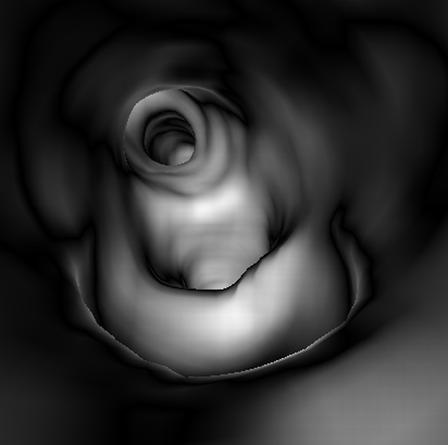
8 Non-calcified plaques Non-calcified plaques are commonly shown on VIE as smooth, protruding sign arising from the coronary wall (Fig 5). In order to demonstrate the non-calcified plaque in relation to the surrounding coronary artery ostia, attention should be paid to changing the CT threshold when the viewing position is moved from the normal coronary artery to the location where plaque is located, as CT attenuation of non-calcified plaque is much lower than that of contrast-enhanced blood in the coronary artery. Figure 6 shows a non-calcified plaque at the proximal segment of LAD on multiplanar reformatted image, while corresponding VIE of the plaque can only be seen clearly when the upper CT threshold is lowered from 140 to 60 HU. Calcified plaques Calcified plaques are usually shown as a protruding sign with smooth appearance on VIE. The position or location of the plaques in relation to the artery wall can be accurately assessed in terms of eccentric (Figs 7, 8) or concentric appearance (Figs 9, 10), or luminal changes regarding the involvement of superior or inferior portion of the wall (Figs 7, 8). Most of the calcified plaques are presented as smooth protruding sign on VIE as shown in Figures However, the presence of extensively calcified plaques along the coronary artery could result in irregular intraluminal changes in the coronary wall. Figure 11 is an example of VIE visualization of a patient with extensively calcified plaque at the LAD with irregular intralumina l appearances in the coronary wall. Mixed plaques 8
9 Irregular intraluminal appearances are commonly seen in mixed plaques on VIE because different components are contained within the plaques. This indicates that the coronary wall undergoes different stages of remodelling process, which consists of both stable stage involving formation of calcified plaques and unstable period involving deposition of non-calcified plaques which contain lipid-ric h components. Figure 12 is an example of mixed plaques at the proximal segment of LAD with noncalcified component representing the majority of the content, with corresponding VIE showing irregular intraluminal changes in the coronary wall caused by the mixed plaques. Figure 13 is another example of mixed plaques at the proximal segment of LAD with calcified component representing the majority of the content, and the corresponding VIE demonstrates irregular intraluminal changes. VIE visualization of coronary stents VIE visualization has been reported to serve as a potential tool for follow-up of endovascular stent grafting and stenting as it allows demonstration of the intraluminal views of the artery wall and stent surface [22, 23, 26-29]. Normal stent appearances Since CT attenuation of coronary stents is much higher than that of contrast-enhanced blood, stents are easily visualized on VIE as high density structure with uniform appearance inside the coronary arteries, depending on the location where the stents are deployed. Figure 14 shows a patent coronary stent placed in the right coronary artery with smooth circular appearance on VIE visualizat ion. Furthermore, VIE visualization also assists detection of stent position in relation to the coronary ostia 9
10 through providing 3D intraluminal views. Figure 15 is an example of left coronary stent which is placed at the proximal segment of LAD, with patency of the coronary ostia arising from the LAD. In-stent restenosis appearances In-stent restenosis appears as low-attenuation area within the high-density metallic stents on 2D CT images. On VIE visualization, the in-stent restenosis is demonstrated as irregular appearances on the coronary wall with subsequent coronary lumen stenosis. Figure 16 shows VIE visualization of in-stent restenosis in the LAD with irregular coronary wall change, while Figure 17 is another example with significant in-stent restenosis in the LAD with corresponding irregular coronary wall changes and lumen stenosis as visualized on VIE. Summary and conclusion In this pictorial essay, we have demonstrated a spectrum of VIE findings of coronary plaques and coronary stents inclusive of both normal lumen appearances and pathological changes caused by plaques or stents. Although VIE is not recommended as a routine tool for detection of coronary plaques or stents, it can be used as a complimentary approach to conventional visualizations for accurate assessment of coronary plaques and stents. 3D VIE visualization shows potential applications for assessment of coronary plaques in terms of plaque characterization and intraluminal appearances, while in coronary stenting, VIE can demonstrate the intralumina l coronary stents in relation to coronary ostia or detect luminal changes due to in-stent restenosis. VIE visualization is considered valuable for enhancing our understanding of the effect of coronary plaques and stents on coronary wall and subsequent clinical outcomes [21, 25, 30]. VIE 10
11 visualization tool presented in this review could contribute to overcome some of the image processing limitations for quantitative assessment of atherosclerotic plaques [31]. The main advantages of VIE visualization are summarised in the table, although further studies are needed to verify the VIE findings and associated clinical value. 11
12 Acknowledgements This work has been supported by research grant from Natural Science Foundation of China (grant ), grant for Excellent Talents from Beijing city (2011D ), and grant for High Levels of Health Technical Personnel in Beijing City Health System ( ). 12
13 References [1] Schuijf JD, Pundziute G, Jukema JW, et al. Diagnostic accuracy of 64-slice multislic e computed tomography in the non-invasive evaluation of significant coronary artery disease. Am J Cardiol 2006; 98: [2] Vanhoenacker P, Heijenbrok-Kal M, Van Heste R, et al. Diagnostic performance of multidetector CT angiography for assessment of coronary artery disease: meta-analysis. Radiology 2007; 244: [3] Sun Z, Lin C, Davidson R, Dong C, Liao Y. Diagnostic value of 64-slice CT angiography in coronary artery disease: A systematic review. Eur J Radiol 2008; 67: [4] Abdulla J, Abildstrom Z, Gotzsche O, Christensen E, Kober L, Torp-Pedersen C. 64-multis lic e detector computed tomography coronary angiography as potential alternative to conventional coronary angiography: a systematic review and meta-analysis. Eur Heart J 2007; 28: [5] Sun Z, Cao Y, Li HF. Multislice computed tomography angiography in the diagnosis of coronary artery disease. J Geriatr Cardiol 2011; 8: [6] Sun Z, Jiang W. Diagnostic value of multislice CT angiography in coronary artery disease: A meta-analysis. Eur J Radiol 2006; 60: [7] Xu L, Zhang Z. Coronary CT angiography with low radiation dose. Int J Cardiovasc Imaging 2010; Suppl 1: [8] Yang L, Zhou T, Zhang R, et al. Meta-analysis: diagnostic accuracy of coronary CT angiography with prospective ECG gating based on step-and-shoot, Flash, and volume modes for 13
14 detection of coronary artery disease. Eur Radiol 2014; May 28 (Epub ahead of print) DOI /s y [9] Sun Z, Ng KH. Diagnostic value of coronary CT angiography with prospective ECG-gating in the diagnosis of coronary artery disease: a systematic review and meta-analysis. Int J Cardiovasc Imaging 2012; 28: [10] von Ballmoos MW, Haring B, Juillert P, Alkadhi H. Meta-analysis: diagnostic performance of low-radiation-dose coronary computed tomography angiography. Ann Intern Med 2011; 154: [11] Rodriguez-Granillo GA, Rosales MA, Degrossi E, Durbano I, Rodriguez AE. Multislice CT coronary angiography for the detection of burden, morphology and distribution of atherosclerotic plaques in the left main bifurcation. Int J Cardiovasc Imaging 2007; 23: [12] Schmid M, Pflederer T, Jang IK, et al. Relationship between degree of remodelling and CT attenuation of plaque in coronary atherosclerotic lesions: an in-vivo analysis by multi-detector computed tomography. Atherosclerosis 2008; 197: [13] Motoyama S, Kondo T, Sarai M, et al. Multislice computed tomographic characteristics of coronary lesions in acute coronary syndromes. J Am Coll Cardiol 2007; 50: [14] Schuijf JD, Beck T, Burgstahler C, et al. Differences in plaque composition and distribution in stable coronary artery disease versus acute coronary syndromes; non-invasive evaluation with multislice computed tomography. Acute Cardiac Care 2007; 9: [15] Motoyama S, Sarai M, Harigaya H, et al. Computed tomographic angiography characteristics of atherosclerotic plaques subsequently resulting in acute coronary syndrome. J Am Coll Cardiol 2009; 54:
15 [16] Burke AP, Kolodgie FD, Farb A, Weber D, Virmani R. Morphological predictors of arterial remodelling in coronary atherosclerosis. Circulation 2012; 105: [17] Ehara S, Kobayashi Y, Yoshiyama M, et al. Spotty calcification typifies the culprit plaque in patients with acute myocardial infarction: an intravascular ultrasound study. Circulation 2004; 110: [18] Maurovich-Horvat P, Hoffmann U, Vorpahl M, Nakano M, Virmani R, Alkadhi H. The napkinring sign: CT signature of high-risk coronary plaques? JACC Cardiovasc Imaging 2010; 3 (4): [19] Sharif F, Murphy RT. Current status of vulnerable plaque detection. Catheter Cardiovasc Interv 2010; 75: [20] Sun Z, Zheng H. Effect of suprarenal stent grafts on the renal artery with ostial calcification observed on CT virtual intravascular endoscopy. Eur J Vasc Endovasc Surg 2004; 28: [21] Sun Z, Dimpudus FJ, Nugroho J, Adipranoto JD. CT virtual intravascular endoscopy assessment of coronary artery plaques: A preliminary study. Eur J Radiol 2010; 75: e112-e119 [22] Sun Z. multislice CT angiography in post-aortic stent grafting: A pictorial essay. Korean J Radiol 2006; 7: [23] Sun Z, Allen YB, Nadkarni S, Knight R, Hartley DE, Lawrence-Brown MM. CT virtual intravascular endoscopy in the visualization of fenestrated stent-grafts. J Endovasc Ther 2008; 15: [24] Sun Z, Al Dosari S, Ng C, al-mumntshari A, Almaliky S. Multislice CT virtual intravascular endoscopy of pulmonary embolism: A pictorial review. Korean J Radiol 2010; 11:
16 [25] Sun Z, Cao Y. Multislice CT angiography assessment of left coronary artery: correlation between bifurcation angle and dimensions and development of coronary artery disease. Eur J Radiol 2011; 79: e90-e95 [26] Sun Z, Winder RJ, Kelly BE, Ellis PK, Kennedy PT, Hirst DG. Diagnostic value of CT virtual intravascular endoscopy in aortic stent grafting. J Endovasc Ther 2004; 11(1): [27] Sun Z, Winder RJ, Kelly BE, Ellis PK, Hirst DG. CT virtual intravascular endoscopy of abdominal aortic aneurysms treated with suprarenal endovascular stent grafting. Abdom Imaging 2003; 28: [28] Sun Z, Gallagher E. Multislice CT virtual intravascular endoscopy for abdominal aortic aneurysm stent grafts. J Vasc Interv Radiol 2004; 15: [29] Almutairi A, Sun Z, Ng C, Al-Safran ZA, Al-Mulla AA, Al-Jamaan AI. Optimal scanning protocols of 64-slice CT angiography in coronary artery stents: An in vitro phantom study. Eur J Radiol 2010; 74(1): [30] Sun Z. Coronary CT angiography in coronary artery disease: correlation between virtual intravascular endoscopic appearances and left bifurcation angulation and coronary plaques. Biomed Res Int 2013; 2013: [31] Jodas DS, Pereira AS, Tavares JRMS. A review of computational method applied for identification and quantification of atherosclerotic plaques in images. Expert Sys Appl 2016;46:


17 Figure legends 17

18 Figure 1. VIE visualization of coronary wall and plaque depends on appropriate selection of CT threshold. Figures A to H are a series of VIE images viewing the LAD and LCx ostia a well as coronary plaque arising from the LAD with selection of CT threshold of 160 HU, 180 HU, 200 HU, 220 HU, 240 HU, 260 HU, 280 HU, 300 HU, respectively. As shown in the images, when upper CT threshold is lower than 200 HU, artifact (arrow) appears in the coronary wall. When upper CT threshold is higher than 240 HU, visualization of coronary plaque and coronary ostia is affected. Thus, the appropriate CT threshold for visualization of coronary ostia and plaque is between 200 and 220 HU. 18


19 Figure 2. Correlation of VIE visualization of coronary anatomy and plaque with orthogonal views. A. The viewing position is placed at the coronary plaque in the LAD ostium, and confirmation of the ostium is determined by checking the axial, coronal and sagittal views. B. The viewing position is focused on the LCx ostium which is confirmed by checking these orthogonal view Figure 3. VIE visualization of normal left coronary ostia. A. VIE view of the left main coronary artery. B and C. VIE demonstrates the left coronary ostia with regard to their intraluminal relationship. 19

 at")

20 Figure 4. VIE visualization of normal right coronary ostium with regular smooth appearance. Figure 5. VIE visualization of non-calcified plaque. A. Curved planar reformatted image shows noncalcified plaque (arrow) at the proximal segment of LAD in a 71-year-old man. B. Corresponding VIE shows the plaque arising from the inferior wall of LAD with smooth appearance, with involvement of the ramus intermedius ostium (arrow). 20

 at the")
 could not be clearly assessed when")

21 Figure 6. VIE visualization of non-calcified plaque with changing CT threshold for clear demonstration of coronary wall and plaque appearances. A. Curved planar reformatted image shows non-calcified plaque (arrow) at the proximal segment of LAD in a 59-year old male. B. Both coronary plaque and LAD ostium (arrows) could not be clearly assessed when the upper CT threshold is selected at 130 HU. C. When the upper CT threshold is reduced from 130 HU to 60 HU, the plaque (arrows) and LAD ostia are clearly visualized on VIE. D. VIE close view (arrows) indicates the relationship between plaque and the LAD ostium with upper CT threshold set at 60 HU. 21


. Figure 8.")

. C.")
22 Figure 7. VIE visualization of eccentric calcified plaques. A. Curved planar reformatted image reveals calcified plaques at the left main stem and LAD coronary arteries in an 80-year-old male. B. VIE shows the calcified plaque (arrows) arising from the superior wall of LAD with involvement of LAD ostium. C. VIE close view shows that the plaque surrounds the ramus intermedium ostium (arrow). Figure 8. VIE visualization of eccentric calcified plaques. A. 2D axial image shows calcified plaques mainly locating in the superior wall of LAD in a 73-year-old female. B. VIE demonstrates a huge intraluminal plaque occupying the LAD ostium (arrows). C. VIE view of distal LAD reveals irregular coronary wall (arrows) caused by the extensively calcified plaque. 22


 surround the LAD osium. Figure 10.")

23 Figure 9. VIE visualization of concentric calcified plaques. A. Curved planar reformatted image shows calcified plaques at the proximal segment of LAD in a 61-year-old man. B. VIE confirms that the concentrically calcified plaques (arrows) surround the LAD osium. Figure 10. VIE visualization of concentric calcified plaque. A. Curved planar reformatted image shows highly calcified plaque at the proximal segment of LAD in a 49-year-old woman. B. VIE shows 23


24 significant stenosis of LAD caused by the calcified plaque (arrow). C. VIE close view of the concentrically calcified plaque covering the LAD ostium. Figure 11. VIE visualization of extensively calcified plaques. A. Curved planar reformatted image shows extensively calcified plaques in the LAD with some low-attenuation areas representing noncalcified component in a 58-year-old male. B. Corresponding VIE demonstrates irregular coronary lumen changes (arrows) caused by the extensively calcified plaques. 24


 due to")
25 Figure 12. VIE visualization of mixed plaques. A. Curved planar reformatted image shows mixed plaques with spotty calcification at the proximal segment of LAD in a 68-year-old male. B. VIE indicates irregular coronary wall changes (arrows) due to different components within the plaques. 25


26 Figure 13. VIE visualization of mixed plaques. A. Curved planar reformatted image shows mixed plaques with calcified components clearly present within the plaques in a 55-year-old male. B. VIE demonstrates irregular coronary wall due to presence of mixed plaques. Figure 14. VIE visualization of patent coronary stents. A. Curved planar reformatted image shows a patent coronary stent in the RCA in a 69-year-old male. B. VIE visualization of the coronary stent when the view is positioned at the proximal segment of RCA. C. Close VIE visualization of stent surface shows smooth circular appearance inside the RCA. 26


27 Figure 15. VIE visualization of coronary stent in relation to the coronary ostia. A. Curved planar reformatted image shows a patent coronary stent in the LAD in a 47-year-old man. B. VIE indicates the circular smooth appearance of coronary stent relative to the LAD ostium (arrows). Figure 16. VIE visualization of coronary in-stent restenosis. A. Curved planar reformatted image shows low-attenuation area (arrows) with suspected in-stent restenosis in the LAD stent in a 65-yearold man. B and C. Distal and proximal VIE views of the LAD lumen confirm the irregular wall changes (arrows) due to in-stent restenosis. Figure 17. VIE visualization of coronary in-stent restenosis. A. Curved planar reformatted image demonstrates low-attenuation area (arrows) in the LAD stent in a 50-year-old woman. B. VIE confirms 27
28 significant lumen stenosis with irregular wall changes (arrows). C. The coronary stent in the LCx remains patent on VIE visualization. 28
Cardiac CT imaging in coronary artery disease: Current status and future directions
 Research Highlight Cardiac CT imaging in coronary artery disease: Current status and future directions Zhonghua Sun Discipline of Medical Imaging, Department of Imaging and Applied Physics, Curtin University,
Impact of SSF on diagnostic performance of coronary CT angiography within one heart beat in patients with high heart rate using a 256-row detector CT
 Impact of SSF on diagnostic performance of coronary CT angiography within one heart beat in patients with high heart rate using a 256-row detector CT Junfu Liang 1,2, Hui Wang 1, Lei Xu 1, Li Dong 1, Zhanming
Multislice CT angiography in abdominal aortic aneurysm treated with endovascular stent grafts: evaluation of 2D and 3D visualisations
 Available online at http://www.biij.org/2007/4/e20 doi: 10.2349/biij.3.4.e20 biij Biomedical Imaging and Intervention Journal ORIGINAL ARTICLE Multislice CT angiography in abdominal aortic aneurysm treated
M Marwan, D Ropers, T Pflederer, W G Daniel, S Achenbach
 Department of Cardiology, University of Erlangen, Erlangen, Germany Correspondence to: Dr M Marwan, Innere Medizin II, Ulmenweg 18, 91054 Erlangen, Germany; mohamed.marwan@ uk-erlangen.de Accepted 17 November
CT Imaging of Atherosclerotic Plaque. William Stanford MD Professor-Emeritus Radiology University of Iowa College of Medicine Iowa City, IA
 CT Imaging of Atherosclerotic Plaque William Stanford MD Professor-Emeritus Radiology University of Iowa College of Medicine Iowa City, IA PREVALENCE OF CARDIOVASCULAR DISEASE In 2006 there were 80 million
Zhonghua Sun, PhD 1 Bibombe P Mwipatayi, MMed 2 Yvonne B Allen, RN 3 David E Hartley, FIR 3 Michael M Lawrence-Brown, FRACS 4
 Multislice CT ngiography of Fenestrated Endovascular Stent Grafting for Treating bdominal ortic neurysms: a Pictorial Review of the 2D/3D Visualizations Zhonghua Sun, PhD 1 ibombe P Mwipatayi, MMed 2 Yvonne
Ultrasound. Computed tomography. Case studies. Utility of IQon Spectral CT in. cardiac imaging
 Ultrasound Computed tomography Case studies Utility of IQon Spectral CT in cardiac imaging Cardiac imaging is a challenging procedure where it is necessary to image a motion-free heart. This requires a
The diagnostic evaluation of dual-source CT (DSCT) in the diagnosis of coronary artery stenoses
 in the diagnosis of coronary artery stenoses") Original Article Open Access The diagnostic evaluation of dual-source CT (DSCT) in the diagnosis of coronary artery stenoses Ziqiao Lei 1, Jin Gu 2, Qing Fu 3, Heshui Shi 4, Haibo Xu 5, Ping Han 6, Jianming
Original Article Open Access The diagnostic evaluation of dual-source CT (DSCT) in the diagnosis of coronary artery stenoses Ziqiao Lei 1, Jin Gu 2, Qing Fu 3, Heshui Shi 4, Haibo Xu 5, Ping Han 6, Jianming
Chapter 4. Department of Cardiology, Leiden University Medical Center, Leiden, The Netherlands. Department of Radiology,
 Chapter 4 Impact of Coronary Calcium Score on Diagnostic Accuracy of Multislice Computed Tomography Coronary Angiography for Detection of Coronary Artery Disease Gabija Pundziute, 1,3 Joanne D. Schuijf,
What every radiologist should know about cardiac CT: A case-based pictorial review
 What every radiologist should know about cardiac CT: A case-based pictorial review Poster No.: C-0555 Congress: ECR 2010 Type: Educational Exhibit Topic: Cardiac Authors: C. M. Capuñay, P. Carrascosa,
Coronary Artery Imaging. Suvipaporn Siripornpitak, MD Inter-hospital Conference : Rajavithi Hospital
 Coronary Artery Imaging Suvipaporn Siripornpitak, MD Inter-hospital Conference : Rajavithi Hospital Larger array : cover scan area Detector size : spatial resolution Rotation speed : scan time Retrospective
Diagnostic accuracy of dual-source computed tomography in the detection of coronary chronic total occlusion: Comparison with invasive angiography
 African Journal of Biotechnology Vol. 10(19), pp. 3854-3858, 9 May, 2011 Available online at http://www.academicjournals.org/ajb DOI: 10.5897/AJB10.983 ISSN 1684 5315 2011 Academic Journals Full Length
Improvement of Image Quality with ß-Blocker Premedication on ECG-Gated 16-MDCT Coronary Angiography
 16-MDCT Coronary Angiography Shim et al. 16-MDCT Coronary Angiography Sung Shine Shim 1 Yookyung Kim Soo Mee Lim Received December 1, 2003; accepted after revision June 1, 2004. 1 All authors: Department
Chapter 43 Noninvasive Coronary Plaque Imaging
 hapter 43 Noninvasive oronary Plaque Imaging NIRUDH KOHLI The goal of coronary imaging is to define the extent of luminal narrowing as well as composition of an atherosclerotic plaque to facilitate appropriate
Triple Rule-out using 320-row-detector volume MDCT: A comparison of the wide volume and helical modes
 Triple Rule-out using 320-row-detector volume MDCT: A comparison of the wide volume and helical modes Poster No.: C-0488 Congress: ECR 2012 Type: Authors: Keywords: DOI: Scientific Exhibit E.-J. Kang,
Eur Heart J. 2011;32:637-45
 Diagnostic Performance of Non-Invasive Multidetector Computed Tomography Coronary Angiography to Detect Coronary Artery Disease using Different Endpoints: Detection of Significant Stenosis versus Detection
Scientific Exhibit. Authors: D. Takenaka, Y. Ohno, Y. Onishi, K. Matsumoto, T.
 The feasibility of biphasic contrast-media-injection-protocol for chest imaging on 320-slice volume MDCT: Direct comparison of biphasic and bolus contrast-media injection protocols on 320-slice volume
TITLE: Multi-Slice Computed Tomography Coronary Angiography for Coronary Artery Disease: A Review of the Clinical Effectiveness and Guidelines
 TITLE: Multi-Slice Computed Tomography Coronary Angiography for Coronary Artery Disease: A Review of the Clinical Effectiveness and Guidelines DATE: 25 February 2009 CONTEXT AND POLICY ISSUES: Coronary
The Final 10-Year Follow-up Results from the Bari Randomized Trial J Am Coll Cardiol (2007) 49;1600-6
 49;1600-6") The Final 10-Year Follow-up Results from the Bari Randomized Trial J Am Coll Cardiol (2007) 49;1600-6 n&list_uids=17433949 64-Multislice Detector Computed Tomography Coronary Angiography as Potential Alternative
The Final 10-Year Follow-up Results from the Bari Randomized Trial J Am Coll Cardiol (2007) 49;1600-6 n&list_uids=17433949 64-Multislice Detector Computed Tomography Coronary Angiography as Potential Alternative
CT angiography techniques. Boot camp
 CT angiography techniques Boot camp Overview Basic concepts Contrast administration arterial opacification Time scan acquisition during the arterial phase Protocol examples Helical non-gated CTA Pulmonary
128-slice dual-source CT coronary angiography using highpitch scan protocols in 102 patients
 128-slice dual-source CT coronary angiography using highpitch scan protocols in 102 patients Poster No.: C-0634 Congress: ECR 2010 Type: Scientific Exhibit Topic: Cardiac Authors: Y. H. Choe, J. W. Lee,
Computer Aided Detection and Diagnosis: Cardiac Imaging Applications
 Computer Aided Detection and Diagnosis: Cardiac Imaging Applications U. Joseph Schoepf, MD, FAHA, FSCBT MR, FSCCT Professor of Radiology, Medicine, and Pediatrics Director of Cardiovascular Imaging Disclosures
Endovascular aneurysm repair alters renal artery movement: a preliminary evaluation using dynamic CTA.
 3/1/2007 o/06-3928 muss diss pag 77 Endovascular aneurysm repair alters renal artery movement: a preliminary evaluation using dynamic CTA. 6 Bart E. Muhs MD 1, Arno Teutelink MD 1, Matthias Prokop MD,
Diagnostic value of 320-slice coronary CT angiography in coronary artery disease: A
 Diagnostic value of 320-slice coronary CT angiography in coronary artery disease: A systematic review and meta-analysis Zhonghua Sun PhD 1, Chenghsun Lin PhD 2 1. Discipline of Medical Imaging, Department
Characteristics of Subclinical Coronary Artery Disease in Diabetic Patients without Known Coronary Artery Disease
 IBIMA Publishing Journal of Research in Diabetes http://www.ibimapublishing.com/journals/diab/diab.html Vol. 2014 (2014), Article ID 322292, 12 pages DOI: 10.5171/2014.322292 Research Article Characteristics
Evidence for myocardial CT perfusion imaging in the diagnosis of hemodynamically significant coronary artery disease
 Editorial Evidence for myocardial CT perfusion imaging in the diagnosis of hemodynamically significant coronary artery disease Zhonghua Sun Discipline of Medical Imaging, Department of Imaging and Applied
Endovascular Aneurysm Repair Alters Renal Artery Movement: A Preliminary Evaluation Using Dynamic CTA
 476 J ENDOVASC THER CLINICAL INVESTIGATION Endovascular Aneurysm Repair Alters Renal Artery Movement: A Preliminary Evaluation Using Dynamic CTA Bart E. Muhs, MD 1 ; Arno Teutelink, MD 1 ; Matthias Prokop,
Simon Nepveu 1, Irina Boldeanu 1, Yves Provost 1, Jean Chalaoui 1, Louis-Mathieu Stevens 2,3, Nicolas Noiseux 2,3, Carl Chartrand-Lefebvre 1,3
 Coronary Artery Bypass Graft Imaging with CT Angiography and Iterative Reconstruction: Quantitave Evaluation of Radiation Dose Reduction and Image Quality Simon Nepveu 1, Irina Boldeanu 1, Yves Provost
Low Dose Era in Cardiac CT
 Low Dose Era in Cardiac CT DIANA E. LITMANOVICH, MD Department of Radiology Beth Israel Deaconess Medical Center Harvard Medical School Disclosures Neither I nor my immediate family members have a financial
Coronary Artery Disease - Reporting and Data System (CAD-RADS)
") A joint publication of the Department of Radiology and Corrigan Minehan Heart Center November 2016 Issue 66 Coronary Artery Disease - Reporting and Data System (CAD-RADS) Sandeep S. Hedgire, MD; Udo Hoffmann,
A joint publication of the Department of Radiology and Corrigan Minehan Heart Center November 2016 Issue 66 Coronary Artery Disease - Reporting and Data System (CAD-RADS) Sandeep S. Hedgire, MD; Udo Hoffmann,
Computed Tomography of the Coronary Arteries
 Cardiology Update DAVOS 2011 Computed Tomography of the Coronary Arteries Anders Persson M.D., Ph.D Director, Assoc. Professor Center for Medical Image Science and Visualization Linköping University SWEDEN
THE ROLE OF HIGH END MULTI DETECTOR CT IN CORONARY IMAGING ESSAY
 THE ROLE OF HIGH END MULTI DETECTOR CT IN CORONARY IMAGING ESSAY Submitted for partial fulfillment of Master degree in Radiodiagnosis By Ahmed Yehia Ahmed (M.B.B.Ch., Cairo University) Supervisors Prof.
Multislice coronary computed tomographic angiography in emergency department presentations of unsuspected acute myocardial infarction
 Journal of Cardiovascular Computed Tomography (2009) 3, 272 278 Case Report Multislice coronary computed tomographic angiography in emergency department presentations of unsuspected acute myocardial infarction
Calcium Scoring and Cardiac CT
 Calcium Scoring and Cardiac CT John C. Finley, MD, FACC, FASE Medical Director, CT Department; Alaska Heart and Vascular Institute February 9, 2018 1. Calcium Scoring 2. CT Coronary Angiography 3. Use
Cardiac Computed Tomography
 Cardiac Computed Tomography Authored and approved by Koen Nieman Stephan Achenbach Francesca Pugliese Bernard Cosyns Patrizio Lancellotti Anastasia Kitsiou Contents CARDIAC COMPUTED TOMOGRAPHY Page 1.
Lei Zhao 1,2, Fabian Plank 2, Moritz Kummann 2, Philipp Burghard 2, Andrea Klauser 2, Wolfgang Dichtl 3, Gudrun Feuchtner 2.
 Original Article Improved non-calcified plaque delineation on coronary CT angiography by sonogram-affirmed iterative reconstruction with different filter strength and relationship with BMI Lei Zhao 1,2,
Utility of CT angiography for pre-operative evaluation of robotic-assisted minimally invasive mitral valve surgery.
 Utility of CT angiography for pre-operative evaluation of robotic-assisted minimally invasive mitral valve surgery. Poster No.: C-2214 Congress: ECR 2014 Type: Educational Exhibit Authors: M. Muthuvelu,
Assessment of Non-Calcified Coronary Plaques Using 64-Slice Computed Tomography: Comparison With Intravascular Ultrasound
 ORIGINAL ARTICLE DOI 10.4070 / kcj.2009.39.3.95 Print ISSN 1738-5520 / On-line ISSN 1738-5555 Copyright c 2009 The Korean Society of Cardiology Assessment of Non-Calcified Coronary Plaques Using 64-Slice
Impact of Body Mass Index and Metabolic Syndrome on the Characteristics of Coronary Plaques Using Computed Tomography Angiography
 Impact of Body Mass Index and Metabolic Syndrome on the Characteristics of Coronary Plaques Using Computed Tomography Angiography Cardiovascular Division, Faculty of Medicine, University of Tsukuba Akira
Computed tomography in coronary imaging: current status
 7 Computed tomography in coronary imaging: current status ARJUN NAIR AND ANAND DEVARAJ Recent technological advances have led to improvements in the use of computerised tomography for coronary imaging.
Imaging ischemic heart disease: Role of CCTA
 Plenary Session II Ischemic Heart Disease Imaging ischemic heart disease: Role of CCTA Florian Wolf Medical University of Vienna Department of Biomedical Imaging and Image Guided Therapy Division of Cardi0vascular
CARDIAC IMAGING FOR SUBCLINICAL CAD
 CARDIAC IMAGING FOR SUBCLINICAL CAD WHY DON'T YOU ADOPT MORE SMART TECHNIQUE? Whal Lee, M.D. Seoul National University Hospital Department of Radiology We are talking about Coronary artery Calcium scoring,
Disclosure Information
 Coronary CTA Pearls and Pitfalls Ricardo C. Cury, MD, FSCCT, FAHA, FACC Chairman of Radiology Radiology Associates of South Florida Director of Cardiac Imaging Miami Cardiac and Vascular Institute Past-President
The diagnosis of coronary plaque stability by multi-slice computed tomography coronary angiography
 Original Article The diagnosis of coronary plaque stability by multi-slice computed tomography coronary angiography Feng-Xiang Song 1 *, Jun Zhou 2 *, Jian-Jun Zhou 3, Yu-Xin Shi 1, Meng-Su Zeng 3, Zhi-Yong
Left main coronary artery (LMCA): The proximal segment
: The proximal segment") Anatomy and Pathology of Left main coronary artery G Nakazawa Tokai Univ. Kanagawa, Japan 1 Anatomy Difinition Left main coronary artery (LMCA): The proximal segment RCA AV LAD LM LCX of the left coronary
Anatomy and Pathology of Left main coronary artery G Nakazawa Tokai Univ. Kanagawa, Japan 1 Anatomy Difinition Left main coronary artery (LMCA): The proximal segment RCA AV LAD LM LCX of the left coronary
Index. radiologic.theclinics.com. Note: Page numbers of article titles are in boldface type.
 Index Note: Page numbers of article titles are in boldface type. A ALCAPA. See Anomalous left coronary artery from the pulmonary artery. Angiosarcoma computed tomographic assessment of, 809 811 Anomalous
Journal of the American College of Cardiology Vol. 47, No. 8, by the American College of Cardiology Foundation ISSN /06/$32.
 Journal of the American College of Cardiology Vol. 47, No. 8, 2006 2006 by the American College of Cardiology Foundation ISSN 0735-1097/06/$32.00 Published by Elsevier Inc. doi:10.1016/j.jacc.2006.01.041
Original Article Application of flash dual-source CT at low radiation dose and low contrast medium dose in triple-rule-out (tro) examination
 examination") Int J Clin Exp Med 2015;8(11):21898-21905 www.ijcem.com /ISSN:1940-5901/IJCEM0015005 Original Article Application of flash dual-source CT at low radiation dose and low contrast medium dose in triple-rule-out
Int J Clin Exp Med 2015;8(11):21898-21905 www.ijcem.com /ISSN:1940-5901/IJCEM0015005 Original Article Application of flash dual-source CT at low radiation dose and low contrast medium dose in triple-rule-out
3D multislice CT angiography in post aortic stent grafting: A pictorial essay
 3D multislice CT angiography in post aortic stent grafting: A pictorial essay Zhonghua Sun PhD Lecturer in Medical Imaging, Discipline of Medical Imaging, Department of Imaging and Applied Physics, Curtin
Performance of dual-source CT with high pitch spiral mode for coronary stent patency compared with invasive coronary angiography
 Journal of Geriatric Cardiology (2016) 13: 817 823 2016 JGC All rights reserved; www.jgc301.com Research Article Open Access Performance of dual-source CT with high pitch spiral mode for coronary stent
Feasibility of contrast agent volume reduction on 640-slice CT coronary angiography in patients with low heart rate
 Feasibility of contrast agent volume reduction on 640-slice CT coronary angiography in patients with low heart rate Poster No.: B-0742 Congress: ECR 2013 Type: Authors: Keywords: DOI: Scientific Paper
Imaging congestive heart failure: role of coronary computed tomography angiography (CCTA)
") Imaging congestive heart failure: role of coronary computed tomography angiography (CCTA) Gianluca Pontone, MD, PhD, FESC, FSCCT Director of MR Unit Deputy Director of Cardiovascul CT Unit Clinical Cardiology
Imaging congestive heart failure: role of coronary computed tomography angiography (CCTA) Gianluca Pontone, MD, PhD, FESC, FSCCT Director of MR Unit Deputy Director of Cardiovascul CT Unit Clinical Cardiology
Fundamentals, Techniques, Pitfalls, and Limitations of MDCT Interpretation and Measurement
 Fundamentals, Techniques, Pitfalls, and Limitations of MDCT Interpretation and Measurement 3 rd Annual Imaging & Physiology Summit November 20-21, 21, 2009 Seoul, Korea Wm. Guy Weigold, MD, FACC Cardiovascular
Diagnosis of coronary stenosis with CT angiography comparison of automated computer diagnosis with expert readings.
 Thomas Jefferson University Jefferson Digital Commons Department of Radiology Faculty Papers Department of Radiology 3-1-2011 Diagnosis of coronary stenosis with CT angiography comparison of automated
Jinling Zhang, 1 Zhehao Lv, 1 Deli Zhao, 1 Lili Liu, 1 Yong Wan, 1 Tingting Fan, 1 Huimin Li, 1 Ying Guan, 1 Bailu Liu, 1 and Qi Yang 2,3
 Diabetes Research Volume 2016, Article ID 4365156, 6 pages http://dx.doi.org/10.1155/2016/4365156 Research Article Coronary Plaque Characteristics Assessed by 256-Slice Coronary CT Angiography and Association
Coronary revascularization treatment based on dual-source computed tomography
 Eur Radiol (2008) 18: 1800 1808 DOI 10.1007/s00330-008-0959-0 CARDIAC R. Dikkers T. P. Willems L. H. Piers G. J. de Jonge R. A. Tio H. J. van der Zaag-Loonen P. M. A. van Ooijen F. Zijlstra M. Oudkerk
Utility of Variable Helical Pitch CT Scanning Technique for CT Angiography of Aortoiliac and Lower Extremity Arteries
 Utility of Variable Helical Pitch CT Scanning Technique for CT Angiography of Aortoiliac and Lower Extremity Arteries Poster No.: C-0863 Congress: ECR 2015 Type: Scientific Exhibit Authors: A. Nakamoto,
SYMPOSIA. Coronary CTA. Indications, Patient Selection, and Clinical Implications
 SYMPOSIA Indications, Patient Selection, and Clinical Implications Christian Thilo, MD,* Mark Auler, MD,* Peter Zwerner, MD,w Philip Costello, MD,* and U. Joseph Schoepf, MD* Abstract: Recent technical
Hospital, 6 Lukon Road, Lukong Town, Changhua Shien, Taiwan 505, Taiwan.
 Volume 1, Issue 1 Image Article Resolution of Inferior Wall Ischemia after Successful Revascularization of LAD Lesion: The Value of Myocardial Perfusion Imaging in Guiding Management of Multi-vessel CAD
Radiation Dose Reduction and Coronary Assessability of Prospective Electrocardiogram-Gated Computed Tomography Coronary Angiography
 Journal of the American College of Cardiology Vol. 52, No. 18, 2008 2008 by the American College of Cardiology Foundation ISSN 0735-1097/08/$34.00 Published by Elsevier Inc. doi:10.1016/j.jacc.2008.07.048
CT or PET/CT for coronary artery disease
 CT or PET/CT for coronary artery disease Rotterdam 2012 Juhani Knuuti, MD, PhD, FESC Turku PET Centre University of Turku Turku, Finland Juhani.knuuti@utu.fi Turku PET Centre University of Turku Åbo Akademi
Coronary CT angiography with prospective ECG-triggering: an effective alternative to invasive coronary angiography
 Review Article Coronary CT angiography with prospective ECG-triggering: an effective alternative to invasive coronary angiography Zhonghua Sun Discipline of Medical Imaging, Department of Imaging and Applied
RAMA-EGAT Risk Score for Predicting Coronary Artery Disease Evaluated by 64- Slice CT Angiography
 RAMA-EGAT Risk Score for Predicting Coronary Artery Disease Evaluated by 64- Slice CT Angiography Supalerk Pattanaprichakul, MD 1, Sutipong Jongjirasiri, MD 2, Sukit Yamwong, MD 1, Jiraporn Laothammatas,
Pushing the limits of cardiac CT. Steven Dymarkowski Radiology / Medical Imaging Research Centre
 Pushing the limits of cardiac CT Steven Dymarkowski Radiology / Medical Imaging Research Centre 5 X 2013 Introduction Rapid technological advances and new clinical applications in cardiovascular imaging
General Imaging. Imaging modalities. Incremental CT. Multislice CT Multislice CT [ MDCT ]
![General Imaging. Imaging modalities. Incremental CT. Multislice CT Multislice CT [ MDCT ]](/thumbs/76/74079340.jpg "General Imaging. Imaging modalities. Incremental CT. Multislice CT Multislice CT [ MDCT ]") General Imaging Imaging modalities Conventional X-rays Ultrasonography [ US ] Computed tomography [ CT ] Radionuclide imaging Magnetic resonance imaging [ MRI ] Angiography conventional, CT,MRI Interventional
General Imaging Imaging modalities Conventional X-rays Ultrasonography [ US ] Computed tomography [ CT ] Radionuclide imaging Magnetic resonance imaging [ MRI ] Angiography conventional, CT,MRI Interventional
Studies with electron beam computed tomography (EBCT) Imaging
 Imaging") Imaging Predictive Value of 16-Slice Multidetector Spiral Computed Tomography to Detect Significant Obstructive Coronary Artery Disease in Patients at High Risk for Coronary Artery Disease Patient- Versus
Imaging Predictive Value of 16-Slice Multidetector Spiral Computed Tomography to Detect Significant Obstructive Coronary Artery Disease in Patients at High Risk for Coronary Artery Disease Patient- Versus
A new method for radiation dose reduction at cardiac CT with multi-phase data-averaging and non-rigid image registration: preliminary clinical trial
 A new method for radiation dose reduction at cardiac CT with multi-phase data-averaging and non-rigid image registration: preliminary clinical trial Poster No.: C-0595 Congress: ECR 2013 Type: Authors:
Recent developments in cardiac CT
 REVIEW Recent developments in cardiac CT With the introduction of 64-multidetector row CT, coronary CT angiography has become a clinical tool, owing to improved image quality and reduced breath-hold time,
Diagnostic value of coronary CT angiography with prospective ECG-gating in the
 Diagnostic value of coronary CT angiography with prospective ECG-gating in the diagnosis of coronary artery disease: A systematic review and meta-analysis Zhonghua Sun PhD 1, Kwan-Hoong Ng PhD 2 1. Discipline
Is computed tomography angiography really useful in. of coronary artery disease?
 Is computed tomography angiography really useful in screening patients with high risk of coronary artery disease? Myeong-Ki Hong, M.D. Ph D Professor of Medicine Division of Cardiology, Severance Cardiovascular
2004;77:800 4 MSCT OF CORONARY ARTERY BYPASS GRAFTS. Results. CABG With Adequate Diagnostic Quality
 Isotropic Half-Millimeter Angiography of Coronary Artery Bypass Grafts With 16-Slice Computed Tomography Marc Dewey, MD, Alexander Lembcke, MD, Christian Enzweiler, MD, Bernd Hamm, MD, and Patrik Rogalla,
B-Flow, Power Doppler and Color Doppler Ultrasound in the Assessment of Carotid Stenosis: Comparison with 64-MD-CT Angiography
 Med. J. Cairo Univ., Vol. 85, No. 2, March: 805-809, 2017 www.medicaljournalofcairouniversity.net B-Flow, Power Doppler and Color Doppler Ultrasound in the Assessment of Carotid Stenosis: Comparison with
Noninvasive Coronary Imaging: Plaque Imaging by MDCT
 Coronary Physiology & Imaging Summit 2007 Noninvasive Coronary Imaging: Plaque Imaging by MDCT Byoung Wook Choi Department of Radiology Yonsei University, Seoul, Korea Stary, H. C. et al. Circulation
Interventional Cardiology. Research article
 Interventional Cardiology Computational fluid dynamic analysis of calcified coronary plaques: correlation between hemodynamic s and cardiac image analysis based on left coronary bifurcation angle and lumen
Isolated congenital coronary anomalies: Evaluation by multislice-ct or MRI
 Isolated congenital coronary anomalies: Evaluation by multislice-ct or MRI B.K. Velthuis, Dept. of Radiology UMC Utrecht, the Netherlands ESC 2010 Coronary artery anomalies CAA Uncommon 0.3-5% normal population
Dual Energy CT of the Heart: Perfusion and Beyond
 Dual Energy CT of the Heart: Perfusion and Beyond U. Joseph Schoepf, MD, FAHA, FSCBT MR, FSCCT Professor of Radiology, Medicine, and Pediatrics Director of Cardiovascular Imaging Disclosures Consultant
IVUS Virtual Histology. Listening through Walls D. Geoffrey Vince, PhD The Cleveland Clinic Foundation
 IVUS Virtual Histology Listening through Walls D. Geoffrey Vince, PhD Disclosure VH is licenced to Volcano Therapeutics Grant funding from Pfizer, Inc. Grant funding from Boston-Scientific Most Myocardial
CT Myocardial Perfusion: Is there Added Value to Coronary CT?
 CT Myocardial Perfusion: Is there Added Value to Coronary CT? U. Joseph Schoepf, MD, FAHA, FSCBT MR, FSCCT Professor of Radiology, Medicine, and Pediatrics Director of Cardiovascular Imaging Disclosures
EXPERIMENTAL AND THERAPEUTIC MEDICINE 9: , 2015
 2384 Dose study of electrocardiogram automatic tube current modulation technology in prospective coronary computed tomography angiography scans of overweight patients GUIRU HE, XIAOPEI LIU, YAN LIU, WEI
Calcium scoring using 64-slice MDCT, dual source CT and EBT: a comparative phantom study
 Int J Cardiovasc Imaging (2008) 24:547 556 DOI 10.1007/s10554-007-9282-0 ORIGINAL PAPER Calcium scoring using 64-slice MDCT, dual source CT and EBT: a comparative phantom study Jaap M. Groen Æ Marcel J.
The 5th Congress of Asian Society of Cardiovascular Imaging
 The 5th Congress of Asian Society of Cardiovascular Imaging June 17 ~ June 19, 2011 HKEC, Hong Kong Number of Participants : 1,032 Country No. of Delegate Country No. of Delegate 1 Australia 15 18 Korea,
Coronary Artery Anomalies from Birth to Adulthood; the Role of CT Coronary Angiography in Sudden Cardiac Death Screening
 Coronary Artery Anomalies from Birth to Adulthood; the Role of CT Coronary Angiography in Sudden Cardiac Death Screening E O Dwyer 1, C O Brien 1, B Loo 1, A Snow Hogan 1, O Buckley1 2, B 1. Department
Culprit Lesion Remodeling and Long-term (> 5years) Prognosis in Patients with Acute Coronary Syndrome
 Prognosis in Patients with Acute Coronary Syndrome") Culprit Lesion Remodeling and Long-term (> 5years) Prognosis in Patients with Acute Coronary Syndrome Hiroyuki Okura*, MD; Nobuya Matsushita**,MD Kenji Shimeno**, MD; Hiroyuki Yamaghishi**, MD Iku Toda**,
Culprit Lesion Remodeling and Long-term (> 5years) Prognosis in Patients with Acute Coronary Syndrome Hiroyuki Okura*, MD; Nobuya Matsushita**,MD Kenji Shimeno**, MD; Hiroyuki Yamaghishi**, MD Iku Toda**,
Optimal image reconstruction intervals for non-invasive coronary angiography with 64-slice CT
 Eur Radiol (2006) 16: 1964 1972 DOI 10.1007/s00330-006-0262-x CARDIAC Sebastian Leschka Lars Husmann Lotus M. Desbiolles Oliver Gaemperli Tiziano Schepis Pascal Koepfli Thomas Boehm Borut Marincek Philipp
Improved Noninvasive Assessment of Coronary Artery Bypass Grafts With 64-Slice Computed Tomographic Angiography in an Unselected Patient Population
 Journal of the American College of Cardiology Vol. 49, No. 9, 2007 2007 by the American College of Cardiology Foundation ISSN 0735-1097/07/$32.00 Published by Elsevier Inc. doi:10.1016/j.jacc.2006.10.066
ROLE OF MULTISLICE COMPUTED TOMOGRAPHY IN CARDIAC IMAGING
 ROLE OF MULTISLICE COMPUTED TOMOGRAPHY IN CARDIAC IMAGING Non-invasive coronary angiography along with multidetector computed tomography or magnetic resonance imaging is attracting increasing interest
Angio-CT: heart and coronary arteries
 European Journal of Radiology 45 (2003) S32/S36 www.elsevier.com/locate/ejrad Angio-CT: heart and coronary arteries Andreas F. Kopp * Tübingen University Hospital, Tübingen, Germany Received 22 November
Low-dose prospective ECG-triggering dual-source CT angiography in infants and children with complex congenital heart disease: first experience
 Low-dose prospective ECG-triggering dual-source CT angiography in infants and children with complex congenital heart disease: first experience Ximing Wang, M.D., Zhaoping Cheng, M.D., Dawei Wu, M.D., Lebin
Low-dose CT coronary angiography in the step-andshoot mode: diagnostic performance
 1 Institute of Diagnostic Radiology, University Hospital Zurich, Zurich, Switzerland; 2 Clinic for Cardiovascular Surgery, University Hospital Zurich, Zurich, Switzerland; 3 Cardiovascular Centre, University
Multislice CTA for Renal Artery Stenting
 Multislice CT for Renal rtery Stenting How CT can be a useful modality for diagnosing and managing renal artery stenosis for stent placement. Y MICHEL WHOLEY, MD, M; JMES WU, ; WILLIM C.L. WU, MD, FCC;
The radiation dose in retrospective
 The radiation dose in retrospective gated tdcoronary computed td tomography (CCT) Saeed AL Ahmari, Ghormallah AL Zahrani, Sumiah AL Helali, Samir AL Dulikan, Abdullah Bafagih, HibaKhashojji Prince Sultan
Interpreting CT Angiography: Three-Dimensional Reconstruction Techniques
 4 Interpreting CT Angiography: Three-Dimensional Reconstruction Techniques Matthew J. Budoff This chapter will take you through the performance and evaluation of the non-invasive coronary angiogram. Using
Evaluation and treatment of intracranial aneurysms using Dual Energy CT Angiography (DECTA) and rotational Digital Subtraction Angiography (DSA).
 and rotational Digital Subtraction Angiography (DSA).") Evaluation and treatment of intracranial aneurysms using Dual Energy CT Angiography (DECTA) and rotational Digital Subtraction Angiography (DSA). L. Testaverde, G. Pelle, A. Saltarelli, P. Rabuffi, M.
Evaluation and treatment of intracranial aneurysms using Dual Energy CT Angiography (DECTA) and rotational Digital Subtraction Angiography (DSA). L. Testaverde, G. Pelle, A. Saltarelli, P. Rabuffi, M.
Case 9799 Stanford type A aortic dissection: US and CT findings
 Case 9799 Stanford type A aortic dissection: US and CT findings Accogli S, Aringhieri G, Scalise P, Angelini G, Pancrazi F, Bemi P, Bartolozzi C Department of Diagnostic and Interventional Radiology, University
FFR and intravascular imaging, which of which?
 FFR and intravascular imaging, which of which? Ayman Khairy MD, PhD, FESC Associate professor of Cardiovascular Medicine Vice Director of Assiut University Hospitals Assiut, Egypt Diagnostic assessment
Vascular Imaging Original Research
 MDCT Angiography of Renal Arteries Vascular Imaging Original Research Adam D. Talenfeld 1 Ryan B. Schwope Huntley J. Alper Emil I. Cohen Robert A. Lookstein Talenfeld AD, Schwope RB, Alper HJ, Cohen EI,
Zurich Open Repository and Archive University of Zurich Main Library Strickhofstrasse 39 CH-8057 Zurich
 Zurich Open Repository and Archive University of Zurich Main Library Strickhofstrasse CH-0 Zurich www.zora.uzh.ch Year: 01 Image quality and radiation dose comparison of prospectively triggered low-dose
An Unreported Type of Coronary Artery Anomaly in Congenitally Corrected Transposition of Great Arteries 선천성수정대혈관전위환자에서새롭게보고된관상동맥변이
 Case Report pissn 1738-2637 / eissn 2288-2928 J Korean Soc Radiol 2016;75(1):62-67 http://dx.doi.org/10.3348/jksr.2016.75.1.62 An Unreported Type of Coronary Artery Anomaly in Congenitally Corrected Transposition
Validation of CT Perfusion Imaging Against Invasive Angiography and FFR on a 320-MDCT Scanner
 Validation of CT Perfusion Imaging Against Invasive Angiography and FFR on a 320-MDCT Scanner Zhen Qian, Gustavo Vasquez, Sarah Rinehart, Parag Joshi, Eric Krivitsky, Anna Kalynych, Dimitri Karmpaliotis,
Banding and Step-Stair Artifacts on the Cardiac-CT Caused By Pseudo-Ectopic Beats
 Banding and Step-Stair Artifacts on the Cardiac-CT Caused By Pseudo-Ectopic Beats Amolak Singh 1*, Yash Sethi 1, Sonya Watkins 1, Angela Youtsey 1, Angie Thomas 1 1. Department of Radiology, University