Imaging congestive heart failure: role of coronary computed tomography angiography (CCTA)
|
|
|
- Leona McCarthy
- 5 years ago
- Views:
Transcription

1 Imaging congestive heart failure: role of coronary computed tomography angiography (CCTA) Gianluca Pontone, MD, PhD, FESC, FSCCT Director of MR Unit Deputy Director of Cardiovascul CT Unit Clinical Cardiology Unit Centro Cardiologico Monzino, IRCCS University of Milan, Italy 0
2 DISCLOSURE SPEAKER BUREAU FOR GENERAL ELECTRIC SPEAKER BUREAU FOR MEDTRONIC SPEAKER BUREAU FOR BRACCO RESEARCH GRANT FROM GENERAL ELECTRIC RESEARCH GRANT FROM HEARTFLOW 1
3 SUMMARY Volume, function and remodelling Rule out coronary artery disease To evaluate LV myocardial damage To evaluate cardiac veins anatomy 2
4 1. VOLUME, FUNCTION AND REMODELLING Global Left Ventricular Function: MDCT vs Echo/SPECT Cury R, J Nucl Cardiol 2007
5 1. VOLUME, FUNCTION AND REMODELLING Global Left Ventricular Function: MDCT vs MRI Raman S, AHJ 2006
6 1. VOLUME, FUNCTION AND REMODELLING Regional Left Ventricular Function: MDCT vs Echo Lessick J AJC 2005

7 1. VOLUME, FUNCTION AND REMODELLING Regional Left Ventricular Function: MDCT and SPECT vs MRI MDCT vs MRI K:0.86 SPECT vs MRI K: 0.51

8 1. VOLUME, FUNCTION AND REMODELLING Clinical implication on myocardial mass estimation Armstrong A JACC CI

9 1. VOLUME, FUNCTION AND REMODELLING Clinical implication on left ventricle volume estimation Armstrong A JACC CI
 as and a long-axis length similar to that of the control.")
10 1. VOLUME, FUNCTION AND REMODELLING Systematic simulation of MR LV remodeling with respect to control. The MR heart has the same LVES dimension (LVESD) as and a long-axis length similar to that of the control. However, there is less curvature from the mid to distal LV segments represented by the dimmer red in the MR patient vs control (bright yellow). These changes in the MR patient contribute to a more spherical LV remodeling and a larger LVES volume. Schiros CG Circulation
11 1. VOLUME, FUNCTION AND REMODELLING The difference between the measured LVESV from summated short-axis images and LVESV calculated from the Bullet formula in MR are plotted vs LVES circumferential curvature at the distal LV. Correlation between left ventricular (LV) end-systolic volume (LVESV) and dimension (LVESD) in mitral regurgitation (MR) patients (A) and control subjects(b). The solid lines represent the fitted model for the LVESD vs LVESV relation with 95% confidence intervals (dash lines), which is cubic in MR patients (n94) and quadratic in control subjects (n51). Schiros CG Circulation


12 1. VOLUME, FUNCTION AND REMODELLING 11

13 1. VOLUME, FUNCTION AND REMODELLING Left Ventricular Function: limitations Temporal Resolution Single Source CT: msec Dual Source CT: 75 msec MRI, Echo < 50 msec -blockade Because -blocker is generally used in MDCT, it can alter the functional parametrs and thus limit the utility of functional analysis Other Mitral Plane and LVOT Radiation Exposure
14 SUMMARY Volume, function and remodelling Rule out coronary artery disease To evaluate LV myocardial damage To evaluate cardiac veins anatomy 13


15 2. RULE OUT CAD Andreini D, Pontone G, JACC
 Group 2 (Control) Number 61 139 Feasibility")
16 2. RULE OUT CAD *: P<0.05 Group 1 (DCM) Group 2 (Control) Number Feasibility 97,2% 96,1% Andreini D, Pontone G, JACC
 Group 2 (Control) Sensitivity 99% 86,1%* Specificity 96,2% 96,4% NPV 99,85 96,4%* PPV")
17 2. RULE OUT CAD *: P<0.05 Group 1 (DCM) Group 2 (Control) Sensitivity 99% 86,1%* Specificity 96,2% 96,4% NPV 99,85 96,4%* PPV 81,2% 86,1% less motion artifacts in DCM population Andreini D, Pontone G, JACC


18 2. RULE OUT CAD LCX LM LAD D1 M1 not-evaluable segments were excluded from the analysis 17

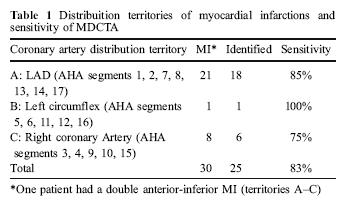
19 2. RULE OUT CAD Andreini D, Pontone G, Circulation CI

20 2. RULE OUT CAD Andreini D, Pontone G, Circulation CI
, first marginal branch (M1), and right coronary artery (RCA).")
21 2. RULE OUT CAD Dilated cardiomyopathy associated with severe CAD. Head-to-head comparison of invasive coronary angiography (left panel) compared with MDCT multiplanar reconstruction (right panel). White arrows show significant stenosis on the proximal segments of left anterior descending artery (LAD), first marginal branch (M1), and right coronary artery (RCA). Andreini D, Pontone G, Circulation CI 2009
 compared with invasive coronary angiography (right panel).")
22 2. RULE OUT CAD Idiopathic form of dilated cardiomyopathy. Head-to-head comparison of MDCT multiplanar reconstruction (left panel) compared with invasive coronary angiography (right panel). LAD indicates left anterior descending artery; LCX, left circumflex artery; RCA, right coronary artery. Andreini D, Pontone G, Circulation CI 2009
23 SUMMARY Volume, function and remodelling Rule out coronary artery disease To evaluate LV myocardial damage To evaluate cardiac veins anatomy 22


24 3. TO EVALUATE LEFT VENTRICLE DAMAGE Thickness Attenuation


25 3. TO EVALUATE LEFT VENTRICLE DAMAGE Late Enhancement with MDCT It is known that MRI can characterize MI with both early and late contrast patterns. First-pass imaging performed immediately after contrast administration may demonstrate areas of hypoenhancement in the endocardial core of the infarct corresponding to microvascular obstruction. Delayed images acquired more than 10 minutes after contrast administration may demonstrate regional hyperenhancement, corresponding to myocardial necrosis or scar. Because iodinated contrast agents used in CT have kinetics similar to gadolinium used in MRI, as discussed later, there is a rationale to believe that DHE-MDCT would be able to identify areas of MI


26 3. TO EVALUATE LEFT VENTRICLE DAMAGE Delayed Time: 5 10 min Tube Voltage: 80 Kv Tube Current: 420 ma Collimation: 64x0.625 mm Gantry Rotaion time: 350 msec ECG-gating: prospective ECG Se Sp NPV PPV 78% 100% 100% 97% Effective Radiation Dose: msv


27 3. TO EVALUATE LEFT VENTRICLE DAMAGE 52 PTS with Acute MI PTCA+Stent CT LE and Tl-SPECT 0 and 6 Month


28 3. TO EVALUATE LEFT VENTRICLE DAMAGE Transmural LE Subend. LE No LE SATO A EHJ 2008

29 3. TO EVALUATE LEFT VENTRICLE DAMAGE Significant increase of LVEDV only in transmural LE Higher incidence of hospitalization only in transmural LE


30 3. TO EVALUATE LEFT VENTRICLE DAMAGE Late Enhancement 0 = no LE Late Enhancement 1: 1% - 25% Late Enhancement 2: 26% - 50% Late Enhancement 3: 51% - 75% Late Enhancement 4: : >75%


31 DELINEATION OF THE ETIOLOGY OF LV DYSFUNCTION

32 DELINEATION OF THE ETIOLOGY OF LV DYSFUNCTION * * * * * * * * Le Polain De Waroix et al EHJ 2008

33 DELINEATION OF THE ETIOLOGY OF LV DYSFUNCTION Se Sp Accuracy 92% 97% 94% Le Polain De Waroix et al EHJ
34 SUMMARY Volume, function and remodelling Rule out coronary artery disease To evaluate LV myocardial damage To evaluate cardiac veins anatomy 33
35 4. CARDIAC VEINS ANATOMY 34
36 4. CARDIAC VEINS ANATOMY Pontone G IJC
37 4. CARDIAC VEINS ANATOMY Pontone G IJC
38 4. CARDIAC VEINS ANATOMY DCM: lower percentage of cardiac veins DCM without specific protocol: more artifacts No differences between DCM and control regard to anatomical details of veins PVLV and LMV are less in all groups Ischemic DCM group shows the less suitable anatomy for CRT 37


39 4. CARDIAC VEINS ANATOMY Normal Cardiac Veins anatomy GCV CS GCV PV MCV LMV GCV PV LEGENDS CS:coronarysinus;MCV:middlecardiacvein;PV:posteriorvein; GCV: great cardiac vein; LMV: left marginal vein; AIV: anterior interventricular vein. AIV LMV


40 4. CARDIAC VEINS ANATOMY Great Cardiac Veins from SVC


41 4. CARDIAC VEINS ANATOMY Fistula between greta cardiac vein and left atrial appendage

42 4. CARDIAC VEINS ANATOMY Giraldi F, Pontone G et al JACC

43 4. CARDIAC VEINS ANATOMY Giraldi F, Pontone G et al JACC

44 4. CARDIAC VEINS ANATOMY Giraldi F, Pontone G et al JACC


45 TAKE HOME MESSAGE Giraldi F, Pontone G et al JACC
46
Coronary Artery Imaging. Suvipaporn Siripornpitak, MD Inter-hospital Conference : Rajavithi Hospital
 Coronary Artery Imaging Suvipaporn Siripornpitak, MD Inter-hospital Conference : Rajavithi Hospital Larger array : cover scan area Detector size : spatial resolution Rotation speed : scan time Retrospective
Imaging and heart failure
 Imaging and heart failure Jeroen J Bax Dept of Cardiology Leiden Univ Medical Center The Netherlands Davos, feb 2013 Research grants: Medtronic, Biotronik, Boston, St Jude, BMS imaging, GE Healthcare,
Rational use of imaging for viability evaluation
 EUROECHO and other imaging modalities 2011 Rational use of imaging for viability evaluation Luc A. Pierard, MD, PhD, FESC, FACC Professor of Medicine Head, Department of Cardiology, CHU Liège, Belgium
Detection and Assessment of MI: Use of Imaging Methods. Robert O. Bonow, M.D.
 Detection and Assessment of MI: Use of Imaging Methods Robert O. Bonow, M.D. Detection and Assessment of MI: Use of Imaging Methods Robert O. Bonow, M.D. No Relationships to Disclose Expert Consensus Document
Disclosures. GETTING TO THE HEART OF THE MATTER WITH MULTIMODALITY CARDIAC IMAGING Organ Review Meeting 25 September. Overview
 GETTING TO THE HEART OF THE MATTER WITH MULTIMODALITY CARDIAC IMAGING Organ Review Meeting 25 September Disclosures None relevant to this presentation Mini Pakkal Assistant Professor of Radiology University
Multisclice CT in combination with functional imaging for CAD. Temporal Resolution. Spatial Resolution. Temporal resolution = ½ of the rotation time
 Multisclice CT in combination with functional imaging for CAD Prof. Juhani Knuuti, MD, FESC Turku University Hospital and University of Turku Turku, Finland MSCT and functional imaging for CAD Practical
Hybrid cardiac imaging Advantages, limitations, clinical scenarios and perspectives for the future
 Hybrid cardiac imaging Advantages, limitations, clinical scenarios and perspectives for the future Prof. Juhani Knuuti, MD, FESC Turku, Finland Disclosure: Juhani Knuuti, M.D. Juhani Knuuti, M.D. has financial
Coronary Artery Anomalies from Birth to Adulthood; the Role of CT Coronary Angiography in Sudden Cardiac Death Screening
 Coronary Artery Anomalies from Birth to Adulthood; the Role of CT Coronary Angiography in Sudden Cardiac Death Screening E O Dwyer 1, C O Brien 1, B Loo 1, A Snow Hogan 1, O Buckley1 2, B 1. Department
J. Schwitter, MD, FESC Section of Cardiology
 J. Schwitter, MD, FESC Section of Cardiology CMR Center of the CHUV University Hospital Lausanne - CHUV Switzerland Centre de RM Cardiaque J. Schwitter, MD, FESC Section of Cardiology CMR Center of the
CT or PET/CT for coronary artery disease
 CT or PET/CT for coronary artery disease Rotterdam 2012 Juhani Knuuti, MD, PhD, FESC Turku PET Centre University of Turku Turku, Finland Juhani.knuuti@utu.fi Turku PET Centre University of Turku Åbo Akademi
Disclosure Information
 Coronary CTA Pearls and Pitfalls Ricardo C. Cury, MD, FSCCT, FAHA, FACC Chairman of Radiology Radiology Associates of South Florida Director of Cardiac Imaging Miami Cardiac and Vascular Institute Past-President
HIGHLIGHT SESSION. Imaging. J. L. Zamorano Gomez (Madrid, ES) Disclosures: Speaker Philips
 Disclosures: Speaker Philips") Imaging. J. L. Zamorano Gomez (Madrid, ES) Disclosures: Speaker Philips Agenda ECHO Diagnosis & Prognosis : Functional MR Severity Aortic Stenosis CT How to select pts for TAVI Adding prognostic info to
Imaging. J. L. Zamorano Gomez (Madrid, ES) Disclosures: Speaker Philips Agenda ECHO Diagnosis & Prognosis : Functional MR Severity Aortic Stenosis CT How to select pts for TAVI Adding prognostic info to
Diagnostic Accuracy of Multidetector Computed Tomography Coronary Angiography in Patients With Dilated Cardiomyopathy
 Journal of the American College of Cardiology Vol. 49, No. 20, 2007 2007 by the American College of Cardiology Foundation ISSN 0735-1097/07/$32.00 Published by Elsevier Inc. doi:10.1016/j.jacc.2007.01.086
Pearls & Pitfalls in nuclear cardiology
 Pearls & Pitfalls in nuclear cardiology Maythinee Chantadisai, MD., NM physician Division of Nuclear Medicine, Department of radiology, KCMH Principle of myocardial perfusion imaging (MPI) Radiotracer
Current Indications for Cardiac MRI: What You See is What You Get?
 Current Indications for Cardiac MRI: What You See is What You Get? Javier Ganame, MD, PhD, FASE No disclosures Cardiology Update, Niagara, Sept 24th, 2016 The Ideal Diagnostic Technique Easy to apply Accurate
Dr Felix Keng. Imaging of the heart is technically difficult because: Role of Cardiac MSCT. Current: Cardiac Motion Respiratory Motion
 Siemens Philips Dr Felix Keng GE Toshiba Role of Cardiac MSCT Current: Structural / congenital heart imaging Extra-cardiac / Great vessel imaging Volumes and ejection fractions (cine + gating) Calcium
Noncoronary Cardiac MDCT
 Noncoronary Cardiac MDCT David A. Bluemke, M.D., Ph.D. Professor, of Radiology and Medicine Johns Hopkins University School of Medicine Baltimore, Maryland Toshiba Disclosures Grant support Noncoronary
Perfusion, Viability, Edema and Hemorrhage: How it Can (and Should) Change Clinical Practice. Rohan Dharmakumar, Ph.D.
 Change Clinical Practice. Rohan Dharmakumar, Ph.D.") Perfusion, Viability, Edema and Hemorrhage: How it Can (and Should) Change Clinical Practice Rohan Dharmakumar, Ph.D. Director, Translational Cardiac Imaging Research Associate Director, Biomedical Imaging
Perfusion, Viability, Edema and Hemorrhage: How it Can (and Should) Change Clinical Practice Rohan Dharmakumar, Ph.D. Director, Translational Cardiac Imaging Research Associate Director, Biomedical Imaging
Sung A Chang Department of Internal Medicine, Division of Cardiology, Sungkyunkwan University School of Medicine, Samsung Medical Center
 CMR Perfusion and Viability A STICH Out of Time? Sung A Chang Department of Internal Medicine, Division of Cardiology, Sungkyunkwan University School of Medicine, Samsung Medical Center Can Imaging Improve
Improvement of Image Quality with ß-Blocker Premedication on ECG-Gated 16-MDCT Coronary Angiography
 16-MDCT Coronary Angiography Shim et al. 16-MDCT Coronary Angiography Sung Shine Shim 1 Yookyung Kim Soo Mee Lim Received December 1, 2003; accepted after revision June 1, 2004. 1 All authors: Department
The best from Euro-Echo Ischemic heart disease. Fausto Rigo,FESC Department of Cardiology Mestre-Venezia Hospital,Italy
 The best from Euro-Echo 2011 Ischemic heart disease Fausto Rigo,FESC Department of Cardiology Mestre-Venezia Hospital,Italy faustorigo@alice.it DECLARATION OF CONFLICT OF INTEREST No conflict of interest
MRI ACS-ben. Tamás Simor MD, PhD, Med Hab. University of Pécs, Heart Institute
 MRI ACS-ben Tamás Simor MD, PhD, Med Hab Time Course of Changes in Infarct Size, Viable Myocardium, and LV Mass After Reperfused and Nonreperfused MI Blue lines denote reperfused myocardial infarction
Noninvasive Visualization of the Cardiac Venous System Using Multislice Computed Tomography
 Journal of the American College of Cardiology Vol. 45, No. 5, 2005 2005 by the American College of Cardiology Foundation ISSN 0735-1097/05/$30.00 Published by Elsevier Inc. doi:10.1016/j.jacc.2004.11.035
Validation of CT Perfusion Imaging Against Invasive Angiography and FFR on a 320-MDCT Scanner
 Validation of CT Perfusion Imaging Against Invasive Angiography and FFR on a 320-MDCT Scanner Zhen Qian, Gustavo Vasquez, Sarah Rinehart, Parag Joshi, Eric Krivitsky, Anna Kalynych, Dimitri Karmpaliotis,
Case based learning: CMR in Heart Failure
 Case based learning: CMR in Heart Failure Milind Y Desai, MD FACC FAHA FESC Associate Professor of Medicine Heart and Vascular Institute, Cleveland Clinic Cleveland, OH Disclosures: none Use of Gadolinium
The use of Cardiac CT and MRI in Clinical Practice
 The use of Cardiac CT and MRI in Clinical Practice Matthew W. Martinez, MD Assistant Professor of Medicine LVPG - Lehigh Valley Heart Specialists Lehigh Valley Health Network Oct. 3, 2009 DISCLOSURE Relevant
SPECT-CT: Τι πρέπει να γνωρίζει ο Καρδιολόγος
 SPECT-CT: Τι πρέπει να γνωρίζει ο Καρδιολόγος Δρ Αναστασία Κίτσιου Διευθύντρια, Καρδιολογική Κλινική, Σισμανόγλειο ΓΝΑ Chair, Education Committee, Section on Nuclear Cardiology & Cardiac CT, EACVI, ESC
Advanced Imaging MRI and CTA
 Advanced Imaging MRI and CTA Who and why may benefit. Matthew W. Martinez, M.D. FACC Lehigh Valley Health Network Director, Cardiovascular Imaging Learning Objectives Review basics of CMR and CTA Review
1. Department of Cardiology, 2. Department of Radiology, Shanghai Chest Hospital affiliated to Shanghai JiaoTong University, Shanghai , China
 20 Clinical Research Dual-phase contrast-enhancement multislice computed tomography imaging for the assessment of elderly patients with acute myocardial infarction after primary percutaneous coronary intervention
I have no financial disclosures
 Manpreet Singh MD I have no financial disclosures Exercise Treadmill Bicycle Functional capacity assessment Well validated prognostic value Ischemic assessment ECG changes ST segments Arrhythmias Hemodynamic
Stable Angina: Indication for revascularization and best medical therapy
 Stable Angina: Indication for revascularization and best medical therapy Cardiology Basics and Updated Guideline 2018 Chang-Hwan Yoon, MD/PhD Cardiovascular Center, Department of Internal Medicine Bundang
Cardiac Viability Testing A Clinical Perspective Annual Cardiac Imaging Symposium. Lisa M Mielniczuk MD FRCPC University of Ottawa Heart Institute
 Cardiac Viability Testing A Clinical Perspective Annual Cardiac Imaging Symposium Lisa M Mielniczuk MD FRCPC University of Ottawa Heart Institute 62 year old male Anterior STEMI late presentation, occluded
CT Perfusion. U. Joseph Schoepf, MD, FAHA, FSCBT MR, FSCCT Professor of Radiology, Medicine, and Pediatrics Director of Cardiovascular Imaging
 CT Perfusion U. Joseph Schoepf, MD, FAHA, FSCBT MR, FSCCT Professor of Radiology, Medicine, and Pediatrics Director of Cardiovascular Imaging Disclosures Consultant for / research support from Bayer Bracco
1. LV function and remodeling. 2. Contribution of myocardial ischemia due to CAD, and
 1 The clinical syndrome of heart failure in adults is commonly associated with the etiologies of ischemic and non-ischemic dilated cardiomyopathy, hypertrophic cardiomyopathy, hypertensive heart disease,
Impaired Regional Myocardial Function Detection Using the Standard Inter-Segmental Integration SINE Wave Curve On Magnetic Resonance Imaging
 Original Article Impaired Regional Myocardial Function Detection Using the Standard Inter-Segmental Integration Ngam-Maung B, RT email : chaothawee@yahoo.com Busakol Ngam-Maung, RT 1 Lertlak Chaothawee,
Computer Aided Detection and Diagnosis: Cardiac Imaging Applications
 Computer Aided Detection and Diagnosis: Cardiac Imaging Applications U. Joseph Schoepf, MD, FAHA, FSCBT MR, FSCCT Professor of Radiology, Medicine, and Pediatrics Director of Cardiovascular Imaging Disclosures
Impact of SSF on diagnostic performance of coronary CT angiography within one heart beat in patients with high heart rate using a 256-row detector CT
 Impact of SSF on diagnostic performance of coronary CT angiography within one heart beat in patients with high heart rate using a 256-row detector CT Junfu Liang 1,2, Hui Wang 1, Lei Xu 1, Li Dong 1, Zhanming
MSRS 6473 Vascular Noninvasive Imaging Procedures
 MSRS 6473 Vascular Noninvasive Imaging Procedures Rex T. Christensen MHA RT (R) (MR) (CT) (ARRT) CIIP Basic Physics Equipment Cardiac Positioning Perfusion Pathology MRI 1 Animal Magnetism MRI Basic Physics
Form 4: Coronary Evaluation
 Page of 7 Patient Details Hidden Show Show/Hide Annotations Stickies: Toggle All Toggle Open Toggle Resolved Form : Coronary Evaluation Print this Form t Started Date of Coronary Evaluation Coronary Evaluation
The Value of Stress MRI in Evaluation of Myocardial Ischemia
 The Value of Stress MRI in Evaluation of Myocardial Ischemia Dr. Saeed Al Sayari, MBBS, EBCR, MBA Department of Radiology and Nuclear Medicine Mafraq Hospital, Abu Dhabi United Arab Emirates Introduction
Dual Energy CT of the Heart: Perfusion and Beyond
 Dual Energy CT of the Heart: Perfusion and Beyond U. Joseph Schoepf, MD, FAHA, FSCBT MR, FSCCT Professor of Radiology, Medicine, and Pediatrics Director of Cardiovascular Imaging Disclosures Consultant
Managing Hypertrophic Cardiomyopathy with Imaging. Gisela C. Mueller University of Michigan Department of Radiology
 Managing Hypertrophic Cardiomyopathy with Imaging Gisela C. Mueller University of Michigan Department of Radiology Disclosures Gadolinium contrast material for cardiac MRI Acronyms Afib CAD Atrial fibrillation
Form 4: Coronary Evaluation
 Patient Details Hidden Show Show/Hide Annotations Form : Coronary Evaluation Print this Form t Started Date of Coronary Evaluation Coronary Evaluation Indication for Coronary Evaluation Check only one.
The role of Magnetic Resonance Imaging in the diagnosis of viability & Coronary Artery Disease
 The role of Magnetic Resonance Imaging in the diagnosis of viability & Coronary Artery Disease G.P. Spanos, MSc, Phd Head of CardioVascular Imaging Tomographia Diagnostic Center Cardiovascular magnetic
Viability Testing Using Dynamic Echocardiography
 Viability Testing Using Dynamic Echocardiography Theodora A Zaglavara, MD, PhD Director of Echocardiography EUROMEDICA KYANOUS STAVROS HOSPITAL Thessaloniki GREECE Goals of Cardiac Imaging in Coronary
EAE Teaching Course. Magnetic Resonance Imaging. Competitive or Complementary? Sofia, Bulgaria, 5-7 April F.E. Rademakers
 EAE Teaching Course Magnetic Resonance Imaging Competitive or Complementary? Sofia, Bulgaria, 5-7 April 2012 F.E. Rademakers Complementary? Of Course N Engl J Med 2012;366:54-63 Clinical relevance Treatment
Cardiac Imaging Tests
 Cardiac Imaging Tests http://www.medpagetoday.com/upload/2010/11/15/23347.jpg Standard imaging tests include echocardiography, chest x-ray, CT, MRI, and various radionuclide techniques. Standard CT and
Form 4: Coronary Evaluation
 Form : Coronary Evaluation Print this Form t Started Date of Coronary Evaluation Coronary Evaluation Indication for Coronary Evaluation Check only one. Angio NOT DONE: n invasive test performed Followup
Form 4: Coronary Evaluation
 Page of 8 Patient Details Hidden Show Show/Hide Annotations Stickies: Toggle All Toggle Open Toggle Resolved Form : Coronary Evaluation Toggle Question Year/Info Print this Form t Started Date of Coronary
Imaging in Ischemic Heart Disease: Role of Cardiac MRI
 Imaging in Ischemic Heart Disease: Role of Cardiac MRI Chiara Bucciarelli Ducci MD, PhD, FESC, FRCP Consultant Senior Lecturer Cardiologist Bristol Heart Institute, University of Bristol, UK Chair elect,
Radiologic Assessment of Myocardial Viability
 November 2001 Radiologic Assessment of Myocardial Viability Joshua Moss, Harvard Medical School Year III Patient EF 66yo female with a 3-year history of intermittent chest pain previously relieved by sublingual
CT Myocardial Perfusion: Is there Added Value to Coronary CT?
 CT Myocardial Perfusion: Is there Added Value to Coronary CT? U. Joseph Schoepf, MD, FAHA, FSCBT MR, FSCCT Professor of Radiology, Medicine, and Pediatrics Director of Cardiovascular Imaging Disclosures
Cardiac Computed Tomography
 Cardiac Computed Tomography Authored and approved by Koen Nieman Stephan Achenbach Francesca Pugliese Bernard Cosyns Patrizio Lancellotti Anastasia Kitsiou Contents CARDIAC COMPUTED TOMOGRAPHY Page 1.
CASE from South Korea
 CASE from South Korea Bon-Kwon Koo, MD, PhD, Seoul, Korea Outpatient clinic of a non-interventional cardiologist F/56 Chief complaint: Angina with recent aggravation, CCS II~III Brief history: # Stroke
Current and Future Imaging Trends in Risk Stratification for CAD
 Current and Future Imaging Trends in Risk Stratification for CAD Brian P. Griffin, MD FACC Department of Cardiovascular Medicine, Heart and Vascular Institute, Cleveland Clinic Disclosures: None Introduction
Ultrasound. Computed tomography. Case studies. Utility of IQon Spectral CT in. cardiac imaging
 Ultrasound Computed tomography Case studies Utility of IQon Spectral CT in cardiac imaging Cardiac imaging is a challenging procedure where it is necessary to image a motion-free heart. This requires a
What every radiologist should know about cardiac CT: A case-based pictorial review
 What every radiologist should know about cardiac CT: A case-based pictorial review Poster No.: C-0555 Congress: ECR 2010 Type: Educational Exhibit Topic: Cardiac Authors: C. M. Capuñay, P. Carrascosa,
Cardiac Stress MRI: Detection of Ischemia. Disclosures: Dobutamine Stress MR. April 28, 2018
 Cardiac MRI: Detection of Ischemia Cardiac MRI in Today s Clinical Practice Foundations of Cardiovascular Magnetic Resonance Daniel C. Lee, MD, MSc Assistant Professor of Medicine and Radiology Co-Director,
Imaging of Coronary Artery Disease: II
 Acta Radiológica Portuguesa, Vol.XIX, nº 74, pág. 45-51, Abr.-Jun., 2007 Imaging of Coronary Artery Disease: II Jean Jeudy University of Maryland School of Medicine Department of Diagnostic Radiology Armed
Coronary interventions
 Controversial issues in the management of ischemic heart failure Coronary interventions Maciej Lesiak Department of Cardiology, University Hospital in Poznan none DECLARATION OF CONFLICT OF INTEREST CHF
CARDIOMYOPATHY IN CT. Hans- Christoph Becker Professor of Radiology
 CARDIOMYOPATHY IN CT Hans- Christoph Becker Professor of Radiology 1 Cardiomyopathy Heart muscle disease Deterioration of the heart function, heart failure Dyspnea, peripheral edema Risk of arrhythmia,
THE ROLE OF HIGH END MULTI DETECTOR CT IN CORONARY IMAGING ESSAY
 THE ROLE OF HIGH END MULTI DETECTOR CT IN CORONARY IMAGING ESSAY Submitted for partial fulfillment of Master degree in Radiodiagnosis By Ahmed Yehia Ahmed (M.B.B.Ch., Cairo University) Supervisors Prof.
G-CSF ATTENUATES VENTRICULAR REMODELLING AFTER ACUTE STEMI. RESULTS OF STem cells Mobilization in Acute Myocardial Infarction
 ESC CONGRESS 2010 Stockholm 28 August-1 September G-CSF ATTENUATES VENTRICULAR REMODELLING AFTER ACUTE STEMI. RESULTS OF STem cells Mobilization in Acute Myocardial Infarction C. Malafronte MD Alessandro
The Emerging Role of Cardiac CT in Cardiovascular Imaging. Anthony Gemignani, MD Vermont Cardiac Network April 28, 2016
 The Emerging Role of Cardiac CT in Cardiovascular Imaging Anthony Gemignani, MD Vermont Cardiac Network April 28, 2016 Conflict Disclosures I have no significant financial relationship with any companies
Role of echocardiography in the assessment of ischemic heart disease 분당서울대학교병원윤연이
 Role of echocardiography in the assessment of ischemic heart disease 분당서울대학교병원윤연이 Outline Evaluation of Chest pain Evaluation of MI complications Prediction of Outcomes Evaluation of Chest pain Evaluation
New Insight about FFR and IVUS MLA
 New Insight about FFR and IVUS MLA Can IVUS MLA Predict FFR
Cardiac CT and MRI. Ashraf Hamdan, MD. Sheba Medical Center. Sheba Medical Center Tel Hashomer. Leviev Heart Center
 Cardiac CT and MRI Ashraf Hamdan, MD Sheba Medical Center Sheba Medical Center Tel Hashomer Leviev Heart Center Spatial resolution Resolution 4x4mm If the object incidentally placed in one pixel high image
Abnormal, Autoquant Adenosine Myocardial Perfusion Heart Imaging. ID: GOLD Date: Age: 46 Sex: M John Doe Phone (310)
") Background: Reason: preoperative assessment of CAD, Shortness of Breath Symptom: atypical chest pain Risk factors: hypertension Under influence: a beta blocker Medications: digoxin Height: 66 in. Weight:
Background: Reason: preoperative assessment of CAD, Shortness of Breath Symptom: atypical chest pain Risk factors: hypertension Under influence: a beta blocker Medications: digoxin Height: 66 in. Weight:
Je bénéficie régulièrement de fonds privés, dans le cadre de projets de recherche ou d activités de formation.
 Je bénéficie régulièrement de fonds privés, dans le cadre de projets de recherche ou d activités de formation. Ces fonds proviennent essentiellement d industriels travaillant dans les domaines de l imagerie
MR Assessment of Myocardial Viability
 MR Assessment of Myocardial Viability Definition of Viability Clinical Metabolism: Presence of glucose uptake Perfusion / Perfusion reserve Morphology: Wall thickness, wall thickening Contractility: Recovery
Use of Nuclear Cardiology in Myocardial Viability Assessment and Introduction to PET and PET/CT for Advanced Users
 Use of Nuclear Cardiology in Myocardial Viability Assessment and Introduction to PET and PET/CT for Advanced Users February 1 5, 2011 University of Santo Tomas Hospital Angelo King A-V Auditorium Manila,
TITLE: Multi-Slice Computed Tomography Coronary Angiography for Coronary Artery Disease: A Review of the Clinical Effectiveness and Guidelines
 TITLE: Multi-Slice Computed Tomography Coronary Angiography for Coronary Artery Disease: A Review of the Clinical Effectiveness and Guidelines DATE: 25 February 2009 CONTEXT AND POLICY ISSUES: Coronary
Role of CMR in heart failure and cardiomyopathy
 Role of CMR in heart failure and cardiomyopathy Hajime Sakuma Department of Radiology, Mie University Late gadolinium enhancement (LGE) LGE MRI can demonstrate site of necrosis, fibrosis or deposition
Dolore Toracico: Il Corretto Approccio ed il Valore Incrementale de Multimodality Imaging nei Pazienti con Rischio di Malattia Basso-intermedio
 Dolore Toracico: Il Corretto Approccio ed il Valore Incrementale de Multimodality Imaging nei Pazienti con Rischio di Malattia Basso-intermedio Gianluca Pontone, MD, PhD, FESC, FSCCT Director of MR Unit
Imaging of the Heart Todd Tessendorf MD FACC
 Imaging of the Heart Todd Tessendorf MD FACC Outline Imaging Modalities for Structural Heart Disease ECHO, MRI Imaging Modalities for Ischemic Heart Disease SPECT, PET, CCTA Show lots of pretty pictures
Fractional Flow Reserve from Coronary CT Angiography (and some neat CT images)
") Fractional Flow Reserve from Coronary CT Angiography (and some neat CT images) Victor Cheng, M.D. Director, Cardiovascular CT Oklahoma Heart Institute 1 Disclosures Tornadoes scare me 2 Treating CAD Fixing
Fractional Flow Reserve from Coronary CT Angiography (and some neat CT images) Victor Cheng, M.D. Director, Cardiovascular CT Oklahoma Heart Institute 1 Disclosures Tornadoes scare me 2 Treating CAD Fixing
Cardiac MRI: Clinical Application to Disease
 Cardiac MRI: Clinical Application to Disease Jessi Smith, MD Cardiothoracic imaging, Indiana University Slides courtesy of Stacy Rissing, MD Outline Imaging planes Disease findings Pulse sequences used
Presenter Disclosure Information
 Various Morphological Types of Ventricular Premature Beats with Fragmented QRS Waves on 12 Lead Holter ECG had a Positive Relationship with Left Ventricular Fibrosis on CT in Patients with Hypertrophic
Cardiac MRI: Cardiomyopathy
 Cardiac MRI: Cardiomyopathy Laura E. Heyneman, MD I do not have any relevant financial relationships with any commercial interests Cardiac MRI: Cardiomyopathy Laura E. Heyneman, MD Duke University Medical
Pushing the limits of cardiac CT. Steven Dymarkowski Radiology / Medical Imaging Research Centre
 Pushing the limits of cardiac CT Steven Dymarkowski Radiology / Medical Imaging Research Centre 5 X 2013 Introduction Rapid technological advances and new clinical applications in cardiovascular imaging
Low-dose prospective ECG-triggering dual-source CT angiography in infants and children with complex congenital heart disease: first experience
 Low-dose prospective ECG-triggering dual-source CT angiography in infants and children with complex congenital heart disease: first experience Ximing Wang, M.D., Zhaoping Cheng, M.D., Dawei Wu, M.D., Lebin
CT Imaging of Atherosclerotic Plaque. William Stanford MD Professor-Emeritus Radiology University of Iowa College of Medicine Iowa City, IA
 CT Imaging of Atherosclerotic Plaque William Stanford MD Professor-Emeritus Radiology University of Iowa College of Medicine Iowa City, IA PREVALENCE OF CARDIOVASCULAR DISEASE In 2006 there were 80 million
Simon Nepveu 1, Irina Boldeanu 1, Yves Provost 1, Jean Chalaoui 1, Louis-Mathieu Stevens 2,3, Nicolas Noiseux 2,3, Carl Chartrand-Lefebvre 1,3
 Coronary Artery Bypass Graft Imaging with CT Angiography and Iterative Reconstruction: Quantitave Evaluation of Radiation Dose Reduction and Image Quality Simon Nepveu 1, Irina Boldeanu 1, Yves Provost
IAEA. Department of Technical Cooperation. And. Nuclear Medicine Section RAS 6/063
 IAEA Department of Technical Cooperation And Nuclear Medicine Section RAS 6/063 Strengthening the Application of Nuclear Medicine in the Management of Cardiovascular Diseases Cardiac Imaging CT and MR
Declaration of conflict of interest
 Declaration of conflict of interest Electrical activation pattern in Left Bundle Branch Block Patients Angelo Auricchio, MD FESC Director, Cardiac Electrophysiology Programme, Fondazione Cardiocentro Ticino,
Improved Noninvasive Assessment of Coronary Artery Bypass Grafts With 64-Slice Computed Tomographic Angiography in an Unselected Patient Population
 Journal of the American College of Cardiology Vol. 49, No. 9, 2007 2007 by the American College of Cardiology Foundation ISSN 0735-1097/07/$32.00 Published by Elsevier Inc. doi:10.1016/j.jacc.2006.10.066
Noninvasive Fractional Flow Reserve from Coronary CT Angiography
 2016 KSC Annual Spring Scientific Conference Noninvasive Fractional Flow Reserve from Coronary CT Angiography Bon-Kwon Koo, MD, PhD, Seoul, Korea Why the hemodynamics for coronary artery disease? Twinlifemarketing.com.au
Ischemic heart disease
 Ischemic heart disease Introduction In > 90% of cases: the cause is: reduced coronary blood flow secondary to: obstructive atherosclerotic vascular disease so most of the time it is called: coronary artery
Debate Should we use FFR? I will say NO.
 Debate Should we use FFR? I will say NO. Hyeon-Cheol Gwon Cardiac and Vascular Center Samsung Medical Center Sungkyunkwan University School of Medicine Dr. Hyeon-Cheol Gwon Research fund from Abbott Korea
Index. radiologic.theclinics.com. Note: Page numbers of article titles are in boldface type.
 Index Note: Page numbers of article titles are in boldface type. A ALCAPA. See Anomalous left coronary artery from the pulmonary artery. Angiosarcoma computed tomographic assessment of, 809 811 Anomalous
CARDIAC AND CORONARY ARTERY ANATOMY NO DISCLOSURES. Axial Anatomy of Heart. Axial Anatomy of Heart. Axial Anatomy of Heart
 CARDIAC AND CORONARY ARTERY ANATOMY NO DISCLOSURES NASCI MEETING, ORLANDO FLORIDA 2009 KOSTAKI G. BIS, MD, FACR DEPARTMENT OF RADIOLOGY WILLIAM BEAUMONT HOSPITAL Royal Oak, Michigan OBJECTIVES CARDIAC
Cardiac CT Lowering the Dose Dramatically
 Cardiac CT Lowering the Dose Dramatically U. Joseph Schoepf, MD, FAHA, FSCBT MR, FSCCT Professor of Radiology, Medicine, and Pediatrics Director of Cardiovascular Imaging Disclosures Consultant for / research
Cardiac magnetic resonance imaging in rheumatoid arthritis: promising or misleading? Sophie Mavrogeni MD FESC
 Cardiac magnetic resonance imaging in rheumatoid arthritis: promising or misleading? Sophie Mavrogeni MD FESC Onassis Cardiac Surgery Center Athens Greece Nothing to disclose Financial disclosure Cardiac
Φαινόμενο No-Reflow. Απεικόνιση με CMR, κλινική συσχέτιση και προγνωστική σημασία
 Φαινόμενο No-Reflow. Απεικόνιση με CMR, κλινική συσχέτιση και προγνωστική σημασία Θεόδωρος. Καραμήτσος MD PhD Honorary Consultant in Cardiology University of Oxford Centre for Clinical Magnetic Resonance
Cardiac Imaging in abnormal rhythm Role of MDCT
 Cardiac Imaging in abnormal rhythm Role of MDCT Cardiac Imaging in abnormal rhythm Role of MDCT Scope of the problem CT in Atrial Fibrillation CT and pacing Ventricular arrhythmia Other applications 1
Importance of CRT team for optimization of the results: a European point of view
 Importance of CRT team for optimization of the results: a European point of view Matteo Bertini, MD, PhD Arcispedale S. Anna Azienda Ospedaliero-Universitaria Cona-Ferrara No conflict of interest to declare
Cardiac MR Cardiac CT
 Cardiac MR Cardiac CT A selection of recent articles JN Dacher 1, A Manrique 2 and J Caudron 1 1 Rouen University Hospital - Department of Radiology 2 Caen University Hospital - Cyceron Imaging Centre
General Cardiovascular Magnetic Resonance Imaging
 2 General Cardiovascular Magnetic Resonance Imaging 19 Peter G. Danias, Cardiovascular MRI: 150 Multiple-Choice Questions and Answers Humana Press 2008 20 Cardiovascular MRI: 150 Multiple-Choice Questions
Atypical pain and normal exercise test
 Atypical pain and normal exercise test F. Mut, M. Beretta Nuclear Medicine Service, Asociacion Española Montevideo, Uruguay Clinical history 67-year old male with several coronary risk factors. Atypical
CT for Myocardial Characterization of Cardiomyopathy. Byoung Wook Choi, Yonsei University Severance Hospital, Seoul, Korea
 CT for Myocardial Characterization of Cardiomyopathy Byoung Wook Choi, Yonsei University Severance Hospital, Seoul, Korea Cardiomyopathy Elliott P et al. Eur Heart J 2008;29:270-276 The European Society