Pulmonary Microvascular Disease
|
|
|
- Merryl Marsh
- 6 years ago
- Views:
Transcription
1 Pulmonary Microvascular Disease CS Restrepo M.D. (UT Health San Antonio, TX) D. Vargas M.D. (University of Colorado in Denver, CO) Saboo M.D. (UT Southwestern, Dallas TX) A.J. Baxi M.D. (UT Health San Antonio, TX Disclosure: Nothing to disclose
2 Introduction The pulmonary microvasculature (arterioles, venules and capillaries) can be affected in numerous conditions. Congenital malformations, inflammatory conditions (vasculitis), idiopathic, proliferative, neoplastic, thrombotic and non-thrombotic embolisms can affect the small vessels of the lung with significant morbidity and mortality. Overview Small vessel vasculitis Hepatopulmonary syndrome Pulmonary veno-occlusive disease Pulmonary capillary hemangiomatosis Pulmonary lymphangiectasia Hereditary hemorrhagic telangiectasia Intravascular metastases Lymphangitic carcinomatosis Non-thrombotic pulmonary embolism
3 Small vessel vasculitis (SVV) Lung involvement is most commonly seen with the primary, idiopathic, small-vessel, or ANCA associated vasculitides; Wegener's granulomatosis, microscopic polyangiitis, and Churg-Strauss syndrome. However, primary, idiopathic medium and large-vessel vasculitis, primary immune complex mediated vasculitis, and secondary vasculitis are all capable of presenting with lung involvement. Imaging spectrum is wide, from extensive air-space consolidation to airway involvement, pulmonary nodules and cavitation. Hansell DM. Radiology 2002;225:
 ANCA vasculitis in a 63")


4 Microscopic polyangiitis (MPA) ANCA vasculitis in a 63 year old female with hemoptysis. CT shows multifocal parenchymal opacities with airspace consolidation in the lower lobes.
 Churg-Strauss")


5 Eosinophilic granulomatosis with polyangiitis (EGPA) Churg-Strauss syndrome in a 48 y/o male with hystory of asthma and nasal polyposis. Graoundglass and patchy consolidation are noted in the bilateral upper lobes.
")


6 Granulomatosis with polyangiitis (GPA) Granulomatosis with polyangiitis (Wegener s ) in a 57 y/o male with cough and hemoptysis. CT shows cavitary and non-cavitary nodular opacities.
7 Hepatopulmonary syndrome (HPS) Clinical syndrome characterized by the triad of hypoxemisa caused by intrapulmonary vascular dilations (IPVDs) in the setting of chronic liver disease, portal hypertension, or congenital portosystemic shunts. Prevalence: 25 % of patients with chronic liver disease Pathopysiology: Chronic liver disease increases the circulatory level of nitric oxide (NO) and endothelin-1 (ET-1), potent pulmonary vasodilators. Pathology: Gross dilation of the pulmonary capillary vessels with an absolute increase in the number of capillaries. Physiology: Ventilation-Perfusion (V/Q) mismatch, Diffusion- Perfusion impairment, and anatomic shunt. Only effective treatment is liver transplantation.
8 Imaging findings in HPS Chest radiograph may reveal increased interstitial markings due to increased IPVDs CT angiography: Most significant imaging findings is the presence of IPVDs and include: - Dilated peripheral subpleural pulmonary vessels that do not taper normally and extend to the pleural surface (most common finding, 85%) - Vessels with increased pulmonary artery to bronchus ratio - Small peripheral arteriovenous malformations (rare15%) Nuclear medicine: Tc99m MAA (micro-aggregated albumin) may show uptake in the kidneys or brain secondary to intrapulmonary shunt. Kim YK, Kim Y, Shim SS. RadioGraphics 2009;29(3):


9 Hepatopulmonary syndrome in a 63 y/o cirrhotic male with hypoxemia. CT shows dilated vessels in the lung periphery.


10 Hepatopulmonary Syndrome VQ scan in a 46 y/o male with alcoholic cirrhosis and hypoxemia. Perfusion exam with 99m Tc MAAT demonstrated abnormal uptake within the brain, bilateral kidneys, bowels, spleen and liver. Also noted is thyroid and salivary gland uptake without activity in the stomach, which may related to small amount of free pertechnetate in the blood pool
11 Pulmonary Veno-Occlusive Disease (PVOD) Rare cause of pulmonary hypertension (PH), roughly represent 5%-10% of all cases of idiopathic PH Characterized by occlusion or narrowing of pulmonary veins and venules by collagen-rich fibrous tissue. Alveolar capillaries become dilated and engorged. Capillary proliferation may occur mimicking capillary hemangiomatosis. Most cases are diagnosed in infants or young adults Etiology remains unknown. May be idiopathic or associated with other conditions like HIV, connective tissue disease, sarcoidosis, chemotherapy and bone marrow transplantation. Poor prognosis with median survival <3 years
12 PVOD: Imaging findings Evidence of pulmonary hypertension (RV enlargement etc.) Signs of post-capillary venous hypertension (septal thickening, mosaic attenuation, multiple small nodules, pleural effusion, pericardial effusion) Centrilobular ground-glass opacities Mediastinal lymphadenopathy Normal-sized large pulmonary veins and left atrium Montani D, et al. Eur Respir J 2009;33(1):


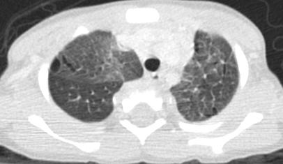
13 PVOD in a young female patient with arterial pulmonary hypertension. CECT shows ground-glass opacities, mosaic pattern of lung attenuation, and interlobular septal thickening in addition to enlarged pulmonary arteries.
14 Pulmonary Capillary Hemangiomatosis (PCH) PCH is a rare cause of PH, characterized by extensive proliferation of pulmonary capillaries within alveolar septae, by endothelial cells with no mitotic activity. PCH is clinically and radiographically indistinguishable from PVOD, making microscopic diagnosis essential for diagnosis. Also considered an idiopathic disease although numerous associations have been reported Similar to PVOD prognosis is poor and lung transplantation is the only definitive treatment. Frazier AA, et al. RadioGraphics 2007;27:


15 Pulmonary Capillary Hemangiomatosis 38 y/o male with Pulmonary hypertension 2ary to pulmonary capillary hemangiomatosis; brother also had the same condition. Both received bilateral lung transplant. Explanted lungs confirmed the diagnosis originally made from open lung biopsy.


16 Pulmonary Capillary Hemangiomatosis PCH in a young adult male. CECT reveal lymphadenopathy with prominent peripheral and peribronchovascular lymphatics.
17 Pulmonary Lymphangiectasia Pathologic dilation of pulmonary subpleural, interlobular, perivascular and peribronchial lymphatics 0.5% prevalence in infants who are stillborn or die in the neonatal period Associated with congenital chylothorax and nonimmune hydrops fetalis Poor prognosis before effective mechanical ventilation became available Improvement after the first year of life Barker PM et al. Eur Respir J 2004;24:413 Bellini C et al. Lymphology 2005;38:111


18 Primary Pulmonary Lymphangiectasia Primary pulmonary lymphangiectasia in a 3 week old male with respiratory distress. Chest x ray and CT shows a diffuse interstitial process with interlobular septal thickening.


19 Congenital Lymphangiectasia
20 Hereditary Hemorrhagic Telangiectasia (Osler-Weber-Rendu) Hereditary hemorrhagic telangiectasia is a vascular dysplasia that can involve the lungs, brain upper gastrointestinal tract and liver 5-15% of patients have pulmonary AVM that present as solitary or multiple masses on chest radiographs Most commonly in lower lobes of lung Few patients present a diffuse form of AVM involving every subsegmental artery of at least one lobe, which has higher morbidity and mortality Prevalence of 1:10,000 Autosomal dominant linked to two loci Mutations of ENG, long arm of chromosome 9 (HHT1) Mutations in gene encoding for activan A receptor, type II kinase, long arm of chromsome 12 Faughnan ME, et al. CHEST 2000;117:31-38 Pierucci P et al. CHEST 2008;133:
")

21 Hereditary Hemorrhagic Telangiectasia (HHT) 18-month-old girl with hemoptysis and numerous bilateral arteriovenous malformations.

22 Lymphangitic carcinomatosis More commonly seen in adenocarcinomas Tumoral spread into the lymphatic usually by hematogenous seeding or by retrograde spread from the hilar / mediastinal lymph nodes Both peripheral and central lymphatics may be involved Manifest as nodular and irregular interlobular septal thickening and / or thicken peribronchovascular interstitium Lymphangitic carcinomatosis in a 54 y/o female with lung adenocarcinoma.
23 Intravascular pulmonary metastases Seen in patients with invasive tumors including renal cell, hepatic and pancreatic carcinomas and sarcomas. At CT affected patients show multifocal dilation and beading of peripheral subsegmental pulmonary arteries. Follow-up studies show interval enlargement and proliferation of the intravascular tumor, different from tumor embolism in which the embolized lesion shows no growth. Thrombotic microangiopathy with fibrocellular intimal hyperplasia of small pulmonary arteries and arterioles induced by tumor microemboli can also occur. Ting PT et al. J Can Assoc Radiol 2005;56:214 Franquet T et al. AJR 2002;179:897


24 Intravascular metastases in a 55 y/o male with advanced renal cell carcinpma. CT shows beaded branching tubular lesions in the bilateral lungs.
25 Nonthrombotic Pulmonary Embolism Biologic agents Septic embolism Fat embolism Amniotic fluid embolism Tumor embolism Hydatid embolism Nonbiologic agents Talc embolism Silicone embolism Cement embolism Glue embolism Catheter embolism Gas embolism Lipiodol embolism Bach AG, Restrepo CS, et al. Imaging of nonthrombotic pulmonary embolism: Biological materials, nonbiological materials, and foreign bodies. European Journal of Radiology 2013;82(3):e120-41
26 Fat Embolism Pathophysiology Long bone fractures (1%-20%) Hemoglobinopathies Severe burn Soft tissue injury Diabetes Pancreatitis Neoplasm Imaging Findings Bilateral opacities that resemble pulmonary edema or ARDS 1=3 days after trauma Normal heart size No redistribution No pleural effusion


27 60 y/o F Day 1 Day 3
28 Talc granulomatosis Talc (magnesium silicate) is an insoluble filler used in several oral medications. When oral medications are crushed, dissolved, and injected intravenously, talc particles embolize small pulmonary vessels. The talc particles may then migrate into the pulmonary interstitium, where they induce a foreign-body reaction and fibrosis. Acute injection may result in acute right ventricular failure and shock. Chronic injection can result in emphysema, pulmonary hypertension, and cor pulmonale Nandavaram S et al. Vasc Med Surg 2016;4(3):272


29 Talc embolism in a 29 year old female, with history of IV drug abuse. CECT obtained few days before she died, shows innumerable small pulmonary nodules bilaterally with denser opacities in the posterior aspect of the lungs. Autopsy demonstrated diffuse embolization of pulmonary arteries and arterioles with endovascular talc particles throughout the pulmonary vasculature as well as granulomas.
30 Glue embolization (Glue lung) Embolization therapy of vascular malformations and tumors with cyanoacrilate glue - Isobutyl-2-cyanoacrylate (IBCA) - N-butyl-2-cyanoacrylate (NBCA) Pulmonary embolism is frequent (35%) Usually asymptomatic Respiratory distress, lung infarct, pleural effusion and death can occur Kjellin IB et al. Pediatr Radiol 2000; 30:279 Pelz DM, et al. AJNR 1995;16:19


31 Extensive pulmonary glue embolization during endovascular embolization of a vein of Galen cerebral malformation in a 7 year old male. Patient developed cough and respiratory symptoms during the procedure.

32 Silicone and Oil Pulmonary Embolism Illicit injection for cosmetic purpose Fluid silicone Non-medical silicone (sealant) Respiratory symptoms 15 min-2 days after injection Fever in 50%, ARDS in 60% Death in 20% Peripheral GGO and interlobular septal thickening Peripheral air-space disease in 70% ARDS in a transsexual, 28 y/o patient with SOB two days after gluteal injection of undisclosed amount of silicone. Restrepo CS, et al. J Computer Assist Tomogr 2009;33:233


33 A C B D Silicone pulmonary embolism in two different patients. A an B. 36 y/o female with bilateral silicone gluteal injection three days earlier. C, D. 32 y/o male with pectoralis muscle injection 12 hours earlier.
34 SUMMARY Pulmonary microvascular disease The pulmonary microvasculature (arterioles, venules and capillaries) can be affected in numerous conditions. Congenital malformations, inflammatory conditions (vasculitis), idiopathic, proliferative, neoplastic, thrombotic and non-thrombotic embolisms can affect the small vessels of the lung with significant morbidity and mortality. Overview Small vessel vasculitis Hepatopulmonary syndrome Pulmonary veno-occlusive disease Pulmonary capillary hemangiomatosis Pulmonary lymphangiectasia Hereditary hemorrhagic telangiectasia Intravascular metastases Lymphangitic carcinomatosis Non-thrombotic pulmonary embolism
:825-837 Montani D, et al. pulmonary veno-acclussive disease. Eur Respir J 2009;33(1):189-200 Frazier AA, et al.")

35 References Hansell DM. Small-vessel disease of the lung: CT-pathologic coreelates.radiology 2002;225: Kim YK, Kim Y, Shim SS. Thoracic complications of liver cirrhosis: radiologic findings. RadioGraphics 2009;29(3): Montani D, et al. pulmonary veno-acclussive disease. Eur Respir J 2009;33(1): Frazier AA, et al. Pulmonary veno-occlussive disease and pulmonary capillary hemangiomatosis.radiographics 2007;27: Faughnan ME, et al. Diffuse pulmonary arteriovenous malformation: characteristics and prognosis. CHEST 2000;117:31-38 Bach AG, Restrepo CS, et al. Imaging of nonthrombotic pulmonary embolism: Biological materials, nonbiological materials, and foreign bodies. European Journal of Radiology 2013;82(3):e Restrepo CS, et al. Silicone pulmonary embolism: report of 10 cases and review of the literature. J Computer Assist Tomogr 2009;33:233 Presenting author contact information: Carlos S. Restrepo M.D. RestrepoC@UTHSCSA.edu
Systemic lupus erythematosus (SLE): Pleuropulmonary Manifestations
: Pleuropulmonary Manifestations") 08/30/10 09/26/10 Systemic lupus erythematosus (SLE): Pleuropulmonary Manifestations Camila Downey S. Universidad de Chile, School of Medicine, Year VII Harvard University, School of Medicine Sept 17,
08/30/10 09/26/10 Systemic lupus erythematosus (SLE): Pleuropulmonary Manifestations Camila Downey S. Universidad de Chile, School of Medicine, Year VII Harvard University, School of Medicine Sept 17,
An Introduction to Radiology for TB Nurses
 An Introduction to Radiology for TB Nurses Garold O. Minns, MD September 14, 2017 TB Nurse Case Management September 12 14, 2017 EXCELLENCE EXPERTISE INNOVATION Garold O. Minns, MD has the following disclosures
Acute and Chronic Lung Disease
 KATHOLIEKE UNIVERSITEIT LEUVEN Faculty of Medicine Acute and Chronic Lung Disease W De Wever, JA Verschakelen Department of Radiology, University Hospitals Leuven, Belgium Clinical utility of HRCT To detect
HRCT in Diffuse Interstitial Lung Disease Steps in High Resolution CT Diagnosis. Where are the lymphatics? Anatomic distribution
 Steps in High Resolution CT Diagnosis Pattern of abnormality Distribution of disease Associated findings Clinical history Tomás Franquet MD What is the diagnosis? Hospital de Sant Pau. Barcelona Secondary
Case of the Day Chest
 Case of the Day Chest Darin White MDCM FRCPC Department of Radiology, Mayo Clinic 76 th Annual Scientific Meeting Canadian Association of Radiologists Montreal, QC April 26, 2013 2013 MFMER slide-1 Disclosures
Pulmonary veno-occlusive disease
 Disclosure Objectives Pulmonary veno-occlusive disease Tilman Humpl The Hospital for Sick Children University of Toronto, Canada Advisor/Research Grants Actelion Pfizer Historical aspects Epidemiology/Genetics
Case 1: Question. 1.1 What is the main pattern of this HRCT? 1. Intralobular line 2. Groundglass opacity 3. Perilymphatic nodule
 HRCT WORK SHOP Case 1 Case 1: Question 1.1 What is the main pattern of this HRCT? 1. Intralobular line 2. Groundglass opacity 3. Perilymphatic nodule Case 1: Question 1.2 What is the diagnosis? 1. Hypersensitivity
Replacement of air with fluid, inflammatory. cells or cellular debris. Parenchymal, Interstitial (Restrictive) and Vascular Diseases.
 and Vascular Diseases.") Parenchymal, Interstitial (Restrictive) and Vascular Diseases Alain C. Borczuk, M.D. Dept of Pathology Replacement of air with fluid, inflammatory cells Pulmonary Edema Pneumonia Hemorrhage Diffuse alveolar
Parenchymal, Interstitial (Restrictive) and Vascular Diseases Alain C. Borczuk, M.D. Dept of Pathology Replacement of air with fluid, inflammatory cells Pulmonary Edema Pneumonia Hemorrhage Diffuse alveolar
Vascular Lung Diseases
 Vascular Lung Diseases SESSION SPECIFIC OBJECTIVES List the major types of vascular lung disease Recognize and describe the pathology of vascular lung disease: Pulmonary embolism, thrombosis, hypertension,
Resident Case Review CHEST. Daria Manos CAR 2016
 Resident Case Review CHEST CAR 2016 Daria Manos Disclosure Speakers bureau, Roche CAR 2016 Daria Manos 1. Recognize common and critical chest radiograph and computed tomography signs and use these clues
Pictorial essay of unusual radiologic manifestations of pulmonary and airway metastasis at initial presentation of lung cancer
 Pictorial essay of unusual radiologic manifestations of pulmonary and airway metastasis at initial presentation of lung cancer Poster No.: C-2297 Congress: ECR 2012 Type: Educational Exhibit Authors: Y.
Pathology of pulmonary vascular disease. Dr.Ashraf Abdelfatah Deyab. Assistant Professor of Pathology Faculty of Medicine Almajma ah University
 Pathology of pulmonary vascular disease Dr.Ashraf Abdelfatah Deyab Assistant Professor of Pathology Faculty of Medicine Almajma ah University Pulmonary vascular disease Type of pulmonary circulation: Types
Definition: HPS is a disease process with a triad of: 1- Liver disease. 2- Widespread intrapulmonary vasodilatation. 3- Gas exchange abnormality prese
 Hepatopulmonary syndrome (HPS) By Alaa Haseeb, MS.c Definition: HPS is a disease process with a triad of: 1- Liver disease. 2- Widespread intrapulmonary vasodilatation. 3- Gas exchange abnormality presenting
TB Radiology for Nurses Garold O. Minns, MD
 TB Nurse Case Management Salina, Kansas March 31-April 1, 2010 TB Radiology for Nurses Garold O. Minns, MD April 1, 2010 TB Radiology for Nurses Highway Patrol Training Center Salina, KS April 1, 2010
Congenital Lung Malformations: Radiologic-Pathologic Correlation
 Acta Radiológica Portuguesa, Vol.XVIII, nº 70, pág. 51-60, Abr.-Jun., 2006 Congenital Lung Malformations: Radiologic-Pathologic Correlation Marilyn J. Siegel Mallinckrodt Institute of Radiology, Washington
TB Intensive Houston, Texas
 TB Intensive Houston, Texas October 15-17, 17 2013 Diagnosis of TB: Radiology Rosa M Estrada-Y-Martin, MD MSc FCCP October 16, 2013 Rosa M Estrada-Y-Martin, MD MSc FCCP, has the following disclosures to
October 2012 Imaging Case of the Month. Michael B. Gotway, MD Associate Editor Imaging. Department of Radiology Mayo Clinic Arizona Scottsdale, AZ
 October 2012 Imaging Case of the Month Michael B. Gotway, MD Associate Editor Imaging Department of Radiology Mayo Clinic Arizona Scottsdale, AZ Clinical History: A 65-year-old non-smoking woman presented
Lung Allograft Dysfunction
 Lung Allograft Dysfunction Carlos S. Restrepo M.D. Ameya Baxi M.D. Department of Radiology University of Texas Health San Antonio Disclaimer: We do not have any conflict of interest or financial gain to
Web Chapter 3. Image Gallery: Lesion detection on low dose chest CT
 Web Chapter 3 Image Gallery: Lesion detection on low dose chest CT Sarabjeet Singh, MD Mannudeep K. Kalra, MD *Eugene J. Mark, MD *James Stone, MD James H. Thrall, MD Department of Radiology and *Department
How to Analyse Difficult Chest CT
 How to Analyse Difficult Chest CT Complex diseases are:- - Large lesion - Unusual or atypical pattern - Multiple discordant findings Diffuse diseases are:- - Numerous findings in both sides 3 basic steps
Bronkhorst colloquium Interstitiële longziekten. Katrien Grünberg, klinisch patholoog
 Bronkhorst colloquium 2013-2014 Interstitiële longziekten De pathologie achter de CT Katrien Grünberg, klinisch patholoog K.grunberg@vumc.nl Preparing: introduction and 3 cases The introduction on microscopic
Cystic Lung Disease. Cristopher A. Meyer, MD
 Cystic Lung Disease Cristopher A. Meyer, MD Air filled structure with definable wall typically less than 1 mm thick Cris A. Meyer, M.D. Professor of Radiology University of Wisconsin School of Medicine
THE MANY FACES OF PULMONARY TUMOR EMBOLISM
 THE MANY FACES OF PULMONARY TUMOR EMBOLISM D Preciado, MD; E Castañer, MD; M Andreu, MD; X Gallardo, MD; I Costa, MD; V P Beltran, MD; B Consola, MD, J M Mata, MD,PhD. Department of Radiology, Section
11/10/2014. Multi-disciplinary Approach to Diffuse Lung Disease: The Imager s Perspective. Radiology
 Multi-disciplinary Approach to Diffuse Lung Disease: The Imager s Perspective Radiology Pathology Clinical 1 Role of HRCT Diagnosis Fibrosis vs. inflammation Next step in management Response to treatment
September 2014 Imaging Case of the Month. Michael B. Gotway, MD. Department of Radiology Mayo Clinic Arizona Scottsdale, AZ
 September 2014 Imaging Case of the Month Michael B. Gotway, MD Department of Radiology Mayo Clinic Arizona Scottsdale, AZ Clinical History: A 57-year-old non-smoking woman presented to her physician as
PULMONARY PATHOLOGY FOR FORENSIC PATHOLOGISTS. David Moffat Pathology Update 2019
 PULMONARY PATHOLOGY FOR FORENSIC PATHOLOGISTS David Moffat Pathology Update 2019 This session will be in the form of a slide seminar, reflecting 12 years of referral practice from forensic autopsies. The
Atlas of the Vasculitic Syndromes
 CHAPTER e40 Atlas of the Vasculitic Syndromes Carol A. Langford Anthony S. Fauci Diagnosis of the vasculitic syndromes is usually based upon characteristic histologic or arteriographic findings in a patient
Parenchymal, Interstitial i (Restrictive) i and Vascular Diseases
 i and Vascular Diseases") Pulmonary Diseases: Structure-Function Correlation II Parenchymal, Interstitial i (Restrictive) i and Vascular Diseases Alain C. Borczuk, M.D. Dept of Pathology Pulmonary Diseases: Structure-Function Correlation
Pulmonary Diseases: Structure-Function Correlation II Parenchymal, Interstitial i (Restrictive) i and Vascular Diseases Alain C. Borczuk, M.D. Dept of Pathology Pulmonary Diseases: Structure-Function Correlation
Financial disclosure COMMON DIAGNOSES IN HRCT. High Res Chest HRCT. HRCT Pre test. I have no financial relationships to disclose. Anatomy Nomenclature
 Financial disclosure I have no financial relationships to disclose. Douglas Johnson D.O. Cardiothoracic Imaging Gaston Radiology COMMON DIAGNOSES IN HRCT High Res Chest Anatomy Nomenclature HRCT Sampling
24. An infant with recurrent pneumonia underwent a frontal chest radiograph (Fig 24-A) followed by
 followed by") 24. An infant with recurrent pneumonia underwent a frontal chest radiograph (Fig 24-A) followed by diagnosis? ndings, what is the most likely A. Pulmonary sequestration B. Congenital pulmonary airway malformation
24. An infant with recurrent pneumonia underwent a frontal chest radiograph (Fig 24-A) followed by diagnosis? ndings, what is the most likely A. Pulmonary sequestration B. Congenital pulmonary airway malformation
Multimodality imaging for PAH: Is CT better than MRI?
 UNIVERSITÀ DEGLI STUDI DI TORINO Facoltà di Medicina e Chirurgia Dipartimento di Scienze Chirurgiche Istituto di Radiologia Azienda Ospedaliera Universitaria Città della Salute e della scienza di Torino
Chest Radiology Interpretation: Findings of Tuberculosis
 Chest Radiology Interpretation: Findings of Tuberculosis Get out your laptops, smart phones or other devices pollev.com/chestradiology Case #1 1 Plombage Pneumonia Cancer 2 Reading the TB CXR Be systematic!
Pulmonary Manifestations of Systemic Lupus Erythematosus 1
 Pulmonary Manifestations of Systemic Lupus Erythematosus 1 Kee Hyuk Yang, M.D., Yo Won Choi, M.D., Seok Chol Jeon, M.D., Choong Ki Park, M.D., Kyung in Joo, M.D., Chang Kok Hahm, M.D., Seung Ro Lee, M.D.
ACUTE RESPIRATORY DISTRESS SYNDROME
 ACUTE RESPIRATORY DISTRESS SYNDROME Angel Coz MD, FCCP, DCE Assistant Professor of Medicine UCSF Fresno November 4, 2017 No disclosures OBJECTIVES Identify current trends and risk factors of ARDS Describe
Case 1 : Question. 1.1 What is the intralobular distribution? 1. Centrilobular 2. Perilymphatic 3. Random
 Interesting case Case 1 Case 1 : Question 1.1 What is the intralobular distribution? 1. Centrilobular 2. Perilymphatic 3. Random Case 1: Answer 1.1 What is the intralobular distribution? 1. Centrilobular
Differential diagnosis
 Differential diagnosis Idiopathic pulmonary fibrosis (IPF) is part of a large family of idiopathic interstitial pneumonias (IIP), one of four subgroups of interstitial lung disease (ILD). Differential
Clinical History. 29 yo woman with polyhydramnios Cardiac mass at fetal ultrasound At 35 weeks, newborn died 30 minutes after delivery
 CASE 1 a Clinical History 29 yo woman with polyhydramnios Cardiac mass at fetal ultrasound At 35 weeks, newborn died 30 minutes after delivery Interface between tumor and normal myocardium Smaller well-demarcated
Variable Pulmonary Manifestations in Hemodialysis Patients 1
 Variable Pulmonary Manifestations in Hemodialysis Patients 1 Yookyung Kim, M.D., Sung Shine Shim, M.D., Jung Hee Shin, M.D., Gyu ock Choi, M.D. 2, Kyung Soo Lee, M.D. 3, Chin- Yi, M.D. 3, Yu-Whan Oh, M.D.
Tuberculosis: The Essentials
 Tuberculosis: The Essentials Kendra L. Fisher, MD, PhD THORACIC TUBERCULOSIS: THE BARE ESSENTIALS Kendra Fisher MD, FRCP (C) Department of Radiology Loma Linda University Medical Center TUBERCULOSIS ()
Interesting Cases. Pulmonary
 Interesting Cases Pulmonary 54M with prior history of COPD, hep B/C, and possible history of TB presented with acute on chronic dyspnea, and productive cough Hazy opacity overlying the left hemithorax
Comparison of High-resolution CT Findings between Miliary Metastases and Miliary Tuberculosis 1
 Comparison of High-resolution CT Findings between Miliary Metastases and Miliary Tuberculosis 1 Chan Sung Kim, M.D., Ki-Nam Lee, M.D., Jin Hwa Lee, M.D. Purpose: To compare the findings of high-resolution
An Image Repository for Chest CT
 An Image Repository for Chest CT Francesco Frajoli for the Chest CT in Antibody Deficiency Group An Image Repository for Chest CT he Chest CT in Antibody Deficiency Group is an international and interdisciplinary
The Imaging Analysis of Pulmonary Sarcodiosis
 www.cancercellresearch.org ISSN: 2161-2609 Article The Imaging Analysis of Pulmonary Sarcodiosis Xin He, Chuanyu Zhang* Department of Radiology, Affiliated Hospital of Qingdao University, Qingdao, China
5/9/2015. Multi-disciplinary Approach to Diffuse Lung Disease: The Imager s Perspective. No, I am not a pulmonologist! Radiology
 Multi-disciplinary Approach to Diffuse Lung Disease: The Imager s Perspective No, I am not a pulmonologist! Radiology Pathology Clinical 1 Everyone needs a CT Confidence in diagnosis Definitive HRCT +
Pulmonary arterio-venous fistulas. Pulmonary arterio-venous fistulas. Pulmonary angiogram. Typical patient presentation
 Pulmonary arterio-venous fistulas Pulmonary arterio-venous fistulas No conflicts of interest to declare Julien I.E.Hoffman Department of Pediatrics UCSF Typical patient presentation KB was cyanotic at
Prenatal and Postnatal Evaluation of Lymphatic Disorders
 Prenatal and Postnatal Evaluation of Lymphatic Disorders David M Biko, MD Director, Section of Cardiovascular and Lymphatic Imaging Children s Hospital of Philadelphia Assistant Professor of Radiology
When to suspect Wegener Granulomatosis: A radiologic review
 When to suspect Wegener Granulomatosis: A radiologic review Poster No.: P-0038 Congress: ESTI 2015 Type: Educational Poster Authors: A. Tilve Gómez, R. Díez Bandera, P. Rodríguez Fernández, M. Garcia Vazquez-Noguerol,
Epidermiology Early pulmonary embolism
 Epidermiology Early pulmonary embolism Sitang Nirattisaikul Faculty of Medicine, Prince of Songkla University 3 rd most common cause of cardiovascular death in the United States, following ischemic heart
Imaging Spectrum of Allergic Lung Disease: Hypersensitivity Reactions on the Lung Parenchyma
 Imaging Spectrum of Allergic Lung Disease: Hypersensitivity Reactions on the Lung Parenchyma Moon Sung Kim 1, Ki-Nam Lee 1, Won Jin Choi 1, Bo Ra Kim 1, Eun-Ju Kang 1 1 Department of Radiology, Dong-A
Exam 2 Respiratory Disorders
 Exam 2 Respiratory Disorders Common Cold Common Cold Pathology Common Cold Consequences Rhinosinusitis Rhinosinusitis Pathology Rhinosinusitis ostia can close due to Influenza (Flu) Influenza Pathology
Case 5 15-year-old male
 Case 5 15-year-old male Present illness: Six months ago, abnormality of ECG was incidentally detected by annual health check. His blood level of γ-gtp, HbA1c and norepinephrine were elevated; however,
Bronchioloalveolar Carcinoma Mimicking DILD:
 Bronchioloalveolar Carcinoma Mimicking DILD: A Case Report 1 Ju Young Lee, M.D., In Jae Lee, M.D., Dong Gyu Kim, M.D. 2, Soo Kee Min, M.D. 3, Min-Jeong Kim, M.D., Sung Il Hwang, M.D., Yul Lee, M.D., Sang
Thoracic Imaging: A Case of Metastatic Adenocarcinoma of Unknown Primary
 January 28, 2009 Thoracic Imaging: A Case of Metastatic Adenocarcinoma of Unknown Primary Kristina Mirabeau-Beale, Harvard Medical School Year III Gillian Lieberman, MD Agenda Introduce Patient RS Discuss
Thoracic CT pattern in lung cancer: correlation of CT and pathologic diagnosis
 19 th Congress of APSR PG of Lung Cancer (ESAP): Update of Lung Cancer Thoracic CT pattern in lung cancer: correlation of CT and pathologic diagnosis Kazuma Kishi, M.D. Department of Respiratory Medicine,
Nonthrombotic pulmonary embolism
 Nonthrombotic pulmonary embolism Poster No.: C-1861 Congress: ECR 2011 Type: Educational Exhibit Authors: D. Upegui, J. A. Carrillo Bayona, A. L. rivera, P. ojeda, C. S. 1 3 1 1 2 2 1 3 Restrepo ; Bogota/CO,
Lung diseases of Vascular Origin. By: Shefaa Qa qqa
 Lung diseases of Vascular Origin By: Shefaa Qa qqa Pulmonary Hypertension Pulmonary hypertension is defined as a mean pulmonary artery pressure greater than or equal to 25 mm Hg at rest. Based on underlying
Diagnosis of TB: Radiology David Finlay, MD
 TB Intensive Tyler, Texas June 2-4, 2010 Diagnosis of TB: Radiology David Finlay, MD June 3, 2010 2stages stages- Tuberculosis 1. primary infection 2. reactivation, or post primary disease 2 1 Primary
The crazy-paving pattern: A radiological-pathological correlated and illustrated overview
 The crazy-paving pattern: A radiological-pathological correlated and illustrated overview Poster No.: C-0827 Congress: ECR 2010 Type: Educational Exhibit Topic: Chest Authors: W. F. M. De Wever, J. Coolen,
Beyond Pulmonary Emboli: Acquired Abnormalities of the Pulmonary Arteries on CTA
 Beyond Pulmonary Emboli: Acquired Abnormalities of the Pulmonary Arteries on CTA Sean Cleary, MD and Katherine Kaproth-Joslin, MD/PhD University of Rochester Medical Center Rochester, NY Financial Disclosures
Granulomatosis with Polyangiitis (Wegener s)
") August 2011 Granulomatosis with Polyangiitis (Wegener s) Andrew Noll, Harvard Medical School, Year III Agenda Patient presentation Overview of granulomatosis with polyangiitis (Wegener s), abbreviated
August 2011 Granulomatosis with Polyangiitis (Wegener s) Andrew Noll, Harvard Medical School, Year III Agenda Patient presentation Overview of granulomatosis with polyangiitis (Wegener s), abbreviated
Radiology of the respiratory/cardiac diseases (part 2)
") Cardiology Cycle - Lecture 6 436 Teams Radiology of the respiratory/cardiac diseases (part 2) Objectives Done By Team Leaders: Khalid Alshehri Hanin Bashaikh Team Members: Leena Alwakeel Aroob Alhuthail
Cardiology Cycle - Lecture 6 436 Teams Radiology of the respiratory/cardiac diseases (part 2) Objectives Done By Team Leaders: Khalid Alshehri Hanin Bashaikh Team Members: Leena Alwakeel Aroob Alhuthail
HEMOPTYSIS. Prof. G. Zuliani
 HEMOPTYSIS Prof. G. Zuliani HEMOPTYSIS Hemoptysis is the expectoration of blood, that can range from blood-streaking of sputum (Hemoptoe) to the presence of gross blood in the absence of any accompanying
Surgical indications: Non-malignant pulmonary diseases. Punnarerk Thongcharoen
 Surgical indications: Non-malignant pulmonary diseases Punnarerk Thongcharoen Non-malignant Malignant as a pathological term: Cancer Non-malignant = not cancer Malignant as an adjective: Disposed to cause
Vascular Malformations of the Brain. William A. Cox, M.D. Forensic Pathologist/Neuropathologist. September 8, 2014
 Vascular Malformations of the Brain William A. Cox, M.D. Forensic Pathologist/Neuropathologist September 8, 2014 Vascular malformations of the brain are classified into four principal groups: arteriovenous
CT findings in multifocal or diffuse non-mucinous bronchioloalveolar carcinoma (BAC)
") CT findings in multifocal or diffuse non-mucinous bronchioloalveolar carcinoma (BAC) Poster No.: C-2192 Congress: ECR 2014 Type: Educational Exhibit Authors: I. Sandu, A. R. Popita, I.-A. Brumboiu; Cluj-Napoca/RO
CT findings in multifocal or diffuse non-mucinous bronchioloalveolar carcinoma (BAC) Poster No.: C-2192 Congress: ECR 2014 Type: Educational Exhibit Authors: I. Sandu, A. R. Popita, I.-A. Brumboiu; Cluj-Napoca/RO
PULMONARY TUBERCULOSIS RADIOLOGY
 PULMONARY TUBERCULOSIS RADIOLOGY RADIOLOGICAL MODALITIES Medical radiophotography Radiography Fluoroscopy Linear (conventional) tomography Computed tomography Pulmonary angiography, bronchography Ultrasonography,
objectives Pitfalls and Pearls in PET/CT imaging Kevin Robinson, DO Assistant Professor Department of Radiology Michigan State University
 objectives Pitfalls and Pearls in PET/CT imaging Kevin Robinson, DO Assistant Professor Department of Radiology Michigan State University To determine the regions of physiologic activity To understand
10/17/2016. Nuts and Bolts of Thoracic Radiology. Objectives. Techniques
 Nuts and Bolts of Thoracic Radiology October 20, 2016 Carleen Risaliti Objectives Understand the basics of chest radiograph Develop a system for interpreting chest radiographs Correctly identify thoracic
100 Chest X Rays for Study Group. by Dr. Suneet Khurana
 100 Chest X Rays for Study Group by Dr. Suneet Khurana Approach to - Chest X Ray (shadow of the viscera on a photographic plate) Gas appears Black Fat appears Dark Grey Water Appears as Light Grey Bone
Imminent Cardiac Collapse: The Catastrophe You Cannot Afford To Miss
 Imminent Cardiac Collapse: The Catastrophe You Cannot Afford To Miss Presenting Authors Ameya J Baxi, MD (baxi@uthscsa.edu) Carlos Restrepo, MD Disclaimer: We do not have any conflict of interest or financial
August 2018 Imaging Case of the Month: Dyspnea in a 55-Year-Old Smoker. Michael B. Gotway, MD
 August 2018 Imaging Case of the Month: Dyspnea in a 55-Year-Old Smoker Michael B. Gotway, MD Department of Radiology Mayo Clinic Arizona Scottsdale, AZ USA Clinical History: A 55 year old woman presented
Atopic Pulmonary Disease: Findings on Thoracic Imaging
 July 2003 Atopic Pulmonary Disease: Findings on Thoracic Imaging Rebecca G. Breslow Harvard Medical School Year IV Churg-Strauss Syndrome Hypersensitivity Pneumonitis Asthma Atopic Pulmonary Disease Allergic
Outline Definition of Terms: Lexicon. Traction Bronchiectasis
 HRCT OF IDIOPATHIC INTERSTITIAL PNEUMONIAS Disclosures Genentech, Inc. Speakers Bureau Tadashi Allen, MD University of Minnesota Assistant Professor Diagnostic Radiology 10/29/2016 Outline Definition of
CT findings of high-attenuation pulmonary abnormalities
 Insights Imaging (2010) 1:287 292 DOI 10.1007/s13244-010-0039-2 PICTORIAL REVIEW CT findings of high-attenuation pulmonary abnormalities Naim Ceylan & Selen Bayraktaroglu & Recep Savaş & Hudaver Alper
Respiratory Pathology. Kristine Krafts, M.D.
 Respiratory Pathology Kristine Krafts, M.D. Normal lung: alveolar spaces Respiratory Pathology Outline Acute respiratory distress syndrome Obstructive lung diseases Restrictive lung diseases Vascular
2009 H1N1 Influenza Infection: Spectrum Of Chest CT Findings, With Radiologic- Pathologic Correlation
 ISPUB.COM The Internet Journal of Radiology Volume 12 Number 2 2009 H1N1 Influenza Infection: Spectrum Of Chest CT Findings, With Radiologic- Pathologic Correlation A Nachiappan, E Weihe, B Akkanti, V
Imaging Cancer Treatment Complications in the Chest
 Imaging Cancer Treatment Complications in the Chest Michelle S. Ginsberg, MD Objectives Imaging Cancer Treatment Complications in the Chest To understand the mechanisms of action of different classes of
Section IX Nuclear Radiology
 Figure 1A Figure 1B 366. A 34-year-old male presented with symptoms of irregular heartbeat and tremor. Thyroid function tests revealed an elevated serum T3, normal T4 and reduced TSH. The 24 hour I-123
8/14/2017. Objective: correlate radiographic findings of common lung diseases to actual lung pathologic features
 What is that lung disease? Pulmonary Patterns & Correlated Pathology Dr. Russell Tucker, DACVR Objective: correlate radiographic findings of common lung diseases to actual lung pathologic features Improved
Recruitment of pre-existing vessels. versus. Angiogenesis
 Pulmonary Arteriovenous Malformations After the Bidirectional Glenn and the Role of VEGF Background PAVMs first recognised during follow-up after classical Glenn shunt, in ipsilateral lung N.Sreeram. Heart
ARDS - a must know. Page 1 of 14
 ARDS - a must know Poster No.: C-1683 Congress: ECR 2016 Type: Authors: Keywords: DOI: Educational Exhibit M. Cristian; Turda/RO Education and training, Edema, Acute, Localisation, Education, Digital radiography,
Pulmonary Complications of Antineoplastic Agents : Era of Targeted Therapy
 Pulmonary Complications of Antineoplastic Agents : Era of Targeted Therapy Poster No.: C-1230 Congress: ECR 2013 Type: Educational Exhibit Authors: H. Y. Kim, J. H. Hwang, Y.-W. Chang, J. Y. Moon; Seoul/KR
Liebow and Carrington's original classification of IIP
 Liebow and Carrington's original classification of IIP-- 1969 Eric J. Stern MD University of Washington UIP Usual interstitial pneumonia DIP Desquamative interstitial pneumonia BIP Bronchiolitis obliterans
OFF THE BEATEN TRACK: A Pictorial Review of Atypical Features of Pulmonary Metastases
 OFF THE BEATEN TRACK: A Pictorial Review of Atypical Features of Pulmonary Metastases Megan Hora, MD Chi Wan Koo, MD Christian Cox, MD Department of Diagnostic Radiology Thoracic Imaging Section Mayo Clinic
PULMONARY IMAGING: GETTING THE MOST INFORMATION FROM THORACIC RADIOGRAPHS
 PULMONARY IMAGING: GETTING THE MOST INFORMATION FROM THORACIC RADIOGRAPHS Peter Scrivani, DVM, DACVR Cornell University College of Veterinary Medicine, Ithaca, NY Outline Pulmonary Imaging Pulmonary anatomy
Cryptogenic Organizing Pneumonia Diagnosis Approach Based on a Clinical-Radiologic-Pathologic Consensus
 Cryptogenic Organizing Pneumonia Diagnosis Approach Based on a Clinical-Radiologic-Pathologic Consensus Poster No.: C-1622 Congress: ECR 2012 Type: Scientific Exhibit Authors: C. Cordero Lares, E. Zorita
RADPrimer Curriculum Breast Topics Covered Basic Intermediate 225
 Breast Anatomy & Normal Variants 11 Breast Imaging Modalities 13 BI RADS Lexicon 3 Mammography: Masses 9 Mammography: Calcifications 17 Mammography: Additional Findings 8 Ultrasound Features 10 Ultrasound
Liver Tumors. Prof. Dr. Ahmed El - Samongy
 Liver Tumors Prof. Dr. Ahmed El - Samongy Objective 1. Identify the most important features of common benign liver tumors 2. Know the risk factors, diagnosis, and management of hepatocellular carcinoma
4/17/2010 C ini n ca c l a Ev E a v l a ua u t a ion o n of o ILD U dat a e t e i n I LDs
 Update in ILDs Diagnosis 101: Clinical Evaluation April 17, 2010 Jay H. Ryu, MD Mayo Clinic, Rochester MN Clinical Evaluation of ILD Outline General aspects of ILDs Classification of ILDs Clinical evaluation
Pulmonary Sarcoidosis - Radiological Evaluation
 Original Research Article Pulmonary Sarcoidosis - Radiological Evaluation Jayesh Shah 1, Darshan Shah 2*, C. Raychaudhuri 3 1 Associate Professor, 2 1 st Year Resident, 3 Professor and HOD Radiology Department,
Manish Powari Regional Training Day 10/12/2014
 Manish Powari Regional Training Day 10/12/2014 Large number of different types of Interstitial Lung Disease (ILD). Most are very rare Most patients present with one of a smaller number of commoner diseases
Community-Acquired Acinetobacter baumannii Pneumonia: Initial Chest Radiographic Findings and Follow-up CT Findings in Helping Predict Patient Outcome
 Community-Acquired Acinetobacter baumannii Pneumonia: Initial Chest Radiographic Findings and Follow-up CT Findings in Helping Predict Patient Outcome Jeong Joo Woo, Dong Hyun Lee, Jin Kyung An Department
OBJECTIVES. Solitary Solid Spiculated Nodule. What would you do next? Case Based Discussion: State of the Art Management of Lung Nodules.
 Organ Imaging : September 25 2015 OBJECTIVES Case Based Discussion: State of the Art Management of Lung Nodules Dr. Elsie T. Nguyen Dr. Kazuhiro Yasufuku 1. To review guidelines for follow up and management
Introduction to Radiology for TB Nurses
 Introduction to Radiology for TB Nurses Juzar Ali, MD; FRCP(C); FCCP May 4, 2018 Essential Skills for the TB Nurse Case Manager Little Rock, AR May 3 4, 2017 Juzar Ali, MD; FRCP(C); FCCP has the following
Eosinophilic lung diseases - what the radiologist needs to know
 Eosinophilic lung diseases - what the radiologist needs to know Poster No.: C-0803 Congress: ECR 2014 Type: Authors: Keywords: DOI: Educational Exhibit E.-M. Heursen, R. Reina Cubero, F. Japon Sola; Cádiz/ES
PET IMAGING (POSITRON EMISSION TOMOGRAPY) FACT SHEET
 FACT SHEET") Positron Emission Tomography (PET) When calling Anthem (1-800-533-1120) or using the Point of Care authorization system for a Health Service Review, the following clinical information may be needed to
Positron Emission Tomography (PET) When calling Anthem (1-800-533-1120) or using the Point of Care authorization system for a Health Service Review, the following clinical information may be needed to
Chest X rays and Case Studies. No disclosures. Outline 5/31/2018. Carlo Manalo, M.D. Department of Radiology Loma Linda University Children s Hospital
 Chest X rays and Case Studies Carlo Manalo, M.D. Department of Radiology Loma Linda University Children s Hospital No disclosures. Outline Importance of history Densities delineated on radiography An approach