Acute and Chronic Lung Disease
|
|
|
- Arnold Houston
- 6 years ago
- Views:
Transcription


1 KATHOLIEKE UNIVERSITEIT LEUVEN Faculty of Medicine Acute and Chronic Lung Disease W De Wever, JA Verschakelen Department of Radiology, University Hospitals Leuven, Belgium
2 Clinical utility of HRCT To detect lung disease patients who have symptoms suggestive of diffuse or acute lung disease patients who have normal or questionable radiographic abnormalities To make a specific diagnosis or limit the differential diagnosis To follow patients who are being treated, to monitor the success or failure of treatment As a guide for the need or optimal site and type of lung biopsy
3 Clinical utility of HRCT To detect lung disease patients who have symptoms suggestive of diffuse or acute lung disease patients who have normal or questionable radiographic abnormalities To make a specific diagnosis or limit the differential diagnosis To evaluate disease activity To follow patients who are being treated, to monitor the success or failure of treatment As a guide for the need or optimal site and type of lung biopsy
4 Assessment of disease activity and prognosis with HRCT HRCT may be used to distinguish between reversible (acute or active) lung disease and irreversible (fibrotic) lung disease A number of signs have been described as indicative of active or reversible and of chronic or irreversible lung disease
5 Signs on HRCT Acute lung diseases Chronic lung diseases Halo sign Air-crescent sign Ground-glass opacity Cystic lesions Consolidation Bronchiectasis Bronchial cuff sign Mucous plugging Traction bronchiectasis Tree-in-bud sign Nodules (centrilobular) Mosaic perfusion Nodules (perilymphatic) Linear and reticular opacities (smooth, without lung distortion) Linear and reticular opacities Nodular or irregular, with lung distortion) Honeycombing
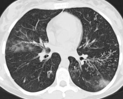
6 Ground-glass opacity hazy increase in density of the lung with preservation of bronchial and vascular margins a number of pathologic conditions: incomplete filling of the alveolar space by cells or fluid minimal thickening of the interstitium or walls increased blood volume an ongoing, dynamic process that is potentially treatable

7 Clinical symptoms!!
8 Ground-glass opacity in the early stages of certain infectious processes in chronic diffuse lung diseases» Alveolar proteinosis» Chronic eosinophilic pneumonia in a number of interstitial diseases» IPF» Connective tissue diseases» EAA» Sarcoidosis» Drug induced pulmonary toxicity
9 Infectious disease Patients with altered immunity due to neoplasm and its treatment, AIDS or organ transplantation are subject to a wide variety of lung injuries that may be rapidly fatal if unrecognized Pneumocystic jirovecii pneumonia:» Spectrum of abnormalities» Ground-glass opacification is the earliest abnormality» Perihilar, asymmetric, mosaic pattern» Slight prominence of the interlobular interstitium, in the proximity of foci of ground-glass opacities» Cystic changes superimposed on ground-glass or consolidations are very suggestive

10
11 Infectious disease Patients with altered immunity due to neoplasm and its treatment, AIDS or organ transplantation are subject to a wide variety of lung injuries that may be rapidly fatal if unrecognized Bacterial and fungal infections:» CT features are not discriminating» Halo-sign Suggestive for early invasive aspergillosis Zone of hemorrhage or edema surrounding the fungal nodule or infarct» Air-crescent sign Recovery of granulocyte function


12 30/7/2003 3/1/2003


13 11/7/2003


14 18/8/ /8/ /10/ /10/2003
15 Acute radiation pneumonitis Earliest findings within the irradiated field: ground-glass opacities heterogeneous consolidation Some degree of injury is typical with doses of 40 Gy or more


16 19/4/2002 4/9/ /9/2002
17 Interstitial disease


18 5/6/2002


19 11/9/2003
20 EAA Acute stage: Corresponding to the initial exposure to the offending antigen HRCT can be normal or may reveal poorly defined air-space consolidations Subacute stage: Resolution of the acute abnormalities Small poorly marginated nodules from 1 5 mm diameter or scattered areas of ground-glass opacities bilateral Multi-local and random without preferential involvement of the central and peripheral lung regions Mid- and upper lobes are frequently affected Chronic stage: Interstitial fibrosis Irregular reticulation and nodularity Mid- and upper lung regions


21 29/12/2003


22 24/2/2004


23 2/6/2004
24 Bronchial and peribronchial abnormalities Abnormalities indicating airway disease Tree-in-bud pattern Acute disease Bronchiectasis Mosaic perfusion Acute - chronic disease Chronic disease
25 Tree-in-bud sign bronchiolar dilatation and filling by mucus, pus, fluid or cells resembling a branching or budding tree usually visible in the lung periphery patchy in distribution indicative of airways disease endobronchial spread of infection mucoid impaction (cystic fibrosis asthma ABPA)


26
27 Bronchiectasis Features of bronchiectasis: dilatation of the lumen signet ring sign airways visible in the peripheral one third of the lung bronchial wall thickening cystic and cylindric bronchiectasis traction bronchiectasis


28 8/1/2004


29
30 Mosaic perfusion Mixed areas of increased and decreased lung attenuation Primary vascular insult versus primary bronchial pathology


31


32
33 Disease Nodular opacities Acute Subacute Chronic Distribution Perilymphatic Centrilobular At random
34 Disease Nodular opacities Acute Subacute Chronic Distribution Perilymphatic patchy patchy Centrilobular diffuse or patchy diffuse or patchy At random diffuse diffuse
35 Disease Nodular opacities Acute Subacute Chronic Distribution Perilymphatic lymphangitic carcinomatosa sarcoidosis silicosis lymphangitic carcinomatosa Centrilobular infectious bronchiolitis EAA histiocytosis X - BAC At random miliary tbc miliary fungal disease metastasis
36 Linear and reticular opacities Smooth or regular (septal) lines without sings of lung distortion inflammatory changes, filling changes acute / subacute disease Nodular or irregular (septal) lines in combination with cystic changes, honeycombing and sings of lung distortion irreversible pulmonary fibrosis chronic disease
37 Linear and reticular opacities Smooth or regular (septal) lines without sings of lung distortion inflammatory changes, filling changes» pulmonary edema, lymphangitic carcinomatosis hemorrhage, amyloidosis, alveolar proteinosis Nodular or irregular (septal) lines in combination with cystic changes, honeycombing and sings of lung distortion irreversible pulmonary fibrosis» UIP, pneumoconiosis, sarcoidosis, asbestosis
38


39


40


41 20/11/2002


42 28/5/2003


43 26/11/2004
44 Cystic lesions Definition a thin-walled well-defined and circumscribed, air- or fluid-containing lesion, with an epithelial or fibrous wall A cystic pattern infectious disease (PJP) histiocytosis X lymphangioleiomyomatosis honeycombing


45
46 Conclusion HRCT may be used to distinguish between reversible (acute or active) lung disease and irreversible (fibrotic) lung disease A number of signs have been described as indicative of active or reversible and of chronic or irreversible lung disease
47 Signs on HRCT Acute lung diseases Chronic lung diseases Ground-glass opacity Consolidation Cystic lesions Mosaic perfusion Bronchial abnormalities Nodules (centrilobular) Nodules (perilymphatic) Linear and reticular opacities (smooth, without lung distortion) Linear and reticular opacities Nodular or irregular, with lung distortion) Honeycombing
48 Conclusion Radiologic interpretation of the different patterns Nodular Linear Increased lungattentuation Decreased lungattenuation Combined patterns Associated findings Radiologic interpretation within the context of clinical information
HRCT in Diffuse Interstitial Lung Disease Steps in High Resolution CT Diagnosis. Where are the lymphatics? Anatomic distribution
 Steps in High Resolution CT Diagnosis Pattern of abnormality Distribution of disease Associated findings Clinical history Tomás Franquet MD What is the diagnosis? Hospital de Sant Pau. Barcelona Secondary
Case 1 : Question. 1.1 What is the intralobular distribution? 1. Centrilobular 2. Perilymphatic 3. Random
 Interesting case Case 1 Case 1 : Question 1.1 What is the intralobular distribution? 1. Centrilobular 2. Perilymphatic 3. Random Case 1: Answer 1.1 What is the intralobular distribution? 1. Centrilobular
Case 1: Question. 1.1 What is the main pattern of this HRCT? 1. Intralobular line 2. Groundglass opacity 3. Perilymphatic nodule
 HRCT WORK SHOP Case 1 Case 1: Question 1.1 What is the main pattern of this HRCT? 1. Intralobular line 2. Groundglass opacity 3. Perilymphatic nodule Case 1: Question 1.2 What is the diagnosis? 1. Hypersensitivity
An Image Repository for Chest CT
 An Image Repository for Chest CT Francesco Frajoli for the Chest CT in Antibody Deficiency Group An Image Repository for Chest CT he Chest CT in Antibody Deficiency Group is an international and interdisciplinary
11/10/2014. Multi-disciplinary Approach to Diffuse Lung Disease: The Imager s Perspective. Radiology
 Multi-disciplinary Approach to Diffuse Lung Disease: The Imager s Perspective Radiology Pathology Clinical 1 Role of HRCT Diagnosis Fibrosis vs. inflammation Next step in management Response to treatment
Daria Manos RSNA 2016 RC 401. https://medicine.dal.ca/departments/depar tment-sites/radiology/contact/faculty/dariamanos.html
 Daria Manos RSNA 2016 RC 401 https://medicine.dal.ca/departments/depar tment-sites/radiology/contact/faculty/dariamanos.html STEP1: Is this fibrotic lung disease? STEP 2: Is this a UIP pattern? If yes:
Financial disclosure COMMON DIAGNOSES IN HRCT. High Res Chest HRCT. HRCT Pre test. I have no financial relationships to disclose. Anatomy Nomenclature
 Financial disclosure I have no financial relationships to disclose. Douglas Johnson D.O. Cardiothoracic Imaging Gaston Radiology COMMON DIAGNOSES IN HRCT High Res Chest Anatomy Nomenclature HRCT Sampling
I have no relevant conflicts of interest to disclose
 I have no relevant conflicts of interest to disclose Diffuse parenchymal lung disease (DPLD) and its associations Secondary lobular anatomy DPLD History, clinical findings, temporal evolution, and exposures
Differential diagnosis
 Differential diagnosis Idiopathic pulmonary fibrosis (IPF) is part of a large family of idiopathic interstitial pneumonias (IIP), one of four subgroups of interstitial lung disease (ILD). Differential
The crazy-paving pattern: A radiological-pathological correlated and illustrated overview
 The crazy-paving pattern: A radiological-pathological correlated and illustrated overview Poster No.: C-0827 Congress: ECR 2010 Type: Educational Exhibit Topic: Chest Authors: W. F. M. De Wever, J. Coolen,
Outline Definition of Terms: Lexicon. Traction Bronchiectasis
 HRCT OF IDIOPATHIC INTERSTITIAL PNEUMONIAS Disclosures Genentech, Inc. Speakers Bureau Tadashi Allen, MD University of Minnesota Assistant Professor Diagnostic Radiology 10/29/2016 Outline Definition of
Usual Interstitial pneumonia and Nonspecific Interstitial Pneumonia. Nitra and the Gangs.
 Usual Interstitial pneumonia and Nonspecific Interstitial Pneumonia Nitra and the Gangs. บทน ำและบทท ๓, ๑๐, ๑๒, ๑๓, ๑๔, ๑๕, ๑๗ Usual Interstitial Pneumonia (UIP) Most common & basic pathologic pattern
HYPERSENSITIVITY PNEUMONITIS
 HYPERSENSITIVITY PNEUMONITIS A preventable fibrosis MOSAVIR ANSARIE MB., FCCP INTERSTITIAL LUNG DISEASES A heterogeneous group of non infectious, non malignant diffuse parenchymal disorders of the lower
Manish Powari Regional Training Day 10/12/2014
 Manish Powari Regional Training Day 10/12/2014 Large number of different types of Interstitial Lung Disease (ILD). Most are very rare Most patients present with one of a smaller number of commoner diseases
Bronkhorst colloquium Interstitiële longziekten. Katrien Grünberg, klinisch patholoog
 Bronkhorst colloquium 2013-2014 Interstitiële longziekten De pathologie achter de CT Katrien Grünberg, klinisch patholoog K.grunberg@vumc.nl Preparing: introduction and 3 cases The introduction on microscopic
PULMONARY TUBERCULOSIS RADIOLOGY
 PULMONARY TUBERCULOSIS RADIOLOGY RADIOLOGICAL MODALITIES Medical radiophotography Radiography Fluoroscopy Linear (conventional) tomography Computed tomography Pulmonary angiography, bronchography Ultrasonography,
Imaging Small Airways Diseases: Not Just Air trapping. Eric J. Stern MD University of Washington
 Imaging Small Airways Diseases: Not Just Air trapping Eric J. Stern MD University of Washington What we are discussing SAD classification SAD imaging with MDCT emphasis What is a small airway? Airway with
Radiologists toolbox to differentiate alveolar versus interstitial lung diseases
 Radiologists toolbox to differentiate alveolar versus interstitial lung diseases Dr Sumer Shikhare, Dr Trishna Shimpi, Dr Ashish Chawla Khoo Teck Puat Hospital Singapore. Relevant financial disclosures
Lung Allograft Dysfunction
 Lung Allograft Dysfunction Carlos S. Restrepo M.D. Ameya Baxi M.D. Department of Radiology University of Texas Health San Antonio Disclaimer: We do not have any conflict of interest or financial gain to
Mimics in chest disease: interstitial opacities
 Insights Imaging (2013) 4:9 27 DOI 10.1007/s13244-012-0207-7 PICTORIAL REVIEW Mimics in chest disease: interstitial opacities Anastasia Oikonomou & Panos Prassopoulos Received: 19 June 2012 / Revised:
Typical and atypical findings of pulmonary sarcoidosis at high resolution CT
 Typical and atypical findings of pulmonary sarcoidosis at high resolution CT Poster No.: C-0169 Congress: ECR 2013 Type: Educational Exhibit Authors: L. Raposo Rodríguez, C. Mejía, B. Escobar Mallada,
Radiologic-pathologic correlation of pulmonary diseases
 The 1578 th Chest Conference/ 3 rd Biennial Clinical- Radiologic-Pathologic Correlation Radiologic-pathologic correlation of pulmonary diseases Harumi Itoh, M.D. University of Fukui, Japan Centriacinar
A Review of Interstitial Lung Diseases. Paul J. Wolters, MD Associate Professor Department of Medicine University of California San Francisco
 A Review of Interstitial Lung Diseases Paul J. Wolters, MD Associate Professor Department of Medicine University of California San Francisco Outline Overview of diagnosis in ILD Why it is important Definition/Classification
Imaging Spectrum of Allergic Lung Disease: Hypersensitivity Reactions on the Lung Parenchyma
 Imaging Spectrum of Allergic Lung Disease: Hypersensitivity Reactions on the Lung Parenchyma Moon Sung Kim 1, Ki-Nam Lee 1, Won Jin Choi 1, Bo Ra Kim 1, Eun-Ju Kang 1 1 Department of Radiology, Dong-A
Liebow and Carrington's original classification of IIP
 Liebow and Carrington's original classification of IIP-- 1969 Eric J. Stern MD University of Washington UIP Usual interstitial pneumonia DIP Desquamative interstitial pneumonia BIP Bronchiolitis obliterans
Bronchiectasis: An Imaging Approach
 Bronchiectasis: An Imaging Approach Travis S Henry, MD Associate Professor of Clinical Radiology Cardiac and Pulmonary Imaging Section University of California, San Francisco Large Middle Small 1 Bronchiectasis
Interesting Cases. Pulmonary
 Interesting Cases Pulmonary 54M with prior history of COPD, hep B/C, and possible history of TB presented with acute on chronic dyspnea, and productive cough Hazy opacity overlying the left hemithorax
Micronodular lung pattern - Differential diagnosis
 Micronodular lung pattern - Differential diagnosis Poster No.: P-0074 Congress: ESTI 2015 Type: Educational Poster Authors: P. Ninitas, F. Marinho, P. Campos, I. Távora ; Lisbon/PT, 1 2 2 3 1 1 3 Funchal/PT,
Micronodular Lung Disease an algorithm
 Micronodular Lung Disease an algorithm H. Page McAdams, MD Department of Radiology Duke University Medical Center Durham, NC USA page.mcadams@duke.edu Question Which of the following lung diseases is MOST
A Review of Interstitial Lung Diseases
 Outline A Review of Interstitial Lung Diseases Paul J. Wolters, MD Associate Professor Department of Medicine University of California San Francisco Overview of diagnosis in ILD Why it is important Definition/Classification
Diagnostic Imaging of Diffuse Infiltrative Disease of the Lung
 Thematic Review Series Respiration 2004;71:4 19 DOI: 10.1159/000075642 Diagnostic Imaging of Diffuse Infiltrative Disease of the Lung Maurizio Zompatori a Claudio Bnà a Venerino Poletti c Enrica Spaggiari
Radiologic Approach to Smoking Related Interstitial Lung Disease
 Radiologic Approach to Smoking Related Interstitial Lung Disease Poster No.: C-1854 Congress: ECR 2013 Type: Educational Exhibit Authors: K.-N. Lee, J.-Y. Han, E.-J. Kang, J. Kang; Busan/KR Keywords: Toxicity,
10/17/2016. Nuts and Bolts of Thoracic Radiology. Objectives. Techniques
 Nuts and Bolts of Thoracic Radiology October 20, 2016 Carleen Risaliti Objectives Understand the basics of chest radiograph Develop a system for interpreting chest radiographs Correctly identify thoracic
Non-neoplastic Lung Disease II
 Pathobasic Non-neoplastic Lung Disease II Spasenija Savic Prince Pathology Program Systematic approach to surgical lung biopsies with ILD Examples (chronic ILD): Idiopathic interstitial pneumonias: UIP,
5/9/2015. Multi-disciplinary Approach to Diffuse Lung Disease: The Imager s Perspective. No, I am not a pulmonologist! Radiology
 Multi-disciplinary Approach to Diffuse Lung Disease: The Imager s Perspective No, I am not a pulmonologist! Radiology Pathology Clinical 1 Everyone needs a CT Confidence in diagnosis Definitive HRCT +
Epidemiology and classification of smoking related interstitial lung diseases
 Epidemiology and classification of smoking related interstitial lung diseases Šterclová M. Department of Respiratory Diseases, Thomayer Hospital, Prague, Czech Republic Supported by an IGA Grant No G 1207
RADIOLOGICALL ANALYSIS OF INTERSTITIAL LUNG DISEASES
 Original Research Article RADIOLOGICALL ANALYSIS OF INTERSTITIAL LUNG DISEASES Meraj Rentia 1*, Himanshu Singla 1, Divya Malpani 2, Tushar Vaishnav 3, Pradeep Jhala 3 1 1 st year Resident, 2 3 rd year
Restrictive lung diseases
 Restrictive lung diseases Restrictive lung diseases are diseases that affect the interstitium of the lung. Interstitium of the lung is the very thin walls surrounding the alveoli, it s formed of epithelium
Role of Computed Tomography in Diagnosis of Diffuse Lung Diseases Chauhan Jayant 1*, Panchal Pankaj 2, Faruqui Tehzeeb 3
 ORIGINAL ARTICLE Role of Computed Tomography in Diagnosis of Diffuse Lung Diseases Chauhan Jayant 1*, Panchal Pankaj 2, Faruqui Tehzeeb 3 1 MD,DTCD,Additional Professor& HOD, 2,3 MBBS, 3 rd year resident
American Thoracic Society European Respiratory Society Classification of the Idiopathic Interstitial Pneumonias: Advances in Knowledge since 20021
 This copy is for personal use only. To order printed copies, contact reprints@rsna.org American Thoracic Society European Respiratory Society Classification of the Idiopathic Interstitial Pneumonias: Advances
Thoracic lung involvement in rheumatoid arthritis: Findings on HRCT
 Thoracic lung involvement in rheumatoid arthritis: Findings on HRCT Poster No.: C-2488 Congress: ECR 2015 Type: Educational Exhibit Authors: R. E. Correa Soto, M. J. Martín Sánchez, J. M. Fernandez 1 1
Uses, limitations and interpretation of CT in pulmonary infections: A practical approach
 Uses, limitations and interpretation of CT in pulmonary infections: A practical approach Canadian Association of Radiologists 2013 DISCLOSURES Speakers honorarium, Siemens Canada Objectives 1. Recognize
Pulmonary Complications of Antineoplastic Agents : Era of Targeted Therapy
 Pulmonary Complications of Antineoplastic Agents : Era of Targeted Therapy Poster No.: C-1230 Congress: ECR 2013 Type: Educational Exhibit Authors: H. Y. Kim, J. H. Hwang, Y.-W. Chang, J. Y. Moon; Seoul/KR
T he diagnostic evaluation of a patient with
 546 REVIEW SERIES Challenges in pulmonary fibrosis? 1: Use of high resolution CT scanning of the lung for the evaluation of patients with idiopathic interstitial pneumonias Michael B Gotway, Michelle M
CT findings in multifocal or diffuse non-mucinous bronchioloalveolar carcinoma (BAC)
") CT findings in multifocal or diffuse non-mucinous bronchioloalveolar carcinoma (BAC) Poster No.: C-2192 Congress: ECR 2014 Type: Educational Exhibit Authors: I. Sandu, A. R. Popita, I.-A. Brumboiu; Cluj-Napoca/RO
CT findings in multifocal or diffuse non-mucinous bronchioloalveolar carcinoma (BAC) Poster No.: C-2192 Congress: ECR 2014 Type: Educational Exhibit Authors: I. Sandu, A. R. Popita, I.-A. Brumboiu; Cluj-Napoca/RO
Cryptogenic Organizing Pneumonia Diagnosis Approach Based on a Clinical-Radiologic-Pathologic Consensus
 Cryptogenic Organizing Pneumonia Diagnosis Approach Based on a Clinical-Radiologic-Pathologic Consensus Poster No.: C-1622 Congress: ECR 2012 Type: Scientific Exhibit Authors: C. Cordero Lares, E. Zorita
NONE OVERVIEW FINANCIAL DISCLOSURES UPDATE ON IDIOPATHIC PULMONARY FIBROSIS/IPF (UIP) FOR PATHOLOGISTS. IPF = Idiopathic UIP Radiologic UIP Path UIP
 FOR PATHOLOGISTS. IPF = Idiopathic UIP Radiologic UIP Path UIP") UPDATE ON IDIOPATHIC PULMONARY FIBROSIS/IPF () FOR PATHOLOGISTS Thomas V. Colby, M.D. Professor of Pathology (Emeritus) Mayo Clinic Arizona FINANCIAL DISCLOSURES NONE OVERVIEW IPF Radiologic Dx Pathologic
UPDATE ON IDIOPATHIC PULMONARY FIBROSIS/IPF () FOR PATHOLOGISTS Thomas V. Colby, M.D. Professor of Pathology (Emeritus) Mayo Clinic Arizona FINANCIAL DISCLOSURES NONE OVERVIEW IPF Radiologic Dx Pathologic
September 2014 Imaging Case of the Month. Michael B. Gotway, MD. Department of Radiology Mayo Clinic Arizona Scottsdale, AZ
 September 2014 Imaging Case of the Month Michael B. Gotway, MD Department of Radiology Mayo Clinic Arizona Scottsdale, AZ Clinical History: A 57-year-old non-smoking woman presented to her physician as
Radiological Imaging of Drug-Induced Pulmonary Lesions
 Review Article imedpub Journals www.imedpub.com Journal of Clinical Radiology and Case Reports Radiological Imaging of Drug-Induced Pulmonary Lesions D souza M *, Rajiah P, Khan A and Irion K Department
How to identify interstitial pneumonias.
 How to identify interstitial pneumonias. Poster No.: C-0804 Congress: ECR 2014 Type: Educational Exhibit Authors: S. claret loaiza, M. C. Cañete Moslero, R. Carreño Gonzalez, C. de la Torre; Malaga/ES
INTERSTITIAL LUNG DISEASE. Radhika Reddy MD Pulmonary/Critical Care Long Beach VA Medical Center January 5, 2018
 INTERSTITIAL LUNG DISEASE Radhika Reddy MD Pulmonary/Critical Care Long Beach VA Medical Center January 5, 2018 Interstitial Lung Disease Interstitial Lung Disease Prevalence by Diagnosis: Idiopathic Interstitial
August 2018 Imaging Case of the Month: Dyspnea in a 55-Year-Old Smoker. Michael B. Gotway, MD
 August 2018 Imaging Case of the Month: Dyspnea in a 55-Year-Old Smoker Michael B. Gotway, MD Department of Radiology Mayo Clinic Arizona Scottsdale, AZ USA Clinical History: A 55 year old woman presented
Atopic Pulmonary Disease: Findings on Thoracic Imaging
 July 2003 Atopic Pulmonary Disease: Findings on Thoracic Imaging Rebecca G. Breslow Harvard Medical School Year IV Churg-Strauss Syndrome Hypersensitivity Pneumonitis Asthma Atopic Pulmonary Disease Allergic
Eosinophilic lung diseases - what the radiologist needs to know
 Eosinophilic lung diseases - what the radiologist needs to know Poster No.: C-0803 Congress: ECR 2014 Type: Authors: Keywords: DOI: Educational Exhibit E.-M. Heursen, R. Reina Cubero, F. Japon Sola; Cádiz/ES
I don t need you. Disclosure Statement. Pathology Approach to ILD 11/5/2016. Kirk D. Jones, MD UCSF Dept of Pathology
 Pathology Approach to ILD Disclosure Statement Relevant financial relationships with a commercial interest: Boeringer Ingleheim, speaker Kirk D. Jones, MD UCSF Dept of Pathology kirk.jones@ucsf.edu I don
Disease spectrum. IPA Invasive pulmonary aspergillosis
 Aspergillus & ABPA Disease spectrum IPA Invasive pulmonary aspergillosis ABPA ABPA pathophysiology conidia of Aspergillus trapped in mucous and narrowed airways of asthmatics/cf germinate to form hyphae
Radiologic findings of drug-induced lung disease
 Radiologic findings of drug-induced lung disease Poster No.: P-0115 Congress: ESTI 2015 Type: Educational Poster Authors: A. I. C. Santos, A. F. Roque, R. Mamede, L. Oliveira, T. Saldanha; Lisbon/PT Keywords:
IPF: Epidemiologia e stato dell arte
 IPF: Epidemiologia e stato dell arte Clinical Classification Diffuse parenchimal lung diseases Exposure-related: - occupational - environmental - medication Desquamative interstitial pneumonia Idiopathic
Diagnostic challenges in IPF
 Medicine, Nursing and Health Sciences Diagnostic challenges in IPF Dr Ian Glaspole Central and Eastern Clinical School, Alfred Hospital and Monash University March 2015 Disclosures Consultancy fees from
Smoking-related Interstitial Lung Diseases: High-Resolution CT Findings
 Smoking-related Interstitial Lung Diseases: High-Resolution CT Findings Poster No.: C-2358 Congress: ECR 2013 Type: Educational Exhibit Authors: V. Cuartero Revilla, M. Nogueras Carrasco, P. Olmedilla
Progress in Idiopathic Pulmonary Fibrosis
 Progress in Idiopathic Pulmonary Fibrosis David A. Lynch, MB Disclosures Progress in Idiopathic Pulmonary Fibrosis David A Lynch, MB Consultant: t Research support: Perceptive Imaging Boehringer Ingelheim
Imaging of the Lung. István Battyány
 Imaging of the Lung István Battyány Anatomy of airways: (asszimetric dichotomy) trachea Main bronchus Lobar bronchus }lung, lobe 1. segmental bronchus subsegmental bronchus bronchus lobularis bronchiolus
Pulmonary manifestations of Rheumatoid Arthritis: what is there waiting to be found?
 Pulmonary manifestations of Rheumatoid Arthritis: what is there waiting to be found? Poster No.: C-1795 Congress: ECR 2015 Type: Educational Exhibit Authors: M. S. C. Rodrigues, R. Correia, A. Carvalho,
Residents Section Pattern of the Month
 Residents Section Pattern of the Month Gosset et al. Tree-In-Bud Pattern Residents Section Pattern of the Month Residents inradiology Natacha Gosset 1 Alexander A. Bankier Ronald L. Eisenberg Gosset N,
October 2012 Imaging Case of the Month. Michael B. Gotway, MD Associate Editor Imaging. Department of Radiology Mayo Clinic Arizona Scottsdale, AZ
 October 2012 Imaging Case of the Month Michael B. Gotway, MD Associate Editor Imaging Department of Radiology Mayo Clinic Arizona Scottsdale, AZ Clinical History: A 65-year-old non-smoking woman presented
Bronchiolitis: A Schematic Diagnostic Approach with Radiologic-pathologic Correlation
 Bronchiolitis: A Schematic Diagnostic Approach with Radiologic-pathologic Correlation Mariana Benegas Urteaga 1, MD; M Sanchez 1, MD; J Ramirez 2, MD; D Barnes 1, MD; T de Caralt 1, MD; R J Perea 1, MD
Immunocompromised patients. Immunocompromised patients. Immunocompromised patients
 Value of CT in Early Pneumonia in Immunocompromised Patients Nantaka Kiranantawat, PSU Preventative Factors Phagocyts Cellular immunity Humoral immunity Predisposing Factors Infection, Stress, Poor nutrition,
Hypersensitivity Pneumonitis: Spectrum of High-Resolution CT and Pathologic Findings
 CT of Hypersensitivity Pneumonitis Chest Imaging Pictorial Essay C. Isabela S. Silva 1 ndrew Churg 2 Nestor L. Müller 1 Silva CIS, Churg, Müller NL Keywords: high-resolution CT, hypersensitivity pneumonitis,
The radiological differential diagnosis of the UIP pattern
 5th International Conference on Idiopathic Pulmonary Fibrosis, Modena, 2015, June 12th The radiological differential diagnosis of the UIP pattern Simon Walsh King s College Hospital Foundation Trust London,
Cystic Lung Disease. Cristopher A. Meyer, MD
 Cystic Lung Disease Cristopher A. Meyer, MD Air filled structure with definable wall typically less than 1 mm thick Cris A. Meyer, M.D. Professor of Radiology University of Wisconsin School of Medicine
Workshop Cyst & Lucency. How to Approach
 Workshop Cyst & Lucency How to Approach To Approach Cystic Lung Disease True cysts? Cavitary disease Cystic bronchiectasis Mosaic attenuation Subpleural cysts Bullae Paraseptal emphysema Honeycombing Birt
Thoracic Manifestations of Sarcoidosis Using Multi-Slice CT
 IOSR Journal of Dental and Medical Sciences (IOSR-JDMS) e-issn: 2279-0853, p-issn: 2279-0861.Volume 14, Issue 9 Ver. II (Sep. 2015), PP 63-68 www.iosrjournals.org Thoracic Manifestations of Sarcoidosis
Dr.kassim.m.sultan F.R.C.P
 Dr.kassim.m.sultan F.R.C.P inflammatory disorder of the lung, involving alveolar walls and terminal airways, that is induced, in a susceptible host, by repeated inhalation of a variety of organic agents.
The Imaging Analysis of Pulmonary Sarcodiosis
 www.cancercellresearch.org ISSN: 2161-2609 Article The Imaging Analysis of Pulmonary Sarcodiosis Xin He, Chuanyu Zhang* Department of Radiology, Affiliated Hospital of Qingdao University, Qingdao, China
Imaging findings in Hypersensitivity Pneumonitis - a pictorical review.
 Imaging findings in Hypersensitivity Pneumonitis - a pictorical review. Poster No.: C-1655 Congress: ECR 2014 Type: Educational Exhibit Authors: B. M. Araujo, A. F. S. Simões, M. S. C. Rodrigues, J. Pereira;
ACUTE PULMNARY INFECTIONS: UNDERSTANDING THE CHEST RADIOGRAPH. Leonard E. Swischuk, M.D. University of Texas Medical Branch
 ACUTE PULMNARY INFECTIONS: UNDERSTANDING THE CHEST RADIOGRAPH Leonard E. Swischuk, M.D. University of Texas Medical Branch AUTHOR HAS NOTHING TO DECLARE LEARNING OBJETIVES Understand the pathophysiology
4/17/2010 C ini n ca c l a Ev E a v l a ua u t a ion o n of o ILD U dat a e t e i n I LDs
 Update in ILDs Diagnosis 101: Clinical Evaluation April 17, 2010 Jay H. Ryu, MD Mayo Clinic, Rochester MN Clinical Evaluation of ILD Outline General aspects of ILDs Classification of ILDs Clinical evaluation
ARTICLE IN PRESS. Ahuva Grubstein a, Daniele Bendayan b, Ithak Schactman c, Maya Cohen a, David Shitrit b, Mordechai R. Kramer b,
 Respiratory Medicine (2005) 99, 948 954 Concomitant upper-lobe bullous emphysema, lower-lobe interstitial fibrosis and pulmonary hypertension in heavy smokers: report of eight cases and review of the literature
Role of High Resolution Computed Tomography in Evaluation of Pulmonary Diseases
 International J. of Healthcare & Biomedical Research, Volume:, Issue:, April, Pages 9-96 Role of High Resolution Computed Tomography in Evaluation of Pulmonary Diseases Dr. Abhijeet D. Nagapurkar*, Dr.
Glossary of terms in thoracic imaging and clinicalradiological correlation. What every radiologist should
 Glossary of terms in thoracic imaging and clinicalradiological correlation. What every radiologist should know. Poster No.: C-1565 Congress: ECR 2012 Type: Educational Exhibit Authors: A. Moreno Pastor,
Systemic lupus erythematosus (SLE): Pleuropulmonary Manifestations
: Pleuropulmonary Manifestations") 08/30/10 09/26/10 Systemic lupus erythematosus (SLE): Pleuropulmonary Manifestations Camila Downey S. Universidad de Chile, School of Medicine, Year VII Harvard University, School of Medicine Sept 17,
08/30/10 09/26/10 Systemic lupus erythematosus (SLE): Pleuropulmonary Manifestations Camila Downey S. Universidad de Chile, School of Medicine, Year VII Harvard University, School of Medicine Sept 17,
Pictorial essay of unusual radiologic manifestations of pulmonary and airway metastasis at initial presentation of lung cancer
 Pictorial essay of unusual radiologic manifestations of pulmonary and airway metastasis at initial presentation of lung cancer Poster No.: C-2297 Congress: ECR 2012 Type: Educational Exhibit Authors: Y.
Spectrum of Cystic Lung Disease and its Mimics. Kathleen Jacobs MD and Elizabeth Weihe MD UC San Diego Medical Center, Department of Radiology
 Spectrum of Cystic Lung Disease and its Mimics Kathleen Jacobs MD and Elizabeth Weihe MD UC San Diego Medical Center, Department of Radiology No Financial Disclosures Learning Objectives 1. Review the
Radiation Pneumonitis Joseph Junewick, MD FACR
 Radiation Pneumonitis Joseph Junewick, MD FACR 03/19/2010 History 16 year old with history of relapsed stage IV-A Hodgkin disease. Prior pulmonary involvement was irradiated. Diagnosis Radiation Pneumonitis
Pulmonary Manifestations of Systemic Lupus Erythematosus 1
 Pulmonary Manifestations of Systemic Lupus Erythematosus 1 Kee Hyuk Yang, M.D., Yo Won Choi, M.D., Seok Chol Jeon, M.D., Choong Ki Park, M.D., Kyung in Joo, M.D., Chang Kok Hahm, M.D., Seung Ro Lee, M.D.
ARDS - a must know. Page 1 of 14
 ARDS - a must know Poster No.: C-1683 Congress: ECR 2016 Type: Authors: Keywords: DOI: Educational Exhibit M. Cristian; Turda/RO Education and training, Edema, Acute, Localisation, Education, Digital radiography,
Pneumocystis jirovecci pneumonia: from mild disease to a real disaster. A pictorial review of the different radiologic patterns in acute settings
 Pneumocystis jirovecci pneumonia: from mild disease to a real disaster. A pictorial review of the different radiologic patterns in acute settings Poster No.: C-1425 Congress: ECR 2017 Type: Educational
The role of high-resolution computed tomography in the follow-up of diffuse lung disease
 SERIES RADIOLOGY The role of high-resolution computed tomography in the follow-up of diffuse lung disease Brett M. Elicker, Kimberly G. Kallianos and Travis S. Henry Number 2 in the Series Radiology Edited
Chest imaging II. Interstitial lung diseases
 Chest imaging II. Interstitial lung diseases Dávid L. Tárnoki MD, PhD Ádám D. TárnokiMD, PhD Department of Radiology Semmelweis University Topics 1. Interstitial lung diseases 2. Occupational lung diseases
Chest Radiology Interpretation: Findings of Tuberculosis
 Chest Radiology Interpretation: Findings of Tuberculosis Get out your laptops, smart phones or other devices pollev.com/chestradiology Case #1 1 Plombage Pneumonia Cancer 2 Reading the TB CXR Be systematic!
Idiopathic interstitial pneumonias (IIPs) are a group of
 are a group of") SYMPOSIA C. Isabela S. Silva, MD, PhD and Nestor L. Müller, MD, PhD Abstract: The idiopathic interstitial pneumonias (IIPs) are a group of diffuse parenchymal lung diseases of unknown etiology characterized
SYMPOSIA C. Isabela S. Silva, MD, PhD and Nestor L. Müller, MD, PhD Abstract: The idiopathic interstitial pneumonias (IIPs) are a group of diffuse parenchymal lung diseases of unknown etiology characterized
Tuberculosis: The Essentials
 Tuberculosis: The Essentials Kendra L. Fisher, MD, PhD THORACIC TUBERCULOSIS: THE BARE ESSENTIALS Kendra Fisher MD, FRCP (C) Department of Radiology Loma Linda University Medical Center TUBERCULOSIS ()
CLEARING THE AIR ON DIFFUSE PARENCHYMAL (INTERSTITIAL) LUNG DISEASE (ILD)
 LUNG DISEASE (ILD)") CLEARING THE AIR ON DIFFUSE PARENCHYMAL (INTERSTITIAL) LUNG DISEASE (ILD) David Northrop MBA, RRT Assistant Director of Respiratory Therapy Services The University of Kansas Health System Clinical Assistant
CLEARING THE AIR ON DIFFUSE PARENCHYMAL (INTERSTITIAL) LUNG DISEASE (ILD) David Northrop MBA, RRT Assistant Director of Respiratory Therapy Services The University of Kansas Health System Clinical Assistant
How to Analyse Difficult Chest CT
 How to Analyse Difficult Chest CT Complex diseases are:- - Large lesion - Unusual or atypical pattern - Multiple discordant findings Diffuse diseases are:- - Numerous findings in both sides 3 basic steps
Lung CT: Part 2, The Interstitial Pneumonias Clinical, Histologic, and CT Manifestations
 Integrative Imaging Review Ferguson and Berkowitz CT of Interstitial Pneumonia Integrative Imaging Review CME SAM Lung CT FOCUS ON: Emma C. Ferguson 1 Eugene A. Berkowitz 2 Ferguson EC, Berkowitz EA Keywords:
Histopathologic Approach to Interstitial Lung Disease
 Histopathologic Approach to Interstitial Lung Disease Kirk D. Jones, MD UCSF Dept of Pathology kirk.jones@ucsf.edu Disclosures I have nothing to disclose 1 Why? Much of interstitial lung disease biopsies
Imaging: how to recognise idiopathic pulmonary fibrosis
 REVIEW IDIOPATHIC PULMONARY FIBROSIS Imaging: how to recognise idiopathic pulmonary fibrosis Anand Devaraj Affiliations: Dept of Radiology, St George s Hospital, London, UK. Correspondence: Anand Devaraj,