Optimal Method for Stenosis Assessment with CT
|
|
|
- Phoebe Simon
- 6 years ago
- Views:
Transcription
1 Optimal Method for Stenosis Assessment with CT Jonathon Leipsic MD FRCPC FSCCT ViceChairman ofradiologyubc Disclosures Speaker s bureau: GE Healthcare and Edwards LifeSciences Grant Support CIHR, GE Healthcare Advisory Board GE Healthcare, Vital Images and Edwards LifeSciences Equity Stakeholder TC3 1


2 CCTA in Routine Clinical Practice in 2013 Is there a severe anatomic stenosis? Left anterior descending artery CCTA Findings: >70% stenosis LAD 25-49% stenosis LCx <25% stenosis RCA Left circumflex artery Right coronary artery 2
3 Three (3) Prospective Multicenter Studies Sensitivity Specificity PPV NPV ACCURACY N=230, Stable Chest Pain; No known CAD; No exclusion (CACS, HR, BMI); CAD prevalence 13% CorE N=291, Stable Chest Pain; No known and Known CAD; Exclusion CACS>600; CAD prevalence 56% Meijboom N=360, Acute and Stable Chest Pain; No known CAD; CAD prevalence 68% Budoff et al. JACC 2008; Miller et al. NEJM 2008; Meijboom et al. JACC 2009 Diagnosis of obstructive CAD Test Sensitivity Specificity Exercise ECG treadmill 1 68% 77% Exercise Echo treadmill 2 86% 81% Dobutamine Echo treadmill 2 ~85% ~85% Exercise nuclear treadmill 3 87% 73% Pharmacologic nuclear 3 89% 75% Cardiac CTA 4 95% 83% 1. ACC/AHA 2002 Guideline Update for Exercise Testing 2. ACC/AHA/ASE 2003 Guideline Update for the Clinical Application of Echocardiography 3. ACC/AHA/ASNC Guidelines for the Clinical Use of Cardiac Radionuclide Imaging 4. ACCURACY study, presented at 2008 ACC Scientific Sessions 3


4 Tips from Dr Achenbach- Data Acquisition Rule-outs/normal coronary arteries can usually be evaluated even at higher heart rates. Especially in difficult cases: Low heart rates maximize accuracy. 3 Calcium For Evaluation of calcified segments: Careful multiplanar reconstruction 4


5 3. Calcium For evaluation of calcified segments: Careful multiplanar recnstruction 3. Calcium For evaluation of calcified segments: Careful multiplanar recnstruction 5


6 The diagnostic performance of CCTA is improving with each generation of technology Dual source CT Improved temporal resolution 75 ms resolution 320 slice MDCT Improved volume coverage Whole heart acquisition in 1 beat High Definition CT Improved spatial resolution Enhanced calcified plaque and metal imaging Reduced radiation Single source vs. Dual source CCTA 200 patients with suspected CAD randomized to SSCT vs. DSCT. Sub-randomization for HR control vs. no HR control. For sensitivity of detection of obstructive stenosis, SSCT improved with HRT (78% vs. 57%, p=0.04); DSCT unaffected by HRC (87% vs. 93%, p=ns). Source: Achenbach et al. JACC Img


7 High Definition CCTA Cordis velocity 2.5mm Cordis velocity 2.5mm Cordis cypher 2.75mm Cordis cypher 2.75mm Non-HDCT HDCT Source: Min et al. JCCT 2009 High Definition CCTA Multicenter (4 sites) study of 70 consecutive patients undergoing CCTA by HDCT and ICA. Obstructive CAD prevalence 48%. Median Estimated Effective Dose of 2.3 msv Heydari et al. CJC


8 Dual Energy CCTA Prospective gating only (increased available padding to 300 msec) 64 x.625 mm 0.5 sec switching 80 to 140 kv Iterative Reconstuction Projection Based Monochromatic and material il decomposition images fully available Potential Advantages of Projection Based Dual NRG CCTA 77 yr old male New chest pressure + Family History CAD CCTA 9/13/2011 Prospective gating; CCTA DLP 350 mgy cm CS 1301 >50% LAD disease, dense calcified plaque distal to 2 nd diagonal Rapid kvp Switching Dual Energy CCTA 9/20/2011 Prospective gating; CCTA DLP 394 mgy cm Less calcium blooming 8


9 90% stenosis Patent stent Reduced d blooming >50% stenosis Conventional 120 kvp CCTA Exam 9/13/2011 GSI CCTA 65 kev MC Images Exam 9/20/ kev 90 kev 50 kev 100 kev 60 kev 110 kev 70 kev 120 kev 80 kev 130 kev 9
10 Disclosures Research Support: NHLBI (R01HL U01 HL [Contract]) QNRF (NPRP ) 3 089) GE Healthcare (significant) Philips Healthcare (modest) Vital Images (modest) Equity Interest: TC3 MDDX Cedars-Sinai Medical Center Medical Advisory Board: GE Healthcare Arineta 60 y/o with atypical angina scheduled to undergo intra-abdominal surgery Symptoms: 60 YM atypical angina and associated with shortness of breath; under a lot of stress ; exercises regularly CAD risk factors: Dyslipidemia, Diabetes, FH, HTN Medications: Aspirin, Beta blocker, Diuretic, Ezetimibe, HMG CoA reductate inhibitor Other factors: BMI 29; ECG- Normal Sinus Rhythm A CCTA is performed
11 How Severe is the Stenosis and What Should be done? Stenosis Severity 0-24% 25-49% 50-69% >70% 100% Cheng et al. JACC CV Imaging (Jul 2008) Limitations of CCTA Stenosis severity by CCTA demonstrates an unreliable relationship to lesion specific ischemiawith the majority of obstructive stenosis detected by CCTA not causal of ischemia CCTA cannot predict global myocardial perfusion 26 11
 Ischemia Matters log Hazard Ratio 6 2 3 4 5")

12 Perfusion Imaging Coronary anatomy is a poor predictor of the hemodynamic significance of CAD (Blankstein, Di Carli, Nature Reviews 2010) Ischemia Matters log Hazard Ratio Cedars-Sinai Nuclear Registry 10,627 patients No CAD 1.9 yr cardiac death * Medical Rx Revasc * *p< % 25% 32.5% 50% Source: Hachamovitch, et al Circulation 2003 % Myocardium Ischemic 12

 nitrates with the for use")


13 Courage Nuclear Sub study 1. Comparison Improvements of the in effectiveness angina class of PCI for ischemia reduction added to optimal 2. medical Less reliance therapy on (OMT) nitrates with the for use symptom of MPI. relief 3. Greater anti ischemic benefit of PCI was with more severe ischemia at baseline. 33% with > 5% Ischemia 18.9% with > 5% Ischemia 4. If > 5% Reduction reduction (p=0.0004) in ischemia in either group, there Reduction was a trend for reduced rate of death or MI (NS) PCI + OMT OMT Shaw et al., Circulation 2008; 117:


14 Stress CT Protocol Scout Images Test Bolus Start ~ 5 Minutes Stress Perfusion Rest Scan HR ~ 79 HR ~ 69 Adenosine Contrast Contrast ~ 7 Minutes Delayed Enhancement End Stress Perfusion Rest Scan Delayed Enhancement Data Stress perfusion, coronary CTA, function Rest Perfusion Delayed enhancement Triggering Retrospective Prospective Prospective Contrast 60 80cc 60 80cc None Tube current modulation Yes (60 70) N/A N/A kv 100 or or Blankstein Blankstein R et al. JACC 2009;54: R et al. JACC 2009;54: CT Perfusion (stress) Coronary CT Angiography Stress SPECT MPI Rest 14


")

15 Studies utilizing cardiac computed tomography perfusion (CTP) imaging as compared with (MPI) CTP vs Stress Perfusion MRI 15

16 CTP/ CCTA vs Cath Measured FFR Diagnostic Performance of Combined Noninvasive Coronary Angiography and Myocardial Perfusion Imaging Using 320 row Detector Computed Tomography: The CORE320 Multicenter International Study João A.C. Lima, M.D. Professor of Medicine and Radiology Johns Hopkins Hospital For the CORE320 Investigators 16
17 Primary Hypothesis CTA 50% Stenosis ICA 50% Stenosis vs CTP Perfusion Defect SPECT Perfusion Defect Vavere A, et al, JCCT, 2011, 5(6): Patient Based Results Incremental Value of Most Severe CTA-CTP over CTA (Reference Standard: 50% by ICA with SPECT-MPI defect) Sensitivity P<0.001 CTA-CTP ROC Area = % CI [ ] CTA ROC Area = % CI [ ] Combined Most Severe CTA-CTP and CTA alone vs. Reference Standard (ICA 50% with SPECT-MPI defect) CTA+CTP N=381 CTA alone Prevalence = 38% 1-Specificity 17


18 Summary Combined CTA and CTP can detect hemodynamically significant stenosis (50% or greater) by ICA with an associated SPECT MPI defect. CT myocardial perfusion adds to the diagnostic power of CT angiography alone. The combination of CTA & CTP in one non invasive examination is useful to identify patients who benefit from revascularization and guide management of CAD. Rocha-Filho et al. Radiology 2010 Feb. 18


19 Case 1: 70-year-old man with diabetes, hypertension, old infarct Reading Stress Step 1 CTA Proximal stent restenosis Step 2 ct perfusion Normal perfusion Step 3 final call Non significant stenosis Rest Gold standard Mild proximal stent stenosis Case 2: 63 y-o male, history of CAD, episode of syncope. Hypertension and hyperlipidemia Reading Stress Step 1 CTA RCA Non interpretable LCX Possible stenosis Step 2 ct perfusion Step 3 final call RCA Normal perfusion LCX Abnormal perfusion RCA Non-significant stenosis LCX Significant stenosis Rest Gold standard RCA Non significant stenosis LCX Significant stenosis 19
 FFR at the time of invasive coronary angiography (ICA) is the only method for")

20 What About Lesion Specific Ischemia? Reference Gold Standard for Ischemia: Fractional Flow Reserve (FFR) FFR at the time of invasive coronary angiography (ICA) is the only method for specific determination of the hemodynamic significance of coronary artery lesions (lesion-specific ischemia) FFR = Ratio of maximal myocardial blood flow through a diseased artery to the blood flow in the hypothetical case that this artery is normal Values <0.80 or <0.75 considered diagnostic of lesion-specific ischemia Source: Pijls NH et al. J Am Coll Cardiol. 2007; Pijls NH et al. J. Am. Coll. Cardiol

21 Assessment of Lesion-specific Ischemia by FFR is the Only Method To Improve Event-free Survival Source: Tonino et al. NEJM 2009;360:213-24; Pijls et al. J Am Coll Cardiol


 and density of blood Calculated data: Velocity and pressure of blood in coronary arteries")
22 Computational Fluid Dynamics Computational fluid dynamics (CFD) quantifies fluid pressure and velocity, based on physical laws of mass conservation and momentum balance CFD for Patient-Specific Models of Coronary Arteries 1. Numerical method approximates governing equations 2. Obtain solution for velocity / pressure at finite (but very large) number of points 3. Simultaneously solve millions of non-linear equations and repeating process for thousands of time intervals within cardiac cycle Images courtesy of Prof. Charbel Farhat, Dept. of Aeronautics & Astronautics, Stanford University An Analogy for Computing FFR CT from a Static CT Flow over a Wing Input data: Geometry from design specs Boundary conditions Velocity of incoming air relative to wing Atmospheric pressure, P=P atm Flow through an Artery Input data: Geometry high quality 64 slice CT Boundary conditions Blood pressure Resting coronary flow calculated from myocardial mass Coronary microcirculatory resistance determined from size of feeding vessel Boundary conditions Boundary conditions Fluid Properties viscosity and density of air Fluid properties viscosity (from hematocrit) and density of blood Calculated data: Velocity and pressure of blood in coronary arteries FFR CT Calculated data: Velocity and pressure of air in front of, around, behind wing Lift and drag FFR CT 84 CCM A Courtesy: C. Farhat, Dept Aeronautics, Stanford 22


23 Patient-Specific Computation of FFR CT (1) (2) (3) (4) (5) (6) 140 mcg/kg/mi n 1. Image-Based Modeling Segmentation of patient-specific arterial geometry 2. Heart-Vessel Interactions Allometric scaling laws relate caliber to pressure and flow 3. Microcirculatory resistance Mophometry laws relate coronary dimension to resistance 4. Left Ventricular Mass Lumped-parameter model couples pulsatile coronary flow to timevarying myocardial pressure 5. Physiologic Conditions Blood as Newtonian fluid adjusted to patient-specific viscosity 6. Induction of Hyperemia Compute maximal coronary vasodilation 7. Fluid Dynamics Navier-Stokes equations applied for coronary pressure Simulating coronary blood flow uses similar principles but is even more complicated Patient-specific geometry Heart-Vessel Interactions Complex fluid Properties Inputs Accurate coronary geometric models including branching structure and pathology Physiologic models personalized using minimal measured data Boundary Conditions Heart/vascular interaction Aortic impedance Time-varying coronary resistance related to intramyocardial pressure Models to simulate hyperemia Flow demand related to myocardial mass, cardiac work, etc Microcirculatory resistance depends on complex vessel structure Physiology changes due to administration of drugs to induce hyperemia Numerical Methods Anisotropic, adaptive, boundary layer mesh generation to reduce computation time Tight coupling between heart model and aorta/coronary model High performance parallel incompressible flow solver 23


24 Case Examples: Obstructive CAD CT ICA and FFR FFR CT Case 1 LAD stenosis FFR 0.65 = Lesion-specific ischemia FFR CT 0.62 = Lesion-specific ischemia CT ICA and FFR FFR CT Case 2 RCA stenosis FFR 0.86 = No ischemia FFR CT 0.87 = No ischemia Examples DISCOVER-FLOW CCTA FFR CT Invasive angiography FFR FFR 0.74 >50% diameter stenosis FFR CT 0.74 ischemia >50% diameter stenosis FFR 0.74 ischemia FFR 0.85 >50% diameter stenosis FFR CT 0.85 no ischemia >50% diameter stenosis FFR 0.84 no ischemia 24


25 I KNOW A TIGHT LESION I KNOW A TIGHT LESION WHEN I SEE ONE.. 25


26 I know a tight lesion when I see one I know a tight lesion when I see one LAD FFR Negative FFR CT = 0.83 Invasive=0.83 LAD FFR Negative FFR CT = 0.81 Invasive=0.81 LAD FFR Positive FFR CT = 0.72 Invasive=0.75 LAD FFR Negative FFR CT = 0.93 Invasive=0.95 LAD FFR Negative FFR CT = 0.84 Invasive=0.84 LAD FFR Positive FFR CT = 0.74 Invasive=


27 What about intermediate stenoses? 50-70% 50-70% Source: Tonino PA et al. J Am Coll Cardiol 27

 Diagnostic Performance of")


28 Moderate vs. Severe Stenosis: Moving Beyond a Binary Approach Stenosis Severity 0-24% 25-49% 50-69% >70% 100% Cheng et al. JACC CV Imaging (Jul 2008) Diagnostic Performance of FFRCT for 30-69% Stenoses by CCTA FFR CT Accuracy Sensitivity Specificity PPV NPV FFR CT CCTA Accuracy Sensitivity Specificity PPV NPV Leipsic et al. RSNA


29 Diagnostic performance of FFR CT compared to invasive measured FFR for lesions of intermediate stenosis severity FFR CT Accuracy (%) Sensitivity (%) Specificity (%) PPV (%) NPV (%) CCTA stenosis >50%* FFR CT 0.80 (95% CI) 56.1 ( ) 90.3 ( ) 25.7 ( ) 51.9 ( ) 75.0 ( ) 35% improvement in overall accuracy with nearly ( ) 60% improvement ( ) ( ) e in specificity ( ) c ( ) FFR CT 0.75 (95% CI) 83.3 ( ) 77.3 ( ) 86.4 ( ) 73.9 ( ) 88.4 ( ) 29
30 Conclusions CCTA is an accurate test for the detection and exclusion of obstructive CAD with significant recent advancements CT perfusion is a developing tool with promising data from single center studies The optimal protocol has yet to be defined dfi d CORE 320 first multicenter again with promising results but may be vendor specific FFR CT - Conclusions 1. Demonstrates high diagnostic performance compared to invasive measured FFR with 6 fold reduction in false positives as compared to CCTA 2. >40% improvement in diagnostic accuracy compared to CCTA 3. Robust discrimination of ischemia-causing lesions 4. Good correlation to invasive FFR FFR i t ti l th d th t b FFR CT is a new computational method that may be considered for the physiologic assessment of intermediate stenoses identified by CCTA 30

31 Awaiting Results from Larger Multicenter Trials Thank you. 31
")


32 Iodine (Water) MD Iodine (HAP) MD 32


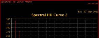
33 Spectral HU Curve 40 kev 50 kev 60 kev 70 kev 80 kev 90 kev 100 kev 120 kev 140 kev 33
34 Moderate vs. Severe Stenosis: Moving Beyond a Binary Approach Stenosis Severity 0-24% 25-49% 50-69% >70% 100% Cheng et al. JACC CV Imaging (Jul 2008) Pre-Test Likelihood: ACC/AHA Clinical Practice Guidelines Pre-test obstructive CAD probability Nonanginal CP Atypical Angina Typical Angina Age (y) (%) (%) (%) (%) (%) (%) Table recreated from: Gibbons, et al. ACC/AHA 2002 Guideline Update for the Management of Patients with Chronic Stable Angina. Diamond GA, Forrester JS. N Eng J Med 1979;300: Chaitman, BR. Circulation 1981;64:
Diagnostic Accuracy of Fractional Flow Reserve from Anatomic Computed TOmographic Angiography: The DeFACTO Study
 Diagnostic Accuracy of Fractional Flow Reserve from Anatomic Computed TOmographic Angiography: The DeFACTO Study James K. Min 1 ; Jonathon Leipsic 2 ; Michael J. Pencina 3 ; Daniel S. Berman 1 ; Bon-Kwon
Fractional Flow Reserve (FFR)
") Non-invasive FFR using coronary CT angiography and computational fluid dyn amics predicts the hemodynamic signifi cance of coronary lesions First in man experience with CT-Flow Andrejs Erglis, Sanda Jegere,
Non-invasive FFR using coronary CT angiography and computational fluid dyn amics predicts the hemodynamic signifi cance of coronary lesions First in man experience with CT-Flow Andrejs Erglis, Sanda Jegere,
The Latest on CT Fractional Flow Reserve. Dimitris Mitsouras, Ph.D.
 The Latest on CT Fractional Flow Reserve Dimitris Mitsouras, Ph.D. Assistant Professor of Radiology Harvard Medical School Director, Applied Imaging Science Lab Brigham and Women s Hospital Disclosures
Disclosure Information
 Coronary CTA Pearls and Pitfalls Ricardo C. Cury, MD, FSCCT, FAHA, FACC Chairman of Radiology Radiology Associates of South Florida Director of Cardiac Imaging Miami Cardiac and Vascular Institute Past-President
Current and Future Imaging Trends in Risk Stratification for CAD
 Current and Future Imaging Trends in Risk Stratification for CAD Brian P. Griffin, MD FACC Department of Cardiovascular Medicine, Heart and Vascular Institute, Cleveland Clinic Disclosures: None Introduction
Fractional Flow Reserve from Coronary CT Angiography (and some neat CT images)
") Fractional Flow Reserve from Coronary CT Angiography (and some neat CT images) Victor Cheng, M.D. Director, Cardiovascular CT Oklahoma Heart Institute 1 Disclosures Tornadoes scare me 2 Treating CAD Fixing
Fractional Flow Reserve from Coronary CT Angiography (and some neat CT images) Victor Cheng, M.D. Director, Cardiovascular CT Oklahoma Heart Institute 1 Disclosures Tornadoes scare me 2 Treating CAD Fixing
Hybrid cardiac imaging Advantages, limitations, clinical scenarios and perspectives for the future
 Hybrid cardiac imaging Advantages, limitations, clinical scenarios and perspectives for the future Prof. Juhani Knuuti, MD, FESC Turku, Finland Disclosure: Juhani Knuuti, M.D. Juhani Knuuti, M.D. has financial
CASE from South Korea
 CASE from South Korea Bon-Kwon Koo, MD, PhD, Seoul, Korea Outpatient clinic of a non-interventional cardiologist F/56 Chief complaint: Angina with recent aggravation, CCS II~III Brief history: # Stroke
CT FFR: Are you ready to totally change the way you diagnose Coronary Artery Disease?
 CT FFR: Are you ready to totally change the way you diagnose Coronary Artery Disease? Madan Mohan MD MRCP FACC CQO, Division of Cardiovascular Medicine University Hospitals Case Medical Center Assistant
Dual Energy CT of the Heart: Perfusion and Beyond
 Dual Energy CT of the Heart: Perfusion and Beyond U. Joseph Schoepf, MD, FAHA, FSCBT MR, FSCCT Professor of Radiology, Medicine, and Pediatrics Director of Cardiovascular Imaging Disclosures Consultant
Multisclice CT in combination with functional imaging for CAD. Temporal Resolution. Spatial Resolution. Temporal resolution = ½ of the rotation time
 Multisclice CT in combination with functional imaging for CAD Prof. Juhani Knuuti, MD, FESC Turku University Hospital and University of Turku Turku, Finland MSCT and functional imaging for CAD Practical
Conflict of Interest Disclosure
 Comparative Advantages of PET Over SPECT: Is PET Really Better? Timothy M. Bateman M.D. Co-Director, Cardiovascular Radiologic Imaging Mid America Heart Institute Professor of Medicine University of Missouri-Kansas
Coronary Artery Imaging. Suvipaporn Siripornpitak, MD Inter-hospital Conference : Rajavithi Hospital
 Coronary Artery Imaging Suvipaporn Siripornpitak, MD Inter-hospital Conference : Rajavithi Hospital Larger array : cover scan area Detector size : spatial resolution Rotation speed : scan time Retrospective
FFR-CT Not Ready for Primetime
 FFR-CT Not Ready for Primetime Leslee J. Shaw, PhD, MASNC, FACC, FAHA, FSCCT R. Bruce Logue Professor of Medicine Co-Director, Emory Clinical CV Research Institute Emory University School of Medicine Atlanta,
Physiologic Assessment by Cardiac CT
 Physiologic Assessment by Cardiac CT Mouaz Al-Mallah, MD MSc FACC Associate Professor of Medicine Division Head, Cardiac Imaging King Abdul-Aziz Cardiac Center National Guard Health Affairs Riyadh, Saudia
Diagnostic Accuracy of Fractional Flow Reserve from Anatomic Computed TOmographic Angiography: The DeFACTO Study
 Diagnostic Accuracy of Fractional Flow Reserve from Anatomic Computed TOmographic Angiography: The DeFACTO Study James K. Min 1 ; Jonathon Leipsic 2 ; Michael J. Pencina 3 ; Daniel S. Berman 1 ; Bon-Kwon
Is computed tomography angiography really useful in. of coronary artery disease?
 Is computed tomography angiography really useful in screening patients with high risk of coronary artery disease? Myeong-Ki Hong, M.D. Ph D Professor of Medicine Division of Cardiology, Severance Cardiovascular
Optimal testing for coronary artery disease in symptomatic and asymptomatic patients
 Optimal testing for coronary artery disease in symptomatic and asymptomatic patients Alexandre C Ferreira, MD Clinical Chief of Cardiology Jackson Health System Director, Interventional Cardiology Training
CT Myocardial Perfusion: Is there Added Value to Coronary CT?
 CT Myocardial Perfusion: Is there Added Value to Coronary CT? U. Joseph Schoepf, MD, FAHA, FSCBT MR, FSCCT Professor of Radiology, Medicine, and Pediatrics Director of Cardiovascular Imaging Disclosures
CT or PET/CT for coronary artery disease
 CT or PET/CT for coronary artery disease Rotterdam 2012 Juhani Knuuti, MD, PhD, FESC Turku PET Centre University of Turku Turku, Finland Juhani.knuuti@utu.fi Turku PET Centre University of Turku Åbo Akademi
Validation of CT Perfusion Imaging Against Invasive Angiography and FFR on a 320-MDCT Scanner
 Validation of CT Perfusion Imaging Against Invasive Angiography and FFR on a 320-MDCT Scanner Zhen Qian, Gustavo Vasquez, Sarah Rinehart, Parag Joshi, Eric Krivitsky, Anna Kalynych, Dimitri Karmpaliotis,
The Emerging Role of Cardiac CT in Cardiovascular Imaging. Anthony Gemignani, MD Vermont Cardiac Network April 28, 2016
 The Emerging Role of Cardiac CT in Cardiovascular Imaging Anthony Gemignani, MD Vermont Cardiac Network April 28, 2016 Conflict Disclosures I have no significant financial relationship with any companies
Cardiac CT Lowering the Dose Dramatically
 Cardiac CT Lowering the Dose Dramatically U. Joseph Schoepf, MD, FAHA, FSCBT MR, FSCCT Professor of Radiology, Medicine, and Pediatrics Director of Cardiovascular Imaging Disclosures Consultant for / research
CT Perfusion. U. Joseph Schoepf, MD, FAHA, FSCBT MR, FSCCT Professor of Radiology, Medicine, and Pediatrics Director of Cardiovascular Imaging
 CT Perfusion U. Joseph Schoepf, MD, FAHA, FSCBT MR, FSCCT Professor of Radiology, Medicine, and Pediatrics Director of Cardiovascular Imaging Disclosures Consultant for / research support from Bayer Bracco
Combining Coronary Artery Calcium Scanning with SPECT/PET Myocardial Perfusion Imaging
 Combining Coronary Artery Calcium Scanning with SPECT/PET Myocardial Perfusion Imaging Daniel S. Berman, MD Director, Cardiac Imaging Cedars-Sinai Heart Institute Professor of Medicine and Imaging Cedars-Sinai
Noninvasive Fractional Flow Reserve from Coronary CT Angiography
 2016 KSC Annual Spring Scientific Conference Noninvasive Fractional Flow Reserve from Coronary CT Angiography Bon-Kwon Koo, MD, PhD, Seoul, Korea Why the hemodynamics for coronary artery disease? Twinlifemarketing.com.au
2/20/2013. Why use imaging in CV prevention? Update on coronary CTA in 2013 Coronary CTA for 1 0 prevention: pros and cons Are we there yet?
 Evolving Role of Coronary CTA in Primary Cardiovascular Disease Prevention: Are We There Yet? Ron Blankstein, M.D., F.A.C.C. Co-Director, Cardiovascular Imaging Training Program Associate Physician, Preventive
Benefit of Performing PCI Based on FFR
 Benefit of Performing PCI Based on FFR William F. Fearon, MD Associate Professor Director, Interventional Cardiology Stanford University Medical Center Benefit of FFR-Guided PCI FFR-Guided PCI vs. Angiography-Guided
Fractional Flow Reserve: Basics, FAME 1, FAME 2. William F. Fearon, MD Associate Professor Stanford University Medical Center
 Fractional Flow Reserve: Basics, FAME 1, FAME 2 William F. Fearon, MD Associate Professor Stanford University Medical Center Conflict of Interest Advisory Board for HeartFlow Research grant from St. Jude
Management of stable CAD FFR guided therapy: the new gold standard
 Management of stable CAD FFR guided therapy: the new gold standard Suleiman Kharabsheh, MD Director; CCU, Telemetry and CHU Associate professor of Cardiology, Alfaisal Univ. KFHI - KFSHRC Should patients
Disclosures. GETTING TO THE HEART OF THE MATTER WITH MULTIMODALITY CARDIAC IMAGING Organ Review Meeting 25 September. Overview
 GETTING TO THE HEART OF THE MATTER WITH MULTIMODALITY CARDIAC IMAGING Organ Review Meeting 25 September Disclosures None relevant to this presentation Mini Pakkal Assistant Professor of Radiology University
Testing the Asymptomatic CAD Patient: When and Why?
 Testing the Asymptomatic CAD Patient: When and Why? Timothy M. Bateman M.D. Co-Director, Cardiovascular Radiologic Imaging Mid America Heart Institute Professor of Medicine University of Missouri-Kansas
Imaging congestive heart failure: role of coronary computed tomography angiography (CCTA)
") Imaging congestive heart failure: role of coronary computed tomography angiography (CCTA) Gianluca Pontone, MD, PhD, FESC, FSCCT Director of MR Unit Deputy Director of Cardiovascul CT Unit Clinical Cardiology
Imaging congestive heart failure: role of coronary computed tomography angiography (CCTA) Gianluca Pontone, MD, PhD, FESC, FSCCT Director of MR Unit Deputy Director of Cardiovascul CT Unit Clinical Cardiology
Which Test When? Avoid the Stress of Stress Testing. Marc Newell, MD, FACC, FSCCT Minneapolis Heart Institute
 Which Test When? Avoid the Stress of Stress Testing Marc Newell, MD, FACC, FSCCT Minneapolis Heart Institute Outline Understand the importance of coronary artery disease assessment Understand the basics
Advanced Imaging MRI and CTA
 Advanced Imaging MRI and CTA Who and why may benefit. Matthew W. Martinez, M.D. FACC Lehigh Valley Health Network Director, Cardiovascular Imaging Learning Objectives Review basics of CMR and CTA Review
Imaging ischemic heart disease: role of SPECT and PET. Focus on Patients with Known CAD
 Imaging ischemic heart disease: role of SPECT and PET. Focus on Patients with Known CAD Hein J. Verberne Academic Medical Center, University of Amsterdam, Amsterdam, Netherlands International Conference
MEDICAL UNIVERSITY of SOUTH CAROLINA
 U. Joseph Schoepf, MD Prof. (h.c.), FAHA, FSCBT-MR, FNASCI, FSCCT Professor of Radiology, Medicine, and Pediatrics Director, Division of Cardiovascular Imaging MEDICAL UNIVERSITY of SOUTH CAROLINA Disclosures
Diagnostic and Prognostic Value of Coronary Ca Score
 Diagnostic and Prognostic Value of Coronary Ca Score Dr. Ghormallah Alzahrani Cardiac imaging division, Adult Cardiology department Prince Sultan Cardiac Center ( PSCC) Madina, June 2 Coronary Calcium
New Insight about FFR and IVUS MLA
 New Insight about FFR and IVUS MLA Can IVUS MLA Predict FFR
I have no financial disclosures
 Manpreet Singh MD I have no financial disclosures Exercise Treadmill Bicycle Functional capacity assessment Well validated prognostic value Ischemic assessment ECG changes ST segments Arrhythmias Hemodynamic
CARDIOLOGY GRAND ROUNDS
 CARDIOLOGY GRAND ROUNDS Title: Fractional flow reserve (FFR) Computed tomography (CT) Speaker: John R. Lesser, MD Senior Consulting Cardiologist, Medical Director CT/CMR Minneapolis Heart Institute at
Debate Should we use FFR? I will say NO.
 Debate Should we use FFR? I will say NO. Hyeon-Cheol Gwon Cardiac and Vascular Center Samsung Medical Center Sungkyunkwan University School of Medicine Dr. Hyeon-Cheol Gwon Research fund from Abbott Korea
Alessandro Albonico Philips
 Alessandro Albonico Philips Alessandro.albonico@philips.com Noise (Standard Deviation in HU) Virtually noise-free Characteristic of a true knowledge-based IR 80 70 Standard Recon idose4 Level6 1 mm Slice
CLINICAL CONSEQUENCES OF THE
 CLINICAL CONSEQUENCES OF THE FAME STUDY TCT ASIA Seoul, Korea, april 26 th, 2012 Nico H. J. Pijls, MD, PhD Catharina Hospital, Eindhoven, The Netherlands GUIDELINES ESC SEPTEMBER 2010 FFR UPGRADED TO LEVEL
The 2016 NASCI Keynote: Trends in Utilization of Cardiac Imaging: The Coronary CTA Conundrum. David C. Levin, M.D.
 The 2016 NASCI Keynote: Trends in Utilization of Cardiac Imaging: The Coronary CTA Conundrum David C. Levin, M.D. October 16, 2016 MPI Utilization Rates/1000[includes PET] total radiologists 2014 total
Can Angiographic Complete Revascularization Improve Outcomes for Patients with Decreased LV Function? NO!
 Can Angiographic Complete Revascularization Improve Outcomes for Patients with Decreased LV Function? NO! Young-Hak Kim, MD, PhD Heart Institute, University of Ulsan College of Medicine Asan Medical Center,
Pushing the limits of cardiac CT. Steven Dymarkowski Radiology / Medical Imaging Research Centre
 Pushing the limits of cardiac CT Steven Dymarkowski Radiology / Medical Imaging Research Centre 5 X 2013 Introduction Rapid technological advances and new clinical applications in cardiovascular imaging
Low-dose and High-resolution Cardiac Imaging with Revolution CT
 GE Healthcare Case study Low-dose and High-resolution Cardiac Imaging with Revolution CT Prof. Philipp A. Kaufmann, M.D. Ronny R. Buechel, M.D. Fran Mikulicic, M.D. Dominik C. Benz, M.D. University of
Use of Nuclear Cardiology in Myocardial Viability Assessment and Introduction to PET and PET/CT for Advanced Users
 Use of Nuclear Cardiology in Myocardial Viability Assessment and Introduction to PET and PET/CT for Advanced Users February 1 5, 2011 University of Santo Tomas Hospital Angelo King A-V Auditorium Manila,
New Technologies for Cardiac CT. Geoffrey D. Rubin, MD, MBA, FACR, FNASCI Duke University
 1996 New Technologies for Cardiac CT Geoffrey D. Rubin, MD, MBA, FACR, FNASCI Duke University New Technology The Long View Levels of Efficacy Endpoint Examples 1: Technical Imaging resolution 2: Diagnostic
FFR in Multivessel Disease
 FFR in Multivessel Disease April, 26 2013 Coronary Physiology in the Catheterization Laboratory Location: European Heart House, Nice, France Pim A.L. Tonino, MD, PhD Hartcentrum, Eindhoven, the Netherlands
Hospital, 6 Lukon Road, Lukong Town, Changhua Shien, Taiwan 505, Taiwan.
 Volume 1, Issue 1 Image Article Resolution of Inferior Wall Ischemia after Successful Revascularization of LAD Lesion: The Value of Myocardial Perfusion Imaging in Guiding Management of Multi-vessel CAD
Multimodality Cardiac Imaging: Its Use in the Era of Value-based Reimbursement
 Multimodality Cardiac Imaging: Its Use in the Era of Value-based Reimbursement Daniel S., MD Director, Cardiac Imaging Cedars-Sinai Heart Institute Professor of Medicine UCLA School of Medicine ACC DC
SPECT or PET for Cardiovascular Screening in High-Risk Patients
 SPECT or PET for Cardiovascular Screening in High-Risk Patients Paeng, Jin Chul MD PhD Department of Nuclear Medicine Seoul National University Hospital Contents Recent Development in SPECT and PET Technology
Recent Advances in Nuclear Cardiology, Cardiac CT, and Cardiac MRI: Applications for CAD in the Era of Value-based Imaging
 Recent Advances in Nuclear Cardiology, Cardiac CT, and Cardiac MRI: Applications for CAD in the Era of Value-based Imaging Daniel S. Berman, MD Director, Cardiac Imaging Cedars-Sinai Heart Institute Professor
Atypical pain and normal exercise test
 Atypical pain and normal exercise test F. Mut, M. Beretta Nuclear Medicine Service, Asociacion Española Montevideo, Uruguay Clinical history 67-year old male with several coronary risk factors. Atypical
Diagnosis of CAD S Richard Underwood
 Diagnosis of CAD S Richard Underwood Professor of Cardiac Imaging Royal Brompton Hospital & Imperial College Faculty of Medicine London, UK The history and diagnosis 89% Non-cardiac chest pain 50% Atypical
Risk Stratification for CAD for the Primary Care Provider
 Risk Stratification for CAD for the Primary Care Provider Shimoli Shah MD Assistant Professor of Medicine Directory, Ambulatory Cardiology Clinic Knight Cardiovascular Institute Oregon Health & Sciences
Ultrasound. Computed tomography. Case studies. Utility of IQon Spectral CT in. cardiac imaging
 Ultrasound Computed tomography Case studies Utility of IQon Spectral CT in cardiac imaging Cardiac imaging is a challenging procedure where it is necessary to image a motion-free heart. This requires a
Cardiac CT Angiography
 Cardiac CT Angiography Dr James Chafey, Radiologist Why do we need a better test for C.A.D? 1. CAD is the leading cause of death in the US CAD 31% Cancer 23% Stroke 7% 2. The prevalence of atherosclerosis
Patient referral for elective coronary angiography: challenging the current strategy
 Patient referral for elective coronary angiography: challenging the current strategy M. Santos, A. Ferreira, A. P. Sousa, J. Brito, R. Calé, L. Raposo, P. Gonçalves, R. Teles, M. Almeida, M. Mendes Cardiology
Calcium scoring Clinical and prognostic value
 Calcium scoring Clinical and prognostic value Matthijs Oudkerk Professor and Chair of Radiology University Medical Center Groningen, University of Groningen Groningen, The Netherlands Sofia 2011 13 May
Evaluation of Intermediate Coronary lesions: Can You Handle the Pressure? Jeffrey A Southard, MD May 4, 2013
 Evaluation of Intermediate Coronary lesions: Can You Handle the Pressure? Jeffrey A Southard, MD May 4, 2013 Disclosures Consultant- St Jude Medical Boston Scientific Speaker- Volcano Corporation Heart
Stable Angina: Indication for revascularization and best medical therapy
 Stable Angina: Indication for revascularization and best medical therapy Cardiology Basics and Updated Guideline 2018 Chang-Hwan Yoon, MD/PhD Cardiovascular Center, Department of Internal Medicine Bundang
Low Dose Era in Cardiac CT
 Low Dose Era in Cardiac CT DIANA E. LITMANOVICH, MD Department of Radiology Beth Israel Deaconess Medical Center Harvard Medical School Disclosures Neither I nor my immediate family members have a financial
Welcome! To submit questions during the presentation: or Text:
 Welcome! To participate in the interactive Q & A please do the following: 1. Download the Socrative Student App 2. Enter Teacher s Room Code: ZD0F3X5Q 3. Select Quiz: Intermountain Cardiac Stress Testing
Εξελίξεις και νέες προοπτικές στην καρδιαγγειακή απεικόνιση CT. Σταμάτης Κυρζόπουλος Ωνάσειο Καρδιοχειρουργικό Κέντρο
 Εξελίξεις και νέες προοπτικές στην καρδιαγγειακή απεικόνιση CT Σταμάτης Κυρζόπουλος Ωνάσειο Καρδιοχειρουργικό Κέντρο No conflict of interest to disclose Noninvasive Cardiac Imaging Unresolved Issues-Future
Management of Stable Ischemic Heart Disease. Vinay Madan MD February 10, 2018
 Management of Stable Ischemic Heart Disease Vinay Madan MD February 10, 2018 1 Disclosure No financial disclosure. 2 Overview of SIHD Diagnosis Outline of talk Functional vs. Anatomic assessment Management
Maria Angela S. Cruz-Anacleto, MD
 Maria Angela S. Cruz-Anacleto, MD 57/Female Menopausal Non-HTN, non-dm Hypothyroid (s/p RAI 1997) Levothyroxine 100 ug OD 5 Months PTA Chest discomfort Stress Echocardiography 5 Months PTA Chest discomfort
Cardiology for the Practitioner Advanced Cardiac Imaging: Worth the pretty pictures?
 Keenan Research Centre Li Ka Shing Knowledge Institute Cardiology for the Practitioner Advanced Cardiac Imaging: Worth the pretty pictures? Howard Leong-Poi, MD, FRCPC Associate Professor of Medicine St.
CT Imaging of Atherosclerotic Plaque. William Stanford MD Professor-Emeritus Radiology University of Iowa College of Medicine Iowa City, IA
 CT Imaging of Atherosclerotic Plaque William Stanford MD Professor-Emeritus Radiology University of Iowa College of Medicine Iowa City, IA PREVALENCE OF CARDIOVASCULAR DISEASE In 2006 there were 80 million
FFR Incorporating & Expanding it s use in Clinical Practice
 FFR Incorporating & Expanding it s use in Clinical Practice Suleiman Kharabsheh, MD Consultant Invasive Cardiology Assistant professor, Alfaisal Univ. KFHI - KFSHRC Concept of FFR Maximum flow down a vessel
Between Coronary Angiography and Fractional Flow Reserve
 Visual-Functional Mismatch Between Coronary Angiography and Fractional Flow Reserve Seung-Jung Park, MD., PhD. University of Ulsan, College of Medicine Asan Medical Center, Seoul, Korea Visual - Functional
FFR-Guided PCI. 4 th Imaging and Physiology Summit October 29 th, 2010 Seoul, Korea. Stanford
 4 th Imaging and Physiology Summit October 29 th, 2010 Seoul, Korea FFR-Guided PCI William F. Fearon, M.D. Associate Professor Division of Cardiovascular Medicine University Medical Center Disclosure Statement
CT-based myocardial ischemia evaluation: quantitative angiography, myocardial perfusion, and CT-FFR
 CT-based myocardial ischemia evaluation: quantitative angiography, myocardial perfusion, and CT-FFR Poster No.: C-2641 Congress: ECR 2015 Type: Educational Exhibit Authors: H. J. Koo, D. H. Yang, J.-W.
Fundamentals, Techniques, Pitfalls, and Limitations of MDCT Interpretation and Measurement
 Fundamentals, Techniques, Pitfalls, and Limitations of MDCT Interpretation and Measurement 3 rd Annual Imaging & Physiology Summit November 20-21, 21, 2009 Seoul, Korea Wm. Guy Weigold, MD, FACC Cardiovascular
Σεμινάριο Ομάδων Εργασίας Fractional Flow Reserve (FFR) Σε ποιούς ασθενείς; ΔΗΜΗΤΡΗΣ ΑΥΖΩΤΗΣ Επιστ. υπεύθυνος Αιμοδυναμικού Τμήματος, Βιοκλινική
 Σε ποιούς ασθενείς; ΔΗΜΗΤΡΗΣ ΑΥΖΩΤΗΣ Επιστ. υπεύθυνος Αιμοδυναμικού Τμήματος, Βιοκλινική") ΕΛΛΗΝΙΚΗΚΑΡΔΙΟΛΟΓΙΚΗΕΤΑΙΡΕΙΑ Σεμινάριο Ομάδων Εργασίας 2011 Fractional Flow Reserve (FFR) Σε ποιούς ασθενείς; ΔΗΜΗΤΡΗΣ ΑΥΖΩΤΗΣ Επιστ. υπεύθυνος Αιμοδυναμικού Τμήματος, Βιοκλινική GUIDELINES ON MYOCARDIAL
ΕΛΛΗΝΙΚΗΚΑΡΔΙΟΛΟΓΙΚΗΕΤΑΙΡΕΙΑ Σεμινάριο Ομάδων Εργασίας 2011 Fractional Flow Reserve (FFR) Σε ποιούς ασθενείς; ΔΗΜΗΤΡΗΣ ΑΥΖΩΤΗΣ Επιστ. υπεύθυνος Αιμοδυναμικού Τμήματος, Βιοκλινική GUIDELINES ON MYOCARDIAL
Coronary interventions
 Controversial issues in the management of ischemic heart failure Coronary interventions Maciej Lesiak Department of Cardiology, University Hospital in Poznan none DECLARATION OF CONFLICT OF INTEREST CHF
Pearls & Pitfalls in nuclear cardiology
 Pearls & Pitfalls in nuclear cardiology Maythinee Chantadisai, MD., NM physician Division of Nuclear Medicine, Department of radiology, KCMH Principle of myocardial perfusion imaging (MPI) Radiotracer
Evidence for myocardial CT perfusion imaging in the diagnosis of hemodynamically significant coronary artery disease
 Editorial Evidence for myocardial CT perfusion imaging in the diagnosis of hemodynamically significant coronary artery disease Zhonghua Sun Discipline of Medical Imaging, Department of Imaging and Applied
Impact of SSF on diagnostic performance of coronary CT angiography within one heart beat in patients with high heart rate using a 256-row detector CT
 Impact of SSF on diagnostic performance of coronary CT angiography within one heart beat in patients with high heart rate using a 256-row detector CT Junfu Liang 1,2, Hui Wang 1, Lei Xu 1, Li Dong 1, Zhanming
Abnormal, Autoquant Adenosine Myocardial Perfusion Heart Imaging. ID: GOLD Date: Age: 46 Sex: M John Doe Phone (310)
") Background: Reason: preoperative assessment of CAD, Shortness of Breath Symptom: atypical chest pain Risk factors: hypertension Under influence: a beta blocker Medications: digoxin Height: 66 in. Weight:
Background: Reason: preoperative assessment of CAD, Shortness of Breath Symptom: atypical chest pain Risk factors: hypertension Under influence: a beta blocker Medications: digoxin Height: 66 in. Weight:
21st Annual Contemporary Therapeutic Issues in Cardiovascular Disease
 21st Annual Contemporary Therapeutic Issues in Cardiovascular Disease Noninvasive Evaluation of Coronary Artery Disease: Anatomical, Functional, Clinical May 5, 2018 Mark Hansen MD FRCPC Cardiologist,
Fractional Flow Reserve. A physiological approach to guide complex interventions
 Fractional Flow Reserve A physiological approach to guide complex interventions What is FFR? Fractional Flow Reserve (FFR) is a lesion specific, physiological index determining the hemodynamic severity
Potential recommendations for CT coronary angiography in athletes
 Potential recommendations for CT coronary angiography in athletes B.K. Velthuis Dept. of Radiology UMC Utrecht, the Netherlands EuroPRevent 15 April 2011 Declaration of interest Philips Medical Systems
FRACTIONAL FLOW RESERVE: STANDARD OF CARE
 FRACTIONAL FLOW RESERVE: FROM INVESTIGATIONAL TOOL TO STANDARD OF CARE TCT ASIA Seoul, Korea, april 26 th, 2012 Nico H. J. Pijls, MD, PhD Catharina Hospital, Eindhoven, The Netherlands FRACTIONAL FLOW
Cardiac Stress MRI: Detection of Ischemia. Disclosures: Dobutamine Stress MR. April 28, 2018
 Cardiac MRI: Detection of Ischemia Cardiac MRI in Today s Clinical Practice Foundations of Cardiovascular Magnetic Resonance Daniel C. Lee, MD, MSc Assistant Professor of Medicine and Radiology Co-Director,
Coronary stenting: the appropriate use of FFR
 Coronary stenting: the appropriate use of FFR Morton J. Kern, MD Professor of Medicine Chief of Cardiology LBVA Associate Chief Cardiology University California Irvine Orange, California To treat or not
Nuclear Cardiology Cardiac Myocardial Perfusion with 82 Rb. Dominique Delbeke, MD, PhD Vanderbilt University Medical Center Nashville, TN
 Nuclear Cardiology Cardiac Myocardial Perfusion with 82 Rb Dominique Delbeke, MD, PhD Vanderbilt University Medical Center Nashville, TN VUMC PET/CT conference 2009 82 Rb Cardiac Perfusion PET 82 Rb is
2/17/2010. Grace Lin, MD Assistant Professor of Medicine University of California, San Francisco
 Modern Management of Patients with Stable Coronary Artery Disease Grace Lin, MD Assistant Professor of Medicine University of California, San Francisco Scope of the Problem Prevalence of CAD: 17.6 million
New Stable Chest Pain Guidance in the UK NICE to have, difficult to implement
 New Stable Chest Pain Guidance in the UK NICE to have, difficult to implement Dr Tim Fairbairn MBChB, MRCP, PhD Consultant Imaging Cardiologist Liverpool Heart and Chest Hospital, United Kingdom 2010 Risk
Cardiac CT - Coronary Calcium Basics Workshop II (Basic)
") Cardiac CT - Coronary Calcium Basics Workshop II (Basic) J. Jeffrey Carr, MD, MSCE Dept. of Radiology & Public Health Sciences Wake Forest University School of Medicine Winston-Salem, NC USA No significant
Cardiac CT - Coronary Calcium Basics Workshop II (Basic) J. Jeffrey Carr, MD, MSCE Dept. of Radiology & Public Health Sciences Wake Forest University School of Medicine Winston-Salem, NC USA No significant
Choosing the Appropriate Stress Test: Brett C. Stoll, MD, FACC February 24, 2018
 Choosing the Appropriate Stress Test: Brett C. Stoll, MD, FACC February 24, 2018 Choosing the Appropriate Stress Test: Does it Really Matter? Brett C. Stoll, MD, FACC February 24, 2018 Conflicts of Interest
Evaluating Clinical Risk and Guiding management with SPECT Imaging
 Evaluating Clinical Risk and Guiding management with SPECT Imaging Raffaele Giubbini Chair and Nuclear Medicine Unit University & Spedali Civili Brescia- Italy U.S. Congressional Budget Office. Technological
Overview. Health and economic burden of coronary artery disease (CAD) Pitfalls in care of patients suspected of having CAD
 Pitfalls in care of patients suspected of having CAD") Quality Challenges and Pitfalls in the Evaluation of Patients with Suspected Heart Disease Joseph A. Ladapo, MD, PhD Assistant Professor of Medicine Department of Population Health NYU School of Medicine
Quality Challenges and Pitfalls in the Evaluation of Patients with Suspected Heart Disease Joseph A. Ladapo, MD, PhD Assistant Professor of Medicine Department of Population Health NYU School of Medicine
Cardiac CT saves money and time as first-line heart test 4/1/2008
 Cardiac CT saves money and time as first-line heart test 4/1/2008 Two studies presented on Monday at the 2008 American College of Cardiology (ACC) meeting in Chicago are emblematic of a new wave of evidence
General Cardiovascular Magnetic Resonance Imaging
 2 General Cardiovascular Magnetic Resonance Imaging 19 Peter G. Danias, Cardiovascular MRI: 150 Multiple-Choice Questions and Answers Humana Press 2008 20 Cardiovascular MRI: 150 Multiple-Choice Questions
Myocardial Perfusion: Positron Emission Tomography
 Myocardial Perfusion: Positron Emission Tomography TH. Schindler, MD University Hospitals of Geneva, Cardiovascular Center, Geneva, Switzerland ESC 2010 Stockholm Personal Disclosure Research Grant support
Computer Aided Detection and Diagnosis: Cardiac Imaging Applications
 Computer Aided Detection and Diagnosis: Cardiac Imaging Applications U. Joseph Schoepf, MD, FAHA, FSCBT MR, FSCCT Professor of Radiology, Medicine, and Pediatrics Director of Cardiovascular Imaging Disclosures
Assessment Of Myocardial Viability
 Assessment Of Myocardial Viability James K. Min, MD FACC President, Society of Cardiovascular Computed Tomography Associate Professor of Medicine, UCLA School of Medicine Associate Professor of Medicine