Upper Extremity Venous Duplex. Michigan Sonographers Society Fall Ultrasound Symposium October 15, 2016
|
|
|
- Francine Tate
- 6 years ago
- Views:
Transcription

1 Upper Extremity Venous Duplex Michigan Sonographers Society Fall Ultrasound Symposium October 15, 2016 Patricia A. (Tish) Poe, BA RVT FSVU Director of Quality Assurance Navix Diagnostix
2 Patricia A. Poe Discloses no relevant financial relationships with commercial interests
3 Upper Extremity Venous Duplex Five top technical tips Know the anatomy Know as much as possible about patient history Become comfortable with central vein evaluation Watch for collateral veins Focus on Doppler waveforms and symmetry between the limbs
4 Upper Extremity Venous Duplex Deep veins of the UE Superior vena cava Brachiocephalic: right and left Internal and external jugular Subclavian Axillary Brachial Radial, ulnar, interosseous
5 Upper Extremity Venous Duplex Superficial veins of the UE Cephalic Basilic Median cubital
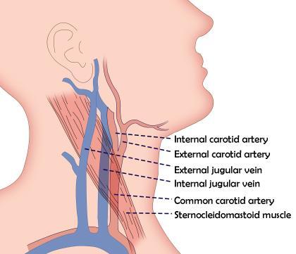
6 Anatomy
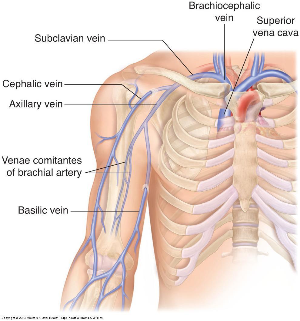
7 Anatomy
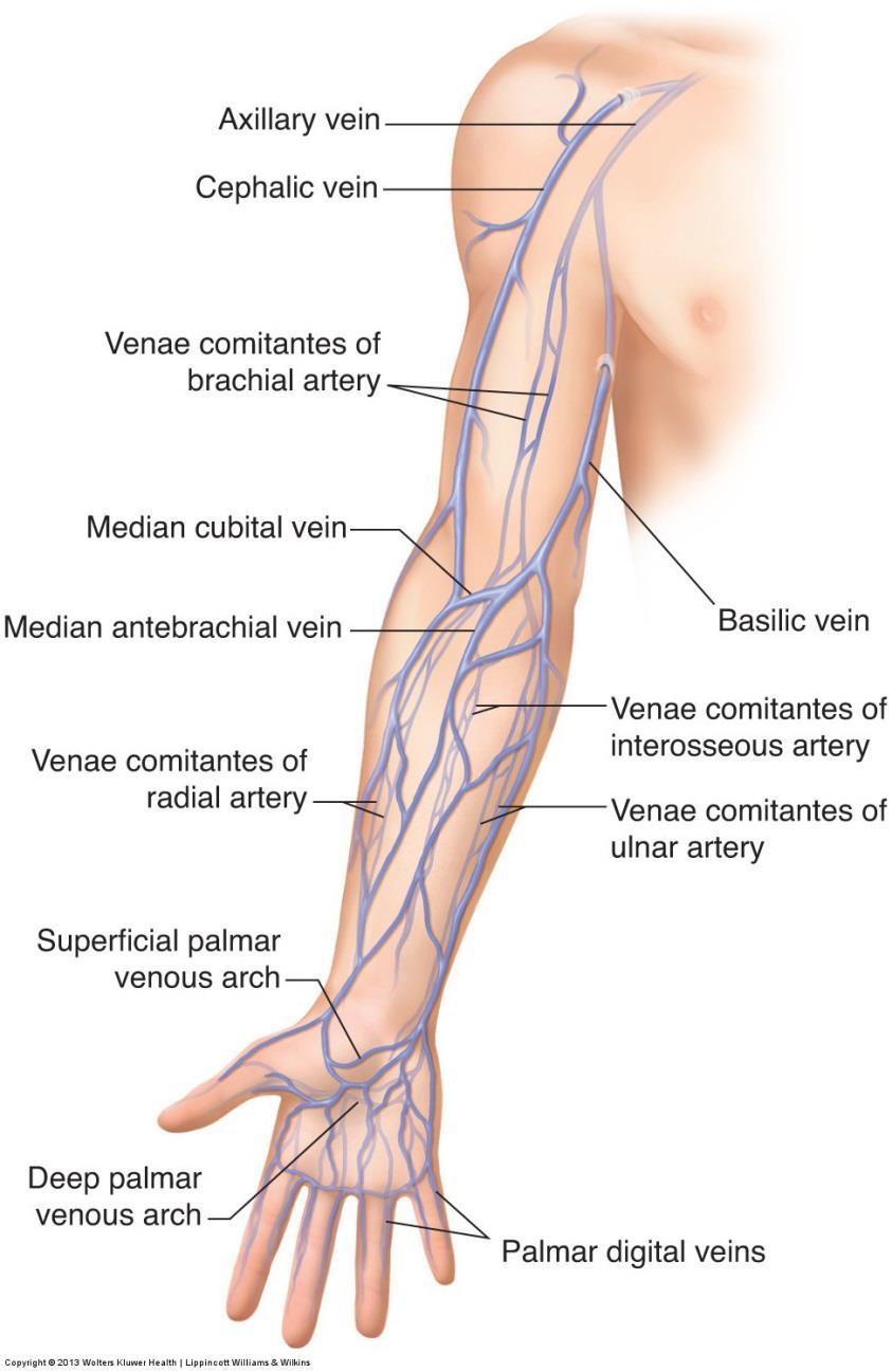
8 Anatomy
9 Upper Extremity Venous Anatomy Anatomy of the veins is variable Level of the brachial artery bifurcation varies
10 Upper Extremity Venous Anatomy Cephalic vein may drain through the antecubital vein into the basilic or a brachial vein and be very small or essentially absent in the upper arm. Basilic vein confluence with deep system may be in the axilla or upper arm.


11 Upper Extremity Anatomy Normal bifurcation level at antecubital fossa.43 cm Identify the level of the brachial bifurcation

12 Upper Extremity Anatomy High bifurcation, axillary level
13 Upper Extremity Venous Duplex Examination Protocol Vessels to be examined include: IJV Brachiocephalic Subclavian Axillary Brachial Cephalic & basilic Forearm vessels if indicated

14 Examination Protocol Transverse views with compression Brachiocephalic, subclavian, and axillary can not be typically compressed confirm patency with color or spectral Doppler Spectral Doppler signals should be recorded from all major vessels If a unilateral study is performed, record contralateral subclavian vein

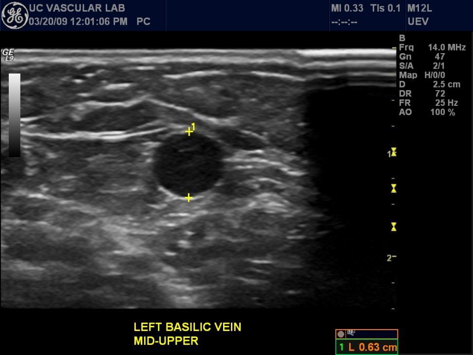
15 Venous Evaluation Compressibility Basilic vein 6.3 mm

16 Identify Arteries with Deep Veins


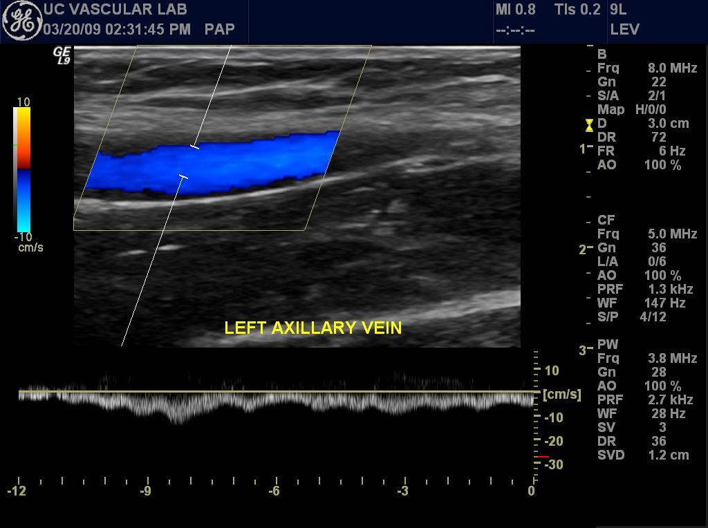
17 Venous Evaluation UE Central Vein Doppler Pulsatile with respirophasicity. Pulsatility decreases distally Pulsatile/Phasic Continuous flow pattern consistent with more central obstruction Abnormal/Continuous

18 UE Central Vein Doppler Signals Abnormal, continuous signal


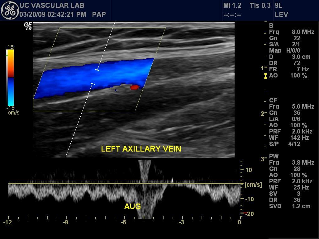
19 Axillary Vein Thrombosis Document absence of Doppler signal
20 Upper Extremity Venous Thrombosis Primary and Secondary
21 Upper Extremity Venous Thrombosis Primary Idiopathic Cancer, hypercoagulable states Thoracic Outlet Syndrome Compression (1 st rib, cervical rib) Effort Thrombosis (Paget-Schroetter) Related to strenuous arm activity
22 Upper Extremity Venous Thrombosis Secondary Central venous catheters (CVC) Account for 75% of all UEDVT cases Higher incidence if catheter tip in axillosubclavian or brachiocephalic as opposed to SVC placement Pacemaker / Defibrillator wires
23 Upper Extremity Venous Duplex Besides thrombosis Fibrosis: can result from radiation therapy with subclavian and axillary involvement frequently seen in patients treated for breast cancer.
24 Upper Extremity Venous Thrombosis Shoulder or neck discomfort / pain Arm and hand edema Symptoms and Signs Prominent superficial veins over the involved arm, chest or neck Jugular venous distention If SVC occluded facial edema, blurred vision, dyspnea
25 Upper Extremity Venous Thrombosis Symptoms and Signs Occluded central venous catheter Infusion difficulty with indwelling catheter Pulmonary embolus Palpable cord in the arm

26 Upper Extremity Venous Duplex Catheter-Associated Thrombus Catheter Thrombus

27 Upper Extremity Venous Duplex Defibrillator wire

28 Upper Extremity Venous Duplex Post PICC line removal

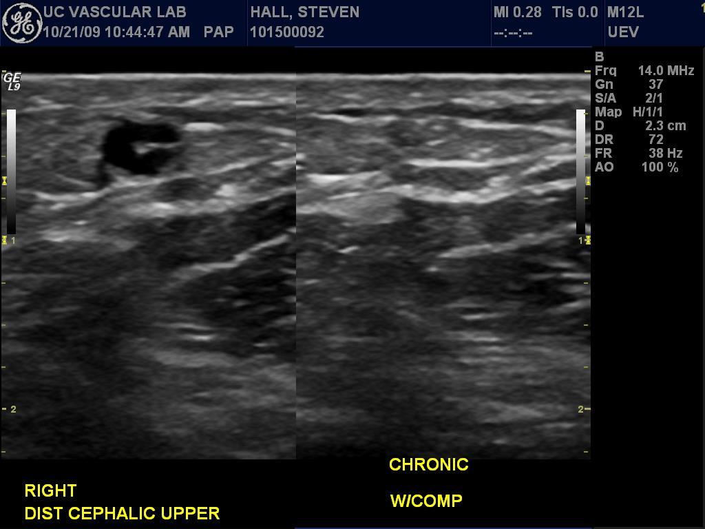
29 Chronic changes
30 Venous Evaluation Central Vein Evaluation Current or previous subclavian catheter placement Current or previous transvenous pacemaker Previous arm, neck, or chest trauma or surgery Multiple previous accesses in an extremity planned as an access site

31 Central Veins

32 Central Veins
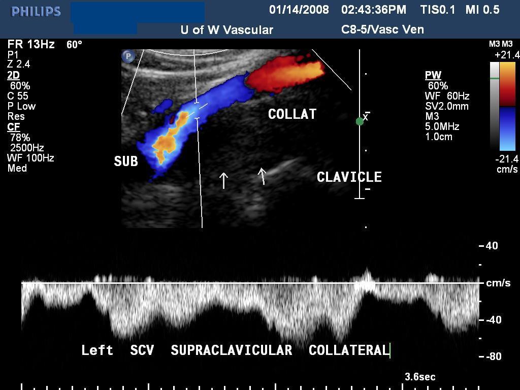
33 Collateral vein

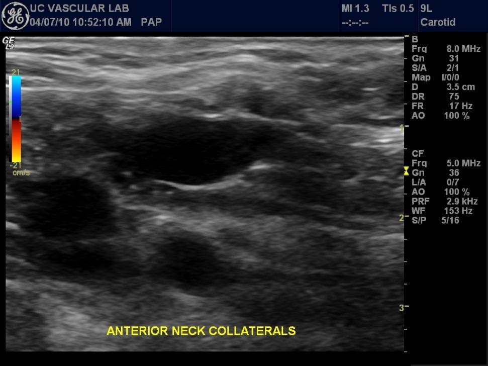
34 Collateral veins across anterior neck

35 UE Central Vein Doppler Signals

36 Central Vein Obstruction

37 Central Vein Obstruction

38 Case example

39 Case example

40 Case example

41 Case example
42 If mapping veins..

43 Venous Evaluation Explore veins > 2mm Veins >2.5 mm optimal


44 Venous Evaluation Cephalic vein 1.3 mm Cephalic vein 5.1 mm Measurement
45 Venous Evaluation Hemodynamics

46 Venous Evaluation Chronic changes


47 Venous Evaluation Chronic changes, deep venous system
48 Thank you! Questions?
For exam: VL DUPLEX EXTREMITY VEINS UNILAT LT
 For exam: VL DUPLEX EXTREMITY VEINS UNILAT LT - 8870390 METHOD/TECHNIQUE: The veins of the left upper extremity were studied at multiple For exam: VL DUPLEX EXTREMITY VEINS UNILAT RT - 8870400 METHOD/TECHNIQUE:
Pseudothrombosis of the Subclavian Vein
 416507JDMXXX10.1177/8756479311416507Wash ko et al.journal of Diagnostic Medical Sonography Pseudothrombosis of the Subclavian Vein Journal of Diagnostic Medical Sonography 27(5) 231 235 The Author(s) 2011
Sonography Evaluation of the Upper Extremity Venous System Evaluation for Deep and Superficial Venous Thrombosis
 Sonography Evaluation of the Upper Extremity Venous System Evaluation for Deep and Superficial Venous Thrombosis Wayne C Leonhardt, BA, RDMS, RVT Mission Imaging Asheville, North Carolina Disclosure Information
Upper Extremity Venous Duplex Evaluation
 VASCULARTECHNOLOGY PROFESSIONAL PERFORMANCE GUIDELINES Upper Extremity Venous Duplex Evaluation This Guideline was prepared by the Professional Guidelines Subcommittee of the Society for Vascular Ultrasound
THE VESSELS OF BLOOD CIRCULATION
 THE VESSELS OF BLOOD CIRCULATION scientistcindy.com /the-vessels-of-blood-circulation.html NOTE: You should familiarize yourself with the anatomy of the heart and have a good understanding of the flow
YOU MUST BRING GLOVES FOR THIS ACTIVITY
 ACTIVITY 10: VESSELS AND CIRCULATION OBJECTIVES: 1) How to get ready: Read Chapter 23, McKinley et al., Human Anatomy, 5e. All text references are for this textbook. 2) Observe and sketch histology slide
3 Circulatory Pathways
 40 Chapter 3 Circulatory Pathways Systemic Arteries -Arteries carry blood away from the heart to the various organs of the body. -The aorta is the longest artery in the body; it branches to give rise to
VENOUS DRAINAGE O US F UPPER UPPER LIM B BY dr.fahad Ullah
 VENOUS DRAINAGE OF UPPER LIMB BY dr.fahad Ullah Venous drainage of the supper limb The venous system of the upper limb drains deoxygenated blood from the arm, forearm and hand It can anatomically be divided
VESSELS: GROSS ANATOMY
 ACTIVITY 10: VESSELS AND CIRCULATION OBJECTIVES: 1) How to get ready: Read Chapter 23, McKinley et al., Human Anatomy, 4e. All text references are for this textbook. 2) Observe and sketch histology slide
Treatment of acute thrombosis of axillo-subclavian vein
 Treatment of acute thrombosis of axillo-subclavian vein Yang Jin Park Vascular Surgery, Samsung Medical Center Sungkyunkwan University School of Medicine CASE A 32-year-old male patient 3-day history of
10/14/2018 Dr. Shatarat
 2018 Objectives To discuss mediastina and its boundaries To discuss and explain the contents of the superior mediastinum To describe the great veins of the superior mediastinum To describe the Arch of
Guidelines, Policies and Statements D20 Statement on Peripheral Venous Ultrasound
 Guidelines, Policies and Statements D20 Statement on Peripheral Venous Ultrasound Disclaimer and Copyright The ASUM Standards of Practice Board have made every effort to ensure that this Guideline/Policy/Statement
Carotid Doppler: Doppler wave forms obtained from the common, external and internal carotid arteries. As well as the vertebral and subclavian
 Competency Carotid Doppler: Doppler wave forms obtained from the common, external and internal carotid arteries. As well as the vertebral and subclavian arteries. Preferred angle is 60 degrees or less.
Risk stratification and incidence of acute complications in upper extremity deep vein thrombosis (UEDVT) patients.
 patients.") Risk stratification and incidence of acute complications in upper extremity deep vein thrombosis (UEDVT) patients. Dr. Santosh Yatam Ganesh MBBS, MPH., Mentors: Dr. Khalid J. Qazi MD, MACP., Dr. Paul Anain
Risk stratification and incidence of acute complications in upper extremity deep vein thrombosis (UEDVT) patients. Dr. Santosh Yatam Ganesh MBBS, MPH., Mentors: Dr. Khalid J. Qazi MD, MACP., Dr. Paul Anain
VENOUS DOPPLER SONOGRAPHY OF THE EXTREMITIES
 VENOUS DOPPLER SONOGRAPHY OF THE EXTREMITIES The 35 th Annual Vanderbilt Diagnostic Sonography Symposium July 23-24, 2011 E. JAMES ANDREWS, JR., M.D., FACR, FACC DEPARTMENT OF RADIOLOGY AND RADIOLOGICAL
Candidate s instructions Look at this cross-section taken at the level of C5. Answer the following questions.
 Section 1 Anatomy Chapter 1. Trachea 1 Candidate s instructions Look at this cross-section taken at the level of C5. Answer the following questions. Pretracheal fascia 1 2 5 3 4 Questions 1. Label the
A A U
 PVD Venous AUC Rating Sheet 2nd Round 1 2 3 4 5 6 7 8 9 10 11 12 13 14 15 Median I NI MADM Rating Agree Disagree Upper Extremity Venous Evaluation Table 1. Venous Duplex of the Upper Extremities for Patency
Day 5 Respiratory & Cardiovascular: Respiratory System
 Day 5 Respiratory & Cardiovascular: Respiratory System Be very careful not to damage the heart and lungs while separating the ribs! Analysis Questions-Respiratory & Cardiovascular Log into QUIA using your
Axilla and Brachial Region
 L 4 A B O R A T O R Y Axilla and Brachial Region BRACHIAL PLEXUS 5 Roots/Rami (ventral rami C5 T1) 3 Trunks Superior (C5, C6) Middle (C7) Inferior (C8, T1) 3 Cords Lateral Cord (Anterior Superior and Anterior
Fascial Compartments of the Upper Arm
 Fascial Compartments of the Upper Arm The upper arm is enclosed in a sheath of deep fascia and has two fascial septa: 1- Medial fascial septum (medial intermuscular septum): attached to the medial supracondylar
Al-Balqa Applied University
 Al-Balqa Applied University Faculty Of Medicine *You can use this checklist as a guide to you for the lab. the items on this checklist represent the main features of the models that you have to know for
Prevention of thrombosis
 Prevention of thrombosis Massimo Lamperti MD, MBA Chief of General Anaesthesia Department Anaesthesiology Institute Cleveland Clinic Abu Dhabi Clinical Professor of Anaesthesiology Cleveland Clinic Lerner
HUMAN HEART. Learn the following structures on the heart models.
 HUMAN HEART Learn the following structures on the heart models. The human heart has four chambers that consist of the right atrium, left atrium, right ventricle, and left ventricle. The atria are smaller
Misc Anatomy. Upper Limb! 2. Lower Limb! 5. Venous Drainage! Head & neck! 8
 Misc Anatomy Upper Limb! 2 Arteries!... 2 Veins!... 2 Spaces!... 4 Lower Limb! 5 Arteries!... 5 Venous Drainage!... 6 Spaces!... 7 Head & neck! 8 Artery!... 8 Ultrasound View for IJ CVL!... 8 Arteries
region of the upper limb between the shoulder and the elbow Superiorly communicates with the axilla.
 1 region of the upper limb between the shoulder and the elbow Superiorly communicates with the axilla. Inferiorly, a number of important structures pass between arm & forearm through cubital fossa. 2 medial
Large veins of the thorax Brachiocephalic veins
 Large veins of the thorax Brachiocephalic veins Right brachiocephalic vein: formed at the root of the neck by the union of the right subclavian & the right internal jugular veins. Left brachiocephalic
The arm: *For images refer back to the slides
 The arm: *For images refer back to the slides Muscles of the arm: deltoid, triceps (which is located at the back of the arm), biceps and brachialis (it lies under the biceps), brachioradialis (it lies
Selection of Permanent Hemodialysis Vascular Access
 Selection of Permanent Hemodialysis Vascular Access TABLE OF CONTENTS 1.0 Scope...1 2.0 Recommendations & Rationale... 2 3.0 References... 3 4.0 Sponsors... 9 5.0 Effective Date... 10 Appendix 1: Key Elements
Veins that are firm to
 Intravenous cannulation is a technique in which a cannula is placed inside a vein to provide venous access. Venous access allows sampling of blood as well as administration of fluids, medications, parenteral
Vascular Surgery and Transplant Unit University of Catania. Pierfrancesco Veroux
 Vascular Surgery and Transplant Unit University of Catania Pierfrancesco Veroux Bologna-Palazzo dei Congressi, 23 Ottobre 2017 Disclosure Speaker name: Prof. Pierfrancesco Veroux I have the following potential
Copy Right- Hongqi ZHANG-Department of Anatomy-Fudan University. Systematic Anatomy. Angiology Part 4. Veins. Dr.Hongqi Zhang ( 张红旗 )
") Systematic Anatomy Angiology Part 4 Veins Dr.Hongqi Zhang ( 张红旗 ) Email: zhanghq58@126.com 1 General introduction of the veins Vessel which return the blood back to atrium No pulsation,veneous blood, metabolic
Systematic Anatomy Angiology Part 4 Veins Dr.Hongqi Zhang ( 张红旗 ) Email: zhanghq58@126.com 1 General introduction of the veins Vessel which return the blood back to atrium No pulsation,veneous blood, metabolic
Ultrasound Guided Vascular Access. 7/25/2016
 Ultrasound Guided Vascular Access 7/25/2016 www.ezono.com 1 Objectives Indications for insertion of central and peripheral lines Complications associated with procedures Role of ultrasound in vascular
HD Scanning: Velocities and Volume Flow
 HD Scanning: Velocities and Volume Flow Non-Invasive Lab Symposium West Orange, NJ April 27, 2018 Volume Flow Cindy Sturt, MD, FACS, RVT 500,000 Americans on dialysis 20-25% annual mortality 65% 5 year
Physician s Vascular Interpretation Examination Content Outline
 Physician s Vascular Interpretation Examination Content Outline (Outline Summary) # Domain Subdomain Percentage 1 2 3 4 5 6 Cerebrovascular Abdominal Peripheral Arterial - Duplex Imaging Peripheral Arterial
You have a what, inside you?
 Costal Emergency Medicine Conference You have a what, inside you? Less than mainstream medical devices encountered in the ED. Eric Ossmann, MD, FACEP Associate Professor Duke University Medical Center
You have a what, inside you?
 Costal Emergency Medicine Conference You have a what, inside you? Less than mainstream medical devices encountered in the ED. Eric Ossmann, MD, FACEP Associate Professor Duke University Medical Center
Certificate in Clinician Performed Ultrasound (CCPU) Syllabus. Vascular Access (venous (peripheral and central) and arterial)
 Syllabus. Vascular Access (venous (peripheral and central) and arterial)") Certificate in Clinician Performed Ultrasound (CCPU) Syllabus Vascular Access (venous (peripheral and central) and arterial) Page 1 of 8 04/16 Vascular Access (venous (peripheral and central) and arterial)
Certificate in Clinician Performed Ultrasound (CCPU) Syllabus Vascular Access (venous (peripheral and central) and arterial) Page 1 of 8 04/16 Vascular Access (venous (peripheral and central) and arterial)
CARDIOVASCULAR DANIL HAMMOUDI.MD
 CARDIOVASCULAR DANIL HAMMOUDI.MD 18 Systemic Circulation Figure 19.19 Pulmonary Circulation Figure 19.18b 1. Thyroid gland 2. Trachea 3. Brachiocephalic 4. Common carotid 5. Internal jugular 6. Superior
Which Artery am I? I am one of two smaller arteries that arise from the brachial. I supply blood to the medial aspect of the forearm.
 I am one of two smaller arteries that arise from the brachial. I supply blood to the medial aspect of the forearm. A. I supply blood to the head and neck. I am large and will branch into two smaller arteries.
Bare Metal Stents vs Stent Grafts
 Bare Metal Stents vs Stent Grafts ASDIN 12th Annual Scientific Meeting Phoenix, AZ, February 20, 2016 Dirk Hentschel, MD Director, Interventional Nephrology Brigham and Women s Hospital Disclosure Consultant:
Eco-color-doppler of the upper extremity: technique, indications and semeiotics
 Eco-color-doppler of the upper extremity: technique, indications and semeiotics Poster No.: C-0029 Congress: ECR 2012 Type: Educational Exhibit Authors: S. Pollice, P. Pollice, R. Stanzione, P. Maggi,
THORACIC OUTLET SYNDROME (T.O.S.) Prof J van Marle
 Prof J van Marle") THORACIC OUTLET SYNDROME (T.O.S.) Prof J van Marle 1. Definition Clinical syndrome caused by compression of the neurovascular bundle as it passes through the thoracic outlet, a narrow space bordered by
THORACIC OUTLET SYNDROME (T.O.S.) Prof J van Marle 1. Definition Clinical syndrome caused by compression of the neurovascular bundle as it passes through the thoracic outlet, a narrow space bordered by
PTA 106 Unit 1 Lecture 3
 PTA 106 Unit 1 Lecture 3 The Basics Arteries: Carry blood away from the heart toward tissues. They typically have thicker vessels walls to handle increased pressure. Contain internal and external elastic
Breathing. Heart Rate
 Breathing Heart Rate Inspiration Expiration (Pressos not Stretched) Heart Rate increases with inspiration (Pressos Stretched) Heart Rate decreases with expiration Upside Down (Pressos Stretched) HR Decreases
Case #1. Case #1- Possible codes. Unraveling the -59 modifier. Principles of Interventional. CASE 1: Simple angioplasty
 Unraveling the -59 modifier Principles of Interventional Coding Donald Schon, MD, FACP Debra Lawson, CPC, PCS Distinct or independent from other services performed on the same day Normally not reported
Vascular Sonography Examination
 Vascular Sonography Examination The purpose of The American Registry of Radiologic Technologists (ARRT ) Vascular Sonography Examination is to assess the knowledge and cognitive skills underlying the intelligent
5/5/15. Overview. Deep vein thrombosis of the upper extremity (UEDVT) Classification of UEDVT Epidemiology. Primary UEDVT.
 Classification of UEDVT Epidemiology. Primary UEDVT.") Deep vein thrombosis of the upper extremity (UEDVT) Hong H. Keo, MD, MSc Angiologie Kantonsspital Aarau 07.05.2015 06. Symposium venöse Insuff. und arterielle Gefässerkr. Glarus Overview Epidemiology Classification/Risk
Deep vein thrombosis of the upper extremity (UEDVT) Hong H. Keo, MD, MSc Angiologie Kantonsspital Aarau 07.05.2015 06. Symposium venöse Insuff. und arterielle Gefässerkr. Glarus Overview Epidemiology Classification/Risk
The Arm and Cubital Fossa
 The Arm and Cubital Fossa Dr. Andrew Gallagher School of Anatomical Sciences University of the Witwatersrand Introduction The ARM (BRACHIUM) is the most proximal segment of the upper limb musculoskeletal
Michigan Vascular Association 2012 Conference Case studies from Massachusetts General Hospital. Our lab
 Michigan Vascular Association 2012 Conference Case studies from Massachusetts General Hospital Kathleen Hannon, MS, RVT, RDMS khannon@partners.org Our lab #1 in the nation! 15 full time RVT s 11 MD s IAC
Slides of Anatomy. Spring Dr. Maher Hadidi, University of Jordan
 Slides of Anatomy Please note : These slides are Dr. Maher Hadidi s slides of spring 2016 and were edited by the Premed Academic Team to fit the slides of spring 2019. Spring 2019 Dr. Maher Hadidi, University
STRUCTURED EDUCATION REQUIREMENTS IMPLEMENTATION DATE: JULY 1, 2016
 STRUCTURED EDUCATION REQUIREMENTS Vascular Sonography The purpose of structured education is to provide the opportunity for individuals to develop mastery of discipline-specific knowledge that, when coupled
Bio& 242, Unit 3/ Lab 4 Blood Vessels, Lymphatic System and Blood Pressure G. Blevins/ G. Brady Summer 2009
 Bio& 242, Unit 3/ Lab 4 Blood Vessels, Lymphatic System and Blood Pressure G. Blevins/ G. Brady Summer 2009 Major Arteries and for arteries and veins with common names your answer must include either artery
Indications: following: embolization. artery that has diseases 5. The evaluation. of suspected. such entities. a cold hand. biopsy
 Peripheral Arterial Ultrasound Protocol Using Color and Spectral Doppler Reviewed by: Mark Yuhasz, MD Last Review Date: January 2015 Contact: (866) 761 4200, Option 1 Indications: The indications for peripheral
*the Arm* -the arm extends from the shoulder joint (proximal), to the elbow joint (distal) - it has one bone ; the humerus which is a long bone
, to the elbow joint (distal) - it has one bone ; the humerus which is a long bone") *the Arm* -the arm extends from the shoulder joint (proximal), to the elbow joint (distal) - it has one bone ; the humerus which is a long bone - muscles in the arm : *brachialis muscle *Biceps brachii
*the Arm* -the arm extends from the shoulder joint (proximal), to the elbow joint (distal) - it has one bone ; the humerus which is a long bone - muscles in the arm : *brachialis muscle *Biceps brachii
Overview of CVADs. Type of device commonly used. Dwell time Flushing requirement Associated complications. lumens
 Source: Clinical Skills Management of Vascular Access Devices Pre-course handbook. Adapted with permission from NHS Lothian Employee and Education Development Team. Overview of CVADs Type of device Veins
Arterial Map of the Thorax, Abdomen and Pelvis 2017 Edition
 Arterial Map of the Thorax, Abdomen and Pelvis Angiography 75605 (-26) Aortography, thoracic 75625 (-26) Aortography, abdominal by serialography 75630 (-26) Aortography, abdominal + bilat iliofemoral 75705
INDICATION: Patients in renal failure who require an arterio-venous fistula for hemodialysis.
 Duplex of Upper Extremity Vessels prior to AVF Surgery OLYMPIC VASCULAR LAB SURGICAL ASSOCIATES Chris Griffith MD, James Reus MD, Kevin Robinson MD, Richard Krug MD Diane Seagroves RVT MEMORIAL NEPHROLOGY
Image Formation (10) 2 Evaluation and Selection of Representative Images (10)
 2 Evaluation and Selection of Representative Images (10)") STRUCTURED SELF ASSESSMENT CONTENT SPECIFICATIONS SSA LAUNCH DATE: JANUARY 1, 2018 Vascular Sonography The purpose of continuing qualifications requirements (CQR) is to assist registered technologists
STRUCTURED SELF ASSESSMENT CONTENT SPECIFICATIONS SSA LAUNCH DATE: JANUARY 1, 2018 Vascular Sonography The purpose of continuing qualifications requirements (CQR) is to assist registered technologists
Thoracic Central Venous Obstruction (T-CVO) A New Look at an Old Problem. (This is what we say about T-CVO!)
 A New Look at an Old Problem. (This is what we say about T-CVO!)") Thoracic Central Venous Obstruction (T-CVO) These are ALL examples of T-CVO Paget- Schroetter Syndrome A New Look at an Old Problem Bart Dolmatch, MD, FSIR but each one is a different Cellulitis in swollen
Thoracic Central Venous Obstruction (T-CVO) These are ALL examples of T-CVO Paget- Schroetter Syndrome A New Look at an Old Problem Bart Dolmatch, MD, FSIR but each one is a different Cellulitis in swollen
As with any intervention, selection of an appropriate
 DVT: ccess Decisions for Interventions ssessing the advantages and disadvantages of venous access options is crucial for safe and successful DVT intervention. Y JOHN. KUFMN, MD, MS, FSIR, FH, FCIRSE, EIR
Subclavian and Axillary Artery Aneurysms
 Subclavian and Axillary Artery Aneurysms April 2008 Francesco A Aiello, M.D. Assistant Professor of Surgery Division of Vascular Endovascular Surgery University of Massachusetts Medical School None DISCLOSURES
BOGOMOLETS NATIONAL MEDICAL UNIVERSITY. Department of Human Anatomy GUIDELINES. The theme of the lesson The vessels of the upper limb.
 BOGOMOLETS NATIONAL MEDICAL UNIVERSITY Department of Human Anatomy GUIDELINES Academic discipline HUMAN ANATOMY Module 2 The theme of the lesson The vessels of the upper limb. Course Faculties І Medical
Int J Adv Med. For your questions please send message to
 Int J Adv Med SPECTRUM OF VASCULAR ABNORMALITIES IN COLOR DOPPLER EXAMINATION OF UPPER EXTREMITIES TESTED FOR SUITABLITY FOR AV FISTULA CREATION IN PATIENTS OF RENAL FAILURE. Journal Name : International
G24: Shoulder and Axilla
 G24: Shoulder and Axilla Syllabus - Pg. 2 ANAT 6010- Medical Gross Anatomy David A. Morton, Ph.D. Objectives Upper limb Systemically: Bones (joints) Muscles Nerves Vessels (arteries/veins) Fascial compartments
Lower Extremity Venous Insufficiency Evaluation
 VASCULAR TECHNOLOGY PROFESSIONAL PERFORMANCE GUIDELINES Lower Extremity Venous Insufficiency Evaluation This Protocol was prepared by members of the Society for Vascular Ultrasound (SVU) as a template
The Upper Limb III. The Brachial Plexus. Anatomy RHS 241 Lecture 12 Dr. Einas Al-Eisa
 The Upper Limb III The Brachial Plexus Anatomy RHS 241 Lecture 12 Dr. Einas Al-Eisa Brachial plexus Network of nerves supplying the upper limb Compression of the plexus results in motor & sensory changes
Long-term complications of cuffed tunneled central
 IMAGING TEACHING CASE Epistaxis as a Rare Complication of Catheter-Related Central Venous Stenosis Jin-Ju Tsai, MD, 1 Ching-Chih Hsia, MD, 1,2 Dong-Ming Tsai, MD, 1 Wei-Tsung Chen, MD, 3 and Yung-Hsuen
Intravenous Catheter Complications
 Vascular Access Device-Related Infection Inadequate skin antisepsis prior to VAD insertion Acute onset of fever, chills, and hypotension. No other apparent source of Notify Prescriber immediately Obtain
Venous Doppler Sonography of the Extremities: A Window to Pathology of the Thorax, Abdomen, and Pelvis
 Vascular and Interventional Radiology linical Perspective Selis and Kadakia Doppler Sonography of the Extremities Vascular and Interventional Radiology linical Perspective Downloaded from www.ajronline.org
c) What is the name of RBC (erythrocyte) formation? Where do blood cells form?
 What is the name of RBC (erythrocyte) formation? Where do blood cells form?") UNIT 6: CARDIOVASCULAR SYSTEM 1) List the three general functions of BLOOD. REVIEW QUESTIONS Blood 2) a) What are the three formed elements /cellular elements in blood? b) Describe the composition of the
UNIT 6: CARDIOVASCULAR SYSTEM 1) List the three general functions of BLOOD. REVIEW QUESTIONS Blood 2) a) What are the three formed elements /cellular elements in blood? b) Describe the composition of the
Vascular Technology Examination Content Outline
 Vascular Technology Examination Content Outline (Outline Summary) # Domain Subdomain Percentage 1 Normal Anatomy, Perfusion, and Function Evaluate normal anatomy, perfusion, function 2 Pathology, Perfusion,
Upper limb Arm & Cubital region 黃敏銓
 Upper limb Arm & Cubital region 黃敏銓 1 Arm Lateral intermuscular septum Anterior (flexor) compartment: stronger Medial intermuscular septum Posterior (extensor) compartment 2 Coracobrachialis Origin: coracoid
AVF 2010 OLYMPIC VASCULAR LAB SURGICAL ASSOCIATES
 Duplex of Upper Extremity Vessels prior to AVF Surgery Revised January 2010 OLYMPIC VASCULAR LAB SURGICAL ASSOCIATES Chris Griffith MD, James Reus MD, Kevin Robinson MD, Richard Krug MD Diane Seagroves
Brachial plexuses and axillary lymph nodes
 Brachial plexuses and axillary lymph nodes Introduction about nervous system nervous system central nervous system periphral nervous system brain spinal cord 31 pairs of spinal nerves 12 paris of cranial
Gateway to the upper limb. An area of transition between the neck and the arm.
 Gateway to the upper limb An area of transition between the neck and the arm. Pyramidal space inferior to shoulder @ junction of arm & thorax Distribution center for the neurovascular structures that serve
3 Mohammad Al-Mohtasib Areej Mosleh
 3 Mohammad Al-Mohtasib Areej Mosleh ***Muscles Connecting the Upper Limb to the Vertebral Column 1.Trapezius Muscle ***The first muscle on the back is trapezius muscle, it s called so according
Thoracic Outlet Syndrome
 Thoracic Outlet Syndrome Part 1: The Scalene Triangle TOS: Vascular Symptom Presentation Venous persistent/intermittent edema heaviness and fatigue deep pain in neck/shoulder increased pain at night warm
Steal Syndrome: The Role of the Vascular Lab
 Steal Syndrome: The Role of the Vascular Lab Eighth Overlook Noninvasive Vascular Lab Symposium Larry A. Scher, M.D. Professor of Surgery Division of Vascular Surgery Montefiore Medical Center Albert Einstein
5/24/16. Matthew Rennels, DO Ryan Szepiela, MD Promedica Toledo Hospital Primary Care Sports Medicine Fellowship
 Matthew Rennels, DO Ryan Szepiela, MD Promedica Toledo Hospital Primary Care Sports Medicine Fellowship! The patient is a 26-year-old male professional baseball pitcher (righthanded) who presented with
Central Venous Line Insertion
 Central Venous Line Insertion Understand the indications and risks of CVC insertion Understand and troubleshoot the seldinger technique Understand available sites and select the appropriate site for clinical
Artery 1 Head and Thoracic Arteries. Arrange the parts in the order blood flows through them.
 Artery 1 Head and Thoracic Arteries 1. Given the following parts of the aorta: 1. abdominal aorta 2. aortic arch 3. ascending aorta 4. thoracic aorta Arrange the parts in the order blood flows through
Treatment of Axillosubclavian Vein Thrombosis: A Novel Technique for Rapid Removal of Clot Using Low-Dose Thrombolysis
 J ENDOVASC THER 733 RAPID COMMUNICATION Treatment of Axillosubclavian Vein Thrombosis: A Novel Technique for Rapid Removal of Clot Using Low-Dose Thrombolysis Frank R. Arko, MD; Paul Cipriano, MD; Eugene
BILATERAL RARE NEURO VASCULAR VARIATIONS OF UPPER LIMB A CASE REPORT
 BILATERAL RARE NEURO VASCULAR VARIATIONS OF UPPER LIMB A CASE REPORT *N. B. S. Parimala Department of Anatomy, Dr. Pinnamaneni Siddhartha Institute of Medical Sciences & Research Foundation, Chinnaoutpalli,
Lecture 2: Clinical anatomy of thoracic cage and cavity II
 Lecture 2: Clinical anatomy of thoracic cage and cavity II Dr. Rehan Asad At the end of this session, the student should be able to: Identify and discuss clinical anatomy of mediastinum such as its deflection,
Vascular access device selection & placement. Alisa Seangleulur, MD Anesthesiology Department, Faculty of Medicine, Thammasat University
 Vascular access device selection & placement Alisa Seangleulur, MD Anesthesiology Department, Faculty of Medicine, Thammasat University How to make the right choice of vascular access device.. Peripheral
Approach to the Swollen Arm With Chronic Dialysis Access
 PICTORIAL ESSAY Approach to the Swollen Arm With Chronic Dialysis Access It s Not Just Deep Vein Thrombosis Shilpa N. Reddy, MD, Meghan C. Boros, MD, Mindy M. Horrow, MD The purposes of this pictorial
Peripheral Vascular Examination. Dr. Gary Mumaugh Western Physical Assessment
 Peripheral Vascular Examination Dr. Gary Mumaugh Western Physical Assessment Competencies 1. Inspection of upper extremity for: size symmetry swelling venous pattern color Texture nail beds Competencies
Mary Lou Garey MSN EMT-P MedFlight of Ohio
 Mary Lou Garey MSN EMT-P MedFlight of Ohio Function Prolonged and frequent access to venous circulation Allows for patient to carry on normal life; decrease number of needle sticks Medications, parenteral
inerve Guide to Nerves 2009
 inerve Guide to Nerves 2009 A guide to self learning and self assessment Context: The following guide is intended to help interpret the sono-anatomy and follow a systematic stepwise approach to the practice
Evaluation of hemodialysis arteriovenous fistula before and after surgery: Teaching points
 Evaluation of hemodialysis arteriovenous fistula before and after surgery: Teaching points Poster No.: C-0625 Congress: ECR 2014 Type: Educational Exhibit Authors: L. C. C. Chierighini, P. C. Francolin,
Sample page. POWER UP YOUR CODING with Optum360, your trusted coding partner for 32 years. Visit optum360coding.com.
 2018 Complete Guide for Interventional Radiology An in-depth guide to interventional radiology coding, billing, and reimbursement for facilities and physicians POWER UP YOUR CODING with Optum360, your
Surface anatomy of Cardiovascular system
 Surface anatomy of Cardiovascular system Prof. Abdulameer Al-Nuaimi E-mail: a.al-nuaimi@sheffield.ac.uk E. mail: abdulameerh@yahoo.com The lines cover the front, side, and back of the thorax Midsternal
Ultrasound and the dialysis patient
 Ultrasound and the dialysis patient Poster No.: C-1765 Congress: ECR 2011 Type: Educational Exhibit Authors: T. M. O. Couto, H. Matos, Â. Moreira, A. Estevao ; vila conde/ 1 2 2 2 1 2, Coimbra/ Keywords:
MUSCLES. Anconeus Muscle
 LAB 7 UPPER LIMBS MUSCLES Anconeus Muscle anconeus origin: distal end of dorsal surface of humerus insertion: lateral surface of ulna from distal margin of the semilunar notch to proximal end of the olecranon
A Rare Case of Bilateral Jugular Venous Malformation
 JOURNAL OF CASE REPORTS 2013;3(2):326-330 A Rare Case of Bilateral Jugular Venous Malformation Prasanna LC, Alva R, D Souza AS, Bhat KMR Department of Anatomy, Kasturba Medical College, Manipal University,
Carotid Abnormalities Coils, Kinks and Tortuosity David Lorelli M.D., RVT, FACS Michigan Vascular Association Conference Saturday, October 20, 2012
 Carotid Abnormalities Coils, Kinks and Tortuosity David Lorelli M.D., RVT, FACS Michigan Vascular Association Conference Saturday, October 20, 2012 Page 1 Table of Contents Carotid Anatomy Carotid Duplex
ACR Ultrasound Accreditation Program Exam Requirements
 ACR Ultrasound Accreditation Program Exam Requirements OBSTETRICAL ULTRASOUND EXAMINATIONS... 3 First Trimester... 3 Second Trimester... 3 Third Trimester... 4 GYNECOLOGICAL ULTRASOUND EXAMINATIONS...
Value Life Lifecath Midli n e uide to Lifecath Midline rse s G u N A
 Value Life Lifecath Midl ine A Nurse s Guide to Lifecath Midline www.vygon.co.uk vygon@vygon.co.uk Useful Information Lifecath Midline Lifecath Midline Catheter Code: 1296 Peelable Cannula Introducer
ESIM 2014 Clinical Case Presentation Israel. Ben-Sasson Maayan Bnei-Zion medical center Haifa
 ESIM 2014 Clinical Case Presentation Israel Ben-Sasson Maayan Bnei-Zion medical center Haifa Presentation A 24 YO male,a ping-pong player, presented to the ER with acute onset of right upper extremity
Central Line Care and Management
 Central Line Care and Management What is a Central Line/ CVAD? (central venous access device) A vascular infusion device that terminates at or close to the heart or in one of the great vessels (aorta,