MALE REPRODUCTIVE DISORDERS AND FERTILITY TRENDS: INFLUENCES OF ENVIRONMENT AND GENETIC SUSCEPTIBILITY
|
|
|
- Cori Walton
- 6 years ago
- Views:
Transcription
1 Physiol Rev 96: 55 97, 2016 Published November 18, 2015; doi: /physrev MALE REPRODUCTIVE DISORDERS AND FERTILITY TRENDS: INFLUENCES OF ENVIRONMENT AND GENETIC SUSCEPTIBILITY Niels E. Skakkebaek, Ewa Rajpert-De Meyts, Germaine M. Buck Louis, Jorma Toppari, Anna-Maria Andersson, Michael L. Eisenberg, Tina Kold Jensen, Niels Jørgensen, Shanna H. Swan, Katherine J. Sapra, Søren Ziebe, Lærke Priskorn, and Anders Juul Department of Growth & Reproduction and EDMaRC, Rigshospitalet, University of Copenhagen, Copenhagen, Denmark; Division of Epidemiology, Statistics and Prevention Research, Eunice Kennedy Shriver National Institute of Child Health and Human Development, National Institutes of Health, Bethesda, Maryland; Department of Physiology & Pediatrics, University of Turku and Turku University Hospital, Turku, Finland; Male Reproductive Medicine & Surgery Program, Stanford University, Stanford, California; Icahn School of Medicine at Mount Sinai, New York, New York; and The Fertility Clinic, Rigshospitalet, Copenhagen, Denmark L Skakkebaek NE, Rajpert-De Meyts E, Buck Louis GM, Toppari J, Andersson A-M, Eisenberg ML, Jensen TK, Jørgensen N, Swan SH, Sapra KJ, Ziebe S, Priskorn L, Juul A. Male Reproductive Disorders and Fertility Trends: Influences of Environment and Genetic Susceptibility. Physiol Rev 96: 55 97, Published November 18, 2015; doi: /physrev It is predicted that Japan and European Union will soon experience appreciable decreases in their populations due to persistently low total fertility rates (TFR) below replacement level (2.1 child per woman). In the United States, where TFR has also declined, there are ethnic differences. Caucasians have rates below replacement, while TFRs among African-Americans and Hispanics are higher. We review possible links between TFR and trends in a range of male reproductive problems, including testicular cancer, disorders of sex development, cryptorchidism, hypospadias, low testosterone levels, poor semen quality, childlessness, changed sex ratio, and increasing demand for assisted reproductive techniques. We present evidence that several adult male reproductive problems arise in utero and are signs of testicular dysgenesis syndrome (TDS). Although TDS might result from genetic mutations, recent evidence suggests that it most often is related to environmental exposures of the fetal testis. However, environmental factors can also affect the adult endocrine system. Based on our review of genetic and environmental factors, we conclude that environmental exposures arising from modern lifestyle, rather than genetics, are the most important factors in the observed trends. These environmental factors might act either directly or via epigenetic mechanisms. In the latter case, the effects of exposures might have an impact for several generations post-exposure. In conclusion, there is an urgent need to prioritize research in reproductive physiology and pathophysiology, particularly in highly industrialized countries facing decreasing populations. We highlight a number of topics that need attention by researchers in human physiology, pathophysiology, environmental health sciences, and demography. I. INTRODUCTION 55 II. INCIDENCE TRENDS, GENETIC III. INFERTILITY 73 IV. ROLE OF GENETIC BACKGROUND FOR THE V. ENVIRONMENTAL MODULATION OF VI. POSSIBLE ROLE OF MALE VII. AFTERWORD: RESEARCH CHALLENGES 83 I. INTRODUCTION During the 20th century, populations of industrialized countries all over the world have experienced a decline in total fertility rates (TFR, average number of live births per woman) far below 2.1, which is the rate considered necessary to sustain a population size at current numbers. At the same time a spectacular rise in testicular germ cell cancer (TGCC) has occurred in all parts of the World. In addition, other male reproductive problems, of which several are linked to testicular cancer, including disorders of spermatogenesis, are widespread and have recently been the focus for many basic and clinical research projects. As shown in FIGURE 1, the total fertility rate has fallen significantly in European Union (EU), Japan, and the United States (US). Also, Hong Kong and Singapore have for decades had TFR significantly below replacement level (now between 1.0 and 1.5). Fertility has therefore become a sig /16 Copyright 2016 the American Physiological Society 55
2 SKAKKEBAEK ET AL. Total Fertility Rate (per woman) European Union Japan USA Replacement level FIGURE 1. Total Fertility Rates (TFR), European Union, Japan and United States, Dotted line represents a fertility rate of 2.1, below which a population cannot be sustained. From the World Bank: selectvariables.aspx?source world-development-indicators. nificant political theme (122). Social and economic factors combined with the advent of effective contraceptive methods and access to induced abortions have contributed substantially to decreasing TFR, although the decline in birth rate started decades before the contraceptive pill and legal abortion were introduced (FIGURE 2). Surprisingly little is known about biological factors that might have changed human fecundity (capacity to conceive) and thereby influenced the TFR. The lack of knowledge in this area probably reflects the fact that research into human fecundity is difficult. Fecundity depends not only on a number of physiological and pathophysiological factors in both partners of a couple. Studies on rates of conceptions are also confounded by social, economic, and psychological factors that might change over time. In addition, TFR can be a poor marker of fecundity as it can be skewed by multiple factors, including induced abortion rates, availability of contraception, desire for pregnancy, and access to assisted reproduction, for example, before 1990 several Eastern European countries had higher abortion rates than birth rates. Total pregnancy rate, which includes both live births and induced abortions, might be more informative of changes in fecundity in a population than TFR (230). The aim of this review is to analyze some global trends in male reproductive health problems and their potential effects on male fecundity as well as the possible etiological roles of environmental, epigenetic, and genetic factors for these trends. Besides testicular cancer, we shall review recent studies on infertility, semen quality, cryptorchidism, and hypospadias and how these disorders might be interrelated with testicular cancer and with each other, through a testicular dysgenesis syndrome (TDS) (FIGURE 3). We shall also review trends in sex ratio and the potential roles of male reproductive disorders for couple fecundity and fertility rates. Finally, we will present some urgent research needs within the field of physiology and pathophysiology of male reproduction. II. INCIDENCE TRENDS, GENETIC SUSCEPTIBILITY, AND PATHOGENESIS OF REPRODUCTIVE DISORDERS A. Testicular Germ Cell Cancer The strongest evidence for adverse trends in male reproductive health comes from epidemiological studies of TGCC, including both seminoma and nonseminoma (231, 481). The increase was first noted in developed countries, where incidence rates have been highest in countries with North- 4.5 Total fertility rate (per woman) FIGURE 2. Total Fertility Rate (TFR), Denmark From Statistics Denmark: statistikbanken.dk/statbank5a/default.asp?w Dotted line represents a fertility rate of 2.1. Note that a downwards trend in TFR started long before introduction of the contraceptive pill in the 1960s. Apparently the trend was interrupted by the First and Second World Wars
3 FERTILITY TRENDS, ENVIRONMENT, AND GENETICS Fetal germ cells Fetal Leydig cells Environmental exposure Genetic defects and polymorphisms Sertoli cells Lifestyle factors Epigenetic factors Decreased Leydig cell function Decreased INSL3 production Decreased testosterone production Testicular dysgenesis Disturbed Sertoli cell function Impaired germ cell differentiation FIGURE 3. The hypothesis of testicular dysgenesis syndrome (TDS) and signs that might be linked to it: poor spermatogenesis, testicular cancer, hypospadias, cryptorchidism, and short ano-genital distance (AGD). The single symptoms and combinations thereof are risk factors for reduced fecundity. [Updated from Skakkebaek et al. (387).] Hypospadias Short AGD Cryptorchidism Decreased testosterone production Impaired spermatogenesis GCNIS Testicular cancer Reduced male fecundity influencing pregnancy rates ern European ancestry, including Denmark, Norway, and New Zealand (341). In the US, the highest incidences have been found among descendants of Europeans in the Midand North West (269). Interestingly, recent studies have shown that countries with previously low rates of TGCC, such as Finland, Italy, and Spain, are now catching up (FIG- URE 4), while high incidence countries such as Denmark and Switzerland now report smaller increases or stabilization of the high rates (231, 481). 1. Genetic aspects TGCC has a strong genetic component; brothers and sons of TGCC patients have significantly higher rates of TGCC (162). Studies have shown that incidence among Caucasians is much higher than among Afro-Americans living in the same area, confirming a role of genetic disposition to TGCC (FIGURE 4). In line with these epidemiological studies, several recent genome-wide association studies (GWAS) have identified a number of gene variants that might predispose to TGCC. Interestingly, most of the significantly associated genes are functionally linked to gonadal development and germ cell function, although pathways more typical for any cancer, for example, chromosome aggregation, microtubule assembly, telomerase function, and DNA repair, have also been associated with TGCC risk. The strongest association has been found with the KITLG locus on 12q22 (203, 349), which encodes for the KIT receptor ligand. The KIT/KITLG signaling pathway is indispensable for germ cell migration and survival and is highly expressed in testicular germ cell neoplasia in situ (GCNIS) and malignant TGCC, except somatically differentiated nonseminomas (347, 402). The significance of KIT/KITLG pathway for the TGCC risk, both in the sporadic and familial cases, has been confirmed by subsequent studies in different populations, and by associations with genes functionally linked to this pathway, such as SPRY, BAK1, or PDE11A (32, 86, 116, 215, 233, 337). Among other biologically interesting gene polymorphisms associated with an increased risk of TGCC, we highlight here four genes, all encoding for proteins involved in sex and germ cell development: DMRT1, PRDM14, DAZL, and HPGDS (77, 204, 215, 367, 432). DMRT1 is a transcription factor needed for sex differentiation and regulation of the onset of germ cell specific meiosis in male and female germ cells (185, 218, 265). Deletions encompassing DMRT1 and DMRT2 loci on chromosome 9p have been found in individuals with gonadoblastoma (244), a germ cell malignancy associated with disorders of sex development and gonadal dysgenesis. On the other hand, amplification of the DMRT1 locus has been detected in spermatocytic tumor, a germ cell tumor of older men not associated with GCNIS (247). PRDM14 is involved in germ cell specification and epigenetic reprogramming (469) and controls expression of the pluripotency genes POU5F1 (OCT4), and NANOG, which are expressed in GCNIS and TGCC (248, 345). Involvement in germ cell specification and embryonic meiosis regulation has also been evidenced for DAZL (206, 237). Of note for the hypothesis linking TGCC and some 57
4 SKAKKEBAEK ET AL. Northern Europe The Americas Asia Age standardized (World) incidence rate per Norway Denmark UK, Scotland* Ireland Iceland Finland Estonia Latvia Lithuania Age standardized (World) incidence rate per Canada* Ecuador* Colombia* USA White* Costa Rica USA Black* Age standardized (World) incidence rate per China* New Zealand Australia Israel Japan* Singapore Philippines* India* Year Year Year *: Regional registries Eastern, Southern and Western Europe Age standardized (World) incidence rate per Czech Republic Slovakia Bulgaria Poland* Belarus Russian Federation Age standardized (World) incidence rate per Spain* Slovenia Croatia Italy* Age standardized (World) incidence rate per Switzerland* Germany* The Netherlands UK, England* France* Year Year Year *: Regional registries FIGURE 4. Trends in testicular cancer; age-standardized (world) incidence (regional or national), all ages. [Modified from Znaor et al. (481). Courtesy of Dr. Arinana Znaor and statistician Mathieu Laversanne, M.Sc., WHO, International Agency for Research in Cancer (IARC), Lyon, France.] forms of male infertility to a TDS (see FIGURE 6, below), some DAZL variants and epigenetic promoter changes (301) have been associated with defective spermatogenesis, but the reports are inconsistent and might depend on the ethnic background of the studied cohort (434, 473). The inconsistent results are likely related to the confounding effect of a variable copy number of DAZ, a gene functionally related to DAZL in postmeiotic germ cells. DAZ copy number variations within partial AZFc deletions (e.g., gr/gr or b2/b3) of this gene are very common in some populations, and gr/gr deletion has been associated with both male subfertility (245, 366) and TGCC (300). The GWAS-detected association of testicular cancer risk with the HPGDS locus is biologically relevant, because this gene encodes for hematopoietic prostaglandin D synthase, an enzyme involved in sex determination in several mammalian species (284, 461). Recent experimental studies provided evidence that this pathway might be a target for endocrine disruption (267), thus giving an example of a pos- 58
5 FERTILITY TRENDS, ENVIRONMENT, AND GENETICS sible gene-environment interaction relevant to the pathogenesis of TGCC. Although a total of 19 genetic polymorphisms associated with TGCC risk have been identified to date, it appears that 25% of TGCC cases, even in the highly susceptible sons of TGCC cases, can be explained by hereditary genetic factors (240, 367). This percentage will likely increase after more genes have been identified in the ongoing large association studies and meta-analyses. Nevertheless, the large increase in sporadic TGCC cases observed worldwide within a generation or two is predominantly caused by an augmented negative influence of yet to be identified environmental factors. 2. Fetal origin of TGCC Basic studies (319, 344, 384) and epidemiological trends (162, 282) favor the hypothesis that TGCC is of fetal origin and should be considered a late-onset disease due to failure of normal fetal programming of the differentiation of primordial germ cells through a gonocyte stage into spermatogonia (FIGURE 5). The expression pattern of human gonocytes resembles that of embryonic stem cells because of the retention of pluripotency genes, such as POUF5/OCT4 and NANOG, but also includes expression of the KIT receptor, AP-2 (TFAP2C), podoplanin (PDPN/D2-40), and a number of germ cell specific genes (11, 190, 347, 396). The expression of the pluripotency genes is gradually lost during the second half of pregnancy and is rarely present after birth, although it might occasionally be seen in a few spermatogonia in normal testicles of boys younger than 1 yr (188). However, in individuals with a high risk of developing germ cell cancer, e.g., patients with mutations in SRY or the androgen receptor gene (AR), and in patients with 45X/46 XY mosaicism, undifferentiated gonocytes might persist during childhood Birth Puberty Spermatids Normal Development Normal Sertoli and Leydig cell function Migration Gonocyte Spermatogonia Spermatocytes PGC Fetal testis Prepubertal testis Adult testis Migration Delayed gonocyte pre-gcnis Adaptation Proliferation SEMINOMA Impaired Development Impaired Sertoli or Leydig cell function Gonocyte Arrest Genomic aberrations Re-programming GCNIS with invasive capacity NONSEMINOMAS Environmental insults Gene mutations EC Somatic differentiation FIGURE 5. Model for the pathogenesis of testicular germ cell tumors of young adults, which are derived from germ cell neoplasia in situ (GCNIS), previously known as carcinoma in situ testis (CIS). These tumors are an example of developmental cancer and are thought to be caused by a combination of adverse environmental and genetic factors (multifactorial and polygenic). The key pathogenetic event is insufficient masculinization and impaired function of the testicular somatic cell niche, which in fetal life is mainly composed of Sertoli and Leydig cells. The insufficient stimulation of developing germ cells causes arrest of gonocyte differentiation to spermatogonia and prolonged expression of pluripotency genes (depicted by red nuclei in all pluripotent cell types in the figure). The delayed gonocytes (pre-gcnis cells) then gradually acquire secondary genomic aberrations (including polyploidization and gain of chromosome 12p), while adapting to the changing niche, especially during and after pubertal hormonal stimulation of the testis. Increased proliferation results in malignant transformation of GCNIS cells into an invasive tumor, either a seminoma or nonseminoma (the latter through the reprogrammed pluripotent stage of embryonal carcinoma, EC). Normal germ cell development is shown in the top part in the figure (PGC, primordial germ cells) on the green background which symbolizes a normal testicular somatic cell niche. [Updated and modified from Rajpert-De Meyts (344).] 59

6 SKAKKEBAEK ET AL. A D M M B i ii 7 60
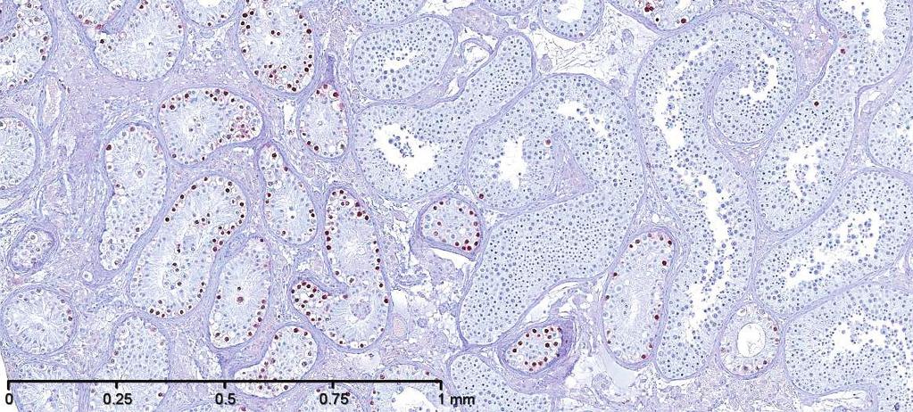
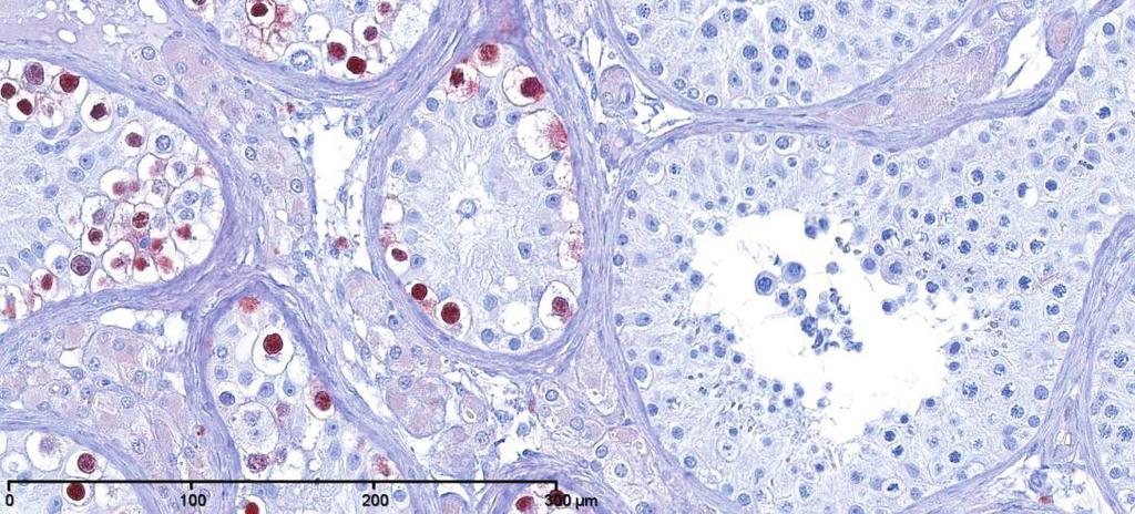
7 FERTILITY TRENDS, ENVIRONMENT, AND GENETICS and subsequently in adulthood develop into the abnormal intratubular cell pattern termed GCNIS (238, 344, 382, 384) (FIGURE 5). GCNIS cells are precursors of both seminoma and nonseminoma, although invasive TGCC might not develop in a testis harboring GCNIS until several years after detection of the cells by testicular biopsy (385). Epidemiological evidence also supports the idea of fetal origin of germ cell cancer. First, the fact that TGCC incidence peaks in young adulthood (between ages 20 and 45 yr) suggests an early onset of the malignant process (40, 79). Second, several epidemiological studies have shown a birth cohort effect in the incidence of TGCC (40, 107) so that men born in later calendar years have higher incidence rates. Third, immigration studies have shown that young men moving from countries with low or high risk of TGCC to a country with an intermediate risk developed TGCC with the same incidence as men of their home countries, while their sons born abroad acquired the risk of the host county (163, 297). Furthermore, the fetal hypothesis is in line with clinical studies of patients with disorders of sex differentiation (DSD) and congenital malformations, such as cryptorchidism and hypospadias, who are at significantly increased risk of developing TGCC (95, 373, 378). In addition, a study has shown that mothers exposure to persistent chemicals was associated with increased risk of TGCC in their sons (157). 3. Testicular cancer as a sign of TDS The heterogeneous group of above-mentioned conditions with reported increased risk of TGCC might have one thing in common: compromised development and function of the fetal Leydig and Sertoli cells. Several studies have investigated the role of these cells in the pathogenesis of TGCC and convincingly demonstrated that testicles harboring TGCC, and biopsies from men with cryptorchidism, hypospadias, and men with poor semen quality, often show evidence of dysgenesis in parts of the testicular tissue, including clusters of incompletely differentiated Sertoli cells, microliths, and Leydig cells clumps (sometimes called micro-nodules) (FIGURE 6) (368, 386, 387). These histological observations in addition to strong epidemiological evidence that TGCC, impairment of spermatogenesis, cryptorchidism and hypospadias are linked together in a risk factor network have prompted us to propose the existence of a TDS of fetal origin (FIGURE 3) (387). The disorders that constitute the condition complex of TDS might have one more thing in common, namely, feminization of the ano-genital distance (AGD) which is normally % longer in males than in females (369). Interestingly, some studies have shown that males with cryptorchidism, low sperm counts, low androgen levels, or hypospadias have decreased AGD (101, 102, 106, 408, 412, 415). It has been confirmed in animal studies that shorter AGD in males with congenital abnormalities of their genitalia reflects decreased androgen levels during the fetal period, as the shorter AGD is already visible after birth. Interestingly, in a large study of boys with cryptorchidism and hypospadias, short AGD was also associated with smaller penis size, confirming the association with neonatal androgen action (415). TGCC, cryptorchidism, hypospadias, and sperm count are not only risk factors for each other at an individual level, but they also seem associated at the population level. Indeed, a French group reviewed international data on TGCC, sperm count, hypospadias, and cryptorchidism and found correlations between the signs of TDS and geographical location, lending support to the unifying concept of the TDS hypothesis (379). B. Cryptorchidism Cryptorchidism is one of the most common birth defects, affecting 2 9% of boys born full term (47). The testes normally descend to the bottom of the scrotum before birth, and if one or both of them fail to do that, the condition is called congenital cryptorchidism. Once fully descended, the testes can later ascend to a cryptorchid position (443), which is called acquired cryptorchidism or ascending testis: its frequency is variable with the highest reported incidence nearly equal to that of congenital cryptorchidism (3). Epidemiological studies that have used registries as a data source usually combine these two groups together, because they are not separated in any International Classification of Diseases (ICD) classification. This adds some confusion to literature. Incidence rates that are based on numbers of orchidopexy typically reflect the frequency of both congenital and acquired cryptorchidism. The most reliable data on the incidence of congenital cryptorchidism come from cohort studies where the boys have been examined with standardized techniques and clear diagnostic criteria. Many clinical cohort studies with a long follow-up have used the classification of Scorer (377) to divide congenital cryptorchidism into subgroups based on the lowest position of the testis by physical examination: nonpalpable, inguinal, FIGURE 6. Examples of testicular dysgenesis in biopsy materials from men with abnormal spermatogenesis. A: specimen showing dysgenetic seminiferous tubules (D) containing numerous undifferentiated Sertoli cells and several microliths (M), but no germ cells. Tubules containing all types of germ cells, including spermatocytes and spermatids, are seen to the right. Hematoxylin-eosin staining was used. B: i) Immunostaining with OCT4, an embryonic marker, of testicular biopsy specimen with a mixture of GCNIS and normal spermatogenesis. ii) Same, higher magnification showing details of tubules with GCNIS and normal spermatogenesis, respectively. Note that OCT4 is only expressed in the nuclei of the GCNIS cells. 61
8 SKAKKEBAEK ET AL. supra-scrotal, high scrotal, and normal scrotal. English studies using this classification showed an increase of the incidence of cryptorchidism from 2.7% at the end of the 1950s (377) to 3.8% at the end of the 1980s (184), and further up to 5.0% in the early 2000s in boys with birth weight above 2,500 g (3) (FIGURE 7). Similarly, studies in Copenhagen showed an increase in the cryptorchidism rate from 1.8% in to 8.5% in (47). Interestingly, in Finland, the incidence of cryptorchidism remained at a low level (2.1%) (47) (FIGURE 7). Some pediatric surgeons have challenged the disease definition by discounting high scrotal cryptorchidism as a defect (81). It is evident, however, that the proper place of the testis is at the fully descended position, although high scrotal testes are usually not treated surgically as are all other, more severe cryptorchid cases (356). Cryptorchid testes are brought down to the scrotum surgically to preserve their spermatogenic capacity and to facilitate cancer surveillance. Spermatogenic cells suffer and start to disappear early in childhood unless the testes are in the proper position (211). Cryptorchidism is a risk factor for infertility, testis cancer, and hypospadias (373, 374), suggesting that these conditions share similar causes affecting fetal testicular development. However, although early orchidopexy (surgical treatment) improves the fertility chances, it might not decrease the risk of testicular cancer (296, 452), although this has been suggested in a few studies (330). 1. Causes of cryptorchidism Testicular descent is hormonally regulated. The key regulatory hormones are testosterone and insulin-like peptide 3 (INSL3), both of which are secreted by Leydig cells in the Incidence of cryptorchidism (%) Denmark United Kingdom Finland Year FIGURE 7. Incidence of cryptorchidism at birth on the basis of prospective clinical studies from the 1950s to the 2000s in Denmark, Finland, and United Kingdom. The data points are marked on the year of the publication of the study which represents the preceding incidence rate (3, 47, 61, 184, 377) testis (38). Pituitary luteinizing hormone (LH) stimulates Leydig cell differentiation and hormone secretion. In the presence of a decrease in these hormones, or defects in their receptors, the testes remain incompletely descended. A number of genetic defects in hormone synthesis and receptors have been described over the last 30 years and are often associated with cryptorchidism as part of a syndrome, but they are found very rarely in patients with isolated cryptorchidism (i.e., without other genital abnormalities) (263). It is noteworthy that isolated cryptorchidism might be caused by gene mutations that physically hamper the testicular descent, such as mutations of the AMH gene or its receptor (AMHR2) in the Persistent Müllerian Duct Syndrome (1, 192). Children with 46,XY karyotype and androgen insensitivity typically have testes either in the abdominal or inguinal position, i.e., they have not undergone inguinoscrotal transfer in utero. Mice with INSL3 deficiency, or with RXFP2/ LGR8 (INSL3 receptor) inactivating mutation, have testes in a high abdominal position, which led to a hypothesis that the early trans-abdominal descent would depend on this hormone (302, 479). However, it seems apparent that both androgens and INSL3 act in the whole process. They act on the gubernaculum, which is an actively transforming fetal organ first attaching the testis to the inner opening of the inguinal canal and then guiding it through the canal to the scrotum, and finally dissolving away. Mutations in INSL3 or RXFP2 have been detected in surprisingly few cryptorchid patients (44, 108). Also, although some polymorphisms have also been described, they have appeared only in a heterozygous manner and have also been reported in healthy individuals. In recent years, however, GWA studies have begun to shed some light on possible gene polymorphisms that predispose to problems with testis descent, either as part of TDS or nonsyndromic cryptorchidism. A study of several TDS components, including cryptorchidism, which combined GWAS with systems biology approaches, found weak associations with gene variants within TGFBR3 and BMP7 loci (86). Associations with other SNPs located in or near TGFBR3 locus have recently been confirmed in a larger study, which also found decreased expression of TGFBR3 protein in the gubernaculum of cryptorchid rats (34). However, until larger studies are performed, the most commonly identified genetic defects associated with cryptorchidism will remain those that affect androgen production or action. Cryptorchidism is clustered in families, which suggests a genetic or intrafamilial environmental cause. This has been analyzed in large registry-based epidemiological studies comparing the incidence in first-degree relatives. Monozygotic and dizygotic twin brothers have similar concordance for cryptorchidism, suggesting a minor role for genetic factors (375). Full brothers have a lower risk than twin brothers if one of the boys is cryptorchid, but 62
9 FERTILITY TRENDS, ENVIRONMENT, AND GENETICS their risk is higher than that of half-brothers. Furthermore, maternal half-brothers have a higher risk than paternal ones. All these findings implicate the importance of maternal environment during pregnancy. 2. Roles of hormonal exposures Animal experiments show that anti-androgens and estrogens can cause cryptorchidism. Androgen action at a specific male programming window during rat embryonic days is critical for proper masculinization, and failure at this developmental phase causes an irreversible undermasculinization, including cryptorchidism, that becomes apparent much later (454). Human development is of course different in timing, but the same principles seem to work there also. In the human, critical male programming occurs at gestational weeks 7 15, i.e., in early and midpregnancy (344, 387). The list of emerging anti-androgens is growing. The compounds that act at the receptor level as antagonists or partial agonists include widely spread pesticide congeners such as dichlorodiphenyldichloroethylene (DDE) and fungicides such as vinclozolin and procymidone. Even larger groups of compounds perturb androgen synthesis. Phthalates are well-studied examples of these chemicals. Interestingly, the effects of phthalates show variation in effects on different species (148, 228), whereas the effects of receptor antagonists are similar over these species. Since testing is performed with rodent models, some anti-androgens that might disturb human steroidogenesis without affecting rodents could go unnoticed. A crucial question is whether human exposure to one or a mixture of many of these chemicals is sufficient to cause disruption of male programming. Mixture studies in experimental animals have shown clearly that these chemicals can act in a simple additive manner, rendering even low doses harmful (76). Modeling studies have demonstrated that experimental results from exposing animals to mixtures of chemicals can be predicted on the basis of response curves of the individual chemicals (213). Estrogenic chemicals and dioxins can also cause cryptorchidism. Estrogens can prevent production of INSL3, which might explain its mechanism of action. In humans, exposure to a synthetic estrogen diethylstilbestrol was linked to increased rate of cryptorchidism (321), but environmental estrogens are typically much less potent. However, together with anti-androgens they might act in the same direction. It is uncertain how significant is their impact. Dioxins act via aryl hydrocarbon receptor (AhR). Rodent studies have shown that dioxins can induce cryptorchidism (141), but it is not known how this effect is mediated. Epidemiological studies on relationships between exposures to endocrine disruptors and cryptorchidism have analyzed single chemicals or chemical groups, and only a few have attempted to integrate these data. Exposures have been measured in blood, urine, placenta, and breast milk that serve as a proxy to mother s load of chemicals during pregnancy. In some cohort studies, careful ascertainment of the diagnosis has been combined with exposure measurements. The results vary according to the matrix that has been used for exposure assessment. The breast milk level of polybrominated flame retardants was associated with an increased risk of cryptorchidism, whereas placental levels were not (256). Similarly dioxin levels in breast milk of Danish women were associated with an increased risk of cryptorchidism (222, 223), whereas placental levels did not show an association (446). In Finland, dioxin levels in neither breast milk nor placenta were associated with cryptorchidism. In American studies of dioxins and DDT, no association was observed with maternal serum values and children s cryptorchidism risk (246). French studies found an association with polychlorinated biphenyls (PCB) levels in breast milk and the incidence of cryptorchidism (55), whereas Danish-Finnish studies did not, or showed the opposite (222). Mixture effects have been analyzed in only a few studies. After combining data from several pesticide exposures by permutation analysis, an association between the level of chlorinated pesticides in breast milk and risk of cryptorchidism in offspring was found (89). Greenhouse workers exposed to pesticides during pregnancy were also shown to have an increased risk of producing cryptorchid sons (14). Phthalate levels in breast milk were not associated with cryptorchidism risk, but they were linked to an increased LH-to-free testosterone ratio in the son at the age of 3 mo, suggesting testicular impairment during lactation (257). It is apparent that there are large data gaps, because only few exposures have been analyzed thus far. It is also unlikely that any individual chemical would have a major impact on the incidence of cryptorchidism. Combination of data from several exposures, particularly those that are known to affect the same signaling cascade, is needed to assess the whole chemical load, or exposome as it is called nowadays. 3. Lifestyle factors The significance of lifestyle factors, such as smoking and alcohol consumption, as risk factors for cryptorchidism remains contentious. There is evidence that heavy smoking during pregnancy ( 10 cigarettes per day) is associated with an increased risk of having a bilaterally cryptorchid son (417), while other studies have not shown a link between smoking during pregnancy and cryptorchidism (88). Registry-based studies did not find an association with mother s alcohol consumption and cryptorchidism in their sons (404), whereas a prospective follow-up study demonstrated a dose-dependent increase in the incidence of cryptorchidism in drinking mothers offspring (90). The lowest adverse effect dose was five units of alcohol per week, which is considerably lower than expected. However, the number of mothers in the group with the highest consumption was 63
10 SKAKKEBAEK ET AL. small, which influenced strongly the overall result and might therefore also be a chance finding. Smoking affects the growth of the fetus, and being small for gestational age at birth is a well-established risk factor for cryptorchidism (47). Prematurity is another strong risk factor, because testicular descent occurs normally during the last trimester and therefore might not occur before a premature birth. Gestational diabetes has become more frequent due to the increasing trends in obesity. In women with gestational diabetes, the risk of delivering a cryptorchid son is increased fourfold compared with non-diabetics (447). The underlying mechanism is not known, although early growth delay of the fetus in the first trimester might play a role. Interestingly, even children of diabetic mothers, who are born large due to compensatory growth in last part of pregnancy, have been found to grow poorly in the first trimester (328). In contrast, no association between gestational diabetes and cryptorchidism was found in a registry-based study from Israel (424). C. Hypospadias The penile congenital malformation, in which the urethra opens somewhere on the ventral side of the penis instead of the tip, is called hypospadias. The urethra might remain split over a long distance. The severity of the hypospadias is defined by the location of the opening. In the distal, mild form, the urethra opens in the glans or corona (sulcus) which is the border of the glans and the shaft. Registration of this birth defect varies in the malformation registries, because it does not necessarily require any surgical treatment. Physiological phimosis might also hide this defect in the newborn, and it might become apparent only after the foreskin can be easily retracted (45, 46). This should be considered when incidence data between countries are compared, because ascertainment, reporting, and registering practices vary (266, 421). More severe forms of hypospadias require surgical reconstruction of the penile urethra, and hospital discharge registries give a reliable estimate of their prevalence. In middle, or penile hypospadias, the opening of the urethra is located on the shaft of the penis, while in proximal hypospadias the opening can be found in the penoscrotal area. Sometimes both of these are called proximal in contrast to the distal form. 1. Incidence of hypospadias Increased incidence of hypospadias has been reported in Australia, US, and Europe over different time periods (200, 299, 326, 327, 421). The latest data from Denmark and Sweden also indicate an increasing trend (252, 308, 309). Until the 1990s, many malformation registries suffered from under-reporting; however, after a more active search, the hypospadias rate was found to be much higher than previously reported (161). This was also one reason for the controversies in the debate of incidence rates (5, 66, 97, 118, 334). Prospective clinical studies might be more accurate and therefore show higher rates than registry studies, for example, 1% versus 0.5% in Denmark (46, 253), as more mild cases might be noted in prospective studies. Interestingly, there are great differences between countries, for example, 1% in Denmark versus 0.3% in Finland according to parallel standardized clinical studies (46, 445). A list of incidence data of hypospadias is presented in TABLE 1. Table 1. Incidence of hypospadias in prospective or cross-sectional clinical studies Country Study Type Rate of Hypospadias Reference Nos. USA, Rochester, MN Prospective cohort study (n 4,474) 0.6% (body wt 2,500 g), 0.8% of all 158 boys USA, New York City, NY Prospective study on pregnant women and infants 0.54% of live-born boys 271 USA, collaborative perinatal project Prospective study (n 53,394 consecutive single births) 0.80% of single-born boys (76% of cases detected at birth) Korea, 38 hospitals Prospective study (n 7,990) 0.21% of boys 74 Southern Jordan Clinical study of 1,748 boys (aged 6 to 12 yr) 0.74% of boys 9 Finland,Turku Prospective cohort study (n 1,505); total 0.27% of live-born boys, 0.33% of liveborn 445 hospital cohort (n 5,798) boys Netherlands, Rotterdam Prospective study (n 7,292) 0.73% of newborn boys 332 Denmark, Copenhagen Prospective cohort study (n 1,072) 1.03% of live-born boys (at 3 yr: 4.64% including also milder cases detected after physiological phimosis resolved) 46 Bulgaria, 5 regions Cross-sectional clinical study (n 6,200 boys aged0to19yr) Modified from Toppari et al. (423) % of boys
11 FERTILITY TRENDS, ENVIRONMENT, AND GENETICS 2. Causes of hypospadias Masculinization of the male is driven by androgens during early fetal development. Penile development is regulated by dihydrotestosterone that is produced locally from testosterone by 5- -reductase. Several genetic mutations leading to hypospadias are known, and they are typically linked to disorders of testicular differentiation, testosterone synthesis, conversion of testosterone to dihydrotestosterone, or androgen receptor action (199). The same classification that is used for the severity of androgen insensitivity can also be used for hypospadias (342). Despite this knowledge, a genetic cause can be found only in a minority of hypospadias cases, and an endocrine abnormality can be found only in 20% of the patients (353). Environmental antiandrogens cause hypospadias in experimental animals in the same manner as they induce cryptorchidism (354), which makes it reasonable to search for common causes of these disorders. Genetic defects, other than those of androgen receptor and steroidogenic enzymes, include Homeobox genes HOXA and HOXD, fibroblast growth factor (FGF) 8, FGF10, FGF receptor 2, and bone morphogenetic protein 7 (123, 123, 132, 288, 290, 292). Activating transcription factor (ATF) 3 might also be involved, since its transcript level was found to be elevated more often in the foreskin of boys operated on for hypospadias than in those circumcised (242). The gene is estrogen regulated and influences transforming growth factor- signaling, which might explain why estrogens can also increase the risk of hypospadias (241, 462). HOXA13 mutations can cause the hand-foot-genital syndrome which includes hypospadias (123, 290), and hypospadias can be a part of many other multi-malformation syndromes. Mutations in MAMLD1 and NR5A1/SF1 can cause testicular dysgenesis, with hypospadias (36, 125). Mutations are rare (313), but the genes can be targets of endocrine disruptors as demonstrated for NR5A1 (407). Androgen and estrogen receptor polymorphisms have been associated with varying risk of hypospadias, but the results are not very consistent and require larger study populations than available so far (27, 39, 440, 451). Diacylglycerol kinase polymorphism has also been linked to the risk of hypospadias (439). 3. Roles of prenatal exposures Being small-for-gestational age is a risk factor for both cryptorchidism and hypospadias (6, 7, 26, 331, 331). Both birth defects can be caused by anti-androgens and estrogens, as shown by epidemiological studies following the children of women who used diethylstilbestrol (DES) during pregnancy (422). DES increases the risk of hypospadias even in the second generation, as the sons of in utero-exposed women have a higher prevalence of hypospadias than other males (54, 199, 210). This might reflect an epigenetic effect by DES. DES-related adverse effects are very similar in human and experimental animals (272), and there is no reason to believe that the anti-androgen-related effects would differ. Epidemiological studies on hypospadias have largely relied on registries, because the condition is rather rare and it is difficult to collect enough cases in prospective clinical studies to reach statistical power. As presented earlier, the registry studies on hypospadias are problematic due to several sources of error in classification of cases versus controls. A possible association between the risk of hypospadias and pesticide exposure was assessed in a meta-analysis that showed a small, increased risk of hypospadias in sons if parents were exposed to pesticides. Medical charts, parental interviews, occupation, job exposure matrix, or linkage of agricultural census and birth records were used for the assessment of exposure (360). Pooled risk ratios were 1.36 (95% CI ) and 1.19 (95% CI ) for maternal and paternal exposures, respectively (360). However, the studies could not assess which chemicals were behind the association, because the pesticides included a large number of different chemicals. Recent studies using a job exposure matrix as a proxy for pesticide exposure suggested an association of hypospadias with heavy metals, or maternal exposure to any endocrine disrupting chemical (EDC) (134), but did not show a significant association between pesticide exposure and hypospadias (287, 298, 361). Maternal serum samples were collected during pregnancy in the Collaborative Perinatal Project (CPP) conducted in the US in the 1950s and 1960s (246, 333). Several chemicals were analyzed in these samples, and the children were examined many times before they were 7 yr old. There was no linear association of PCB levels with hypospadias, but there was an increased odds ratio for the sum of some PCBs (270). No significant association was found between hypospadias and chlordane-related contaminants, DDE, -hexachlorocyclohexane, or other pesticides (246, 270, 333, 425). Another study relating the levels of DDT or DDE in pregnancy serum samples from the 1950s and 1960s with hypospadias in the sons also showed no association (42). In a study from the 2000s, an increased risk of hypospadias was associated with above-median level of hexachlorobenzene (HCB) in primiparous women as assessed from serum samples collected several weeks after delivery (134). No significant associations between hypospadias and midpregnancy serum levels of PCBs, PBDEs, HCB, DDT, or DDE were found in the study of Carmichael et al. (66). Serum levels of PBB at the time of conception showed no association with the risk of hypospadias according to a small questionnaire-based study (392). Phthalate metabolite level (mono-4-methyl-7-carboxyheptyl)phthalate (7cx- MMeHP, a DiNP metabolite) in amniotic fluid showed el- 65
12 SKAKKEBAEK ET AL. evated odds ratios for hypospadias (1.69 [0.78 to 3.67]), but was not consistently associated with the amniotic fluid levels of steroid hormones or insulin-like peptide 3 (172). DEHP [di(2-ethylhexyl)phthalate] metabolite levels did not show similar associations (172). A vegetarian diet of mothers was associated with an increased risk for hypospadias in the British ALSPAC study (310). In contrast, a decreased risk was reported for mothers having fish or meat in their diet during pregnancy (6). Also, a phytoestrogen-rich diet was associated with a reduced risk of hypospadias (65). No difference was found in the hypospadias risk of boys whose mothers used, or did not use, organic food diet, but a frequent concurrent consumption of high-fat dairy products (milk, butter) while rarely or never choosing the organic alternatives during pregnancy was associated with an increased odds ratio of hypospadias (adjusted OR 2.18, 95% CI ) (75). The need for assisted reproductive techniques (ART) and subfertility are risk factors for hypospadias (83, 201, 209, 413, 455). While fetal exposure to DES increased the risk of hypospadias, the role of other pharmaceutical sex steroids is controversial. Use of progestins was reported to increase the risk of hypospadias (63, 84). However, according to a meta-analysis of 14 studies, no association between exposure to sex steroids (except DES) during the first trimester and external genital malformations could be found (348). It is apparent that epidemiological studies have difficulties in case ascertainment, exposure assessment, and statistical power. Experimental studies have clearly indicated risks of hypospadias associated with anti-androgenic chemicals, such as phthalates and vinclozolin (76, 208), but epidemiological studies have failed to reach any comprehensive measurement of these compounds in large enough study populations of humans to draw conclusions about their role in hypospadias. D. Onset of Male Puberty While a clear downward trend in timing of puberty has been documented among girls, this trend has not been clear in males until recently. In fact, American data on male puberty ( ) were reviewed by an expert panel in 2006, which failed to find a significant decline, probably due to insufficient data as well as use of different study designs and study populations (111). However, several data have emerged since then suggesting a significant downward trend in male pubertal timing (FIGURE 8). For example, a European study of 21,612 boys born showed a downward trend in age at peak height velocity (PHV) (8), although other European studies did not find such changes (323). Male puberty marks the transitional period during which the infantile boy attains adult reproductive capacity and develops into a mature man. Puberty usually starts at Median years Year FIGURE 8. Recent changes in male pubertal timing. Testicular volume was 3 ml. [From Mouritsen et al. (293).] 11.5 yr of age, although with large interindividual variability (9 14 yr). Pubertal onset in a boy before 9 yr or after 14 yr of age is considered pathological and necessitates further evaluation to exclude underlying pathologies. The timing of puberty is determined by genetic as well as environmental factors such as body composition, physical fitness, nutritional and socioeconomic status, ethnicity, residence, foreign adoption, and exposure to endocrine disrupters (325). The pubertal development of secondary sexual characteristics begins with growth of the testes as a result of follicle stimulating hormone (FSH) stimulation of seminiferous epithelium. When testicular volume exceeds 3 4 ml, it is considered a definite clinical sign of pubertal onset. The stimulation of spermatogenesis involves multiple endocrine and local factors including FSH, LH, testosterone, inhibin B, AMH, etc., and the increasing testicular volume is a marker of spermatogenesis. Leydig cells are stimulated by LH at the onset of puberty, and subsequently start to produce testosterone which influences the growth of the penis (width and length), androgenization of the scrotal sac, and pubic hair development. Alternative pubertal markers include the pubertal growth spurt which is best described by the age at PHV, the point of maximal growth velocity in puberty (8). Age at PHV is a late pubertal marker, and usually occurs when the boy is in genital stages 3 4 when testicular volumes are ml. Another late marker of puberty is voice breaking, which occurs at an average age of 14 yr (195). Age at first emission of spermatozoa (spermarche) could be considered the male counterpart of age at menarche in females. Age at first emission of spermatozoa can be recorded in first morning urine samples (307), or by selfreported involuntary or voluntary emissions (420). 66
13 FERTILITY TRENDS, ENVIRONMENT, AND GENETICS 1. Endocrine regulation of pubertal onset The hypothalamic-pituitary-gonadal (HPG) hormone axis has already been activated in the neonatal period, which results in increase in circulating levels of FSH, LH, testosterone, and inhibin B (a period termed mini-puberty ) (19). The physiological reason for this phenomenon, which lasts a few months, is not known, and the presence of circulating androgens does not result in virilization of genitals, probably because of the relatively short period of androgen exposure, and because androgen receptors are not yet widely expressed. Most of testosterone is bound to sex hormone binding globulin during mini-puberty. Mini-puberty is, however, accompanied by descent of undescended testes (47). Although the factors responsible for this early HPG activation and its subsequent silencing remain unknown, epigenetic factors have been suggested to play a role. After 11 years of postnatal suppression, the HPG axis is reactivated, which results in increases in circulating levels of FSH and LH which in turn stimulates testosterone, Insl3 and inhibin B (18, 183). The mechanisms underlying the pubertal reactivation of the HPG axis are largely unknown, but most likely include physiological and lifestyle factors such as fat mass, physical fitness, nutrition, vitamin D, and psychosocial factors (325). The pubertal period is characterized by very high activity of the growth hormone-insulinlike growth factor axis (194) as well as high insulin levels (397, 398), and resembles in many ways a transient acromegalic state associated with insulin resistance. Signs of current changes in pubertal timing were evident in the cross-sectional Copenhagen Puberty study, where a significant 3 4 mo downward change in age at pubertal onset (testicular volume 3 ml) during a 15-yr period was reported in the very same area of the capital region (399). These findings were confirmed in a longitudinal followup study in the same Copenhagen area (293). In accordance with these findings, much earlier pubic hair development was found in the Avon Longitudinal Study of Parents and Children (ALSPAC) cohort from the United Kingdom (UK) (data collected ) compared with the original UK data collected by Marshall and Tanner (11.4 vs yr) (285). Likewise, a recent US study reported mean ages of beginning of genital and pubic hair growth 6 mo to 2 yr earlier than in older US studies (165). Altogether, it appears that the age at which the Copenhagen male population reaches puberty and attains adult reproductive capacity is decreasing. We do not know the reasons or longterm consequences for these trends, but suggest that the observed changes in age at pubertal onset might represent early warnings of environmental factors influencing male reproductive health. E. Changing Testosterone Levels Testosterone produced by the Leydig cells in the testes is the major male sex steroid. It plays important roles in sex differentiation as well as in male puberty and in adulthood for developing and sustaining the secondary male sex characteristics and spermatogenesis. Production of testosterone is stimulated by LH secreted by the pituitary, which itself is stimulated by gonadotropin releasing hormone (GnRH) from the hypothalamus. On the other hand, circulating testosterone has, together with estrogen, an inhibitory effect on both GnRH and LH secretion. In the adult male, a balance between LH and testosterone level is thus attained through a hormonal negative-feedback loop, which is part of the hypothalamo-pituitary-testis hormone axis (166). Only 1 2% of circulating testosterone occurs free in the bloodstream; the vast majority of circulating sex steroids is bound to serum proteins such as sex steroid binding protein (SHBG), which has a high affinity for binding of both testosterone and estradiol (154). Tissues of the body, including the hypothalamus and pituitary, only see the free (unbound) sex steroids, and SHBG serum level is therefore an important coplayer in the GnRH-LH-testosterone hormonal feedback loop. 1. Age-related changes in male testosterone levels Both total and free testosterone levels decrease in men with increasing age (FIGURE 9), while SHBG and gonadotropin levels increase. There is no doubt that age-related changes in body composition and lifestyle contribute towards this change as overweight, type 2 diabetes, and decreased exercise all are associated with decreased total testosterone levels. Moreover, obesity or more severe metabolic illnesses are also associated with decreased free testosterone (144, 174, 426). When adjusting for some of these covariates, the age trend in total testosterone is attenuated, while declining nm older cohorts cross sectional longitudinal Age FIGURE 9. Average male serum testosterone levels by age (full drawn line) based on healthy men from the general population show only a moderate decline from the age of years. Superimposed (dotted line) is an illustration of the consequence of applying the average rate of decline ( 1.6%/year) observed in a longitudinal study of individual testosterone levels (115) from the age of 22 (year of peak testosterone levels in the cross-sectional material). [From Andersson et al. (15).] 67
14 SKAKKEBAEK ET AL. free testosterone and increasing LH and SHBG with age are seen even with adjustment for BMI, comorbidity, and smoking (468). Declining testosterone with increasing LH levels in aging men suggest an impairment of testicular function with age, which is further supported by the fact that older men have an attenuated testosterone response to LH stimulation (243). In older men, however, declining testosterone is not always accompanied by a reciprocal rise in LH. This reflects the fact that the attenuation of GnRH-LH signaling might influence the ability to compensate for an impairment of the Leydig cells inferred by aging through expected increased LH signaling. This blunting of the HPG axis might further lead to declining testosterone levels (442). Cross-sectional studies on age-related male testosterone levels generally show relatively modest changes in serum testosterone levels between different age groups with estimated changes per year of 0 to 0.8%, while steeper declines in individual total testosterone (e.g., 1.6%/year) have been observed in longitudinal studies (FIGURE 9) (115). The discrepancy in the rate of age-related decline in testosterone levels observed in cross-sectional studies compared with longitudinal studies could be due to a selection bias. Cross-sectional studies might be more likely to include the healthier segment of elderly men while longitudinal studies might be more prone to followup on participants irrespective of health status. However, we and others have suggested that a true age-related rate of decline in testosterone in crosssectional study material could be blunted by the occurrence of a birth cohort related decline in testosterone levels over the generations of men included in the cross-sectional material (15, 115). 2. Secular trends In 2007, a paper reported a population-level decline in male testosterone levels over time (427). The paper was based on a US study of more than 1,300 men, some of whom were examined for up to three times over an 18-yr period. The age-independent secular change in total testosterone levels and bioavailable testosterone corresponded to, respectively, 1.0%/yr and 1.3%/yr over the period (427). A Danish study, which was published shortly after, also reported the observation of a secular decline in male testosterone levels in a study of more than 5,300 men from the general population in four studies conducted at different time points between 1982 and 2001 (15). In the Danish study, the secular decline was significant for total testosterone and SHBG levels but not for free testosterone and the decline could partly, but not exclusively, be explained by a secular increase in BMI. A Swedish study comparing reproductive hormone levels in comparable age groups of men examined in 1995 (n 430) and in 2008 (n 149) found that free testosterone was significantly lower in the men examined in 2008 (430). A trend of lower total testosterone was also observed in the Swedish men examined in 2008 compared with 1995, although this trend did not reach statistical significance. The difference between free testosterone levels in the Swedish men examined in 1995 versus 2008 remained after adjustment for a difference in weight in the oldest age group (430). More recently, secular declines in total testosterone, free testosterone, and SHBG were reported in Finnish men ( 3,000) from the general population (329). These trends remained significant following adjustment for BMI. In the Finnish study, they also observed a secular decline in gonadotropins, indicating that while decline in testosterone might be due to detrimental changes at the gonad level, the hypothalamus-pituitary-axis did not seem to respond appropriately to this change in the examined men (329). In studies on secular trends, as those described above, time period and birth year are completely confounded when adjusted for age because time period, age, and birth year are completely linearly dependent on each other. Thus, while adjusting for the effect of age on reproductive hormone levels, it is impossible to discern whether the remaining differences are due to a time period effect or a birth cohort effect. Irrespectively, it is perturbing that this secular decline is observed at the population level in several industrialized countries over the same period/birth years. While a concurrent increase in obesity and metabolic disturbances and a decrease in number of smokers among men in the same countries might contribute to the declining testosterone levels, these health and lifestyle changes do not seem to fully explain the observed trends (268, 329, 427) (see FIGURE 10). It is tempting to speculate that there is a link between trends of declining male testosterone and increased male reproductive health problems in general. Normal testicular function is dependent on paracrine communication between cells of the different compartments in the testis. Testosterone from the Leydig cells acts on the Sertoli cells and is crucial for Sertoli cell differentiation during sexual maturation and for Sertoli cell supported sperm production in the adult male. Likewise, paracrine factors from the seminiferous tubules and the peritubular cells influence the function of the Leydig cells. Thus the hormones inhibin B and anti-müllerian hormone (AMH) produced by the Sertoli cells both seem to contribute to the regulation of LH-stimulated steroid production of the Leydig cells, with AMH having a suppressive (428, 429) and inhibin B having a putative stimulatory effect (167) on testosterone production, the latter presumably by reversing an inhibiting effect of activin on testosterone production by the Leydig cells. It is therefore not surprising that a compromised function in one of the compartments of the testis is reflected in the function of other compartments of the testis. Accordingly, while individual men with poor sperm concentration might have testosterone levels within the normal range, as a group, subfertile men have lower serum testosterone levels than fertile men (16). They also 68
15 FERTILITY TRENDS, ENVIRONMENT, AND GENETICS A Total Testosterone (ng/dl) Total Testosterone (ng/dl) B Age (years) T1: Age (years) have, in general, higher serum LH levels resulting in an even lower testosterone-to-lh ratio compared with fertile men. This indicates that testosterone levels of subfertile men often are sustained on the basis of a more intense stimulation by gonadotropin (16), indicating that impaired Leydig cell function is more common among men with poor semen quality. F. Sperm Concentration and Other Measures of Semen Quality T2: T3: FIGURE 10. Secular trends in mean total testosterone levels of normal men by age. A: stratified by time period of study. B: stratified by 5-yr birth cohort. Please note the different scales of the y-axes in A and B. [Modified from Travison et al. (427).] The publication of Carlsen et al. in 1992 (64), which concluded that sperm concentration had declined 50% over the previous 50 years, remains controversial (193, 383). As has been summarized elsewhere (411), this controversy centers on three primary concerns. Some authors suggested that poor or highly variable data invalidated any inference about trends in sperm counts (232, 316). Others questioned the validity of the statistical methods used in this analysis (52, 113, 316). Bias due to changing study populations (53) or confounded by factors such as age and abstinence time (time between sample collection and last ejaculation) was also suggested (316, 441). Following the 1992 publication of Carlsen et al. (64), considerable research activity was initiated to address these concerns resulting in numerous studies. Some of these used retrospectively collected and others newly collected data, which we discuss below. Studies relying on retrospectively collected data differed in study design and methods including: 1) semen parameters examined (sperm concentration, semen volume, total sperm count, percent motile sperm, percent morphologically normal sperm); 2) semen collection and analysis methods (sperm counting methods, motility criteria, morphology criteria, abstinence time, season of collection, participation in an external quality control program); 3) variables used to assess temporal and spatial variability (study time period, geographical area); 4) study population/recruitment methods (partners of infertile women undergoing ART procedure, potential semen donors, male partners of subfertile couples, and young men with unknown fertility); and 5) potential confounders controlled in analysis (age, year of birth, abstinence time, sociodemographic variables, lifestyle factors, medical conditions) and sample size. 1. Reanalyses of historical semen quality data A detailed reanalysis in 1997 of data from the 61 studies included in the systematic review by Carlsen et al. in 1992 (64) used multivariate linear models to control for potential sources of bias and confounding factors in those studies (410). The reanalysis showed significant declines in sperm concentration in the United States and Europe/Australia after controlling for abstinence time, age, percent of men with proven fertility, and specimen collection method. Declines in sperm concentration in the United States ( 1.5%/yr) and Europe/Australia ( 3%/yr) were greater than the average decline reported by Carlsen et al. ( 1%/yr). However, there was no evidence of a decline in non-western countries, for which data were very limited. In 2000, an additional independent literature review and updated analysis was performed (411). In this, 47 English language studies published from 1934 to 1996 were added to those analyzed previously. Results of that analysis were consistent with those of the Carlsen study and the 1997 reanalysis by Swan et al. (410). The authors concluded that the trends in sperm concentration previously reported for were also seen in data from 1934 to Retrospective studies of temporal trends in semen quality within countries Since 1992, many studies have examined trends in sperm counts within individual countries (both developed and de- 69
16 SKAKKEBAEK ET AL. veloping countries) using historical data. A large recent study reported a significant decline in sperm concentration and morphology in 26,609 men from the French general population who provided samples between 1989 and 2005 (363). In line with this, another French study showed a decline in total sperm count and morphology among semen donor candidates (400). Declines in sperm concentration have also been observed in Israel (150), Tunisia (114), and Scotland (401). However, no decline was observed in sperm concentration in South Sweden from 1985 to 1995 (41), nor in North-Eastern Spain between 1960 and 1996 (20). 3. Prospectively designed cross-sectional studies of semen quality in partners of pregnant women and unselected young men To overcome some of the problems of historical and crosssectional data, several standardized and coordinated crosssectional studies of semen quality have been undertaken. Two large multicenter studies designed to examine geographical variation of semen quality were conducted in partners of pregnant women. Each of these studies used consistent methods of recruitment and semen analysis and careful quality control to minimize between-center differences. The Study For Future Families (SFF) measured semen parameters in 763 partners of pregnant women in Los Angeles, CA; Minneapolis, MN; Columbia, MO; New York City, NY; and Iowa City, IA (351, 409). This was the first US study to compare semen parameters among study centers using standardized methods and strict quality control. These data suggested that sperm concentration, total count, and motility are reduced in semirural and agricultural areas relative to more urban and less agriculturally exposed areas. A multicenter European study collected semen samples from 1,082 fertile men from four European cities (Copenhagen, Denmark; Paris, France; Edinburgh, Scotland; and Turku, Finland) and demonstrated significant geographical differences in sperm counts, most notably between men living in Turku, Finland and Copenhagen, Denmark (186). Population-based studies of semen parameters in young men conducted in a consistent manner have been ongoing in several European countries, the US, and Japan since the late 1990s (117, 187, 189, 274, 275, 320, 340). These studies provide information about both geographical differences in semen parameters over the past 20 years as well as about temporal trends in the countries that include cohorts across time (189, 191). The temporal trends available to date exhibit considerable geographical variation. Decreases of more than 20% in total sperm counts and sperm concentration were detected among Finnish men between 1998 and 2006 (191), and a decrease of 15% was seen in young Spanish men during the most recent decade (274). Conversely, a Swedish study that was not coordinated in the above studies but basically using the same methods found no significant changes in semen parameters among young Swedes between 2000 and 2010 (31). Increases in total sperm count and sperm concentration of 14 and 12%, respectively, were observed among Danish men between 1996 and 2010 (189). It should be noted that despite the increase in median sperm concentration during this time (from 43 to /ml), sperm concentration in these healthy young men was still markedly lower in 2010 than in Danish men in infertile couples in the 1940s (median above /ml) (See FIGURE 11). It is notable that most recent studies also show a very high frequency of morphologically abnormal spermatozoa. As an example, the recent study of young men from the general Danish population showed that the median percent of morphologically normal spermatozoa is 7%, a number that remained substantially unchanged throughout the 15-yr study period (189). 4. Possible etiological factors underlying geographical and temporal variability in semen quality As discussed in the section on origin of testicular germ cell cancer (FIGURE 3), the hypothesis of the testicular dysgenesis syndrome, proposed in 1993, suggests that reduced spermatogenesis in adulthood can be a consequence of exposure in fetal life to environmental chemicals (381). Environmental chemicals, including endocrine disrupting chemicals such as dioxins and perfluorinated compounds (PFCs), as well as complex mixtures such as those in combustion products, appear to affect negatively both the perinatal and adult testes, emphasizing the importance of environmental and lifestyle factors that have impacts throughout life (380). Western lifestyle (sedentary work/lifestyle, obesity) is also potentially damaging to sperm production (103, 131, 175) as are other lifestyle factors (stress, sleep, smoking, maternal smoking, nutrition) (4, 130, 138, 173, 234). Data on the effects of environmental chemicals, such as pesticides, food additives, DDT, PCBs, or plasticizers, on spermatogenesis both in the perinatal period and in adult men are limited, lacking, or inconsistent (87, 160, 261). However, reports on reduced sperm counts and azoospermia in men exposed to dibromochloropropane (DBCP) (137, 335, 460) and dioxin (280, 281) provide proof of principle that exogenous chemicals during adult life can disturb human spermatogenesis as has been shown in numerous rodent studies (119, 140, 159, 444). In addition, recent studies have indicated that some endocrine disrupters, including ultraviolet filters, might have a direct effect on human sperm functions (289, 371, 414), including effects on CatSper, the calcium ion channel, which is crucial for sperm movements and acrosome reaction (239, 405). Observations from wildlife and animal experiments lend support to the idea that environmental factors can adversely affect male reproduction. An example is the finding that cryptorchidism and other of the symptoms associated with TDS in humans, have also been reported in large numbers among populations of Sitka black-tailed deer in Alaska 70
17 FERTILITY TRENDS, ENVIRONMENT, AND GENETICS A Sperm concentration B Total sperm count % of men % of men < >200 0 < >1500 Sperm concentration (million/ml) Total sperm count (million) Men from general population ( ) Men from infertal couples ( ) FIGURE 11. Distributions of sperm counts in Danish men from the general population, examined from 1996 to 2010 and Danish men examined in an infertility clinic in the 1940s. All men had durations of ejaculation abstinence greater than 48 h. Sperm concentration (A) and total sperm counts (B) are shown. [From Jørgensen et al. (189).] (56). Similar findings from studies of wildlife were also reported by other authors (cf. Ref. 459). 5. Semen quality and fecundity Since 1980, the World Health Organization (WHO) manual for the examination of human semen has served as a standardized protocol for measurement methods and reference values for sperm parameters ( ). In the WHO 2010 manual, the lower reference limit for sperm concentration was decreased from /ml to /ml, the value that had been in use since This value reflects the fifth centile for fertile men (defined as time to pregnancy 12 mo) and reflects the semen characteristics of recent fathers. It is notable that these cut-off points are appreciably below the value of /ml, considered to have been a normal sperm count in the 1940s (153, 255). Furthermore, other studies have shown reduced monthly probability of conception for sperm concentration below /ml (50, 147, 389). Bonde et al. (50) examined the monthly probability of conception in relation to semen parameters in couples attempting pregnancy and found that when sperm concentration was below /ml or the number of motile sperm 70%, the monthly probability of conception decreased. This is consistent with results from a large network study of fertile and infertile couples which found that semen samples with concentration below /ml, motility below 63%, and percent of sperm with normal morphology 9% (using strict criteria methods) were outside the fertile range (147). From an investigation of fertile men, Slama et al. (389) detected decreasing probability of conception with sperm concentrations below /ml and a total sperm count below 145 mill. Despite these population-level results, the ability of single semen parameters to predict fecundity on an individual level is limited, as was concluded by the United States National Cooperative Medicine Network in a large multicenter study comparing sperm parameters in 756 infertile and 696 fertile men (147). This national study concluded that although threshold values for sperm concentration, motility, and morphology can be used to classify men as subfertile or infertile, none of the parameters measured individually was diagnostic of fertility (147). Although a healthy mature man has tens of millions of spermatozoa per ejaculate, it has been estimated that only 1 per million will succeed in contacting the egg within the fallopian tube (100). If these estimates are confirmed, an average semen sample with a total sperm count of 150 million, as currently often seen among young men in Denmark (189), might have as few as 150 spermatozoa capable of fertilization. 6. Conclusion Over the past 25 years, abundant literature has identified important geographical differences in semen parameters between and within countries. While there is considerable variability in trends in sperm counts over the past 20 years, several recent studies report that 20 30% of young men today have sperm concentration below /ml, which is associated with reduced fecundity (50, 147, 389). We therefore estimate that 20 30% of men in the examined cohorts might be at risk of prolonged waiting time to preg- 71
18 SKAKKEBAEK ET AL Sex ratio at birth FIGURE 12. Sex ratio at birth and joinpoint segments, , all mothers. [From Mathews and Hamilton (264).] Year nancy if they want to become fathers, and 10 15% have a sperm count so low that they might require fertility treatment (see sect. III). G. Sex Ratio Sex ratio of offspring may act as in indicator of male reproductive problems. For example, men who were exposed to the pesticide DBCP at their workplace (137) and men who were exposed to dioxin at the Seveso accident had an excess of girls (278). However, several factors may influence sex ratio. It is generally assumed that 105 male births occur for each 100 female births leading to 51.5% of births being male (324). The sex ratio is important as it might reflect important demographic shifts as well as impact economic conditions (143, 264). In the first half of the 20th century, the sex ratio increased in most Western countries due to improved obstetrical care which led to relatively more live births of males. But while thought to be constant over time in the absence of health advances, recent data suggest a decline of the sex ratio in many Western countries (91). An analysis from US birth data demonstrates a decline in the sex ratio beginning around 1940 (91, 264; see FIGURES 12 and 13). Over that period of time, the proportion of male births declined from 51.4 to 51.2%, or 2 fewer males per 1,000 births. In Denmark, the percentage of male births decreased from 51.5 in the 1950s to 51.3 in 1995, while in the Netherlands it declined from 51.6 to 51.3 over this same time period (283, 438). Canada also showed a similar decline in recent decades from 51.5 to 51.3 from 1970 to 1990 (10). However, an examination of 29 coun Joinpoint sex ratio trend Observed sex ratio trend Sex ratio at birth FIGURE 13. Sex ratio at birth and joinpoint segments for births to white mothers, [From Mathews and Hamilton (264).] Year 72
19 FERTILITY TRENDS, ENVIRONMENT, AND GENETICS tries from a WHO database identified some countries where the sex ratio seemed to increase over time including several in southern Europe such as Italy and Spain (324). However, while a universal decline in the sex ratio was not reported for all countries, a majority (16 of the 29) did show a decline, while six showed an increase and seven showed no change. While certain regions and countries might not reflect the recent downward trend in sex ratio, for the remainder of the countries, this concerning index has been explored as a sentinel health indicator. As data suggest a possible decline in male fertility over the past half century, investigators have explored whether a relationship exists between infertility and sex ratio. Hypothesizing that infertile men might have an impaired ability to sire male heirs, Weijin and Olsen (453) found that couples with a longer time to pregnancy had a lower sex ratio (453). However, other investigations have cast doubt on this relationship (105, 171, 180, 394). A Dutch group found that the proportion of male births increases with a longer time to pregnancy (394). As female influences are thought to be a powerful mechanism for gender selection, examining postgestational outcome such as live birth sex might be inadequate to assess the role of the male contribution to the sex ratio. A US group examined the proportion of Y bearing sperm and identified an inverse relationship between the production of Y chromosome bearing sperm and semen quality, suggesting an impaired ability for infertile men to sire male heirs (104). In addition to the fertility of the parents, the health of the parents has also been examined as a factor that might impact sex ratio. Diabetes, non-hodgkin s lymphoma, hepatitis B, and testicular cancer are among the diseases thought to lower sex ratio (70, 171, 318, 357). The impact that environmental exposure can have on sex ratio has prompted concern regarding the recent declines in the sex ratio in several countries. An explosion at a chemical plant in Seveso, Italy in 1976 exposed the local population to high levels of dioxin, a known endocrine disruptor. A subsequent generation of children sired by parents with high exposure levels displayed a lowered sex ratio (278, 279). Workers exposed to the gonadotoxic nematocide DBCP demonstrated reduced sex ratios compared with children born prior to paternal exposure (137, 336). Other exposures including boron and those from aluminium manufacture have also demonstrated decreases in the sex ratio or sperm Y:X ratio (277, 359). These data demonstrating an influence of chemical exposure on sex ratio has laid the foundation for many to hypothesize that environmental exposures might be the driver behind the declining sex ratio. In addition to chemical exposure, environmental stressors in the form of catastrophic events have also been shown to alter the sex ratio. The Kobe earthquake, September 11 attack in New York, economic downturns, and war have all been shown to lower the sex ratio (67, 68, 126, 482). The authors speculated that alterations in semen quality or spontaneous abortion might have contributed, although the definitive etiology remains uncertain. While the preconception and adult environment might play a role in sex ratio, social factors might also impact on sex ratio. Sex-selective abortion in some countries have increased in prevalence based on availability of abortion and early identification of the sex of a fetus (124, 178, 476). Such practices might have a significant impact on the sex ratio of an entire population (29). III. INFERTILITY Given the reported changes in male reproductive health, an immediate question is whether they are associated with an increased prevalence of infertility. This is a challenging question to answer given the absence of population-based monitoring data suitable for assessing temporal patterns of infertility, and the methodological nuances associated with measuring infertility as briefly noted below. A. Definition Infertility has been defined as the inability of a couple to conceive after 1 yr of sexual intercourse without contraception (110). This broad definition does not reflect the considerable heterogeneity of infertility, which comprises both couples without and with prior pregnancies, or so-called primary and secondary infertility, respectively. Approximately half of infertility with an identifiable diagnostic finding is attributed to female factors (e.g., endocrine, tubal, uterine, cervical, and oocyte factors) and another half to male factors (e.g., poor spermatogenesis, cryptorchidism, poor semen quality, cancer, genetic syndromes). Such diagnostic categorization will be dependent on clinical norms and practices, the extent of diagnostic testing that couples undergo, and the sensitivity/specificity of such testing. For example, 66% of fertile couples undergoing standardized infertility evaluations for research purposes were observed to have one or more infertility factors (146). Another consideration with regard to infertility terminology is the uncertain percentage of couples that might have both male and female factors identified, while many others will have unknown or idiopathic infertility. Considerable misclassification of diagnostic subtypes of infertility arises when based on self-reported information (94). Also, an unknown percentage of infertile couples will resolve their infertility either spontaneously or with medical treatment, while others will have unresolved infertility. This observation has prompted authors to define infertility as a continuum of fecundity ending with an absolute inability to conceive (145). 73
20 SKAKKEBAEK ET AL. B. Prevalence/Incidence of Infertility One of the earliest prevalence estimates of infertility estimated that 16% of couples in the United Kingdom were affected (169) followed by estimates that varied by place and time. For example, the prevalence of infertility ranged from 8 to 12% in Asian and Latin American studies (458), with similar ranges reported in parts of sub-saharan Africa. Specifically, prevalence was 9% in Gambia (406), although 20 30% in Nigeria (229, 315). An overall prevalence in Europe was estimated to be 16%, ranging from a low of 10% in Southern Italy to a high of 24% in East Germany (196). Prevalence varied from 15 to 33% in five population-based samples (Denmark, Germany, Italy, Poland, and Spain) of women aged yr reporting for their first pregnancy attempt (205). In a random sample of Scottish women aged yr, 19% reported having experienced infertility with more primary than secondary infertility reported (43). Most recently, prevalence in Canada was reported to range from 12 to 16% when varying the assumptions about factors associated with conception (62). Geographical variation in prevalence is also observed among developing countries, ranging from 4 to 17% in 25 population-based surveys comprising 172,413 women, with lifetime infertility ranging from 12 to 26% (49). Of note is the preponderance of prevalence data based on female rather than male reporting. A recent systematic review revealed wide fluctuations in prevalence depending on definition, choice of referent population, and specification of numerators/denominators underscoring the inability to derive a single prevalence estimate across studies (145). FIG- URE 14 illustrates some of the considerations that are needed when defining infertility (numerator) and selecting the population at risk (denominator), which might affect prevalence estimates. In addition, global prevalence of lifetime infertility has been reported to vary from 6.6% in Norway (364) to 32.6% in the US (372). These estimates of the prevalence of infertility were obtained from cross-sectional surveys, but ideally incidence data are needed. Such data must be derived from prospective cohort studies that recruit couples prior to or upon becoming at risk for pregnancy, such as when discontinuing contraception for purposes of becoming pregnant. Couples are then followed daily through 12 menstrual cycles or months at risk for pregnancy. Three such studies have been conducted (59, 170, 480), while another four preconception cohort studies have followed only female partners for 12 cycles or months (58, 109, 120, 418). Collectively, these prospective cohort studies with preconception enrollment of couples or women suggest that the incidence of infertility ranges between 12 and 18%. Irrespective of study design, it is important to keep in mind that human fertility is dynamic in nature, as infertility does not necessarily imply sterility. For example, while fecund couples have pregnancies resulting in births, many couples with fecundity impairments, such as those experiencing pregnancy losses or 12-mo infertility, will become pregnant and have births either with or without medical assistance as illustrated in FIGURE 15. Another approach for estimating the prevalence of infertility utilizes time-to-pregnancy (TTP) data usually obtained from pregnant women or through record linkages or registries data. TTP is easily obtained from questionnaires asking how long the couple had regular unprotected intercourse before pregnancy occurred and allows for the cate- Numerator considerations Denominator considerations Definitions Retrospective report vs. prospective observation >12 months unprotected intercoursee >12 months trying to conceive Physician diagnosed infertility Treatment for infertility Prevalence Sexual activity Use of reversible contraception Surginal/medical Surgical/medical sterilization FIGURE 14. Roles of definitions, populations, numerators, and denominators of calculated prevalence of infertility. Populations All couples Reproductive-aged couples Sexually active couples Cohabitating couples Married couples Couples with pregnancy All couples Reproductive-aged couples Sexually active couples Cohabitating couples Married couples Couples trying to conceive 74
21 FERTILITY TRENDS, ENVIRONMENT, AND GENETICS Birth Pregnancy Fecund FIGURE 15. Birth Pregnancy (with medical assistance) Impaired fecundity (pregnancy loss, infertility) gorization of couples not only with regard to infertility (TTP 12 cycles/mo) but also in relation to conception delay or so-called impaired fecundity (TTP 6 cycles/mo). Reliance on retrospectively reported TTP requires some caution with regard to interpretation, given its uncertain validity that has only been empirically assessed in two studies using the gold standard of prospectively measured TTP. Specifically, the validity of self-reported TTP was reported to be good for shorter periods of recall (477), but poor for longer periods of recall given notable bidirectional errors in reporting (80). However, reliability of retrospectively reported TTP has been reported to be good (182). Recently, the current duration approach has been developed and offers a novel method for identifying women currently at risk for pregnancy. This cross-sectional approach queries women (or men) regarding the time since stopping contraception or attempting to become pregnant at the time of interview. This method allows for the estimation of a TTP-like distribution that accounts for left censoring (207). Applying this approach in France using a household-based sampling framework yielded a 12-mo infertility prevalence of 24% and a 24-mo prevalence of 11% (390). Recently, the current duration approach was used to estimate infertility prevalence among respondents in the 2002 National Survey of Family Growth (NSFG) conducted in the US. Prevalence was estimated to be 15.5% based on female reporting (416) and 12.0% for male reporting (251). C. Temporal Patterns of Infertility Birth Pregnancy (without medical assistance) Impaired fecundity (pregnancy loss, infertility) Dynamic nature of fecundity and fertility. It is exceedingly difficult with available data to accurately estimate the temporal pattern of infertility as illustrated in a recent systematic review (145). Doing so would require longitudinal assessments using similar methods that are responsive to the nuances underlying pregnancy intentions, periods at risk, and other methodological issues including sources of sampling biases. While several authors have estimated infertility prevalence in various countries for particular time periods as noted above, few attempts have been undertaken to assess temporal patterns. Perhaps the closest available temporal data are derived from the NSFG. This cross-sectional survey was conducted at specific time periods (1982, 1988, 1995, 2002) until continual enrollment began in The NSFG survey interviews a representative sample of US women aged yr (and men commencing in 2006) about many aspects of reproductive health. Infertility is not directly queried but is derived from respondents answers to a series of conditional questions on relationship status, sexual activity, contraceptive use, and pregnancy attempts within the past 12 mo. In 2002, this construct estimated a US prevalence of 7.4% (72), which is half the estimate (15.5%) for this same time period using the current duration approach, as noted above. This might be a function of the survey s continued reliance on a construct measure of infertility rather than direct querying of men/women. Based on repeated cross-sectional NSFG data, the 12-mo infertility prevalence for married women has steadily declined in the US from 8.5% in 1982 to 6.0% in (71); however, a growing percentage (41% in 2011) of US births are to unmarried women (262). We are unaware of data on temporal patterns of infertility for other geographical locations. To our knowledge, there is no population-based monitoring of infertility in any country. This critical data gap is in the context of considerable reported variations in human fecundability, as measured by TTP (176) or semen quality (186, 409), and despite earlier calls for monitoring human fecundity via surveys (179) or more purposeful research initiatives inclusive of the couple (317). Attempts to assess temporal patterns of human fecundity include the following four initiatives presented in chronological order. First, temporal patterns of self-reported TTP, defined as the number of years of involuntary childlessness, were assessed for 832,000 primiparous women aged 20 yr and older with births between 1983 and 2002 as identified in the Swedish Medical Birth Registry (370). This unique investigation accounted for two important sources of bias, namely, truncation and age and calendar time at the initiation of trying. Subfertility was estimated to have decreased for more recent cohorts, although it is important to note that the sampling framework comprised only fertile women giving birth, which underrepresents women with impaired fecundity who either cannot conceive or carry a pregnancy to live birth. Another important consideration is the increased awareness of the fertile window over this time period and the availability of in-home tests aimed at timing intercourse to maximize chances of conception or for identifying pregnancy, which were not as readily available in earlier cohorts. A second initiative assessed temporal patterns in the rates of natural conceptions for 803,435 Danish women born between 1960 and 1984, allowing for an indirect assessment of infertility (230). A gradual decline in both observed and 75
22 SKAKKEBAEK ET AL. projected rates of natural conception was observed. Other important observed trends included declining abortion rates and a slight increase in the percentage of childless women regardless of use of assisted technologies (14.5 to 15.6%). Another investigation assessed trends in childlessness among birth cohorts of males, which is unique in that most research focuses on females. Specifically, 1,359,975 Danish men born between 1945 and 1980 were linked with their children using national birth and ART registries (338). The percentage of childless men at age 45 yr increased from 14.8 to 21.9%. While infertility was not estimated specifically, investigators pooled five European databases to assess fertility time trends for 8,532 combined pregnancies whose mothers were aged yr and for whom valid self-reported TTP was available along with 715 contraception failures occurring between 1953 and 1993 (181). An increasing fertility trend was reported, and attributed to a male cohort effect for TTP and contraception failure. While the authors undertook several analyses, there are noteworthy limitations that might impact on temporal patterns including restricting pregnancies: 1) to those resulting in a live birth, which systematically excludes women unable to conceive or carry a pregnancy to birth, and 2) to the first trying attempt rather than including all such attempts. This latter practice assumes that the first pregnancy attempt is representative of all subsequent trying attempts, which might or might not be true (250). D. Use of ART Indirect information on trends in infertility might be obtained from statistics on assisted reproduction. In Denmark, nationwide activities on ART are registered every year. As seen from FIGURE 16, there has been a significant increase in the use of ART during the past 13 years. While the level of assisted reproduction might seem to have levelled out in recent years, there has at the same time been a 13% drop in the number of Danish women aged yr, which is the age range of the majority of women seeking help to reproduce. Consequently, the proportion of couples/women seeking treatment has continued to increase. While it might appear that there has been a huge increase in the usage of donor semen, it is important to note several possible reasons for this increase. As a consequence of legislative changes, treatment of lesbian and single women was introduced during the time period, thus increasing the number of women seeking this treatment option. Furthermore, complete registration on the number of treatments using donor sperm have only been achieved in recent years. It is important also to recognize that data on birth cohorts always include children conceived through assisted reproduction, and this could mask the real fertility potential in a population. IV. ROLE OF GENETIC BACKGROUND FOR THE RISK OF MALE REPRODUCTIVE DISORDERS It is clear that the quick pace of incidence trends of the above reviewed disorders of male reproductive health is consistent with the relatively recent changes in the environment and lifestyle-related factors, rather than accumulation of inherited genetic aberrations. However, the available data demonstrate that there have been quite striking geographical and ethnic differences in most of these trends. Number of ART cycles Number of women age in the Danish population IUI-D IUI-H OD FER IVF ICSI Number of women age years FIGURE 16. Assisted reproduction (ART) in Denmark, IUI-D, insemination with donor semen; IUI-H, husband semen; OD, oocyte donation; FER, frozen embryo replacement; IVF, in vitro fertilization; ICSI, intracytoplasmatic sperm injection. From The Danish Fertility Society: _dok/dfs2014.pdf. 76
23 FERTILITY TRENDS, ENVIRONMENT, AND GENETICS A. Genetic Polymorphisms Explaining Ethnic Differences The most clear-cut evidence of ethnic differences in disease incidence has been provided by epidemiological studies of testicular cancer, which are briefly reviewed above. The incidence of TGCC worldwide is by far greatest among white people of northern European ancestry and lowest among African men. These prevalence differences are not primarily related to environment, as convincingly illustrated by ethnic differences among the populations in the US (133, 269). Men of African ancestry seem to be protected from testicular cancer, and possibly also have a lower incidence of cryptorchidism. An explanation has been sought in several studies. Because of the association of TGCC with inborn disorders, especially those linked to insufficient masculinization, including cryptorchidism, it was hypothesized that the steroid hormone levels might differ between black and white people. Although no marked differences have been noted in serum testosterone between white and black men, there might be some differences in women. Significantly higher (by 48%) levels of serum testosterone were identified in black women during the first trimester of pregnancy (164). These data are uncertain, because the studied groups were very small, and the genetic polymorphisms responsible for the observed differences have not been identified. It is known that androgen action is modulated to a small extent by the number of trinucleotide CAG or GGC/GGN repeats in exon 1 of the androgen receptor (AR) gene. A highly expanded AR(CAG) 40 is a cause of a serious neurodegenerative disorder (spinal bulbar muscular atrophy, or Kennedy syndrome), which is also associated with progressive failure of spermatogenesis and with hypogonadism (226). A subtle decrease in the transactivation of the AR has been reported in men with either long or very short (CAG)n stretches (303). Conversely, both long and short stretches (CAG)n repeats have been associated with infertility (304), but many other studies did not show any such association (457). On the other hand, the AR polymorphisms might modulate sperm production in normal men (448). The current consensus based on meta-analytic studies is that longer CAG repeats are associated with male subfertility at a cohort level and are considered a contributing factor rather than a cause of infertility (93, 304, 434). In general, no significant associations of the AR(CAG)n repeats polymorphism and testicular cancer have been detected among white men (346), but some weak associations with some TGCC histologies or with certain combinations of the repeats have been reported (93, 128, 135). Shorter AR- (CAG)n repeats have also been associated with cryptorchidism in white males (92). Taken together, these data show that the AR polymorphisms might have a minor modulating effect on testis function and possibly also on the ethnic variability in the risk of reproductive disorders, but cannot explain the huge difference in the TGCC incidence between blacks and whites. However, GWAS of testicular cancer performed in recent years in multi-ethnic populations have identified a robust genetic risk factor, a polymorphic SNP rs in the KITLG locus (203, 349). The frequency of the KITLG risk allele differs significantly between populations, with a sizable majority of Caucasians (81%) carrying it, compared with 25% in African populations (203, 240). This skewed distribution of the polymorphic alleles makes biological sense, as, in addition to germ cell migration and survival, the KITLG/KIT signaling pathway is involved in differentiation of melanocytes and regulation of skin pigmentation; hence, the KITLG has likely undergone positive selection in the European population during adaptation to changed light and temperature conditions (203). B. Genetic Causes of Male Infertility As far as the genetic risk for male subfertility or infertility is concerned, it is clear that sex chromosome aneuploidy, deleterious gene mutations or copy number variations (CNV, especially deletions) that negatively affect testis development, germ cell development, or sperm maturation will cause these phenotypes. Numerous genetic aberrations can be mentioned here, including XXY, XX-male, deletions and rearrangements of the Y-chromosome, mutations in SRY, AR, CFTR, NR5A1, etc. A detailed description exceeds the scope of this review, so the reader should consult recent papers devoted specifically to genetics of male infertility (28, 36, 166, 216, 245, 273). Some of the Y-chromosome deletions often have significant impact on spermatogenesis, but the phenotypes are variable depending on the copy number, other rearrangements, or a constellation of inherited common gene variants. Partial AZFc deletions (e.g., gr/gr), removing some but not all copies of DAZ, CDY, and other coding and noncoding genes are a typical example (245, 352, 366). Genome-wide CNV array studies using modern versions of the comparative genomic hybridization (CGH) technique began to uncover additional CNV linked to male infertility (73, 217, 249, 403, ). Interestingly, a generally increased CNV burden seems to be associated with male infertility (28, 216). Several such aberrations, inculuding mutations within TEX11 gene, have been mapped to the X-chromosome, which houses many germ cell-specific genes, and in analogy to the Y contains palindromic regions that facilitate rearrangements (73, 217, 471). However, knowledge on more subtle regulation of human spermatogenesis by genetic variability remains rather limited, and many single-gene studies based on educated guess, including the above-summarized AR story, did not contribute much (434). One polymorphic pathway is a notable exception: gene variants of FSHB and FSHR have been 77
24 SKAKKEBAEK ET AL. confirmed as biologically relevant by independently performed robust studies (142). Two polymorphisms in this pathway, FSHB 211G T and FSHR 2039A G, have been shown to be associated with serum FSH and testicular volume, and the carriers of a combination of the two less favorable genotypes had a greater risk of oligozoospermia (433). After the appearance of the genome-wide SNP microarrays, several GWA studies and numerous replication attempts assessing single SNPs failed to associate such variants with male infertility. Most of those studies have given inconclusive results due to heterogeneity of patient phenotypes and often insufficient power of studies. It required much larger study populations such as of azoospermic men from China (168, 474) or selecting a genetically related cohort (214) to identify a quite modest number of informative SNPs, which are still of limited clinical relevance (28). To what extent these common variants contribute to modulation of reproductive function remains to be established. In summary, it is clear that the genetic variability between ethnically different populations, e.g., Africans versus Europeans, has a profound effect on the risk of reproductive disorders, which is well documented for TGCC. However, genetic background cannot explain temporary trends within the same ethnic group, which are predominantly environmentally determined. Importantly, common genetic variants can modulate the individual susceptibility within the same population, because not all exposed men develop the same phenotypes. Another important aspect is that inherited traits are not always genetic. Fascinating new research provides evidence that the predominant way of individual adaptation to the changing environment is likely epigenetic, as summarized below. V. ENVIRONMENTAL MODULATION OF EPIGENETIC GERM CELL PROFILE: A POSSIBLE EXPLANATION FOR SOME OF THE REPRODUCTIVE HEALTH TRENDS? Epigenetic processes affect gene expression without changing the gene sequence. There are numerous mechanisms of epigenetic regulation, and only those best described in the literature with regard to reproduction are mentioned here. Since the topic of this review is human fertility, the emphasis is on germ cells, which are remarkably different from the somatic cells in terms of epigenetic regulation during development and maturation. A. DNA Methylation in Germ Cells The best known and probably most common mechanism of gene silencing is DNA methylation, which involves direct alteration of DNA by methylation of cytosine/cg dinucleotides. This process is fundamental for developmental programming and cell differentiation and is best known from parental imprinting and X-chromosome inactivation. DNA methylation is partly determined by DNA sequence because of the nonrandom localization of the CpG islands, which are especially frequent in promoter regions or repetitive sequences (376). Studies in mice demonstrated that soon after fertilization the genome is demethylated to remove paternal marks, and the process of erasure of DNA methylation is repeated again only in primordial germ cells (PGC) (152). The demethylation process requires a set of specialized enzymes (e.g., APOBEC1, TETs) and the base excision repair proteins (MBD4, APEX1, PARP1) (152, 198). Subsequently, the genome is progressively remethylated according to the preprogrammed pattern, including restoration of the imprinting. In human testes, this remethylation process begins when fetal gonocytes gradually mature to prespermatogonia (136, 456). In pathological situations, where the gonocytes fail to mature and become pre-gcnis cells due to gonadal dysgenesis and TDS (described earlier in this review), the genome remains essentially completely demethylated (12, 306, 456), suggesting the maintenance of this status might be an active process inherent to germ cells (220), similar to the one described in mice. In normal male germ cells, the DNA remethylation process continues and is considered final in spermatocytes just before they enter meiosis (312). The remethylation of DNA requires both maintenance and de novo DNA methyltransferases (DNMTs). Inactivation of these genes in mice causes disturbance of maternal and paternal imprinting, loss of spermatogonia, and meiotic catastrophe (51, 202, 225). B. Changes in DNA Methylation in TGCC The regulation of the DNA methylation process in human germ cells has not yet been well described, and even less information is available regarding what causes disturbances of this process. We know, however, what happens if germ cells turn malignant, but interestingly, different types of TGCC show strikingly different patterns of genome methylation (reviewed in Ref. 221). Classical seminoma, which resembles GCNIS, is also characterized by low DNA methylation, whereas the genome of non-seminomas is methylated in a nonrandom manner: highly methylated at Alu repeats, but hypomethylated at LINE1 transposons and imprinted genes, likely due to the secondary genomic changes and vast reprogramming (12, 306, 393, 436, 456). In contrast, spermatocytic tumor, a rare TGCC predominantly of older men that originates from clonally expanding mature spermatogonia with gain-of-function mutations (139), is characterized by completely chaotic and dysregulated DNA methylation (219). 78
25 FERTILITY TRENDS, ENVIRONMENT, AND GENETICS Numerous studies have documented abnormal DNA methylation in spermatozoa of patients with infertility, especially oligo-astheno-zoospermia, but also in some forms of idiopathic azoospermia (reviewed in Ref. 48). These methylation aberrations might occur both at imprinted sites, e.g., IGF2/H19 and promoter regions as well as genome-wide, so it is beyond doubt that the system is essential for sperm function and might fail at many different points. Whether these abnormalities are genetically determined, acquired during development, or caused by environmental factors specifically disturbing spermiogenesis remains to be established in most cases. C. Does Environment Affect DNA Methylation? A direct effect of some environmental factors on DNA methylation has been demonstrated in experimental studies in animal models. Human data are scarce, especially concerning the prenatal development. In a recent study of human fetal tissues matched for maternal smoking, changes of DNA methylation at the imprinted gene IGF2 and the glucocorticoid receptor gene (GR/NR3C1) were found, likely due to alterations in methyl donor availability and changes in 1-carbon metabolism (98). This is of relevance in view of clinical studies reporting an increased risk of cryptorchidism (see sect. IIB), changes in reproductive hormones, earlier puberty, and impaired semen quality in the males exposed in utero to maternal smoking (350). D. Histone Modifications Another main mechanism of gene expression regulation involves posttranslational modifications of histones, which are proteins building the cell s chromatin. The histone tails can be modified by acetylation, methylation, phosphorylation, ubiquitination, crotonylation, and other chemical additions, which change the chromatin structure allowing or prohibiting binding of transcription factors to DNA and thus regulating gene transcription (365). Some of these modifications, such as crotonylation, seem to be specific for haploid germ cells and preferentially positioned in nonrandom chromosomal regions, e.g., sex chromosomes (286). These modifications require the action of specific enzymes, such as histone acetyltransferases (HAT), histone deacetylases (HDAC), histone methyl-transferases (HMT), or histone demethylases (HDM), with the latter encoded by a family of Jumonji genes, expression of which is developmentally regulated and differs among cell types (121). Again, the current knowledge on the dynamics of histone modifications in germ cells is mainly based on rodent studies (151), with few human studies exploring this field. Only a few studies, which analyzed histone modifications in GC- NIS cells and TGCC, also examined a few samples of normal fetal testes, and found some differences between mice and men, including high levels of H2A.Z, HP1, H3K9ac, and H4/H2AR3me2, but low levels of H3K9me2/3 and H3K27me3 (12, 35, 99). The pattern observed in GCNIS cells was characterized by an open structure of chromatin, with high levels of H2A.Z, H3K4me1/2/3, H3K9ac, H3K27ac, and H4/H2AR3me2, and the absence of the restrictive H3K9me2 and H3K27me3, but surprisingly high levels of H3K9me3 (12, 35). Combined with a very low DNA methylation level, the absence of DNA damage response and a high proliferation rate, this permissive chromatin of GCNIS cells may render them vulnerable to exogenous factors, possibly causing chromosomal instability and secondary genomic aberrations (35, 221). E. Sperm Protamination A mechanism specific for haploid germ cells and spermiogenesis is the gradual exchange of histones by protamines, P1 and P2, in preparation for DNA compaction and inactivation in late spermatids. This process is not complete, and a fraction of the compacted chromatin in human spermatozoa retains classical histones. These low-protaminated foci are predominantly associated with genome regions with regulatory functions essential for the early development of the embryo (25, 156). Despite a very marked compaction of chromatin in spermatozoa, the regions that retain histones contain many active RNAs, including protamine transcripts, but also various small RNAs (see below), which are thought to play a role immediately after fertilization (227). Dysregulation of the histone-protamine transition, resulting in increased retention of protamine transcripts (23), low protamination, or an abnormal P1/P2 ratio in sperm, has been described in patients with infertility. Abnormalities of sperm transcriptome (129) and histone retention (155) have also been detected in sperm of infertile men. Whether these abnormalities of sperm protamination in subfertile men are caused by genetic or environmental factors requires more research. So far, only cigarette smoking has been implicated in one study (472). F. Role of Noncoding RNAs A previously unknown mechanism of epigenetic regulation that has emerged more recently is the direct inactivation of transcripts by small noncoding RNAs (sncrnas), comprising microrna (mirna), PIWI-interacting RNA (pirna), endogenous small interfering RNA (endo-sirna), circular RNA (circrna), and others. pirnas are of special relevance in the field of male reproduction, because this wellconserved class of RNA is preferentially present in germ cells (236, 470), and although pirnas have been detected both in male and female germlines, in mammals they seem to be active mainly in testes and during spermatogenesis. Their function has not been completely elucidated, but it is thought that pirnas together with PIWI and other similar 79
26 SKAKKEBAEK ET AL. proteins (Mili/Miwi etc) evolved to silence transposon sequences in germ cells (24, 437) and might also be involved in parental imprinting. Mouse knockout models have revealed that small RNAs and the associated proteins are essential for spermatogenesis, but human studies have only recently begun. One study reported changes in the expression profile of PIWIL2, PIWIL4, MOV10L1, and TDRD9 in testes of cryptorchid boys (149), but these data need to be confirmed in additional studies. In addition to pirnas, male germ cells express yet another small RNA type of unclear biological significance, endo-sirnas, which require DICER1 but no DROSHA/DGCR8, hence they differ from mirnas (395, 478). More robust human data exist on mirnas, which are particularly abundant in mammalian testes and germ cells (470). Mature mirnas are incorporated into RNA-silencing complexes, called RISC, which direct silencing of messenger RNAs (mrnas). It has been hypothesized that mir- NAs can act as a novel class of hormones, because they are often enriched in exosomes which are transported in circulation to remote organs, and this mechanism might be particularly active in pathological conditions, such as cancer (475). Indeed, very important recent studies profiled mir- NAs in patients with TGCC revealed the presence of embryonic mirnas and provided evidence that serum mir- NAs are very specific and robust markers for this malignancy regardless of the age of the patient harboring the tumor (294, 322, 355, 449). Importantly, these mirna are already present in the preinvasive GCNIS cells, thus opening possibilities of an early diagnosis of this disease (311). Specific mirnas are enriched in mammalian testes (358), so an important role for mirnas in regulation of human spermatogenesis is expected, but the data so far are scarce. Several human studies have shown that the mirna profile changes in infertile men, depending on the testis histopathology, especially the presence of germ cells in seminiferous tubules (2, 85, 235, 431), but more studies are needed to dissect the role of these mirnas and the target transcripts. Interestingly, recent mouse studies suggest that the mirna profile of sperm can be affected by paternal stress or trauma, even if sustained early in life, and these changes might be transmitted to the next generation (127, 362). G. Transgenerational Environmental Effects Since it became evident that some epigenetic modifications present in sperm are being transferred to the embryo during conception, and some of these changes are not erased, a long-suspected phenomenon of nongenomic inheritance has become a hot topic of intense research. Transgenerational effects of some environmental exposures or lifestyle habits have been observed in humans. For example, a lower incidence of heart disease and obesity was observed among grandsons of men who experienced famine in childhood (197). These observations have been confirmed and extended in transgenerational animal studies. The field is, however, not without controversy, because some of ground-breaking studies that reported such effects in the second generation of rats treated with the commonly used pesticides vinclozolin or metoxychlor (21, 22) were challenged with lack of reproducibility and other problems, which had to be clarified in an erratum, and even required withdrawal of one paper. However, since then, the group has produced very robust data confirming the transgenerational effects on male reproduction (sperm epigenome) and obesity of several endocrine disrupters used as pesticides or components of plastics, including DDT, metoxychlor, bisphenol A (BPA), and other compounds (78, 259, 260, 388). If these important data can be extrapolated to humans, some of the currently observed trends in male reproductive health might be explained by exposures to chemicals in previous generations. In conclusion, the evidence is growing that numerous endocrine disrupters and probably other lifestyle-related factors, such as smoking, and possibly also diet and stress, are able to exert a direct effect on the human epigenome, both in utero and in adulthood, with germ cells apparently among the most sensitive cells. These effects might be aggravated by the existence of genetic variants predisposing to less optimal function of some endocrine pathways, e.g., FSHR/ FSHB or AR polymorphisms, which might result in pathology, including germ cell cancer and reduced semen quality. VI. POSSIBLE ROLE OF MALE REPRODUCTIVE DISORDERS IN DECREASING PREGNANCY RATES The ultimate end point of normal male reproductive function is conception and delivery of a normal child without use of assisted reproductive techniques. As seen from FIG- URE 17, there has been a remarkable decline in fertility rates in most parts of the world during the past years, although African and some Asian and South American countries and Mexico still have TFR substantially above replacement level. A decline in TFR is also noticeable in recently industrialized developing countries such as Brazil and Chile. These changes can be due to changes in social and economic factors, well described by demographers in their studies on the transition from high-fertility to low-fertility societies. Several countries have had public health policies encouraging couples to use contraception (419), although only China has had a one-child policy (122, 476). However, the early period of the declining TFR started long before the introduction of the pill, which occurred in the late 1960s. In some European countries such as Denmark, the drop in TFR even started 100 years ago (FIGURE 2). 80
27 FERTILITY TRENDS, ENVIRONMENT, AND GENETICS 8 7 North America Canada Mexico USA Replacement level 7 6 Asia Total Fertility Rate (per woman) Total Fertility Rate (per woman) China Hong Kong India Indonesia Japan Korea, Rep. Pakistan Singapore Sri Lanka Turkey Replacement level Total Fertility Rate (per woman) South America Argentina Bolivia Brazil Chile Colombia Ecuador Paraguay Peru Uruguay Venezuela Replacement level Total Fertility Rate (per woman) Europe Denmark Finland France Germany Greece Netherlands Norway Portugal Spain Sweden United Kingdom Replacement level Africa 8 Total Fertility Rate (per woman) Congo, Dem. Rep. Egypt Ethiopia Libya Morocco Somalia South Africa Sudan Zimbabwe Replacement level FIGURE 17. Fertility rates, , across North America, South America, Africa, Asia, and Europe. From the World Bank: source world-development-indicators. There is no doubt that socioeconomic factors are important for fertility rates in modern industrialized countries (254). However, it is a crucial question whether they can fully explain the current fertility rates, which in many countries have constantly been below replacement level for years (FIGURE 17). Importantly, the decline of TFR does not seem to be caused by increasing rates of induced abortions during the past 40 years. On the contrary, a decline in 81

.] abortion rates has been noticed for decades in countries where legal abortions are registered (82) (see FIGURE 18). Thus we seem to be witnessing a true trend with lower pregnancy rates (82).")
28 SKAKKEBAEK ET AL Abortion rates Fertility excluding ART Total natural conception rate Total fertility rate minus ART FIGURE 18. Total pregnancy rate of the total Danish population according to year of birth ( ) of the pregnant women. ART pregnancies not included. Note the declining pregnancy rate. [From Jensen et al. (177).] abortion rates has been noticed for decades in countries where legal abortions are registered (82) (see FIGURE 18). Thus we seem to be witnessing a true trend with lower pregnancy rates (82). Interestingly, in a study based on the entire Danish population, we found a significant birth cohort effect in the trend in pregnancy rates: women born in 1970 had lower rates of pregnancies (including both births and abortions) than those born in 1960 (177, 230). We also found a birth cohort trend in childlessness: Danish men born in 1960 were significantly more often childless than those born in 1945 (22 and 15%, respectively) (338). It has been speculated that increasing age of women at first pregnancy could explain the decreasing fertility rate. However, data from Statistics Denmark clearly show that the average age of delivering women was in fact higher in 1901 than today (FIGURE 19; Blomberg Jensen et al., unpublished data). A crucial question is whether reduced fecundity plays a role for the lower number of pregnancies in the more recently born cohorts. As reviewed above, recent studies have shown clear adverse trends in several aspects of male reproductive health, including an increase in the incidence of TGCC (231, 269) and lower and decreasing serum testosterone levels (15, 329, 427). Also, the incidence of congenital genital abnormalities have become more common (83, 258), and semen quality has deteriorated in many countries (30, 64, 363). Thus there is substantial evidence that a significant proportion of young men from Europe (13), Japan, and the US (212) have semen quality compatible with some degree of subfertility or even infertility. Fortunately, the reproductive capacity of normal, healthy men is very high, as normal semen specimens contain excesses of sperm. However, the evidence presented above suggests that semen quality of a significant proportion of young men in developed countries might be at or below a tipping point, where fecundity might in fact be affected (17). The situation might not yet lead to widespread infertility as moderately lower fecundity might just lead to longer waiting time to pregnancy and not be affecting family sizes of modern couples (391), most of whom only wish for two or three children. However, moderately lower fecundity might still result in a lower chance of unplanned pregnancies among couples in the general population and thereby influence pregnancy rate. Theoretically, this might have a significant effect on TFR, as unplanned (but still accepted) pregnancies without abortion occur very often (291). However, severely reduced male fecundity due to poor semen quality might cause clinical infertility necessitating ART, particularly if the female partner is also subfertile, for example, due to age Age FIGURE 19. Mean ages of Danish women delivering from From Statistics Denmark: statbank5a/default.asp?w
Sensitive and vulnerable risks groups in relation to chemical risk assessment and management
 International Research and Research Training Centre Sensitive and vulnerable risks groups in relation to chemical risk assessment and management Niels E. Skakkebaek Department of Growth and Reproduction
Maldescended testis in Adults. Dr. BG GAUDJI Urologist STEVE BIKO ACADEMIC HOSPITAL
 Maldescended testis in Adults Dr. BG GAUDJI Urologist STEVE BIKO ACADEMIC HOSPITAL Definitions Cryptorchid: testis neither resides nor can be manipulated into the scrotum Ectopic: aberrant course Retractile:
DAX1, testes development role 7, 8 DFFRY, spermatogenesis role 49 DMRT genes, male sex differentiation role 15
 Subject Index N-Acetylcysteine, sperm quality effects 71 Ambiguous genitalia, origins 1, 2 Anti-Müllerian hormone function 13 receptors 13 Sertoli cell secretion 10, 38 Apoptosis assays in testes 73, 74
IN SUMMARY HST 071 NORMAL & ABNORMAL SEXUAL DIFFERENTIATION Fetal Sex Differentiation Postnatal Diagnosis and Management of Intersex Abnormalities
 Harvard-MIT Division of Health Sciences and Technology HST.071: Human Reproductive Biology Course Director: Professor Henry Klapholz IN SUMMARY HST 071 Title: Fetal Sex Differentiation Postnatal Diagnosis
Your environment: Your fertility
 Your environment: Your fertility Strong Fertility Center Education Series September 25, 2008 Shanna H. Swan, PhD Professor Obstetrics & Gynecology University of Rochester School of Medicine Has fertility
W.S. O University of Hong Kong
 W.S. O University of Hong Kong Development of the Genital System 1. Sexual differentiation 2. Differentiation of the gonads a. Germ cells extragonadal in origin b. Genital ridge intermediate mesoderm consisting
DISORDERS OF MALE GENITALS
 Wit JM, Ranke MB, Kelnar CJH (eds): ESPE classification of paediatric endocrine diagnosis. 9. Testicular disorders/disorders of male genitals. Horm Res 2007;68(suppl 2):63 66 ESPE Code Diagnosis OMIM ICD10
Management of gonads in DSD
 Management of gonads in DSD Martine Cools, paediatric endocrinologist, Katja Wolffenbuttel and Piet Hoebeke, paediatric urologists, all at University Hospital Ghent, Belgium Sten Drop, paediatric endocrinologist
Endocrine Disrupter Chemicals: Universal Access to Healthcare
 Endocrine Disrupter Chemicals: the right to know Riana Bornman, School of Health Systems and Public Health, University of Pretoria SAMA Conference 2016 Universal Access to Healthcare Concern started SOS-EDCs
Table 6.1 Summary information for colorectal cancer in Ireland,
 6 Colorectal cancer 6.1 Summary Colorectal cancer is the second most common cancer in Ireland (excluding non-melanoma skin cancer). It accounts for 12% of all malignant neoplasia in females and 15% in
Reproductive Endocrinology. Isabel Hwang Department of Physiology Faculty of Medicine University of Hong Kong Hong Kong May2007
 Reproductive Endocrinology Isabel Hwang Department of Physiology Faculty of Medicine University of Hong Kong Hong Kong May2007 isabelss@hkucc.hku.hk A 3-hormone chain of command controls reproduction with
Hormones of brain-testicular axis
 (Hormone Function) Hormones of brain-testicular axis anterior pituitary drives changes during puberty controlled by GnRH from hypothalamus begins to secrete FSH, LH LH targets interstitial endocrinocytes
What are the main functions of the male reproductive system? 1. Produce sperm 2. Deposit sperm into the female 3. Provide a pathway for the removal
 What are the main functions of the male reproductive system? 1. Produce sperm 2. Deposit sperm into the female 3. Provide a pathway for the removal of urine Where is sperm produced? -In the 2 testes What
Sperm donation Oocyte donation. Hong Kong þ Guideline þ þ Hungary þ þ þ þ Israel þ þ þ þ Italy þ þ þ. Germany þ þ þ þ Greece þ þ þ þ
 CHAPTER 8: Donation Although there has been a reduction in the use of donor sperm because of ICSI and the impact of the removal of anonymity in some countries (1), sperm donation is still used and has
Chapter 7 DEVELOPMENT AND SEX DETERMINATION
 Chapter 7 DEVELOPMENT AND SEX DETERMINATION Chapter Summary The male and female reproductive systems produce the sperm and eggs, and promote their meeting and fusion, which results in a fertilized egg.
Biology of Reproduction-Biol 326
 Biology of Reproduction-Biol 326 READ ALL INSTRUCTIONS CAREFULLY. ANSWER ALL THE QUESTIONS ON THE ANSWER SHEET. THE ANSWER ON THE ANSWER SHEET IS YOUR OFFICIAL ANSWER REGARDLESS OF WHAT YOU MARK ON THE
Endocrine Disrupting Chemicals: New considerations in the toxics space TURI Annual Meeting
 Endocrine Disrupting Chemicals: New considerations in the toxics space TURI Annual Meeting Laura N. Vandenberg, PhD UMass Amherst School of Public Health April 25, 2018 Disclosure statement I am funded
Environmental and Developmental Origins of Ovarian Reserve. Nick Macklon Professor of Obstetrics and Gynaecology, University of Southampton, UK.
 Environmental and Developmental Origins of Ovarian Reserve Nick Macklon Professor of Obstetrics and Gynaecology, University of Southampton, UK. Why worry? Women delaying childbirth Diminished ovarian
Session 5 Children s Health
 2002 Session 5 Children s Health Regional Differences and Temporal Trends in Semen Quality Niels Jørgensen Rigshospitalet, Denmark Recent retrospective studies on secular trends in male reproductive health
Why Reproduce? In order to ensure the continuation of the species and the continuation of life in general by producing offspring
 HUMAN REPRODUCTION Why Reproduce? In order to ensure the continuation of the species and the continuation of life in general by producing offspring Asexual vs Sexual Reproduction Remember: Asexual reproduction:
Table 7.1 Summary information for lung cancer in Ireland,
 7 Lung cancer 7.1 Summary Lung cancer is the third most common cancer in Ireland, accounting for 15% of cancers in men and 9% in women, if non-melanoma skin cancer is excluded (table 7.1). Each year, approximately
Outline. Male Reproductive System Testes and Sperm Hormonal Regulation
 Outline Male Reproductive System Testes and Sperm Hormonal Regulation Female Reproductive System Genital Tract Hormonal Levels Uterine Cycle Fertilization and Pregnancy Control of Reproduction Infertility
Biology of gender Sex chromosomes determine gonadal sex (testis-determining factor)
") Indifferent ducts of embryo Biology of gender Sex chromosomes determine gonadal sex (testis-determining factor) Y chromosome present Y chromosome absent Phenotypic sex is depends on development of external
Indifferent ducts of embryo Biology of gender Sex chromosomes determine gonadal sex (testis-determining factor) Y chromosome present Y chromosome absent Phenotypic sex is depends on development of external
Biology of gender Sex chromosomes determine gonadal sex (testis-determining factor)
") Indifferent ducts of embryo Y chromosome present Y chromosome absent Male Female penis ovary uterus vagina testis Biology of gender Sex chromosomes determine gonadal sex (testis-determining factor) Phenotypic
Indifferent ducts of embryo Y chromosome present Y chromosome absent Male Female penis ovary uterus vagina testis Biology of gender Sex chromosomes determine gonadal sex (testis-determining factor) Phenotypic
Biology of Reproduction- Zool 346 Exam 2
 Biology of Reproduction- Zool 346 Exam 2 ANSWER ALL THE QUESTIONS ON THE ANSWER SHEET. THE ANSWER ON THE ANSWER SHEET IS YOUR OFFICIAL ANSWER. Some critical words are boldfaced. This exam is 7 pages long.
Male Anatomy. testes, genetically determined in mammals - testis releases hormones that then control the development of secondary sex characteristics
 Male Anatomy Male Anatomy Primary Organ testes, genetically determined in mammals - testis releases hormones that then control the development of secondary sex characteristics 1) Secondary Organs internal
Sex Determination and Development of Reproductive Organs
 Sex Determination and Development of Reproductive Organs Sex determination The SRY + gene is necessary and probably sufficient for testis development The earliest sexual difference appears in the gonad
Sexual Development. 6 Stages of Development
 6 Sexual Development 6 Stages of Development Development passes through distinct stages, the first of which is fertilization, when one sperm enters one ovum. To enter an ovum, a sperm must undergo the
Testes (male gonads) -Produce sperm -Produce sex hormones -Found in a sac called the scrotum -Suspended outside of the body cavity for temperature
 -Produce sperm -Produce sex hormones -Found in a sac called the scrotum -Suspended outside of the body cavity for temperature") REPRODUCTION Testes (male gonads) -Produce sperm -Produce sex hormones -Found in a sac called the scrotum -Suspended outside of the body cavity for temperature reduction -Testes wall made of fibrous connective
REPRODUCTION Testes (male gonads) -Produce sperm -Produce sex hormones -Found in a sac called the scrotum -Suspended outside of the body cavity for temperature reduction -Testes wall made of fibrous connective
EOH3101 PRINCIPLES OF ENVIRONMENTAL HEALTH ENVIRONMENTAL HEALTH ISSUES: ENVIROMENTAL HORMONES
 EOH3101 PRINCIPLES OF ENVIRONMENTAL HEALTH ENVIRONMENTAL HEALTH ISSUES: ENVIROMENTAL HORMONES INTRODUCTION Endocrine disruptors are chemicals that may interfere with the body s endocrine system and produce
Clinical evaluation of infertility
 Clinical evaluation of infertility DR. FARIBA KHANIPOUYANI OBSTETRICIAN & GYNECOLOGIST PRENATOLOGIST Definition: inability to achieve conception despite one year of frequent unprotected intercourse. Male
Physiology of Male Reproductive System
 Physiology of Male Reproductive System the anterior pituitary gland serves as the primary control of reproductive function at puberty Ant Pituitary secretes FSH & large amounts of LH (ICSH) FSH & LH cause
TESTOSTERONE DEFINITION
 DEFINITION A hormone that is a hydroxyl steroid ketone (C19H28O2) produced especially by the testes or made synthetically and that is responsible for inducing and maintaining male secondary sex characteristics.
Mohammad Sha ban. Basheq Jehad. Hamzah Nakhleh
 11 Mohammad Sha ban Basheq Jehad Hamzah Nakhleh Physiology of the reproductive system In physiology, we are concerned with the mechanisms in which the system functions, and how the system responds to different
To General Embryology Dr: Azza Zaki
 Introduction To General Embryology The Human Development is a continuous process that begins when an ovum from a female is fertilized by a sperm from a male. Cell division, growth and differentiation transform
Towards 2020: Linda S. Birnbaum, Ph.D., D.A.B.T., A.T.S. Director National Institute of Environmental Health Sciences National Toxicology Program
 Towards 2020: What are the Critical Environmental Health Challenges? Linda S. Birnbaum, Ph.D., D.A.B.T., A.T.S. Director National Institute of Environmental Health Sciences National Toxicology Program
11 Melanoma of the skin
 11 Melanoma of the skin 11.1 Summary Melanoma of the skin is the ninth most common cancer in Ireland, accounting for 2.4 of all malignant neoplasia in men and 4.2 in women, if non-melanoma skin cancers
Cryptorchidism and Hypospadias in The Netherlands: Are Endocrine Disrupters Involved?
 Cryptorchidism and Hypospadias in The Netherlands: Are Endocrine Disrupters Involved? Frank H. Pierik Erasmus University Rotterdam, The Netherlands Thank you Mr Chairmen for your kind introduction, and
SISTEMA REPRODUCTOR (LA IDEA FIJA) Copyright 2004 Pearson Education, Inc., publishing as Benjamin Cummings
 Copyright 2004 Pearson Education, Inc., publishing as Benjamin Cummings") SISTEMA REPRODUCTOR (LA IDEA FIJA) How male and female reproductive systems differentiate The reproductive organs and how they work How gametes are produced and fertilized Pregnancy, stages of development,
SISTEMA REPRODUCTOR (LA IDEA FIJA) How male and female reproductive systems differentiate The reproductive organs and how they work How gametes are produced and fertilized Pregnancy, stages of development,
Action of reproductive hormones through the life span 9/22/99
 Action of reproductive hormones through the life span Do reproductive hormones affect the life span? One hypothesis about the rate of aging asserts that there is selective pressure for either high rate
The Biology of Sex: How We Become Male or Female.
 The Biology of Sex: How We Become Male or Female. Dr. Tamatha Barbeau, Dept. of Biology Guest Lecture for Gender 200 March 2017 Objectives: 1. Sex vs. Gender defined. 2. Biological sex based on inheritance
- production of two types of gametes -- fused at fertilization to form zygote
 Male reproductive system I. Sexual reproduction -- overview - production of two types of gametes -- fused at fertilization to form zygote - promotes genetic variety among members of a species -- each offspring
Hormonal Control of Male Sexual Function
 Hormonal Control of Male Sexual Function A majority of the control of sexual functions in the male (and the female) begins with secretions of gonadotropin-releasing hormone (GnRH) by the hypothalamus.
Lecture 15 (Nov 16 th ): Hormones and Sexual Behavior Lecture Outline. 4) Gender Phenotype : Organizing Effects of Sex Hormones in Utero and Anomalies
: Hormones and Sexual Behavior Lecture Outline. 4) Gender Phenotype : Organizing Effects of Sex Hormones in Utero and Anomalies") Lecture 15 (Nov 16 th ): Hormones and Sexual Behavior Lecture Outline 1) Organs / Glands / Hormonal Communication 2) Sex Hormones: Male vs. Female 3) Genetic Gender (XX, XY) 4) Gender Phenotype : Organizing
Lecture 15 (Nov 16 th ): Hormones and Sexual Behavior Lecture Outline 1) Organs / Glands / Hormonal Communication 2) Sex Hormones: Male vs. Female 3) Genetic Gender (XX, XY) 4) Gender Phenotype : Organizing
Reproductive System Purpose General Structures Male Structures Functions Female Anatomy Structures Functions Clinical Applications
 The Reproductive System: Male, Ch 23 Outline of class lecture After studying the male reproductive system you should be able to: 1. Define the purpose of reproduction and identify the general organs of
Table 9.1 Summary information for stomach cancer in Ireland,
 9 Stomach cancer 9.1 Summary Stomach cancer ranks seventh in terms of the most common cancers in Ireland, accounting for 4.1% of all malignant neoplasia in men and 2.8% in women, when non-melanoma skin
The Reproductive System
 PowerPoint Lecture Slide Presentation by Patty Bostwick-Taylor, Florence-Darlington Technical College The Reproductive System 16PART A The Reproductive System Gonads primary sex organs Testes in males
Endocrine and Reproductive Systems. Chapter 39: Biology II
 Endocrine and Reproductive Systems Chapter 39: Biology II The Endocrine System Made up of glands that release their products into the bloodstream These products broadcast messages throughout the body Chemicals
ENVIRONMENTAL AND LIFE STYLE DETERMINANTS OF SEMEN QUALITY. Jorma Toppari Departments of Physiology and Pediatrics University of Turku
 ENVIRONMENTAL AND LIFE STYLE DETERMINANTS OF SEMEN QUALITY Jorma Toppari Departments of Physiology and Pediatrics University of Turku 2013 Disclosures I have received lecture honoraria from Eli Lilly,
Objectives: 1. Review male & female reproductive anatomy 2. Gametogenesis & steroidogenesis 3. Reproductive problems
 CH. 15 - REPRODUCTIVE SYSTEM Objectives: 1. Review male & female reproductive anatomy 2. Gametogenesis & steroidogenesis 3. Reproductive problems 3. Male Reproductive anatomy and physiology. Testes = paired
D7.1 Report summarising results of survey of EU countries to identify volumes and trends in relation to the import and export of stem cells
 Disclaimer: The content of this Deliverable represents the views of the author only and is his/her sole responsibility; it cannot be considered to reflect the views of the European Commission and/or the
Experimentally induced testicular dysgenesis syndrome originates in the masculinization programming window
 Experimentally induced testicular dysgenesis syndrome originates in the masculinization programming window Sander van den Driesche, 1 Karen R. Kilcoyne, 1 Ida Wagner, 1 Diane Rebourcet, 1 Ashley Boyle,
Male Reproductive Structures I. Overview A. Main functions: 1. Produce a haploid male gamete (sperm) 2. Deposit sperm in the female so fertilization
 2. Deposit sperm in the female so fertilization") Male Reproductive Structures I. Overview A. Main functions: 1. Produce a haploid male gamete (sperm) 2. Deposit sperm in the female so fertilization may occur! A. Scrotum 1. Muscular pouch that holds the
Male Reproductive Structures I. Overview A. Main functions: 1. Produce a haploid male gamete (sperm) 2. Deposit sperm in the female so fertilization may occur! A. Scrotum 1. Muscular pouch that holds the
Chapter 18 Development. Sexual Differentiation
 Chapter 18 Development Sexual Differentiation There Are Many Levels of Sex Determination Chromosomal Sex Gonadal Sex Internal Sex Organs External Sex Organs Brain Sex Gender Identity Gender Preference
Extract from Cancer survival in Europe by country and age: results of EUROCARE-5 a population-based study
 EUROCARE-5 on-line database Data and methods Extract from Cancer survival in Europe 1999 2007 by country and age: results of EUROCARE-5 a population-based study De Angelis R, Sant M, Coleman MP, Francisci
The Reproductive System
 Essentials of Human Anatomy & Physiology Elaine N. Marieb Seventh Edition Chapter 16 The Reproductive System Slides 16.1 16.20 Lecture Slides in PowerPoint by Jerry L. Cook The Reproductive System Gonads
Why Reproduce? In order to ensure the continuation of the species and the continuation of life in general by producing offspring
 Quiz: Evolution Human Reproduction Why Reproduce? In order to ensure the continuation of the species and the continuation of life in general by producing offspring Asexual vs Sexual Reproduction Remember:
STRUCTURE AND FUNCTION OF THE MALE REPRODUCTIVE SYSTEM
 Unit 7A STRUCTURE AND FUNCTION OF THE MALE REPRODUCTIVE SYSTEM LEARNING OBJECTIVES 1. Learn the structures of the male reproductive system. 2. Learn the functions of the male reproductive system. 3. Learn
FLASH CARDS. Kalat s Book Chapter 11 Alphabetical
 FLASH CARDS www.biologicalpsych.com Kalat s Book Chapter 11 Alphabetical alpha-fetoprotein alpha-fetoprotein Alpha-Fetal Protein (AFP) or alpha-1- fetoprotein. During a prenatal sensitive period, estradiol
UNIVERSITY OF MEDICINE AND PHARMACY OF CRAIOVA DOCTORAL SCHOOL. PHD THESIS UNDESCENDED TESTICLE IN CHILD CLINICAL AND THERAPEUTIC ASPECTS - Abstract -
 UNIVERSITY OF MEDICINE AND PHARMACY OF CRAIOVA DOCTORAL SCHOOL PHD THESIS UNDESCENDED TESTICLE IN CHILD CLINICAL AND THERAPEUTIC ASPECTS - Abstract - SCIENTIFIC COORDINATOR: Prof. Dr. Ion GEORGESCU PHD
(Epi)Genetics in normal and malignant germ cell development.
Genetics in normal and malignant germ cell development.") (Epi)Genetics in normal and malignant germ cell development. Leendert Looijenga, Department of Pathology, Lab. Exp. Patho-Oncol. Erasmus MC, Rotterdam OPTIMAL PATIENT CARE MM Basic and Transl. Oncol.,
(Epi)Genetics in normal and malignant germ cell development. Leendert Looijenga, Department of Pathology, Lab. Exp. Patho-Oncol. Erasmus MC, Rotterdam OPTIMAL PATIENT CARE MM Basic and Transl. Oncol.,
The Reproductive System
 16 PART A The Reproductive System PowerPoint Lecture Slide Presentation by Jerry L. Cook, Sam Houston University ESSENTIALS OF HUMAN ANATOMY & PHYSIOLOGY EIGHTH EDITION ELAINE N. MARIEB The Reproductive
The beginning of puberty is marked by the progressive increase in the production of sex hormones.
 Puberty is characterized by the changes that prepare the human body for the ability to reproduce. This stage generally occurs between the ages of 10 and 14 years old. The beginning of puberty is marked
Reproductive physiology. About this Chapter. Case introduction. The brain directs reproduction 2010/6/29. The Male Reproductive System
 Section Ⅻ Reproductive physiology Ming-jie Wang E-Mail: mjwang@shmu.edu.cn About this Chapter The reproductive organs and how they work the major endocrine functions of sexual glands actions of sex hormones
REPRODUCCIÓN. La idea fija. Copyright 2004 Pearson Education, Inc., publishing as Benjamin Cummings
 REPRODUCCIÓN La idea fija How male and female reproductive systems differentiate The reproductive organs and how they work How gametes are produced and fertilized Pregnancy, stages of development, birth
Genetics and Testicular Cancer
 Genetics and Testicular Cancer Division of Cancer Epidemiology and Genetics Clinical Genetics Branch 7/12/05 Tentative Schedule of Visit 1. Description of the research aspects of the study 3. Genetics
Population- based cancer survival estimates
 Population- based cancer survival estimates Represent average prognosis of cancer in a specific population Socio-economic features Health care seeking behaviours Coverage and quality of health care services
Information for Patients. Male infertility. English
 Information for Patients Male infertility English Table of contents What is male infertility?... 3 Diagnosis... 3 Medical history... 3 Physical examination... 3 Hormone blood tests... 3 Semen analysis...
Male Reproduction Organs. 1. Testes 2. Epididymis 3. Vas deferens 4. Urethra 5. Penis 6. Prostate 7. Seminal vesicles 8. Bulbourethral glands
 Outline Terminology Human Reproduction Biol 105 Lecture Packet 21 Chapter 17 I. Male Reproduction A. Reproductive organs B. Sperm development II. Female Reproduction A. Reproductive organs B. Egg development
Unit 15 ~ Learning Guide
 Unit 15 ~ Learning Guide Name: INSTRUCTIONS Complete the following notes and questions as you work through the related lessons. You are required to have this package completed BEFORE you write your unit
Bio 12- Ch. 21: Reproductive System
 Bio 12- Ch. 21: Reproductive System 21.1- Male Reproductive System o Male anatomy o Testes and how they relate to sperm production and male sex hormones o Hormone regulation in males 21.2- Female Reproductive
What is the relationship between genes and chromosomes? Is twinning genetic or can a person choose to have twins?
 WHAT WILL YOU KNOW? What is the relationship between genes and chromosomes? Is twinning genetic or can a person choose to have twins? How could a person have the gene for something that is never apparent?
Table 15.1 Summary information for kidney cancer in Ireland, Ireland RoI NI female male female male female male % of all new cancer cases
 15. KIDNEY CANCER 15.1. SUMMARY Kidney cancer was the twelfth most common cancer in Ireland, accounting for 1.8% of all malignant neoplasms, excluding non-melanoma skin cancer, in women and 2.8% in men
Assisted reproductive technology and intrauterine inseminations in Europe, 2005: results generated from European registers by ESHRE
 Human Reproduction, Vol.1, No.1 pp. 1 21, 2009 doi:10.1093/humrep/dep035 Hum. Reprod. Advance Access published February 18, 2009 ORIGINAL ARTICLE ESHRE Assisted reproductive technology and intrauterine
Epidemiology of Mutations for Cystic Fibrosis
 Appendixes Appendix A Epidemiology of Mutations for Cystic Fibrosis The differential distribution of mutations causing cystic fibrosis (CF) has clear implications for carrier screening. Besides DF508,
11. SEXUAL DIFFERENTIATION. Germinal cells, gonocytes. Indifferent stage INDIFFERENT STAGE
 11. SEXUAL DIFFERENTIATION INDIFFERENT STAGE Early in pregnancy, (within 10-15 % of the pregnancy s expected length) a genital ridge is formed in the sides of the embryonic tissue, ventral to the mesonephros
Let s Talk About Hormones!
 Let s Talk About Hormones! This lesson was created by Serena Reves and Nichelle Penney, with materials from the BCTF and The Pride Education Network. Hormones are responsible for the regulation of many
Endocrine disrupters a role in human health?
 9 In: Endocrine Disrupters T. Grotmol, A. Bernhoft, G.S. Eriksen and T.P. Flaten, eds. Oslo: The Norwegian Academy of Science and Letters, 2006. Endocrine disrupters a role in human health? Erik Dybing
Global EHS Resource Center
 Global EHS Resource Center Understand environmental and workplace safety requirements that affect your global operations. 800.372.1033 bna.com/gelw Global EHS Resource Center This comprehensive research
Spermatogenesis. What is it and what does it look like? How do hormones regulate spermatogenesis?
 Spermatogenesis What is it and what does it look like? How do hormones regulate spermatogenesis? FSH, androgens, growth factors Animal Physiology (Hill, Wise, Anderson): Ch. 15 435-438 1 Spermatogenesis:
to ensure the. Sexual reproduction requires the (from the mother) by a (from the father). Fertilization is the fusion of.
 by a (from the father). Fertilization is the fusion of.") The Reproductive System Fill-In Notes Purpose of life: to ensure the. Stages of Human Development Sexual reproduction requires the (from the mother) by a (from the father). Fertilization is the fusion
The Reproductive System Fill-In Notes Purpose of life: to ensure the. Stages of Human Development Sexual reproduction requires the (from the mother) by a (from the father). Fertilization is the fusion
Strategic delivery: Setting standards Increasing and. Details: Output: Demonstrating efficiency. informing choice.
 Strategic delivery: Setting standards Increasing and informing choice Demonstrating efficiency economy and value Details: Meeting Scientific and Clinical Advances Advisory Committee Agenda item 6 Paper
DOHaD: Role of environmental chemical exposures
 DOHaD: Role of environmental chemical exposures Linda S. Birnbaum, Ph.D., D.A.B.T., A.T.S. Director, National Institute of Environmental Health Sciences and National Toxicology Program PPTox-IV Boston,
Physiologic Anatomy of the Male Sexual Organs
 Reproductive and Hormonal Functions of the Male The reproductive functions of the male can be divided into three major subdivisions: (1) spermatogenesis, which means simply the formation of sperm; (2)
When testes make no testosterone: Identifying a rare cause of 46, XY female phenotype in adulthood
 When testes make no testosterone: Identifying a rare cause of 46, XY female phenotype in adulthood Gardner DG, Shoback D. Greenspan's Basic & Clinical Endocrinology, 10e; 2017 Sira Korpaisarn, MD Endocrinology
Chapter 22 The Reproductive System (I)
") Chapter 22 The Reproductive System (I) An Overview of Reproductive Physiology o The Male Reproductive System o The Female Reproductive System 22.1 Reproductive System Overview Reproductive system = all
Chapter 22 The Reproductive System (I) An Overview of Reproductive Physiology o The Male Reproductive System o The Female Reproductive System 22.1 Reproductive System Overview Reproductive system = all
PHYSIOLOGY AND PATHOLOGY OF SEXUAL DIFFERENTIATION
 PHYSIOLOGY AND PATHOLOGY OF SEXUAL DIFFERENTIATION Prof. Dr med. Jolanta Słowikowska-Hilczer Department of Andrology and Reproductive Endocrinology Medical University of Łódź, Poland Sexual determination
Analysis of the Sex-determining Region of the Y Chromosome (SRY) in a Case of 46, XX True Hermaphrodite
 in a Case of 46, XX True Hermaphrodite") Clin Pediatr Endocrinol 1994; 3(2): 91-95 Copyright (C) 1994 by The Japanese Society for Pediatric Endocrinology Analysis of the Sex-determining Region of the Y Chromosome (SRY) in a Case of 46, XX True
Clin Pediatr Endocrinol 1994; 3(2): 91-95 Copyright (C) 1994 by The Japanese Society for Pediatric Endocrinology Analysis of the Sex-determining Region of the Y Chromosome (SRY) in a Case of 46, XX True
Optimum Time for Orchiopexy in Cryptorchidism
 Optimum Time for Orchiopexy in Cryptorchidism Frank Hinman, Jr., M.D. THE RESULTS of orchiopexy are disappointing. Is it that the surgical procedure is faulty, or can the defect be in the patient or in
Cancer and Fertility Ashley Munchel, MD Assistant Professor of Pediatrics University of Maryland Medical Center
 Cancer and Fertility Ashley Munchel, MD Assistant Professor of Pediatrics University of Maryland Medical Center Trends in Pediatric Cancer Incidence Rates by Site, Ages Birth to 19 Years, 1975 to 2010.
Female Reproductive Physiology. Dr Raelia Lew CREI, FRANZCOG, PhD, MMed, MBBS Fertility Specialist, Melbourne IVF
 Female Reproductive Physiology Dr Raelia Lew CREI, FRANZCOG, PhD, MMed, MBBS Fertility Specialist, Melbourne IVF REFERENCE Lew, R, Natural History of ovarian function including assessment of ovarian reserve
Sperm production. Sperm production. Meiosis. Mitosis. The cells of Leydig in testes secrete
 Sperm production Ductus deferens Epididymis The cells of Leydig in testes secrete Seminiferous testosterone (T) tubules T secreted at puberty produces 2 o sex characteristics, spermatogenesis, & maintain
Sperm production. Sperm production. Controlling sperm production. Meiosis. Mitosis. The cells of Leydig in testes secrete
 Ductus deferens Sperm production Epididymis The cells of Leydig in testes secrete Seminiferous testosterone (T) tubules T secreted at puberty produces 2 o sex characteristics, spermatogenesis, & maintain
Who is a migrant? 12/10/2018. HIV and migrants. Heterogeneous groups of persons with different migration drivers
 HIV and migrants Julia del Amo has received research grants awarded to her institution from Companies BMS, Gilead, ViiV and epidemiology teaching fees from ViiV, Gilead and MSD Dr Julia del Amo National
25 September 2012 Early Years Pathfinder. misuse. Insert name of presentation on Master Slide. Presenter: Dr Sarah J Jones
 25 September 2012 Early Years Pathfinder Project Substance misuse Insert name of presentation on Master Slide Presenter: Dr Sarah J Jones Presentation aim To present the findings of the review Background
Histology of Male Reproductive system (1)
") Histology of Male Reproductive system (1) Prof. Dr. Malak A. Al-yawer Learning Objectives At the end of this lecture, the medical student will be able to: State the organization of the testis Define seminiferous
Histology of Male Reproductive system (1) Prof. Dr. Malak A. Al-yawer Learning Objectives At the end of this lecture, the medical student will be able to: State the organization of the testis Define seminiferous
American Urological Association (AUA) Guideline
 Guideline") 1 Approved by the AUA Board of Directors April 2014 Authors disclosure of potential conflicts of interest and author/staff contributions appear at the end of the article. 2014 by the American Urological
1 Approved by the AUA Board of Directors April 2014 Authors disclosure of potential conflicts of interest and author/staff contributions appear at the end of the article. 2014 by the American Urological
Approach to Disorders of Sex Development (DSD)
") Approach to Disorders of Sex Development (DSD) Old name: The Approach to Intersex Disorders Dr. Abdulmoein Al-Agha, FRCP Ass. Professor & Consultant Pediatric Endocrinologist, KAUH, Erfan Hospital & Ibn
Approach to Disorders of Sex Development (DSD) Old name: The Approach to Intersex Disorders Dr. Abdulmoein Al-Agha, FRCP Ass. Professor & Consultant Pediatric Endocrinologist, KAUH, Erfan Hospital & Ibn