Satiety induced by neuropeptide FF and gastrin in birds. Amanda Lynn Logan
|
|
|
- Ilene French
- 5 years ago
- Views:
Transcription
1 Satiety induced by neuropeptide FF and gastrin in birds Amanda Lynn Logan Thesis submitted to the faculty of the Virginia Polytechnic Institute and State University in partial fulfillment of the requirements for the degree of Master of Science In Animal and Poultry Sciences Mark A. Cline D. Michael Denbow Elizabeth R. Gilbert Dongmin Liu May 9, 2018 Blacksburg, VA Keywords: hypothalamus, appetite, quail, broiler chick, NPFF, gastrin Copyright Amanda L. Logan
2 Satiety induced by neuropeptide FF and gastrin in birds Amanda Lynn Logan ACADEMIC ABSTRACT Mammalian and avian species differ in some appetite-related aspects including how and which neurotransmitters and hormones regulate appetite. The objective of this research was to determine how two satiety-inducing neuropeptides regulate feeding behavior in avian models. Neuropeptide FF (NPFF) was intracerebroventricularly (ICV) injected into Japanese quail and decreased food intake at a dose of 32 nmol. NPFF-injected quail had increased expression levels of hypothalamic melanocortin subtype 3 receptor and decreased expression levels of neuropeptide Y receptor subtype 1 mrnas compared to vehicle-injected controls. In a second study, gastrin was ICV injected into broiler chicks and decreased food intake at a dose of 500 ng (0.12 nmol). There was increased c-fos immunoreactivity in the lateral hypothalamus (LH), paraventricular nucleus (PVN), arcuate nucleus, nucleus of the solitary tract, and area postrema at 1 h post-injection. Although a variety of genes were measured in those activated nuclei, there were only differences in melanin-concentrating hormone mrna in the LH and corticotropinreleasing factor (CRF) mrna in the PVN, suggesting that CRF signaling was involved in the hypothalamic response to gastrin. However, co-injection of gastrin and astressin, a CRF receptor antagonist did not affect gastrin-induced suppression of food intake, implying that the CRF receptors may not be directly associated with gastrin-induced satiety. Identifying the molecular pathways that mediate the effects of anorexigenic neuropeptides in birds will lead to the development of novel treatment options for appetite-related diseases and increased understanding
3 of factors that affect production efficiency in commercial poultry and survival/resource allocation in wild birds.
4 Anorexic effects of neuropeptides in Japanese quail and broiler chicks Amanda Lynn Logan PUBLIC ABSTRACT Compulsive eating behavior associated with obesity, and anorexia nervosa, are appetite-related disorders for which no effective pharmacological treatment options exist. From an agricultural perspective, understanding what drives eating behavior is important for informing management decisions that affect animal health and welfare. From an evolutionary point of view, understanding the neural mechanisms of appetite in different species is important because of the necessity of appetite for survival. This thesis research focused on elucidating the central mechanisms associated with the actions of two neuropeptides that inhibit food intake. In the first experiment, we explored the mechanisms underlying the appetite suppressive effects of neuropeptide FF (NPFF), a neuropeptide known for its morphine-modulating properties, and found that it decreased food intake in Japanese quail, a bird that is more representative of a bird in the wild than the chicken. NPFF affected the gene expression of several appetite-related factors in the hypothalamus, a region of the brain that regulates appetite, providing insights on the associated molecular pathways. In the next study, we examined the effect of gastrin, a digestive hormone known for its role in regulating gastric acid secretion, in broiler chicks. Broiler chickens are meat-type chickens genetically selected for growth and meat production. Gastrin significantly decreased food intake in broiler chicks, and this was associated with changes in the gene expression of melanin-concentrating hormone and corticotropin-releasing factor in the lateral hypothalamus and paraventricular nucleus of the hypothalamus, respectively. Overall, we were able to provide novel insights regarding the hypothalamic mechanisms that
5 regulate the inhibition of food consumption. These findings may lead to the development of a novel appetite suppressant or stimulant to adjust food consumption in individuals with eating disorders and chickens during specific stages of growth.
6 Table of Contents ACADEMIC ABSTRACT... ii PUBLIC ABSTRACT... iv Table of Contents... vi List of Figures... viii List of Tables... ix Chapter 1: Literature Review... 1 Introduction... 1 Energy Homeostasis... 1 Hedonic Food Intake... 2 Mammalian Species... 4 a. Hypothalamic nuclei... 4 b. Factors affecting food intake in the hypothalamus... 7 c. Brainstem... 8 d. Conclusion... 9 Avian Species... 9 a. Differences... 9 Conclusion Chapter 2: Central neuropeptide FF injection is associated with anorexigenic effects in Japanese quail Abstract Keywords Introduction Materials and methods Animals Intracerebroventricular (ICV) injection procedure Experiment 1: food and water intake Experiment 2: hypothalamic mrna abundance of appetite-associated factors Total RNA isolation, reverse transcription and real-time PCR Experiment 3: behavior analysis Results : Food and water intake : Whole hypothalamus mrna abundance Behavior analysis Discussion Chapter 3: Hypothalamic mechanism of gastrin-induced satiety Abstract Keywords Introduction Materials and Methods Animals Intracerebroventricular (ICV) injection procedure vi
7 2.3 Experiment 1: Effect on food and water intake Experiment 2: c-fos immunohistochemistry in hypothalamic nuclei Experiment 3: mrna abundance in hypothalamic nuclei Experiment 4: Food intake following gastrin injection and a CRF receptor antagonist Experiment 5: Behavior measurement Results Food and water intake c-fos immunoreactivity in hypothalamic and brainstem nuclei mrna abundance in specific hypothalamic nuclei Food intake following co-injection of gastrin and a CRF receptor antagonist Behavior measurements Discussion Chapter 4: Epilogue and Future Direction References Appendices Appendix A. c-fos Immunohistochemistry Protocol Appendix B. ANY-maze Behavior Analysis Instructions vii
8 List of Figures Figure 2-1. Food intake following intracerebroventricular injection of NPFF Figure 2-2. Water intake following intracerebroventricular injection of NPFF Figure 2-3. Fold difference in hypothalamic mrna following intracerebroventricular injection of NPFF Figure 3-1. Food intake following intracerebroventricular injection of gastrin Figure 3-2. Water intake following intracerebroventricular injection of gastrin Figure 3-3. Number of c-fos immunoreactive cells following intracerebroventricular injection of gastrin Figure 3-4. Representative photomicrographs of c-fos immunohistochemical staining following intracerebroventricular injection of gastrin Figure 3-5. mrna abundance in specific nuclei following intracerebroventricular injection of gastrin Figure 3-6. Food intake following intracerebroventricular injection of 500 ng gastrin, 6 nmol astressin, or 500 ng gastrin + 6 nmol astressin Figure 3-7. Water intake following intracerebroventricular injection of 500 ng gastrin, 6 nmol astressin, or 500 ng gastrin + 6 nmol astressin viii
9 List of Tables Table 2-1. Primers used for real-time PCR for NPFF Table 2-2. Count-type Behaviors for NPFF Injection Experiment Table 2-3. Timed-type Behaviors for NPFF Injection Experiment Table 3-1. Primers used for real-time PCR for the gastrin experiment Table 3-2. Count-type behaviors for the gastrin injection experiment Table 3-3. Timed-type behaviors for the gastrin injection experiment ix
10 Chapter 1: Literature Review Introduction As obesity becomes more prevalent, the research, medical, and general population continue to be concerned with appetite and food intake regulation. Energy homeostasis refers to the body having a normal balance of energy intake, storage, and expenditure in order to ensure that all physiological processes function to support optimal growth, health, and activity levels. However, a high percentage of the population is experiencing an increase in food intake and a decrease in energy expenditure, thus leading to weight gain and the current obesity epidemic. From an agricultural perspective, the broiler chicken industry is facing similar problems, with obesity causing secondary issues like leg and metabolic disorders and visually unappealing meat products for consumers. Elucidating the hypothalamic and brainstem mechanisms associated with appetite could lead to treatment options for those living with obesity and for those raising broiler chickens. Energy Homeostasis In order to maintain energy homeostasis, the body needs to conserve or expend energy and consume food or stop eating depending on what the body requires in that moment. Following a depletion of energy, the homeostatic pathway increases the body s motivation to eat. When hunger arises, the extra energy that is stored in adipose tissue leads to release of signals from adipose, and the peripheral and central systems communicate with each other via hormones and neurotransmitters to regulate energy usage in order to maintain energy homeostasis [1]. This 1
11 signaling is essential to mammalian and avian species to keep this multiplex regulation of appetite under control. The regulation of energy homeostasis requires two systems to work in unison: the peripheral and central nervous system. The peripheral system includes hormones that have a role in hunger and satiety and are released from a variety of tissues including the pancreas (insulin), adipose tissue (leptin), and gastrointestinal tract (ghrelin, cholecystokinin (CCK), glucagon-like peptide-1, and peptide YY) [2], while the central system includes numerous categories of neurotransmitters including, but not limited to, peptides and amino acids and their derivatives [3]. The hypothalamus, which is of paramount importance in the central nervous system, holds a dominant role in integrating signals from both of these regulatory systems. Hedonic Food Intake Aside from homeostatic purposes, hedonic rewards have a strong influence on appetite and could be a cause of the growing obesity epidemic. This is an essential factor in causing obesity because a person or animal will want to keep eating if the food is stimulating their body s reward circuits and giving them a sense of euphoria. The major neurotransmitter involved in this category of eating is dopamine. The mesocorticolimbic (MCL) system, or reward circuit, consists of the ventral tegmental area (VTA), the medial prefrontal cortex, amygdala, hippocampus, and nucleus accumbens (NAc). Dopamine projects from the VTA to the other regions of the brain. The dopamine is released in response to cues in order to target the MCL [4]. The activation of the MCL system and the output of a feeling of euphoria is the cause of hedonic eating behaviors. 2
12 Palatable foods, or high-energy foods, are known to activate this reward system. There are several reasons that fats and carbohydrates are thought to be more palatable than other foods. Both can be quickly converted into energy. From a hunter-gatherer society perspective, it would be advantageous to eat a large amount when food was available because it provided them with energy [5] and allowed them to have a positive energy balance when food was unavailable [6]. In addition, sugary foods and animal fat were not readily available and were seen as valuable food items in the eyes of early humans and caused modern humans to evolve a motivation to seek out these foods [7]. In modern societies, this evolved mechanism can lead to obesity since these foods are readily available and cravings exist. The sweet taste of sugars plays a role in opioid activation which helps in analgesia [8]. Additionally, sugar was found to be addictive in male rats and the rats exhibited signs of opioid withdrawal when sugar withdrawal was implemented by naloxone injection or food deprivation [9]. These factors are a few of the reasons why the rate of obesity is increasing at such a high rate. In order to control these hedonic eating behaviors, changes in the diet and discovering the mechanisms associated with homeostatic regulation need to be closely reviewed so that proper solutions can be implemented. This literature review will focus on a few neurotransmitters, including peptides and amino acids and their derivatives, that are essential in regulating homeostatic appetite in both mammalian species and avian species. Differences are present between mammals and birds and it is important to recognize the reasons for that in order to properly understand the role of the hypothalamus and brainstem in food intake. In this thesis research, the satiety-inducing mechanisms of neuropeptide FF (NPFF) in Japanese quail and gastrin in broiler chicks were investigated. 3
13 Mammalian Species a. Hypothalamic nuclei The hypothalamus contains many different nuclei that have a role in various physiological processes in the body. In order to achieve energy homeostasis, neurotransmitters, including peptides and amino acids and their derivatives from different areas of the brain, communicate to the rest of the body. There are at least five appetitive nuclei centers in the hypothalamus that form the basis of appetite regulation: ventromedial nucleus of the hypothalamus (VMH), the dorsomedial nucleus (DMN), the paraventricular nucleus (PVN), the lateral hypothalamus (LH), and the arcuate nucleus (ARC). In the mid to late 20 th century, multiple lesioning studies of hypothalamic nuclei were performed which led to the discovery of the roles these nuclei play in appetite regulation. Sometimes these lesions would result in a decrease in food intake, while other times they would cause an increase in food intake, depending on what part of the hypothalamus was lesioned [10]. In 1943, the VMH was bilaterally lesioned in rats which caused an increase in food intake [11]. In 1951, the lesioning of the LH in rats caused a decrease in food intake [10]. These classical lesion studies led to the dual-center hypothesis that the LH is the feeding center and the VMH is the satiety center. However, problems with this hypothesis began to arise when multiple studies showed that lesions to other areas also caused an increase in food intake [12] indicating that food intake regulation is much more complex. Since then, more research has been conducted in order to understand the specific signaling that occurs in each nucleus, some of which is discussed below. 4
14 The mammalian ARC, known as the infundibular nucleus in avian species, is at the base of the hypothalamus [13], located adjacent to the third ventricle in the hypothalamus. It is the site containing leptin receptors which when activated have potent effects on energy homeostasis [14]. Likewise, the VMH, LH, and brainstem contain leptin receptors for circulating leptin [15]. The ARC has at least two groups of neurons. One group contains neuropeptide Y (NPY) and agoutirelated peptide (AGRP)-producing neurons. The other group contains pro-opiomelanocortin (POMC) and cocaine-and amphetamine regulated transcript (CART)-producing neurons. The AgRP and NPY population is orexigenic, while the CART and POMC population is anorexigenic [16]. The neurons in the ARC communicate with neurons in the PVN where thyrotropinreleasing hormone (TRH), corticotropin-releasing factor (CRF), and oxytocin are released. The neurons also communicate with the LH and the perifornical area where orexigenic melaninconcentrating hormone (MCH) and orexins are made. Signals from high levels of adiposity, when the body is in positive energy balance, stimulate the ARC to release anorexigenic peptides, while signals from low levels of adiposity, during times of fasting, would signal the release of the orexigenic peptides [2]. NPY and AgRP are co-expressed in neurons in the medial part of the ARC. The NPY/AGRP neurons cause the release of peptides that stimulate food intake by increasing NPY signaling and increasing the antagonistic effects of AgRP. NPY is one of the most abundant, orexigenic peptides which stimulates food intake and decreases energy expenditure. NPY that is released in the PVN, LH, and perifornical area (PFA) regions in the arcuate nucleus has an orexigenic effect [17]. Thus, an inhibition of the NPY system could be one way to reduce food intake; however, there are many systems are involved in food intake. 5
15 Melanocortins including α-msh are cleaved from POMC, are released in the PVN, and act to decrease food intake [17]. AgRP is a melanocortin receptor antagonist. Fasting increases the expression of AgRP, which in turn decreases the effects of melanocortin [18] by disrupting the melanocortin receptors (MCR) in order to stimulate food intake. Melanocortin subtype 3 receptors are expressed in some of the food intake-regulating hypothalamic nuclei: the ARC, LH, and VMH [19]. Melanocortin subtype 4 receptors (MC4R) are even more widespread. They are found in the ARC, DMN, LH, PVN, and VMH [20]. MC4R-deficient mice result in obesity, hyperphagia, hyperinsulinemia, and hyperglycemia [21]. During times of fasting or low levels of adiposity, orexigenic factors will stimulate AgRP and NPY neurons in the ARC in order to increase food intake to achieve energy homeostasis. On the other hand, the other group of neurons works to inhibit food intake. POMC and CART neurons are co-expressed in the lateral part of the ARC. Fasting inhibits POMC and CART releasing neurons in the ARC so that POMC and CART are not released onto second order neurons, while leptin, released when the body has had enough food, activates anorexigenic POMC and CART neurons in the mammalian model [22]. A deficiency of POMC protein causes obesity and low levels of adrenocorticotropic hormone (ACTH). POMC is the precursor for ACTH and MSH, which are anorexigenic, and beta-endorphin, which is orexigenic [20]. The LH, PVN, VMH and DMN contain second order neurons that respond to signals from the first order neurons in the ARC or directly from the signals themselves. The LH is known as a feeding center and thus destroying it will lead to a loss of regulation and the body will not perceive hunger. The LH contains melanin-concentrating hormone- and orexin-releasing neurons, which are orexigenic factors in mammals. The NPY/AgRP and a-msh-releasing neurons from the ARC project to the MCH- and orexin-releasing neurons in the LH, to stimulate 6
16 or inhibit food intake, respectively [23]. The PVN is next to the superior section of the third ventricle in the anterior hypothalamus located above the DMN, VMH, and ARC. The PVN is responsible for the majority of secretion of the hormones CRH and TRH, which are anorexigenic, and it also reacts to signals coming from the ARC [13]. The PVN has a role in both satiety and hunger. The VMH has a role in energy and glucose homeostasis and can sense circulating glucose, lactate, and leptin. As glucose and lactate levels rise in the blood, glucoseexcited neurons located in the VMH increase their firing of action potentials, which is hypothesized to subsequently affect energy and glucose homeostasis [24]. The VMH also uses leptin signaling via its leptin receptors to regulate food intake [15]. The DMN has a role in both hunger and satiety. It contains many NPY, an orexigenic factor, and a-msh, an anorexigenic factor, terminals from the ARC which are important for regulating food intake [23]. After decades of research, the simple dual-center hypothesis has turned into a complex system of regulation by the hypothalamic nuclei associated with appetite. b. Factors affecting food intake in the hypothalamus There are many factors affecting food intake in the hypothalamus. A few will be discussed here. As briefly discussed already, leptin receptors are expressed in the two groups of ARC neuron centers, NPY/AGRP and POMC/CART [16], the LH, VMH, and brainstem [15], which are essential for leptin signaling. Leptin is released by adipocytes to signal to the hypothalamus to reduce food intake. Therefore, leptin protein deficiency or a deficiency in leptin receptors causes obesity. Leptin was shown to inhibit NPY and POMC expression in mice [25]. Acting in opposition to leptin is ghrelin. Ghrelin is produced in the stomach [26] in X/A-like cells, secreted into blood vessels, and stimulates food intake since the ARC is the main active 7
17 location of ghrelin [27]. Ghrelin binds to growth hormone secretagogue receptors [26] which are found in the ARC and VMH of the hypothalamus, among other body tissues, [28] explaining its ability to effect food intake. Overall, when food restriction occurs, the levels of leptin fall and ghrelin rises activating NPY/AgRP neurons and inhibiting POMC/CART neurons to increase food intake. c. Brainstem In addition to the hypothalamus, the brainstem plays an important role in appetite. The brainstem receives and sends signals from and to the hypothalamus. The PVN of the hypothalamus provides output to areas of the brainstem [29]. Three specific anatomies, the vagus nerve, the area postrema (AP), and the nucleus of the solitary tract (NTS), will be discussed. The vagus nerve serves as a link between the brain and the stomach [23]. Leptin not only activates the hypothalamic neurons but also the vagal pathway when leptin and CCK work together to regulate short-term satiety [30]. A knockdown of the leptin receptor in the NTS and the AP caused mice to experience an increase in food intake [31], showing the role of the leptin receptors in the brainstem to respond to circulating leptin [15]. However, in long term satiety, vagal afferents play a secondary role. The vagal afferent nerve fibers can communicate information regarding mechanical touch [32], tension, and muscle stretch and/or length [33] in the gastrointestinal tract. Circulating amino acids also have the ability to control food intake. L -Arginine, L -lysine, and L -glutamic acid decrease food intake in rats when orally gavaged and injected intragastrically and intravenously. Upon administration of these amino acids, c-fos immunohistochemistry revealed increased c-fos-positive cells in both the AP and NTS [34]. The blood brain barrier 8
18 does not surround the AP and the NTS so these two nuclei receive vagal afferent signals, allowing them to respond to circulating nutrients, like amino acids. The anorexigenic effect seen in rats caused by arginine and glutamic acid require the area postrema neurons to be intact and the anorexigenic effect caused by lysine requires the vagus nerve to be intact [34], demonstrating the areas of the brainstem that respond to circulating amino acids. The brainstem can send and receive information to and from the hypothalamus, and can sense circulating nutrients and peptides in the blood, showing the brainstem as a link between the hypothalamus and the rest of the body. d. Conclusion Many factors play into appetite regulation. However, some neurotransmitters have different effects in avian species compared to their effects in mammalian species. In order to better understand appetite regulation and to properly research neuropeptides, the next part of the review will focus on parts of the central and peripheral appetite regulatory systems that are different between mammalian and avian species. Avian Species a. Differences Some of the neuropeptides and hormones that play a different role in food intake in avian species than in mammals include ghrelin, leptin, orexin, and prolactin-releasing peptide (PrRP). This may be due to differences in amino acid sequences, hypothalamic mechanisms, or other 9
19 physiological differences. Mammalian ghrelin inhibits food intake in neonatal chicks, but stimulates food intake in rats [35]. Avian ghrelin exhibits 54% amino acid identity to human and rat ghrelin [36], which may account for the differences observed. Like mammals, birds use ghrelin for growth hormone (GH) release. Unlike mammals, avian ghrelin has a greater effect on corticosterone release which could be used to further enhance the avian adrenal glands, the site of stress response [37]. In birds, NPY neurons do not respond to ghrelin, whereas in mammals the orexigenic effects of ghrelin is mediated by NPY. Instead, ghrelin activates corticotropinreleasing factor (CRF) to inhibit food intake in avian species [36]. The first avian leptin gene was discovered in zebra finches and is only 26%-29% identical to mammalian leptin [38]. The leptin gene was also discovered in rock doves. It only shares 47% similarity with human leptin [39]. In mice, leptin is mainly expressed in adipose tissue [40] and, when adiposity levels are high, signals to the brain to reduce food intake. In chickens; however, leptin is expressed in low levels in adipose tissue [41]. The avian leptin receptor is highly expressed in the pituitary, while the mammalian leptin receptor is highly expressed in the hypothalamus [41]. In mammals, leptin has an inhibitory effect on food intake; however, in chickens, when centrally injected, food intake was not affected [42], showing that leptin may serve a different role in avian species. As discussed in the mammalian food intake section of this literature review, leptin has a large role in regulation of mammalian food intake; however, this may not be true of avian species. Central injections of both orexin-a and orexin-b stimulate food intake in rats [43]. However, chickens injected with orexin-a (sequence of human, rat, mouse, and bovine) and orexin-b (sequence of rat and mouse) decrease food intake. Human, rat, mouse, and bovine orexin-a share the same amino acid sequence. Rat and mouse orexin-b share the same sequence 10
20 [44]. In the rat, orexin-a and orexin-b are 46% identical in sequence [43]. But, as noticed with leptin, the avian orexin is not 100% identical to mammalian orexin. Chicken orexin A is approximately 85% similar to human, pig, and rat orexin A, while chicken orexin B is approximately 65% similar to human, pig, and rat orexin B [45]. In 12-week old female feedrestricted (25% of ad-libitum from hatch to 12-weeks) broilers, orexin mrna was higher than in ad-libitum fed broilers, suggesting a role in appetite regulation [46], even if it does not directly decrease food intake. Lastly, another neuropeptide that has the opposite effect in avian species is PrRP. Chicken PrRP-20 differs from rat PrRP-20 at only 3 amino acids. Aside from PrRP, PrRP2 was also discovered in chickens, but is not found in mammalian species and did not originate from the same gene as mammalian PrRP [47, 48]. In rats, PrRP is known to decrease food intake; however, when injected with rat PrRP-31, chickens increased food intake [49]. An ICV injection of PrRP into the PVN in rats did not have an effect on food intake; however, an injection into the DMN caused a decrease in food intake, suggesting that the DMN may be a site of action for mammalian PrRP. In addition, it was found that PrRP increased a-msh and neurotensin secretion, which are anorexigenic, and decreased CART release, which is orexigenic, suggesting multiple mediators of mammalian PrRP s effect on food intake [50]. Both ICV injection of rat PrRP and chicken PrRP2 into broiler chickens caused an increase in NPY mrna expression in the diencephalon suggesting that NPY may mediate the orexigenic effects of PrRP and PrRP2 in broiler chicks when fed a high carbohydrate diet [47, 51]. The location of PrRP receptors may account for the differences seen in food intake. In both chickens and mammals, the PrRP receptors are similarly distributed in the central nervous system; however, they vary in location in the periphery. In chickens, PrRP receptor is located in the skeletal muscle, testis, ovary, lung, 11
21 and pituitary, while in mammals, PrRP receptor is located in the spinal cord, thyroid gland, lung, pancreas, kidney, gut, and testis. Further, in chickens the PrRP2 receptor is more widely distributed in the pituitary, heart, lung, liver, pancreas, kidney, gut, skeletal muscle testis, and ovary [47], suggesting that PrRP and PrRP2 may play different physiological roles in the chicken. As noted above, differences between avian and mammalian species exist. Most peptides have a similar effect across the whole avian species, while some show differences in the effect on food intake or in hypothalamic mechanisms that mediate the effects of the peptides. For example, neuropeptide Y has been found to increase food intake in broilers, layers [52], quail [53], white-crowned sparrows [54], and ring doves [55]. However, even within the chicken species, some neurotransmitters can cause opposing effects on food intake. For example, AgRP, when ICV injected in broiler chicks, does not affect food intake 1, 2 or 4 h post-injection; however, when AgRP is ICV injected in layer chicks, food intake is increased at 1, 2, and 4 h post-injection [56]. Another example that occurs within the avian species is differing hypothalamic mechanisms of a-msh-induced satiety. In quail that were ICV injected with a- MSH, there were increased c-fos immunoreactive cells in the ARC, DMN, LH, and PVN [57]. In lines of chickens that have undergone selection for low body weight or high body weight, an ICV injection of a-msh caused increased c-fos immunoreactive cells in the DMN, PVN, and VMH in both the low weight and high weight chickens compared to vehicle-injected, while the low-weights had a greater magnitude of increased c-fos, showing that even between these two lines of chickens, signaling differs [58]. Lastly, in quail that were ICV-injected with a-msh, there was increased c-fos immunoreactive cells in the PVN and VMH [59]. With varying nuclei being activated, it can be speculated that the hypothalamic mechanism of a-msh-induced satiety 12
22 varies between these birds. The differences within the chicken species may be because broiler chickens have undergone intensive genetic selection to increase efficiency and achieve faster growth rates and the low and high body weight lines have also been highly selected for, while Japanese quail are more wild-type birds. Conclusion Figuring out how these processes work in mammals can lead to solutions for obesity and anorexia. While learning about the avian processes could have implications for humans, there are additional benefits in agriculture, like figuring out how to grow birds more efficiently by regulating their appetite. While the location and purpose of most neurons and neuropeptides are similar between avian and mammalian species, some differences like the effects of leptin and PrRP stand out. It is important to note that mammalian and avian forms of neuropeptides and hormones are not completely identical which could account for the differences in functions. Injecting a mammalian version of a neuropeptide into a bird could have different implications than if an avian version of the neuropeptide was injected into a bird or if an avian version was injected into a mammal. Further, avian species have many anatomical differences compared to humans which could lead to the varying effects of neurotransmitters. More research needs to be conducted in order to figure out the mechanisms of these peripheral and central signaling hormones and neuropeptides. 13
23 Chapter 2: Central neuropeptide FF injection is associated with anorexigenic effects in Japanese quail Abstract: Neuropeptide FF (NPFF), a neuropeptide known for pro- and anti- opioid like functions, including an anti-opioid effect on morphine-induced analgesia, decreases food intake in rats and chickens when centrally injected. In the current study, the Japanese quail was used as an avian model that has undergone less artificial selection than the broiler chicken. In Experiment 1, an ICV injection of 16 and 32 nmol NPFF decreased food intake during the first 30 min. In Experiment 2, at 1 h post-icv injection of 32 nmol NPFF we collected the hypothalamus and measured the mrna abundance of appetite-associated factors. NPFF-injected birds had an increased abundance of melanocortin subtype 3 receptor (MC3R) mrna and a decreased abundance of neuropeptide Y receptor subtype 1 (NPYR1) mrna, compared to the vehicle-injected quail. In Experiment 3, a comprehensive behavior analysis was conducted following an injection of 32 nmol NPFF. Quail injected with NPFF displayed a decrease in the the number of steps taken and the distance moved compared to vehicle-injected quail. In summary, NPFF caused a decrease in food intake and this may involve melanocortin and NPY signaling in the hypothalamus. Keywords: Japanese quail, NPFF, appetite, hypothalamus 14
24 1. Introduction Neuropeptide FF (NPFF) was first isolated from the bovine brain in 1985 and shown to have a role in morphine modulation in the rat [60]. NPFF has several functions including roles in pro- and anti-opioid-like effects on pain, food intake, memory, and blood pressure regulation [61]. Although NPFF does not bind directly to opioid receptors, NPFF is involved with pro- and anti-opioid-like effects. It was demonstrated in the broiler chicken that NPFF inhibits neuropeptide Y and B-endorphin, which are ligands of opioid receptors [62], which may account for its roles in both morphine modulation and food intake. In the rat, NPFF is found in high concentrations in the posterior pituitary, spinal cord, hypothalamus, and pons-medulla [61]. NPFF is a member of the RFamide family of peptides, and this family is linked to G-protein-coupled receptors. RFamides have varying effects on appetite with pyroglutamylated RFamide peptide (QRFP) stimulating food intake in rodents and the others, NPFF, neuropeptide AF (NPAF), and prolactin-releasing peptide (PrRP), inhibiting food intake in rodents [63]. However, when mammalian PrRP is injected into chickens, it increased food intake [49]. In regards to appetite regulation, rats significantly decreased food intake following an intracerebroventricular (ICV) injection of NPFF, possibly due to NPFF s anti-opioid function [64]. In a separate group s study involving rats, an ICV injection of 1 nmol or 3 nmol NPFF caused a decrease in food intake that lasted 30 min post-injection and an increase in water intake that lasted 2 h post-injection, speculating that the reduction in food intake was a secondary effect to the potent increase in water intake. When 10 nmol were administered, food intake was decreased for 8 h post-injection and water intake was decreased for 8 h post-injection, The higher doses of NPFF caused barrel rolling effects in rats [60, 65]. 15
25 To further understand the anorexigenic effects of NPFF, an avian model was used. The doses used in both the rat and chicken studies were similar; 0, 3, and 10 nmol for rats and 0, 4.16, 8.32, nmol for chickens. NPFF decreased food intake in Cobb-500 broiler chickens; however, the significant increase in water intake was not observed nor were the barrel rolling behaviors, suggesting differences in the NPFF system between rats and chicks [66]. NPFF injection was associated with activation of the dorsomedial nucleus (DMN), nucleus dorsomedialis posterior thalami, paraventricular nucleus (PVN), and ventromedial hypothalamus (VMH) of the hypothalamus, which have roles in mediating satiety, but failed to activate the LH which is a hunger center, providing insights on pathways involved in decreasing food intake in broiler chickens [66]. In the present study, Japanese quail were ICV injected with NPFF in order to determine the hypothalamic mechanism of NPFF-mediated suppression of food intake in this avian model, which has not been as heavily selected for growth-related traits compared to the broiler chicken. 2. Materials and methods 2.1 Animals Japanese quail were bred and hatched in our vivarium. The breeder flock was established with eggs donated by Dr. Mike Lacy at the University of Georgia. Upon removal from the hatcher, straight run chicks were group caged in a brooder for 4 days, then individually in galvanized wire cages (8 cm wide, 7 cm deep and 8 cm high) in a room at a constant temperature of 35 ± 1 C, 50 ± 5% relative humidity, with a 14-h light/10-h dark period. At all times, unless otherwise noted, chicks had ad libitum access to tap water and a mash starter diet formulated to meet or exceed the recommended nutrient specifications for the starter phase of growth 16
26 (2,900 kcal ME/kg and 24% CP) [67]. The individual cages allowed visual and auditory contact with other chicks. After the chicks were individually caged, they were handled twice daily to reduce the effects of stress on the day of data collection. The handling procedure consisted of the chick being removed from its cage and placed in a small Plexiglas box where it remained momentarily before being transferred to another Plexiglas box where it remained momentarily. The chick was removed from the second Plexiglas box and its head was inserted into a restraining device and held in this position for 5 s. The chick was then placed back into the Plexiglas box momentarily before having its head repositioned in the restraining device for another 5 s. Following removal from the restraining device for the second time the chick was placed back into its home cage. This procedure was conducted twice daily until the day prior to injection. The restraining device was a hardened block of clay that had been molded around the head of a 8 day-old quail chick cadaver and was designed such that the entire area of the frontal bone was not obstructed in order to facilitate free-hand intracerebroventricular (ICV) injection. The restraining device was designed with two air vents positioned to end at the nostrils. All experiments were conducted on day 7 post-hatch straight run quail. All procedures were performed according to the National Research Council publication, Guide for Care and Use of Laboratory Animals and were approved by the Virginia Tech Institutional Animal Care and Use Committee Intracerebroventricular (ICV) injection procedure On the day of the experiment (day 7 post-hatch), chicks were injected using a method adapted from Davis et al. [68] that does not appear to induce physiological stress [69]. The head of the chick was briefly inserted into the aforementioned restraining device. Injection coordinates 17
27 were 2 mm anterior to the coronal suture,.75 mm lateral from the sagittal suture, and 1.5 mm deep targeting the left lateral ventricle. Anatomical landmarks were determined visually and by palpation. Injection depth was controlled by placing a plastic tubing sheath over the needle. The needle remained at injection depth in the un-anesthetized chick for 5 s post-injection to reduce backflow. Chicks were assigned to treatments at random. Neuropeptide FF (American Peptide Company/Bachem, Sunnyvale, California) was dissolved in artificial cerebrospinal fluid [70] as a vehicle for a total injection volume of 5 µl with 0.1% Evans Blue dye to facilitate injection site localization. After data collection, the chick was decapitated and its head sectioned along the frontal plane to determine the site of injection. Any chick without dye present in the lateral ventricle system was eliminated from analysis. Sex of chicks was determined visually by dissection at the time of decapitation following each experiment Experiment 1: food and water intake After a 6 h fast, chicks were randomly assigned to receive either 0 (vehicle only), 8, 16, or 32 nmol neuropeptide FF by ICV injection. After injection, chicks were returned to their individual home cages and given ad libitum access to both food and water. Food and water intake were monitored (0.01 g) every 30 min for 180 min post-injection. Water weight (g) was converted to volume (ml; 1 g = 1 ml). Data were analyzed using analysis of variance (ANOVA) within each time point, with the statistical model including the main effect of dose. When dose effects were significant, Tukey's method of multiple comparisons was used to separate the means within each time point. Statistical significance was set at P < 0.05 for all experiments. Food and water intake results are shown on a cumulative and non-cumulative basis for each experiment. 18
28 2.4. Experiment 2: hypothalamic mrna abundance of appetite-associated factors After a 6 h fast, chicks were randomly assigned to receive either vehicle or 32 nmol NPFF (based on results of Experiment 1) by ICV injection. Each chick was deeply anesthetized at 60 min post-injection with sodium pentobarbital via cardiopuncture, decapitated, and its brain removed. The whole upside-down brain was snap frozen in liquid nitrogen for 9 s. This duration freezes the outermost portion of the brain, providing firmness while leaving the center unfrozen to permit dissection without shattering. Cuts were made visually as per the following anatomy: perpendicular to the midline suture a cut was made at the septopallio-mesencephalic tract and at the third cranial nerves 1.8 mm parallel to the midline two cuts were made and finally the dorsal cut was made from the anterior commissure to 0.8 mm ventral to the posterior commissure [71]. This block (comprised primarily of the hypothalamus) was immediately stored in RNAlater (Qiagen) Total RNA isolation, reverse transcription and real-time PCR The hypothalamus was homogenized using 5 mm stainless steel beads (Qiagen, Valencia, CA, USA), Tri reagent (Molecular Research Center, Inc., Cincinnati, OH, USA) and a Tissue Lyser II (Qiagen), and total RNA was extracted following the manufacturer's instructions (Molecular Research Center). The RNeasy Mini Kit (Qiagen) and RNase-free DNase I (Qiagen) were then used for total RNA purification. The integrity of total RNA samples was evaluated by agarose-formaldehyde gel electrophoresis and concentration and purity assessed by spectrophotometry at 260/280/230 nm with a Nanophotometer Pearl (IMPLEN, Westlake Village, CA, USA). 19
29 First-strand cdna was synthesized in 20 µl reactions from 200 ng of total RNA using the High Capacity cdna Reverse Transcription kit (Applied Biosystems, Carlsbad, CA, USA) following the manufacturer's instructions. Reactions were performed under the following conditions: 25 C for 10 min, 37 C for 120 min and 85 C for 5 min. Primers for real-time PCR were designed with Primer Express 3.0 software (Applied Biosystems) (Table 1) and validated for amplification efficiency before use (95 105%). Real-time PCR was performed as we previously described [71]. Real-time PCR data were analyzed using the ΔΔCT method with β-actin as the reference gene and the average of the vehicle-injected Japanese quail chicks as the calibrator sample [72]. Relative quantities calculated as 2 ΔΔCT were used for statistical analysis. The statistical model included the main effect of treatment Experiment 3: behavior analysis In addition to the injection acclimation procedure, to acclimate chicks to the behavior recording arenas, chicks were placed in mock acrylic recording arenas for 1 h each day. On the day of the observation, chicks were fasted for 6 h then randomly assigned to receive either vehicle or 32 nmol NPFF by ICV injection. Following ICV injection, chicks were immediately placed in a 290 mm 290 mm acrylic recording arena with food and water containers in diagonal corners. Chicks were simultaneously and automatically recorded from three angles for 30 min post-injection on DVD and data were analyzed in 5 min intervals using ANY-maze behavioral analysis software (Stoelting, Wood Dale, IL). At 30 min post-injection, food intake was measured. Locomotion (m traveled), the amount of time spent preening, in deep rest, standing, or sitting, and the number of steps, food pecks, jumps, defecations, exploratory pecks, escape 20
30 attempts, and water pecks were quantified. Food and water pecks were defined as pecks within the corresponding container, whereas any other pecks were counted as exploratory. Deep rest was defined as the eyes closed for greater than 3 s (and its timing starting 3 s after eye closure, ending when eyes reopened). Preening was defined as trimming or dressing down with the beak. Due to non-heterogeneous variance, behavior data were analyzed by the Mann Whitney U test. Pecking efficiency at 30 min post-injection was calculated by dividing food consumed by number of food pecks for each chick. Pecking efficiency and food intake data were analyzed by ANOVA. 3. Results 3.1: Food and water intake Quail injected with 16 and 32 nmol NPFF decreased food intake in the first 30 min of the 180 min duration of the experiment (Fig. 1). There were no significant differences observed in water intake (Fig. 2). 3.2: Whole hypothalamus mrna abundance Quail injected with 32 nmol NPFF showed an increase in melanocortin subtype 3 receptor (MC3R) and a decrease in neuropeptide Y receptor subtype 1 (NPYR1) mrnas relative to chicks injected with the vehicle (Fig. 3). There were no effects of NPFF injection on the mrna abundance of any of the other genes measured. 21
31 3.3 Behavior analysis Quail injected with 32 nmol NPFF had alterations in count-type behaviors (Table 2). The chicks decreased their distance moved 5 min post-injection. Lastly, they decreased their number of steps 10, 25, and 30 min post-injection. The quail did not have alterations in timed-type behaviors (Table 3). In addition, food intake and feed peck efficiency were measured after the 30 min trial. Feed peck efficiency was not different between groups and food intake was decreased as we observed in Experiment Discussion In Experiment 1, the quail injected with 16 and 32 nmol of NPFF decreased food intake in the first 30 min. Unlike the previous study conducted in broiler chicks, the quail did not decrease food intake in response to 8.32 nmol NPFF, showing a dose threshold difference between the two species. In addition, the decrease in food intake lasted 90 min in broiler chicks with doses of 8.32 and 16.6 [66], whereas the decrease in quail food intake lasted 30 min with doses of 16 and 32, once again showing a possible difference in peptide potency between the two species. Similar to the broiler chicks, the quail did not have an increase in their water intake, unlike what was demonstrated in rats [65], suggesting that NPFF s anorexigenic effect is not associated with water intake in avian species. In Experiment 2, the mrna abundance of specific appetite-related genes in the whole hypothalamus was measured. Genes encoding factors that have a known role in regulating appetite in the hypothalamus were evaluated. There was greater MC3R and decreased NPYR1. In broiler chicks, it was concluded that inhibiting the ligands of both opioid receptor mu and 22
32 opioid receptor kappa, rather than the receptors themselves, mediates the anorexigenic effects of NPFF [62] and in rats, it was also demonstrated that NPFF does not bind directly to opioid receptors [73]. a-melanocyte stimulating hormone (a-msh), an anorexigenic neuropeptide, binds to MC3R receptors in the second-order neurons of the hypothalamus like the lateral hypothalamus and ventromedial hypothalamus [74]. In addition, MC3R is found in POMC neurons in the arcuate nucleus, which are known for their role in inhibiting food intake [75]. MC3R may be partly mediating the effects of NPFF on food intake, although data in this study represent mrna abundance at a single time point after injection. Neuropeptide Y (NPY), a potent orexigenic neuropeptide, binds to NPYR1 receptors to exert its effects. The use of an NPYR1 antagonist blocked NPY-induced feeding in mice, showing the importance of this receptor in appetite regulation [76]. NPFF may induce its effects on food intake by downregulating NPYR1 in order to dampen the effects of NPY in quail. In the present study, NPFF-injected quail decreased distance moved and steps. In chickens, a central injection of NPFF caused a decrease in feed pecks, jumps, standing time, and perching time, and an increase in exploratory pecks, sitting time, and sleeping time [66]. This suggests that NPFF may cause behaviors that are competitive with food intake in both avian species. In summary, the present study demonstrated that similar to the chicken and mammalian models, Japanese quail exhibit a decrease in food intake following a central injection of NPFF. The NPFF-induced satiety may be mediated through the upregulation of MC3R and the downregulation of NPYR1 signaling in the hypothalamus. Further research will need to be done to elucidate the exact hypothalamic nuclei that mediate NPFF s appetite effects and to investigate the role of opioid receptors in NPFF-injected quail. 23
.")
33 Figure 2-1. Food intake following intracerebroventricular injection of NPFF. Cumulative (A) and non-cumulative (B) food intake expressed as a percentage of body weight in Japanese quail. Values are means ± standard error; bars with unique letters are different from each other within a time point (P <.05). For this experiment, chicks per treatment were available for the analysis. 24
 and non-cumulative (B) water intake expressed as a percentage")

34 Figure 2-2. Water intake following intracerebroventricular injection of NPFF. Cumulative (A) and non-cumulative (B) water intake expressed as a percentage of body weight in Japanese quail. Values are means ± standard error. For this experiment, chicks per treatment were available for the analysis. 25
. For this experiment, 7-12 chicks per treatment were available for the analysis.")
35 Figure 2-3. Fold difference in hypothalamic mrna following intracerebroventricular injection of NPFF. Values are means ± standard error; bars with * are different from the vehicle-injected group within a gene (P < 0.05). For this experiment, 7-12 chicks per treatment were available for the analysis. AgRP, agouti-related peptide; CRF, corticotropin-releasing factor; MC3R and MC4R, respectively, melanocortin receptors 3 and 4; NPY, neuropeptide Y; NPYR1, NPYR2, and NPYR5, respectively, neuropeptide Y receptors 1, 2, and 5; POMC, pro-opiomelanocortin. 26
36 Table 2-1. Primers used for real-time PCR for NPFF 1 Gene Accession no. Sequences (forward/reverse) b-actin XM_ GTCCACCGCAAATGCTTCTAA/TGCGCATTTATGGGTTTTGTT AgRP XM_ GGTTCTTCAACGCCTTCTGCTA/TTCTTGCCACATGGGAAGGT CRF XM_ TCAGCACCAGAGCCATCACA/GCTCTATAAAAATAAAGAGATGACATCAGA MC3R XM_ GCCTCCCTTTATGTTCACATGT/GCTGCGATGCGCTTCAC MC4R XM_ CATCAGCTTGCTGGAGAACGT/GCGAATGGAGGTTCTTGTTCTT NPY XM_ CATGCAGGGCACCATGAG/CAGCGACAGGGCGAAAGTC NPYR1 XM_ TAGCCATGTCCACCATGCA/GGGCTTGCCTGCTTTAGAGA NPYR2 XM_ TGCCTACATCCGCATTTGG/ATTCCCTGCCCCAGGACTA NPYR5 XM_ GGCTGGCTTTGTGGGAAA/CTGTCTTCTGCTTGCGTTTTGT POMC NM_ GCCAGACCCCGCTGATG/CTTGTAGGCGCTTTTGATGAT 1 Primers were designed with Primer Express, version 3.0 (Applied Biosystems). AgRP, agouti-related peptide; CRF, corticotropin-releasing factor; MC3R and MC4R, respectively, melanocortin receptors 3 and 4; NPY, neuropeptide Y; NPYR1, NPYR2, and NPYR5, respectively, neuropeptide Y receptors 1, 2, and 5; POMC, pro-opiomelanocortin. 27
37 Table 2-2. Count-type Behaviors for NPFF Injection Experiment 1 Time post-injection (min) Behavior Treatment Water pecks (n) Control ± ±2.0 NPFF ±0.4 Feeding pecks (n) Control 32.9± ± ± ± ± ±25.8 NPFF 25.2± ± ± ± ± ±30.1 Exploratory pecks (n) Control 35.2± ± ± ± ± ±34.3 NPFF 25.4± ± ± ± ± ±27.7 Jumps (n) Control 0.5± ± ± ± ± ±0.5 NPFF 0.3± ± ± ± ± ±0.5 Escape attempts (n) Control NPFF Defecations (n) Control 0 0.1± ± ± ± ±0.1 NPFF 0.1± ± ± ± ± ±0.1 Distance moved (m) Control 5.0± ± ± ± ± ± 3.8 NPFF 3.7±0.5* 8.0± ± ± ± ±2.9 Steps (n) Control 192.3± ± ± ± ± ±77.0 NPFF 147.5± ±37.2* 497.1± ± ±83.2* ±96.3* 1 Count-type behaviors recorded 5 to 30 min following ICV injection of 32 nmol NPFF to Japanese quail chicks (Experiment 3). Values are means ± standard error. * denotes difference from vehicle within an observation time. For this experiment, per treatment were available for the analysis. 28
38 Table 2-3. Timed-type Behaviors for NPFF Injection Experiment 1 Time post-injection (min) Behavior Treatment Standing Control 297.4± ± ± ± ± ±6.4 NPFF 288.2± ± ± ± ± ±28.3 Sitting Control ± ± ± ±0.2 NPFF ± ± ± ±0.1 Deep Rest Control NPFF ± ± ± ±0.1 Preening Control 0.2± ± ± ± ± ±2.0 NPFF 0.3± ± ± ± ± ±4.9 1 Timed-type behaviors were recorded 5 to 30 min following ICV injection of 32 nmol NPFF to Japanese quail chicks (Experiment 3). Values are the means ± standard error. * denotes different from vehicle within an observation time. For this experiment, chicks per treatment were available for the analysis. 29
39 Chapter 3: Hypothalamic mechanism of gastrin-induced satiety Abstract: Central injection of gastrin, a digestive hormone, decreases food intake in both mammalian and avian species. The physiological mechanism of gastrin-induced satiety in broiler chicks is unknown. Broiler chicks that were intracerebroventricularly (ICV) injected with 500 ng chicken gastrin decreased food intake for the first 120 min of the 180 min experiment. Chicks ICV injected with gastrin had increased c-fos immunoreactivity in the lateral hypothalamus (LH), paraventricular nucleus (PVN) and arcuate nucleus of the hypothalamus, and area postrema and nucleus of the solitary tract of the brainstem. Injection of gastrin was associated with a decrease in melanin-concentrating hormone (MCH) mrna in the LH and an increase in corticotropin-releasing factor (CRF) mrna in the PVN, suggesting that gastrin is exerting effects on appetite via these pathways. However, co-injection of a CRF receptor antagonist, astressin, with gastrin did not affect gastrin-induced reductions in food intake. Thus, effects of gastrin on food intake in broiler chicks may not directly involve CRF receptor signaling, but are likely mediated via several key areas in the hypothalamus and brainstem. Keywords: Chick, gastrin, appetite, CRF, hypothalamus 30
40 1. Introduction Mammalian gastrin, a digestive hormone, is primarily expressed in gastrin cells (G cells) in the pyloric antrum [77]. In mammals, gastrin regulates gastric acid secretion and proliferation of cells in the gastric epithelium through cholecystokinin-2 (CCK-2) receptors [78]. Likewise, in chickens, gastrin stimulates gastric acid secretion [79] and binds to the CCK-CHR receptor which is similar in structure to CCK-2R, but it is unclear if the function is the same [80], which may be due to the structural differences between chicken and mammalian gastrin. The CCK- CHR receptor is present in the chicken brain and has binding affinity for gastrin [80, 81]. Chicken gastrin, isolated from the antrum, shares the same COOH-terminal containing Typ-Met-Asp-PheNH 2 as mammalian gastrin and CCK. However, chicken gastrin more closely resembles the structure of mammalian CCK due to both having a sulfated tyrosine residue that is seven positions from the COOH-terminal. Although it has a similar structure to mammalian CCK, chicken gastrin has the same function as mammalian gastrin due to its ability to stimulate gastric acid secretion, but not pancreatic secretion [79]. The gastrin/cck family members affect food intake when injected centrally into chickens [82]. Peripheral injection of gastrin-17 does not affect food intake in rats at a dose of 4 mg or 12 mg over a 30 min period [83]. However, a subcutaneous injection of 4 nmol gastrin produced a significant decrease in food intake in 7 day-old chicks for the duration of a 3 h experiment [84]. Both mice and chickens ate less when intracerebroventricularly (ICV) injected with gastrin [85, 86]. While it is evident that gastrin decreases food intake in chicks, the central mechanism is unknown. Specific hypothalamic and brainstem nuclei play a role in food intake. The lateral hypothalamus (LH), paraventricular nucleus (PVN), ventromedial hypothalamus (VMH), 31
41 dorsomedial nucleus (DMN), and the arcuate nucleus (ARC) of the hypothalamus are known for their roles in food intake. The area postrema (AP) and the nucleus of the solitary tract (NTS) of the brainstem are also known to help in the regulation of appetite. In mice, central injection of gastrin upregulated hypothalamic pro-opiomelanocortin (POMC) gene expression at 4 h postinjection, but the gene expression in individual nuclei was not measured [86]. Thus, the purpose of this study was to determine the hypothalamic and brainstem nuclei and corresponding appetite-associated mrna that mediate gastrin s anorexigenic effect in the chicken. 2. Materials and Methods 2.1 Animals Straight run Cobb-500 broiler chicks were obtained from breeders weeks of age from a commercial hatchery on the morning of hatch. On day 1 post-hatch, they were individually caged in a room at 30 ± 2 C and 50 ± 5% relative humidity with ad libitum access to mash diet (22% crude protein and 3000 kcal ME/kg) and tap water. All experiments were performed on day 4 post-hatch broiler chicks. The number of chicks used in each experiment are stated in the corresponding figures and tables. All experimental procedures were performed according to Virginia Tech s Institutional Animal Care and Use Committee. 2.2 Intracerebroventricular (ICV) injection procedure Chicks were injected (day 4 post-hatch) using a method adapted from [68] that does not appear to induce physiological stress [44]. The head of the chick was briefly inserted into a restraining device that left the cranium exposed and allowed for free-hand injection. Injection 32
42 coordinates were 3 mm anterior to the coronal suture, 1 mm lateral from the sagittal suture, and 2 mm deep targeting the left lateral ventricle. Anatomical landmarks were determined visually and by palpation. Injection depth was controlled by placing a plastic tubing sheath over the needle. The needle remained at injection depth in the un-anesthetized chick for 10 s postinjection to reduce backflow. Chicks were assigned to treatments at random. Chicken gastrin ( molecular weight, Phoenix Pharmaceuticals Inc., Burlingame, CA, USA) was dissolved in artificial cerebrospinal fluid [87] as a vehicle for a total injection volume of 5 µl with 0.1% Evans Blue dye to facilitate injection site localization. After data collection, the chick was decapitated and its head sectioned along the frontal plane to determine the site of injection. Any chick without dye present in the lateral ventricle system was eliminated from analysis. Sex of chicks was determined visually by dissection following the experiments. 2.3 Experiment 1: Effect on food and water intake After a 3 h fast with access to water, chicks were randomly assigned to receive either 0 (vehicle only), 125, 250, or 500 ng chicken gastrin by ICV injection. After injection, chicks were returned to their individual home cages and given ad libitum access to both food and water. Food and water intake were monitored (0.01 g) every 30 min for 180 min post-injection. Water weight (g) was converted to volume (ml; 1 g = 1 ml). Data were analyzed using analysis of variance (ANOVA) within each time point. When dose effects were significant, Tukey's method of multiple comparisons was used to separate the means within each time point. For all experiments, data are presented as means ± SE and statistical significance set at P <
43 2.4 Experiment 2: c-fos immunohistochemistry in hypothalamic nuclei Chicks were randomly assigned to receive either vehicle or 500 ng gastrin (based on results of Experiment 1) by ICV injection. Chicks were fasted 3 h prior to injection, but given ad libitum access to water. Sixty min post-injection, as this is the time expected for the most robust c-fos expression [88], chicks were deeply anesthetized with sodium pentobarbital via cardiopuncture, then perfused via the carotid artery with 0.9% NaCl followed by 4% paraformaldehyde in 0.1 M phosphate buffer (PB) containing 0.2% picric acid at ph 7.4. Brains were removed from skulls and post-fixed for 60 min in the same solution, after which they were blocked and placed through a series of graded sucrose incubations, consisting of 20 and 30% in 0.1 M PB, until they sank in each. Several 60 µm coronal sections that contained appetite-related nuclei based on anatomies described by Kuenzel [89] were collected in 0.02 M phosphate buffered saline (PBS) containing 0.1% sodium azide using a cryostat at 15 C [90]. The lateral hypothalamus (LH) was collected corresponding to 8.0 from interaural. The paraventricular nucleus (PVN) was collected corresponding to interaural 7.4 from interaural. The ventromedial hypothalamus (VMH) was collected corresponding to 6.8 from interaural. The dorsomedial nucleus (DMN) and the arcuate nucleus (ARC) were collected at 5.4 from interaural. Lastly, the nucleus of the solitary tract (NTS) and the area postrema (AP) were collected at 2.8 from the interaural. Sections were processed immediately after collection. Procedures for the c-fos immunohistochemistry assay were performed as we described and validated previously [91] using rabbit polyclonal anti-c-fos at a dilution of 1:5,000 (Abcam, AB429) [91, 92]. Anatomy was confirmed and a digital micrograph captured for each section. 34
44 Overlays containing the respective nuclei boundaries were digitally merged with micrographs and the number of c-fos immunoreactive cells within each respective nucleus counted by a technician blind to treatment. Data were analyzed by ANOVA and the model included the main effect of gastrin dose within a nucleus. 2.5 Experiment 3: mrna abundance in hypothalamic nuclei After a 3 h fast with access to water, injections were performed in chicks that were randomly assigned to either vehicle or 500 ng gastrin (based on Experiment 1), and at 1 h postinjection, each chick was deeply anesthetized with sodium pentobarbital via cardiopuncture, then perfused via the carotid artery with 2.5 ml of RNA stabilizing buffer (16.7 mm sodium citrate, 13.3 mm EDTA, and 3.5 M ammonium sulfate; ph = 5.2). Within 30 min of perfusion, brains were sectioned in a cryostat at 10 C into 500 µm thick coronal sections. Sections were collected from the rostral to caudal direction: the LH and PVN corresponding to 8.0 and 7.4 from interaural, respectively, and the DMN and ARC (also lateral to infundibular nucleus) corresponding to 5.4 from interaural based on published anatomy. The NTS and AP were collected from 2.8 from interaural [89] in a single biopsy and evalulated as a single nuclei due to the limitations of the 1 mm size of the punch extraction. We selected to isolate nuclei for total RNA isolation that responded to treatment in Experiment 1 with increased c-fos immunoreactivity. Nuclei biopsies were collected on a metal block housed on dry ice, using sterile disposable biopsy instruments (1 mm, Braintree Scientific Inc., Braintree, MA). The biopsies were immediately submerged in RNA lysis buffer with 1% betamercaptoethanol (Norgen Biotek, Ontario, Canada), vortexed, snap-frozen in liquid nitrogen, and stored at 80 C. 35
45 Total RNA was isolated from biopsies using the Total RNA Purification Micro kit (Norgen Biotek) and optional RNase-Free DNase-I kit (Norgen Biotek), following the manufacturer s instructions with some modifications. After thawing, the biopsies were vortexed vigorously for 30 s and incubated for 5 min at room temperature before the addition of 70% molecular-grade ethanol. Total RNA was eluted in nuclease-free water and assessed by spectrophotometry. RNA integrity was verified using Experion RNA StdSens Chips (Bio- Rad, Hercules, CA). Single-strand cdna was synthesized from 100 ng total RNA in 20 µl reactions with a High Capacity cdna Reverse Transcription kit (Applied Biosystems, Carlsbad, CA) following the manufacturer s instructions. Reactions were performed under the following conditions: 25 C for 10 min, 37 C for 2 h, and 85 C for 5 min. Primers for real time PCR were designed with Primer Express 3.0 (Applied Biosystems) (Table 1). Amplification efficiency was validated for all primer pairs before use (95 100% efficiency). Genes were selected to measure in each nuclei based on a report of the site-specific expression of hypothalamic neuropeptides and their receptors in different nuclei in mammals [93], and based on our previous hypothalamic gene expression studies [94]. Total RNA yield limited the total number of genes that could be measured in a single nucleus. Real-time PCR reactions were performed in duplicate 10 µl reactions that contained 5 µl Fast SYBR Green Master Mix (Applied Biosystems), 0.5 µl primers (0.25 µl of 5 µm forward primer and 0.25 µl of 5 µm reverse primer), 1.5 µl nuclease-free water, and 3 µl 5- fold diluted cdna using a 7500 Fast Real-Time PCR System (Applied Biosystems). PCR was performed under the following conditions: 95 C for 20 s and 40 cycles of 90 C for 3 s plus 60 C for 30 s. A dissociation step consisting of 95 C for 15 s, 60 C for 1 min, 95 C for 15 s 36
46 and 60 C for 15 s was performed at the end of each PCR reaction to ensure amplicon specificity. Factors were considered to be negligibly expressed when CT values were >35 and did not differ between cdna generated from standard reactions and negative control reverse transcription reactions (assumed to represent genomic DNA amplification). All genes evaluated were included in the analysis due to these criteria being met. Data were analyzed using the ΔΔCT method, where ΔCT = CT target gene CT actin, and ΔΔCT = ΔCT target sample ΔCT calibrator [95]. Actin served as the reference gene and vehicle-treated chicks within a nuclei served as the calibrator sample. Relative quantities, calculated as 2 ΔΔCT, were used for ANOVA with the model including the main effect of treatment, using JMP Pro 13 (SAS Ins.) and the Fit Model Platform. Significance was assigned at P < Experiment 4: Food intake following gastrin injection and a CRF receptor antagonist The experimental procedures were identical to those in Experiment 1 except that broilertype chicks were randomly assigned to receive either vehicle only, 500 ng gastrin, 6 nmol astressin ( MW; American Peptide Co., Sunnyvale, CA, USA), or 500 ng gastrin + 6 nmol astressin by ICV injection after a 3 h fast with ad libitum access to water. The dose of astressin was based on previous experiments in broiler chicks [96]. 2.7 Experiment 5: Behavior measurement Chicks were kept in individual cages with auditory but not visual contact with each other (to reduce isolation stress during the observational period), and were randomly assigned to 37
47 receive either vehicle or 500 ng chicken gastrin by ICV injection. Following 3 h of fasting with access to water, injections were performed and chicks were immediately placed in a 290 mm 290 mm acrylic recording arena with food and water containers in diagonal corners. Chicks were simultaneously and automatically recorded from three angles for 30 min postinjection on DVD and data were analyzed in 5 min intervals using ANY-maze behavioral analysis software (Stoelting, Wood Dale, IL). At 30 min post-injection, food intake was measured. Locomotion (m traveled), the amount of time spent standing, sitting, preening, or in deep rest, and the number of jumps, steps, feeding and exploratory pecks, and escape attempts were quantified. Food pecks were defined as pecks within the food container, whereas any other pecks were counted as exploratory. Deep rest was defined as the eyes closed for greater than 3 s (and its timing starting 3 s after eye closure, ending when eyes reopened). Preening was defined as trimming or dressing down with the beak. Due to non-heterogeneous variance, behavior data were analyzed by the Mann Whitney U test. Pecking efficiency at 30 min post-injection was calculated by dividing food consumed by number of food pecks for each chick. Pecking efficiency and food intake were analyzed by ANOVA. Significance was assigned at P < Results 3.1 Food and water intake Chicks injected with 500 ng gastrin decreased food intake relative to vehicle-injected chicks from 30 min to 120 min following injection on a cumulative basis. By 150 min postinjection, the effect subsided and there were no differences among treatments (Fig. 3-1A). On a non-cumulative basis, food intake was significantly decreased by 500 ng of gastrin relative to the 38
48 vehicle during the first 30 min (Fig. 1B). Water intake was not influenced by gastrin during the 180 min observation period on a cumulative basis (Fig. 3-2A), but was decreased at 120 min on a non-cumulative basis relative to that of chicks that were injected with the vehicle (Fig. 3-2B). 3.2 c-fos immunoreactivity in hypothalamic and brainstem nuclei Chicks injected with 500 ng gastrin displayed changes in c-fos expression in the hypothalamus and brainstem (Fig. 3-3 and 3-4). Those treated with gastrin had increased c-fos immunoreactivity in the LH, PVN, ARC, AP, and NTS relative to chicks injected with the vehicle. There were no effects observed in the VMH and DMN (Fig. 3-3). 3.3 mrna abundance in specific hypothalamic nuclei Chicks injected with 500 ng gastrin expressed less melanin-concentrating hormone (MCH) in the LH (Fig. 3-5A) and more corticotropin-releasing factor (CRF) mrnas in the PVN relative to chicks injected with vehicle (Fig. 3-5B). There were no effects of gastrin injection on the mrna abundance of any of the genes measured in the ARC (Fig. 3-5C) or AP/NTS (Fig. 3-5D). 3.4 Food intake following co-injection of gastrin and a CRF receptor antagonist During the first 30 min post-injection, chicks injected with 500 ng gastrin and 500 ng gastrin + 6 nmol astressin ate less than the vehicle-injected group, while the astressin-injected chicks were not different from the vehicle-injected chicks (Fig. 3-6A and 3-6B). On a cumulative basis, at 60 min, chicks injected with astressin consumed a similar amount of food as the vehicle- 39
49 injected chicks, but more than the gastrin and gastrin + astressin-injected chicks (Fig. 3-6A). At 60 min on a non-cumulative basis, there were no significant differences among groups (Fig. 3-6B). During the first 30 min, water intake was similar. However, on both a cumulative and noncumulative basis, at 60 min, the gastrin + astressin-injected chicks drank less than the vehicleinjected chicks (Fig. 3-7C and 3-7D). 3.5 Behavior measurements Chicks injected with 500 ng gastrin displayed changes in timed-type behaviors (Table 2). The chicks injected with gastrin increased the amount of time spent standing at 5, 10, and 15 min following injection and decreased the amount of time spent sitting at 10 and 15 min following gastrin injection. Lastly, they decreased time spent in deep rest at 20 min following injection. There were no differences in count-type behaviors among groups (Table 3). In addition to the behavior analysis, food and water intake were measured after the 30 min period. Gastrin-injected chicks significantly decreased food intake, but did not affect water intake for the 30 min behavior experiment (data not shown). 4. Discussion We measured the effects of ICV-injected gastrin on food intake in fasted broiler chicks. A subcutaneous injection of two doses, 4 and 12 mg/kg gastrin-17, in both Zucker obese and lean rats did not affect food intake over a 30 min period [83]. In mice, an ICV injection of 1 ng of human gastrin-17 resulted in an almost 3-fold decrease in food intake during the 4 h period [86]. However, due to the constraint of the varying lengths of the study, a conclusion cannot be 40
50 reached on the true effect of gastrin on food intake in mammals. Further research will need to be done looking at additional time points. A subcutaneous injection of 4 nmol/kg gastrin in 7 day-old chicks resulted in decreased food intake over a 3 h period [84]; however, only one time point was evaluated so it cannot be compared to the effect of a subcutaneous injection in the rat, especially since the rat study only included food intake data at 30 min post-injection. In 2 day-old chicks, both 275 and 550 ng chicken gastrin decreased food intake for 60 min post-injection, while the 550 ng dose continued to decrease food intake for 120 min. An additional experiment showed that the decrease of food intake associated with the 550 ng gastrin-injected birds started as early as 15 min post-injection [85]. In another study by the same group, 131 pmol chicken gastrin-36 decreased food intake at both 60 and 120 min following injection [82]. Consistent with these findings, we observed that 500 ng gastrin decreased food intake from 30 to 120 min post-injection over a 3 h period; however, the 250 ng dose did not affect food intake. It is important to note the threshold difference in dose between rodents and chicks. During ICV injection of gastrin, in rodents, a dose of 1 ng was used, while in chicks, a dose of 275 to 550 ng was needed to produce a response. The c-fos immunoreactivity was measured in Experiment 2 to identify the appetiteassociated nuclei in the hypothalamus that were likely mediating the food intake effects of gastrin. Of the 7 brain nuclei evaluated, c-fos immunoreactivity was increased in the LH, PVN, ARC, NTS, and AP of gastrin-treated chicks. An intravenous injection of 20 µg gastrin-17 in Wistar rats increased c-fos expression in the LH and NTS-DMX (dorsal motor nucleus of the vagus nerve), but did not increase the c-fos expression in the PVN [97]. Further, an intraperitoneal injection of 195 µmol/kg of pentagastrin in Sprague-Dawley rats increased the 41
51 number of c-fos immunoreactive cells in both the AP and the NTS [98]. Both of these studies are similar to our findings, even though both involved peripheral injections. The hypothalamus is a major center for appetite regulation. The ARC contains first order neurons. These neurons release neuropeptide Y and agouti-related peptide, which increase food intake, and release pro-opiomelanocortin (POMC) and cocaine- and amphetamine-regulated transcript, which decrease food intake. POMC is a precursor protein which can be cleaved into peptides, some of which can reduce food intake, while another can actually increase food intake. When a signal of adiposity (e.g., leptin from the circulation) communicates with the ARC, the anorexigenic first order neurons will signal via projections to second order neurons located in nuclei such as the PVN. In the PVN, neuropeptides such as CRF are released that induce satiety. On the other hand, when hunger-inducing signals are communicated to the ARC, the orexigenic first order neurons will signal via projections to second order neurons located in regions such as the LH to secrete hunger-inducing neuropeptides [2]. The hypothalamus and brainstem also communicate to regulate appetite. For example, the PVN provides output to the brainstem [29]. Additionally, the brainstem receives vagal afferents from the periphery and a knockdown of the leptin receptor in both the AP and NTS caused an increase in food intake in mice [31]. Projections from the NTS innervate with nuclei in the hypothalamus, including the PVN, DMN, ARC, and LH [29]. In the present study, an increase in c-fos immunoreactivity in the ARC indicates an increase in the activity of the first-order neurons associated with satiety. An increase in c-fos immunoreactivity in the PVN suggests that there might be increased anorexigenic signaling via projections from the ARC. We also observed an increase in c-fos immunoreactive cells in the LH of the hypothalamus and AP and NTS of the brainstem. Thus, gastrin-induced satiety may 42
52 involve pathways associated with activation of hypothalamic nuclei leading to activation of brainstem nuclei. For instance, the PVN projects to the caudal brainstem and serration of this projection caused hyperphagia and obesity in adult female rats [99]. The brainstem projects to the ARC to communicate appetite regulatory signals [100]. In mice, an ICV injection with gastrin increased hypothalamic POMC gene expression [84]. POMC production is associated with an increase in anorexigenic signals [2]. In order to more precisely define the molecular mechanisms associated with gastrin-induced reductions in food intake, we measured mrna abundance of appetite-associated factors in the nuclei that were activated in response to gastrin injection. A decrease in MCH mrna was observed in the LH, while an increase in CRF mrna was observed in the PVN. No changes in gene expression were found in the ARC or AP/NTS, which was surprising although we measured a relatively small subset of genes and may have missed factors that were associated with gastrin s effects, or missed the time at which maximal changes in gene expression occurred. Satiety signals are projected to the PVN from the ARC, which causes the PVN to release anorexigenic neuropeptides, including CRF [2]. CRF-immunoreactive perikarya was detected in numerous nuclei of the chicken hypothalamus, including the PVN [101]. CRF injection decreased food intake in fed broilers for 3 h post-icv injection, and decreased food intake at 150 and 180 min post-icv injection in fasted broilers [102]. The activation of the PVN coupled to the upregulation of CRF mrna in the PVN thus suggest that the satiety effects of central gastrin are partly mediated through increased CRF signaling in the PVN. Astressin is a CRF receptor antagonist that binds to CRF receptors with high affinity. [103]. In addition to CRF binding to CRF receptors, urocortin binds with higher affinity than CRF to CRF receptor 2 [104]. Therefore, astressin will block the CRF receptors, and if we see a 43
53 change in response it may be due to blocking CRF, urocortin, or other endogenous ligands. To evaluate if gastrin-induced reductions in food intake were being mediated by CRF receptors in the PVN, we co-injected gastrin and astressin. The dual injection did not affect gastrin-induced reductions in food intake, suggesting that CRF receptors might not be directly involved in gastrin-induced satiety in chicks. The behavior analysis was conducted to determine if the effect of gastrin injection was behaviorally specific to food intake. In 2 day-old chicks, ICV gastrin decreased drinking frequency at 60 min following injection and increased locomotor activity at 60 and 120 min following injection [85]. In the present study, gastrin-injected 4 day-old chicks spent less time sitting and in deep rest, and more time standing during the 30 min trial. However, these significant differences are only present in a few of the time points. Although the dose used in chicks is substantially increased compared to the dose used in rodents, it does not seem that gastrin is producing overdose- or malaise-related behavior, and seems likely that the effects of gastrin are specific to food intake. In summary, ICV injection of gastrin was associated with a decrease in food intake, activation of the LH, PVN, ARC, AP, and NTS, an increase in CRF expression in the PVN, and a decrease in MCH expression in the LH in broiler chicks. Thus, gastrin s effects on food intake likely involve pathways in these key appetite-related nuclei. However, because dual-injection of gastrin and astressin did not affect gastrin-induced inhibition of food intake, it is unlikely that CRF receptor signaling is directly involved in gastrin-induced satiety. Future studies should explore the contribution of more molecular pathways in the hypothalamic and brainstem regions that were activated in response to gastrin injection. 44
.")
54 Figure 3-1. Food intake following intracerebroventricular injection of gastrin. Cumulative (A) and non-cumulative (B) food intake in 4 day-old broiler chicks. Values are means ± standard error; bars with unique letters are different from each other within a time point (P < 0.05). For this experiment, 8-10 chicks per treatment were available for the analysis. 45
 and non-cumulative (B) water intake in 4 day-old broiler chicks.")
.")
55 Figure 3-2. Water intake following intracerebroventricular injection of gastrin. Cumulative (A) and non-cumulative (B) water intake in 4 day-old broiler chicks. Values are means ± standard error; bars with unique letters are different from each other within a time point (P < 0.05). For this experiment, 8-10 chicks per treatment were available for the analysis. 46
56 Figure 3-3. Number of c-fos immunoreactive cells following intracerebroventricular injection of gastrin in 4 day-old broiler chicks at 60 min post-injection in the paraventricular nucleus (PVN), ventromedial hypothalamus (VMH), lateral hypothalamus (LH), dorsomedial nucleus (DMN), arcuate nucleus of the hypothalamus (ARC), and area postrema (AP) and nucleus of the solitary tract (NTS) of the brainstem. Values are means ± standard error; bars with *are different from the vehicle-injected group within a nucleus (P < 0.05). 47
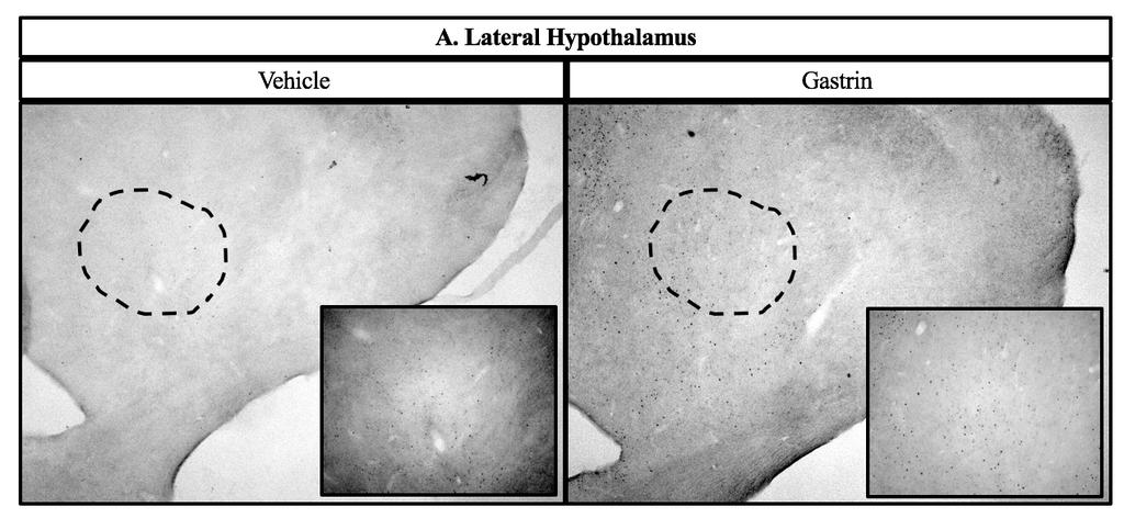
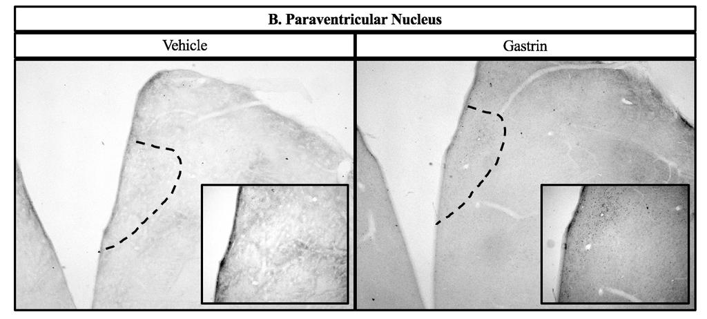
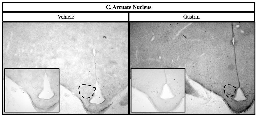
57 48

58 Figure 3-4. Representative photomicrographs of c-fos immunohistochemical staining following intracerebroventricular injection of gastrin in the LH (A), PVN (B), ARC (C), and AP/NTS (D). Photomicrographs taken at greater magnification are located in the bottom corner of each image. Treatment is indicated above each photomicrograph. For this experiment, 5 chicks per treatment were available for the analysis. 49
 (A),")
.")

59 Figure 3-5. mrna abundance in specific nuclei following intracerebroventricular injection of gastrin in 4 day-old broiler chicks at 60 min post-injection in the lateral hypothalamus (LH) (A), paraventricular nucleus (PVN) (B), arcuate nucleus (ARC) (C), and the combined biopsy of the area postrema (AP) and nucleus of the solitary tract (NTS) (D). Values are means ± standard error; bars with *are different from the vehicle-injected group within a gene (P < 0.05). For this experiment, 6-18 chicks per treatment were available for the analysis. AADC, aromatic L-amino acid decarboxylase; AgRP, agouti-related peptide; CART, cocaine and amphetamine-regulated transcript; CRF, corticotropin-releasing factor; CRFR1 and CRFR2, respectively, corticotropinreleasing factor receptor subtypes 1 and 2; GAD1, glutamate decarboxylase isoform 1; MC3R and MC4R, respectively, melanocortin receptors 3 and 4; MCH, melanin-concentrating hormone; MT, mesotocin; NPY, neuropeptide Y; NPYR1, NPYR2, and NPYR5, respectively, neuropeptide Y receptors 1, 2, and 5; ORX, orexin; POMC, pro-opiomelanocortin; PrRP, prolactin-releasing peptide; TH, tyrosine hydroxylase. 50
 and non-cumulative (B) food intake in 4 day-old broiler chicks. Values are means ± standard error; bars with unique letters are different from each other within a time point (P < 0.05).")
60 Figure 3-6. Food intake following intracerebroventricular injection of 500 ng gastrin, 6 nmol astressin, or 500 ng gastrin + 6 nmol astressin. Cumulative (A) and non-cumulative (B) food intake in 4 day-old broiler chicks. Values are means ± standard error; bars with unique letters are different from each other within a time point (P < 0.05). For this experiment, 8-10 chicks per treatment were available for the analysis. 48
Internal Regulation II Energy
 Internal Regulation II Energy Reading: BCP Chapter 16 lookfordiagnosis.com Homeostasis Biologically, what is necessary for life is a coordinated set of chemical reactions. These reactions take place in
CNS Control of Food Intake. Adena Zadourian & Andrea Shelton
 CNS Control of Food Intake Adena Zadourian & Andrea Shelton Controlling Food Intake Energy Homeostasis (Change in body adiposity + compensatory changes in food intake) Background Information/Review Insulin
Motivation 1 of 6. during the prandial state when the blood is filled
 Motivation 1 of 6 I. INTRODUCTION A. Motivation: a condition (usually internal) that initiates, activates, or maintains goal-directed behavior. B. Archery analogy 1. undrawn bow has no potential energy
Neurophysiology of the Regulation of Food Intake and the Common Reward Pathways of Obesity and Addiction. Laura Gunter
 Neurophysiology of the Regulation of Food Intake and the Common Reward Pathways of Obesity and Addiction Laura Gunter The Brain as the Regulatory Center for Appetite The brain is the integration center
FLASH CARDS. Kalat s Book Chapter 10 Alphabetical
 FLASH CARDS www.biologicalpsych.com Kalat s Book Chapter 10 Alphabetical AgRP AgRP Agouti-related peptide; synthesized in hypothalamus. Acts as an appetite stimulator. Also decreases metabolism. aldosterone
Chapter 12. Ingestive Behavior
 Chapter 12 Ingestive Behavior Drinking a. fluid compartments b. osmometric thirst c. volumetric thirst Eating a. energy sources b. starting a meal c. stopping a meal d. eating disordersd Drinking a. fluid
LESSON 3.3 WORKBOOK. How do we decide when and how much to eat?
 Appetite The psychological desire to eat, driven by feelings of pleasure from the brain. Hunger The biological or physiological need to eat, caused by a release of hormones from the digestive tract. LESSON
Νευροφυσιολογία και Αισθήσεις
 Biomedical Imaging & Applied Optics University of Cyprus Νευροφυσιολογία και Αισθήσεις Διάλεξη 16 Κίνητρα Συμπεριφοράς ή Υποκίνηση (Motivation) Introduction Types of behavior Unconscious reflexes Voluntary
Ingestive Behavior: Feeding & Weight Regulation. Hypovolemic vs. Osmotic Thirst
 Ingestive Behavior: Feeding & Weight Regulation 1 Hypovolemic Thirst Receptors, CNS, Responses Salt Appetite Digestive components Glucose Homeostasis: Insulin & Glucagon Diabetes Mellitus 1 & 2 CNS Hypothalamic
Ghrelin mediates stressinduced. behavior in mice. Chuang et al 2011 L3: Love, Lust, Labor
 Ghrelin mediates stressinduced food-reward behavior in mice Chuang et al 2011 L3: Love, Lust, Labor Agenda Introduction What is Ghrelin? Previous Models New model Methods Results Discussion Conclusion
Ingestive Behaviors 33. Introduction. Page 1. control and story lines. (a review of general endocrinology) Integration (or the basic reflex arc model)
 Integration (or the basic reflex arc model)") Ingestive Behaviors 33 (a review of general endocrinology) A neuroendocrine system: components, a reflex arc, the endocrine system, the AN, endocrine / nervous systems as afferents and efferents, the theoretical
Ingestive Behaviors 33 (a review of general endocrinology) A neuroendocrine system: components, a reflex arc, the endocrine system, the AN, endocrine / nervous systems as afferents and efferents, the theoretical
Copyright 2017 The Guilford Press
 This is a chapter excerpt from Guilford Publications. Eating Disorders and Obesity: A Comprehensive Handbook, Third Edition. Edited by Kelly D. Brownell and B. Timothy Walsh. Copyright 2017. Purchase this
Hypothalamus. Small, central, & essential.
 Hypothalamus Small, central, & essential. Summary: You can t live without a hypothalamus. Located at the junction between the brain stem and the forebrain Medial hypothalamus: interface between the brain
Chapter 1. General introduction. Part of this chapter is adapted from Adan et.al., Br.J.Pharmacol. 2006;149:815
 Chapter 1 General introduction Part of this chapter is adapted from Adan et.al., Br.J.Pharmacol. 2006;149:815 Chapter 1 B. Tiesjema, 2007 GENERAL INTRODUCTION NEURAL CIRCUITS INVOLVED IN ENERGY BALANCE
Insulin-Leptin Interactions
 Insulin-Leptin Interactions Ahmed S., Al-Azzam N., Cao B. Karshaleva B., Sriram S., Vu K. If you understand a system, you can predict it. Agenda - Energy homeostasis Overview of leptin and insulin Signaling
Digestion: Endocrinology of Appetite
 Digestion: Endocrinology of Dr. Ritamarie Loscalzo Medical Disclaimer: The information in this presentation is not intended to replace a one on one relationship with a qualified health care professional
Bi156 lecture 2, 1/6/12. Eating and weight regulation
 Bi156 lecture 2, 1/6/12 Eating and weight regulation Introduction: weight regulation in an affluent society In our society much effort and money is expended on regulation of weight. Failure to maintain
Dietary Macronutrient Composition and Exogenous Neuropeptide Y Affect Feed Intake in Broiler Chicks
 Dietary Macronutrient Composition and Exogenous Neuropeptide Y Affect Feed Intake in Broiler Chicks Laura Ashley Nelson Thesis submitted to the Faculty of the Virginia Polytechnic Institute and State University
BIOLOGY - CLUTCH CH.45 - ENDOCRINE SYSTEM.
 !! www.clutchprep.com Chemical signals allow cells to communicate with each other Pheromones chemical signals released to the environment to communicate with other organisms Autocrine signaling self-signaling,
Investigation of the role of nesfatin-1/nucb2 in the central nervous system. Ph.D. thesis Katalin Könczöl
 Investigation of the role of nesfatin-1/nucb2 in the central nervous system Ph.D. thesis Katalin Könczöl Semmelweis University János Szentágothai Doctoral School of Neurosciences Supervisor: Official reviewers:
Testosterone and other male hormones seem to be related to aggressive behavior in some species
 Testosterone and Male Aggression Testosterone and other male hormones seem to be related to aggressive behavior in some species In the fish species Oreochromis mossambicus, elevated levels have been found
Homeostasis Through Chemistry. The Endocrine System Topic 6.6
 Homeostasis Through Chemistry The Endocrine System Topic 6.6 Comparing NS & ES Animals have two systems of internal communication and regulation The nervous system Response time: Fast, quick Signals: electrical
Temperature, Regulation, Thirst, and Hunger
 PSYB64 Lecture 6 Temperature, Regulation, Thirst, and Hunger 1. Homeostasis 2. Temperature 3. Thirst 4. Hunger 5. Obesity & Hunger Disorders HOMEOSTASIS Homeostasis: Physiological equilibrium Motivation:
Energy Balance. Applied Human Metabolism VII. Energy Out. Factors that effect BMR/RMR 17/03/2016
 Energy Balance Applied Human Metabolism VII Weight Regulation The balance of energy taken in or leaving the body determines body mass Energy In = Energy Out Weight Maintenance Energy In < Energy Out Weight
Ingestive Behaviors 21. Introduction. Page 1. control and story lines. (a review of general endocrinology) Integration (or the basic reflex arc model)
 Integration (or the basic reflex arc model)") Ingestive Behaviors 21 (a review of general endocrinology) A neuroendocrine system: components, a reflex arc, the endocrine system, the AN, endocrine / nervous systems as afferents and efferents, the theoretical
Ingestive Behaviors 21 (a review of general endocrinology) A neuroendocrine system: components, a reflex arc, the endocrine system, the AN, endocrine / nervous systems as afferents and efferents, the theoretical
1 Neuroregulation of Appetite
 Chapter 1 / Neuroregulation of Appetite 3 1 Neuroregulation of Appetite Ofer Reizes, PhD, Stephen C. Benoit, PhD, and Deborah J. Clegg, PhD CONTENTS INTRODUCTION THE DUAL-CENTERS HYPOTHESIS CONTROL OF
Understanding obesity by local overexpression of neuropeptides: a viral vector based approach
 Understanding obesity by local overexpression of neuropeptides: a viral vector based approach Cover photo: Chris Timmers Cover design: Marije Tiesjema Print: Gildeprint, Enschede ISBN: 978-90-39344675
Chapter 24 Cholesterol, Energy Balance and Body Temperature. 10/28/13 MDufilho
 Chapter 24 Cholesterol, Energy Balance and Body Temperature 10/28/13 MDufilho 1 Metabolic Role of the Liver Hepatocytes ~500 metabolic functions Process nearly every class of nutrient Play major role in
ENDOCRINOLOGY. Dr.AZZA SAJID ALKINANY 2 nd STAGE
 ENDOCRINOLOGY Dr.AZZA SAJID ALKINANY 2 nd STAGE THE RELATIONSHIP AMONG THE HYPOTHALMUS,POSTERIOR PITUITARY AND TARGET TISSUES. The posterior pituitary does not produce its own hormones, but stores and
Chapter 26. Hormones and the Endocrine System. Lecture by Edward J. Zalisko
 Chapter 26 Hormones and the Endocrine System PowerPoint Lectures for Biology: Concepts & Connections, Sixth Edition Campbell, Reece, Taylor, Simon, and Dickey Copyright 2009 Pearson Education, Inc. Lecture
Neurobiology of Addiction
 Neurobiology of Addiction Domenic A. Ciraulo, MD Director of Alcohol Pharmacotherapy Research Center for Addiction Medicine Department of Psychiatry Massachusetts General Hospital Disclosure Neither I
Peripubertal, leptin-deficient ob/ob female mice were used in an investigation of
 ESSICK-BROOKSHIRE, ELIZABETH ANN, M.S. The Effects of Peripherally Administered 17-β Estradiol and BIBP3226, a NPY Y1 Receptor Antagonist, on Food Intake, Body Mass, Reproductive Development and Behavior
Orexin and Sleep. Team: A Little Bit of Leptin
 Orexin and Sleep Team: A Little Bit of Leptin Intro to Orexin 1997 -Scripps Research Institute gene expression in the hypothalamus Found gene clone 35 - expression limited to the lateral hypothalamus NTs
Hormonal regulation of. Physiology Department Medical School, University of Sumatera Utara
 Hormonal regulation of nutrient metabolism Physiology Department Medical School, University of Sumatera Utara Homeostasis & Controls Successful compensation Homeostasis reestablished Failure to compensate
Chapter 20 Endocrine System
 Chapter 20 Endocrine System The endocrine system consists of glands and tissues that secrete Hormones are chemicals that affect other glands or tissues, many times far away from the site of hormone production
Motility Conference Ghrelin
 Motility Conference Ghrelin Emori Bizer, M.D. Division of Gastroenterology/Hepatology November 21, 2007 Ghrelin: Basics Hormone produced by the A-like A endocrine cells in the oxyntic mucosa (stomach body
Endocrine System. Chapter 20. Endocrine Glands and Hormones. The Endocrine System. Endocrine glands
 Chapter 20 Endocrine System Endocrine Glands and Hormones The endocrine system consists of glands and tissues that secrete hormones Hormones are chemicals that affect other glands or tissues, many times
Chapter Nine. Temperature Regulation, Thirst, and Hunger
 Chapter Nine Temperature Regulation, Thirst, and Hunger Regulating Systems Homeostasis: Physiological equilibrium Epic FAIL with weight? Obesity rates 1980-2000 Motivation: Activating and directing behavior
Central nervous system control of food intake
 insight review article Central nervous system control of food intake Michael W. Schwartz*, Stephen C. Woods, Daniel Porte Jr*, Randy J. Seeley & Denis G. Baskin* Departments of Medicine* and Biological
Chapter 18: Endocrine Glands
 Chapter 18: Endocrine Glands I. Functions of the Endocrine System A. List and describe the eight major functions of the endocrine system: 1. 2. 3. 4. 5. 6. 7. 8. Page 1 of 19 C II. Pituitary Gland and
LESSON 3.4 WORKBOOK. Can you become addicted to food?
 DEFINITIONS OF TERMS Dopamine A compound that sends signals from one neuron to another, and is made from the amino acid tyrosine. Dopamine reward pathway A circuit in the brain that when activated leads
Chapter 20. Endocrine System Chemical signals coordinate body functions Chemical signals coordinate body functions. !
 26.1 Chemical signals coordinate body functions Chapter 20 Endocrine System! Hormones Chemical signals Secreted by endocrine glands Usually carried in the blood Cause specific changes in target cells Secretory
BIOL 2458 A&P II CHAPTER 18 SI Both the system and the endocrine system affect all body cells.
 BIOL 2458 A&P II CHAPTER 18 SI 1 1. Both the system and the endocrine system affect all body cells. 2. Affect on target cells by the system is slow. Affect on target cells by the system is fast. INTERCELLULAR
Ch45: Endocrine System
 Ch45: Endocrine System Endocrine System Homeostasis is the tendency to maintain a stable internal environment. Function = coordinate and control the body with hormones to maintain homeostasis Works with
Hormonal Regulation of Food Intake
 Physiol Rev 85: 1131 1158, 2005; doi:10.1152/physrev.00015.2004. Hormonal Regulation of Food Intake SARAH STANLEY, KATIE WYNNE, BARBARA McGOWAN, AND STEPHEN BLOOM Endocrine Unit, Imperial College Faculty
Endocrine System Hormones. AP Biology
 Endocrine System Hormones 2007-2008 Regulation Why are hormones needed? u chemical messages from one body part to another u communication needed to coordinate whole body u daily homeostasis & regulation
Energy flow in the organism
 I. Parameters of energy metabolism, basal metabolic rate, measurements. II. Control of food intake, hunger and satiety Péter Sántha, 12.02. 2017. Energy flow in the organism NUTRIENTS PHYSICAL WORK HEAT
NROSCI/BIOSC 1070 and MSNBIO 2070 September 11, 2017 Control Mechanisms 2: Endocrine Control
 NROSCI/BIOSC 1070 and MSNBIO 2070 September 11, 2017 Control Mechanisms 2: Endocrine Control Hormones are chemical messengers that are secreted into the blood by endocrine cells or specialized neurons.
Homeostasis and Mechanisms of Weight Regulation
 Homeostasis and Mechanisms of Weight Regulation Purpose In this activity students will investigate how negative feedback mechanisms function to maintain homeostatic balance using a recently discovered
The Players. Liver Thyroid Adrenals Pancreas Reproductive System Pituitary Gut Bacteria
 The Players Part I Quick Review Understanding some of the key systems and their relationship to hormones is the best place to start It will help with some of the hormone interconnections Key to understanding
Chemical Regulation. Chapter 26. Testosterone and Male Aggression: Is There a Link? THE NATURE OF CHEMICAL REGULATION
 Chapter 6 Chemical Regulation PowerPoint Lectures for Biology: Concepts and Connections, Fifth Edition Campbell, Reece, Taylor, and Simon Testosterone and Male Aggression: Is There a Link? Among male animals,
Endocrine System Hormones (Ch. 45)
") Endocrine System Hormones (Ch. 45) Regulation Why are hormones needed? chemical messages from one body part to another communication needed to coordinate whole body daily homeostasis & regulation of large
Endocrine System Hormones (Ch. 45) Regulation Why are hormones needed? chemical messages from one body part to another communication needed to coordinate whole body daily homeostasis & regulation of large
Chapter 16: Endocrine System 1
 Ch 16 Endocrine System Bi 233 Endocrine system Endocrine System: Overview Body s second great controlling system Influences metabolic activities of cells by means of hormones Slow signaling Endocrine glands
Stressin with Ghrelin. Peptide Pods: Lara, Elijah, Karen, Brandon, Milena, Max & Lucile
 Stressin with Ghrelin Peptide Pods: Lara, Elijah, Karen, Brandon, Milena, Max & Lucile Agenda Ghrelin Essentials Review Associations of Ghrelin with eating, stress, metabolic factors, etc. High-fat diet
INTRODUCTION TO THE BIOCHEMISTRY OF HORMONES AND THEIR RECPTORS
 INTRODUCTION TO THE BIOCHEMISTRY OF HORMONES AND THEIR RECPTORS 1 Introduction to the Biochemistry of Hormones and their Receptors Lectuctre1 Sunday 17/2/ Objectives: 1. To understand the biochemical nature
Neurobiology of food intake in health and disease
 Neurobiology of food intake in health and disease Gregory J. Morton, Thomas H. Meek and Michael W. Schwartz Abstract Under normal conditions, food intake and energy expenditure are balanced by a homeostatic
Mechanisms of hypothalamic regulation of food intake in birds. Jinxin Wang. Doctor of Philosophy In Animal and Poultry Sciences. Mark A.
 Mechanisms of hypothalamic regulation of food intake in birds Jinxin Wang Dissertation submitted to the faculty of the Virginia Polytechnic Institute and State University in partial fulfillment of the
Pancreas. Endocrine pancreas - Islets of Langerhans A or alpha cells glucagon B or beta cells insulin Delta cells somatostatin
 Endocrine System Pancreas Endocrine pancreas - Islets of Langerhans A or alpha cells glucagon B or beta cells insulin Delta cells somatostatin Glucagon & Metabolism Produced by beta cells of Islets Primary
Behavioral and Motivational mechanisms of Brain. Limbic system and the Hypothalamus
 Behavioral and Motivational mechanisms of Brain Limbic system and the Hypothalamus 1 General functions 1. Control of behavior 2. Control level of activities in different parts of brain 3. Motivational
processes in the central nervous system (CNS), affecting many of the during the course of ethanol treatment. Ethanol stimulates the release of
, affecting many of the during the course of ethanol treatment. Ethanol stimulates the release of") INTRODUCTION INTRODUCTION Neuroscience research is essential for understanding the biological basis of ethanol-related brain alterations and for identifying the molecular targets for therapeutic compounds
INTRODUCTION INTRODUCTION Neuroscience research is essential for understanding the biological basis of ethanol-related brain alterations and for identifying the molecular targets for therapeutic compounds
Chapter 45-Hormones and the Endocrine System. Simple Hormone Pathways
 Chapter 45-Hormones and the Endocrine System Simple Hormone s Low ph in duodenum Hormones are released from an endocrine, travel through the bloodstream, and interact with the receptor or a target to cause
Psychology - Problem Drill 05: Endocrine System & Influence on Behavior
 Psychology - Problem Drill 05: Endocrine System & Influence on Behavior No. 1 of 10 1. Which of the following statements is FALSE regarding the interaction between the nervous an endocrine systems? (A)
The Emotional Nervous System
 The Emotional Nervous System Dr. C. George Boeree Emotion involves the entire nervous system, of course. But there are two parts of the nervous system that are especially significant: The limbic system
BIOLOGY 2402 Anatomy and Physiology Lecture. Chapter 18 ENDOCRINE GLANDS
 BIOLOGY 2402 Anatomy and Physiology Lecture Chapter 18 ENDOCRINE GLANDS 1 ENDOCRINE GLANDS Homeostasis depends on the precise regulation of the organs and organ systems of the body. Together the nervous
REGULATION OF FOOD INTAKE AND NUTRITIONAL STATE
 REGULATION OF FOOD INTAKE AND NUTRITIONAL STATE INTAKE OUTPUT CENTER OF SATIETY ncl. ventromedialis in hypothalamus - CENTER OF HUNGER (permanently active) lateral hypothalamus (nucleus under fasciculus
Chapter 3. Structure and Function of the Nervous System. Copyright (c) Allyn and Bacon 2004
 Allyn and Bacon 2004") Chapter 3 Structure and Function of the Nervous System 1 Basic Features of the Nervous System Neuraxis: An imaginary line drawn through the center of the length of the central nervous system, from the
Chapter 3 Structure and Function of the Nervous System 1 Basic Features of the Nervous System Neuraxis: An imaginary line drawn through the center of the length of the central nervous system, from the
4/23/2018. Endocrine System: Overview. Endocrine System: Overview
 Endocrine System: Overview With nervous system, coordinates and integrates activity of body cells Influences metabolic activities via hormones transported in blood Response slower but longer lasting than
Endocrine System Notes
 Endocrine System Notes is the tendency to maintain a stable internal environment. - parts of the body that secrete hormones directly into the body. - parts of the body that make secretions which travel
Cephalization. Nervous Systems Chapter 49 11/10/2013. Nervous systems consist of circuits of neurons and supporting cells
 Nervous Systems Chapter 49 Cephalization Nervous systems consist of circuits of neurons and supporting cells Nervous system organization usually correlates with lifestyle Organization of the vertebrate
Prasad S. Dalvi. Copyright by Prasad S. Dalvi 2012
 Molecular Mechanisms Involved in Insulin- and Leptinmediated Regulation of Hypothalamic Proglucagon Gene Expression and Action of Glucagon-like Peptides on Hypothalamic Neuropeptides by Prasad S. Dalvi
Chapter 26 Hormones and the
 Chapter 6 Hormones and the Endocrine System Introduction In lions, the hormone testosterone promotes the development and maintenance of male traits including growth and maintenance of the mane and increased
Hypothalamus. To learn how the brain regulates neuroendocrine secretions NTA Ch 14, pgs Key Figs: 14-3; 14-4,
 Hypothalamus Objectives To learn the general organization of the hypothalamus and the functions of the major nuclei NTA Ch 14, pgs. 419-422 Key Figs: 14-2, 14-3 To learn how the brain regulates neuroendocrine
Endocrine secretion cells secrete substances into the extracellular fluid
 Animal Hormones Concept 30.1 Hormones Are Chemical Messengers Endocrine secretion cells secrete substances into the extracellular fluid Exocrine secretion cells secrete substances into a duct or a body
Chapter 11 - Endocrine System
 Chapter 11 - Endocrine System 11.1 Introduction A. The endocrine system is made up of the cells, tissues, and organs that secrete hormones into body fluids. B. The body has two kinds of glands, exocrine
The Intricate Web Of Hormone Relationships Dr. Ritamarie Loscalzo
 The Intricate Web Of Hormone Relationships Dr. Ritamarie Loscalzo Medical Disclaimer: The information in this presentation is not intended to replace a one-onone relationship with a qualified health care
Refer to the figure below, a diagram of a renal tubule, to answer the following questions.
 1. The digestion and utilization of which nutrient creates the greatest need for osmoregulation by the kidneys? a. protein b. starch c. fat d. oil e. cellulose 2. Which of the following is true of urea?
Neuroendocrinology an integrative approach
 Neuroendocrinology an integrative approach JP Advis DVM, Ph.D. Bartlett Hall, Animal ciences, Cook, (848) 932-9240, advis@aesop.rutgers.edu 06 Course website: rci.rutgers.edu/~advis Material to be covered:
The Nervous System. Functions of the Nervous System input gathering To monitor occurring inside and outside the body Changes =
 The Nervous System Functions of the Nervous System input gathering To monitor occurring inside and outside the body Changes = To process and sensory input and decide if is needed output A response to integrated
3. The function of that hormone. In other words, what change does that hormone facilitate.
 Slide 2 The endocrine operates to regulate internal functions. It does so, via the use of hormones, or chemical messengers. Hormones travel in the blood from the site of production to distant target cells
Injectable GLP 1 therapy: weight loss effects seen in obesity with and without diabetes
 Injectable GLP 1 therapy: weight loss effects seen in obesity with and without diabetes Dr Masud Haq Consultant Lead in Diabetes & Endocrinology Maidstone & Tunbridge Wells NHS Trust & The London Preventative
Unit 3: The Biological Bases of Behaviour
 Unit 3: The Biological Bases of Behaviour Section 1: Communication in the Nervous System Section 2: Organization in the Nervous System Section 3: Researching the Brain Section 4: The Brain Section 5: Cerebral
The Physiology of Weight Regulation: Implications for Effective Clinical Care
 Roundtable on Obesity Solutions The Physiology of Weight Regulation: Implications for Effective Clinical Care Lee M. Kaplan, MD, PhD Obesity, Metabolism & Nutrition Institute Massachusetts General Hospital
Chapter 9 The Endocrine System and Hormone Activity
 Chapter 9 The Endocrine System and Hormone Activity Overview Coordinates and directs the activity of cells. Interacts with the nervous system Uses chemical messengers called hormones released by organs
! acts via the autonomic nervous system. ! maintaining body weight within tight limits. ! ventromedial (VMN) ! arcuate (ARC) ! neuropeptide Y (NPY)
 ! arcuate (ARC) ! neuropeptide Y (NPY)") Brain Regulates energy homeostatis Glucose Sensing in the Brain Seminar 2012 Gareth Price! acts in response to circulating signals of nutrient states! acts via the autonomic nervous system Ensures a balance
Brain Regulates energy homeostatis Glucose Sensing in the Brain Seminar 2012 Gareth Price! acts in response to circulating signals of nutrient states! acts via the autonomic nervous system Ensures a balance
THE ROLE OF INSULIN RECEPTOR SIGNALING IN THE BRAIN. COGS 163 By: Pranav Singh Alexandra Villar
 THE ROLE OF INSULIN RECEPTOR SIGNALING IN THE BRAIN COGS 163 By: Pranav Singh Alexandra Villar INTRODUCTION Insulin is a hormone produced in the pancreas by the islets of Langerhans that regulates the
Ch45: Endocrine System
 Ch45: Endocrine System Endocrine System Homeostasis is the tendency to maintain a stable internal environment. Function = with hormones to maintain homeostasis Works with nervous system Anatomy Location:
BIOLOGY. CONCEPTS & CONNECTIONS Fourth Edition. Neil A. Campbell Jane B. Reece Lawrence G. Mitchell Martha R. Taylor. CHAPTER 26 Chemical Regulation
 BIOLOGY CONCEPTS & CONNECTIONS Fourth Edition Neil A. Campbell Jane B. Reece Lawrence G. Mitchell Martha R. Taylor CHAPTER 26 Chemical Regulation Modules 26.1 26.5 From PowerPoint Lectures for Biology:
Campbell's Biology: Concepts and Connections, 7e (Reece et al.) Chapter 26 Hormones and the Endocrine System Multiple-Choice Questions
 Chapter 26 Hormones and the Endocrine System Multiple-Choice Questions") Campbell's Biology: Concepts and Connections, 7e (Reece et al.) Chapter 26 Hormones and the Endocrine System 26.1 Multiple-Choice Questions 1) Hormones are chemicals produced by the endocrine system that
Campbell's Biology: Concepts and Connections, 7e (Reece et al.) Chapter 26 Hormones and the Endocrine System 26.1 Multiple-Choice Questions 1) Hormones are chemicals produced by the endocrine system that
GENERAL CHARACTERISTICS OF THE ENDOCRINE SYSTEM FIGURE 17.1
 GENERAL CHARACTERISTICS OF THE ENDOCRINE SYSTEM FIGURE 17.1 1. The endocrine system consists of glands that secrete chemical signals, called hormones, into the blood. In addition, other organs and cells
Hypothalamic TLR2 triggers sickness behavior via a microglia-neuronal axis
 Hypothalamic TLR triggers sickness behavior via a microglia-neuronal axis Sungho Jin, *, Jae Geun Kim,, *, Jeong Woo Park, Marco Koch,, Tamas L. Horvath and Byung Ju Lee Department of Biological Sciences,
The Endocrine System. I. Overview of the Endocrine System. II. Three Families of Hormones. III. Hormone Receptors. IV. Classes of Hormone Receptor
 The Endocrine System I. Overview of the Endocrine System A. Regulates long term metabolic processes B. Releases hormones from endocrine cells 1. Hormones are chemicals 2. Alter metabolism of cells 3. Release
The Human Brain. I Think Therefore I am
 The Human Brain I Think Therefore I am The Beginning The simplest creatures have very simple nervous systems made up of nothing but a bunch of nerve cells They have neural nets, individual neurons linked
Unit 4 Homeostasis. The term homeostasis refers to the body s attempt. Your body systems must to maintain a stable internal environment -
 Unit 4 Homeostasis The term homeostasis refers to the body s attempt Your body systems must to maintain a stable internal environment - The body is trying to maintain, through a series of monitored adjustments.
Biological Bases of Behavior. 3: Structure of the Nervous System
 Biological Bases of Behavior 3: Structure of the Nervous System Neuroanatomy Terms The neuraxis is an imaginary line drawn through the spinal cord up to the front of the brain Anatomical directions are
ABSTRACT CONTROL. Long term use of sugars can induce excess caloric intake and/or obesity. To
 ABSTRACT Title of Dissertation: THE EFFECTS OF SUGAR INTAKE ON ENERGY CONTROL Changhui Zhao, Doctor of Philosophy, 2015 Directed By: Professor Thomas W. Castonguay, Department of Nutrition and Food Science
Neural pathways controlling homeostatic and hedonic feeding in rats on free-choice diets van den Heuvel, J.K.
 UvA-DARE (Digital Academic Repository) Neural pathways controlling homeostatic and hedonic feeding in rats on free-choice diets van den Heuvel, J.K. Link to publication Citation for published version (APA):
Human Anatomy and Physiology - Problem Drill 16: The Endocrine System
 Human Anatomy and Physiology - Problem Drill 16: The Endocrine System Question No. 1 of 10 The endocrine system is made up of a number of organs and glands. Which one of the following is not an organ or
The dorsomedial hypothalamic nucleus and its role in ingestive behavior and body weight regulation: Lessons learned from lesioning studies
 Physiology & Behavior 76 (2002) 431 442 The dorsomedial hypothalamic nucleus and its role in ingestive behavior and body weight regulation: Lessons learned from lesioning studies Larry L. Bellinger a,
Empower Preventive Medicine. Timothy J. McCormick, DO, MPH 4221 Baymeadows Suite 6 Jacksonville, FL
 Empower Preventive Medicine Timothy J. McCormick, DO, MPH 4221 Baymeadows Suite 6 Jacksonville, FL 32217 904-367-4005 Drtim@emprevmed.com Obesity Medicine Old paradigm: Obesity was a matter of willpower,
Chapter 8.2 The Endocrine System
 Major Endocrine Organs Hypothalamus Pineal Gland Pituitary Gland Thyroid Gland Thymus Gland Adrenal Glands Pancreas Ovaries (Female) Testis (Male) Chapter 8.2 The Endocrine System The endocrine system