Hemostasis and fibrinolysis the hemostatic balance
|
|
|
- Deborah Conley
- 5 years ago
- Views:
Transcription
1 Hemostasis and fibrinolysis the hemostatic balance Bleeding Clotting The hemostatic balance Reference: Boron & Boulpaep Medical Physiology, 3 rd Ed, Chapter 18 Dr. Ana-Maria Zagrean Carol Davila University of Medicine and Pharmacy
2 Hemostasis and fibrinolysis The normal functioning of the circulatory system depends on: - maintenance of a normal blood fluidity and flow, and - preservation of the blood vessel walls integrity in order to prevent blood leaking These requirements are fulfilled by the hemostatic and fibrinolytic processes. Blood is normally in a liquid state inside blood vessels as long as it does not come into contact with: - negatively charged surfaces (e.g., the collagen beneath endothelial cells) that activate an intrinsic coagulation pathway, - tissue factors (e.g., released from damaged tissue) that activate an extrinsic coagulation pathway. Thrombolytic/fibrinolytic pathways keep the balance of coagulation pathways by lysing blood clots/thrombus (intravascular clot).
3 Hemostasis and fibrinolysis involve the following components: - Vascular / Endothelial: - endothelial cells - vascular smooth muscle - Globular: - platelets - erithrocytes - Plasmatics: - plasmatic proteins, coagulation factors - plasmatic ionic Ca 2+
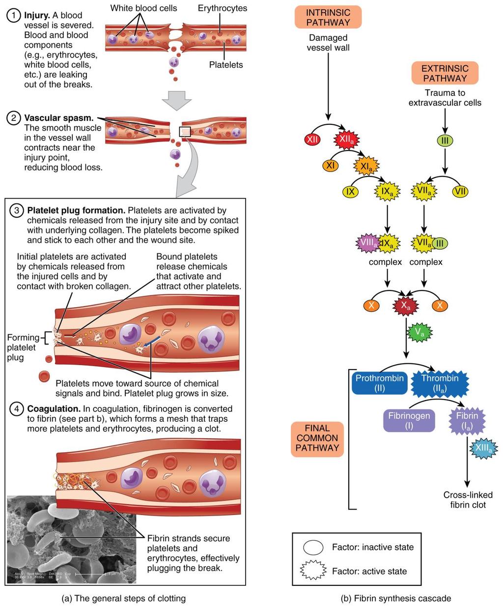
4 Hemostasis and fibrinolysis events (1) local vasoconstriction to collapse vessels with an intravascular pressure below the critical closing pressure (2) increased tissue pressure vessel radius decrease, to diminish blood flow, decrease the hemorrhage (3) adhesion, activation an aggregation of platelets resulting in platelet plug formation, in the case of capillary bleeding/vessel rupture stop the hemorrhage (4) coagulation or clot formation through controlled proteolysis of coagulation proteins fibrin network stabilize the clot (5) anticoagulant processes that prevent excessive hemostasis (6) clot retraction and fibrinolysis that breaks up clots vessel wall repair or fibrous organization of the clot into fibrous tissue
5 Hemostasis and fibrinolysis events (1) local vasoconstriction, to collapse vessels with an intravascular pressure below the critical closing pressure (2) increased tissue pressure vessel radius decrease, to diminish blood flow, decrease the hemorrhage (3) adhesion, activation an aggregation of platelets resulting in platelet plug formation, in the case of capillary bleeding/vessel rupture stop the hemorrhage, (4) coagulation or clot formation through controlled proteolysis of coagulation proteins fibrin network stabilize the clot (5) anticoagulant processes that prevent excessive hemostasis (6) clot retraction and fibrinolysis that breaks up clots vessel wall repair or fibrous organization of the clot into fibrous tissue
6 (1) Local vascular constriction Vasoconstriction raises the critical closing pressure and thus collapses vessels that have an intravascular pressure below the critical closing pressure. Vessel constriction is also promoted by chemical products of platelet plug formation and of coagulation: 1. local myogenic spasm: local myogenic contraction of the blood vessels is initiated by direct damage to the vascular wall. 2. local factors from platelets and endothelium: thromboxane A2 (TXA2), serotonin (5-HT), thrombin-triggered release of endothelin 1 (ET-1) that is one of the most powerful vasoconstrictor 3. nervous reflexes initiated by pain nerve impulses or other sensory impulses that originate from the traumatized vessel or nearby tissues. Vascular spasm is directly related with the degree of vessel injury. The spasm can last for minutes to hours, during which time the processes of platelet plugging & blood coagulation take place. Vasodilatation can occur in the neighboring vessels.
7 Hemostasis and fibrinolysis events (1) vasoconstriction to collapse vessels with an intravascular pressure below the critical closing pressure (2) increased tissue pressure vessel radius decrease, to diminish blood flow, decrease the hemorrhage (3) adhesion, activation an aggregation of platelets resulting in platelet plug formation, in the case of capillary bleeding/vessel rupture stop the hemorrhage, (4) coagulation or clot formation through controlled proteolysis of coagulation proteins fibrin network stabilize the clot (5) anticoagulant processes that prevent excessive hemostasis (6) clot retraction and fibrinolysis that breaks up clots vessel wall repair or fibrous organization of the clot into fibrous tissue

8 (2) Increased tissue pressure Determined by blood extravasation into the interstitial perivascular space Contributes to hemostasis because it decreases transmural pressure, which is the difference between intravascular pressure and tissue pressure. Transmural pressure is the main determinant of blood vessel radius. Given the fourth-power relationship between flow and blood vessel radius, an increase in tissue pressure that causes radius to decrease by a factor of 2 would diminish flow by a factor of 16. That is why pressing a finger against a small cut stop the bleeding; a tourniquet increases extravascular pressure and halt an arterial hemorrhage in a limb. Finally, surgeons routinely make use of this principle when applying hemostatic clamps to close off bleeders. This is the Hagen-Poiseuille equation, where F is the flow, ΔP is the driving pressure, r is the inner radius of the tube, l is its length, and η is the viscosity.
9 Hemostasis and fibrinolysis events (1) vasoconstriction to collapse vessels with an intravascular pressure below the critical closing pressure (2) increased tissue pressure vessel radius decrease, to diminish blood flow, decrease the hemorrhage (3) adhesion, activation an aggregation of platelets resulting in platelet plug formation, in the case of capillary bleeding/vessel rupture stop the hemorrhage, (4) coagulation or clot formation through controlled proteolysis of coagulation proteins fibrin network stabilize the clot (5) anticoagulant processes that prevent excessive hemostasis (6) clot retraction and fibrinolysis that breaks up clots vessel wall repair or fibrous organization of the clot into fibrous tissue
10 (3) Platelet functions: Platelet plug formation by platelet reaction of: - adhesion - activation - aggregation Platelets have a Ca-dependent procoagulant activity through Platelet factor-3 (membrane phospholipid, PF-3) F Xa and thrombin (prothrombinase activity)
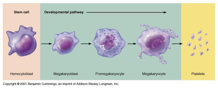
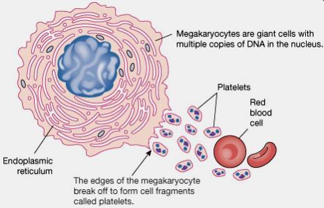
11 Platelets (thrombocytes) produced in bone marrow by fragmentation of megakariocytes time interval from differentiation of the stem cell to the production of platelets (thrombocytopoiesis) ~ 10 days controled by growth inducers and differentiations inducers: thrombopoietin, IL-6, IL-3, Vit B12, GM-CSF (effects: in no. of megakariocytes and in mean volume or nuclear units) release ~ 4,000 platelets/megakariocyte
12 Platelets production Nuclear Cytoplasmic Platelets release replication granulation
13 Platelets production
14 Platelet circulation Half-life in the blood: 8-12 days Normal count: 150, ,000 / ml Young platelets spend up to 36 hrs. in spleen after release from bone marrow Normally not active until damage occur to the vessel walls Eliminated from the circulation mainly by the tissue macrophage system (> than 50% in the spleen) Platelet membrane: glycoproteins coat (repulses adherence to normal endothelium, causes adherence to injured vessel wall) phospholipids that activate blood-clotting reactions Platelet antigens: - specific surface AG: HPA1-5 (human platelet alloantigens) - also express ABO and HLA class I antigens
15 Platelet structure Disk-like shape, Φ= 1-2 µm, vol=5.8 fl, colourless, without nucleus Circumferential skeleton of microtubules that maintains the normal circulating discoid shape and consists in residual Golgi & ER (contain [Ca], site of different enzymes, PG and TXA2 synthesis) The cytoplasm contains mitochondria (oxidative phosphorylation ATP, ADP synthesis), smooth ER, lysosomes (hydrolytic enzymes), peroxisomes (catalase), fibrin-stabilizing factor (F XIII) and the following kinds of granules: - electron-dense granules (Ca 2+, ADP, serotonin) - a-granules (heparin antagonist PF 4, vwf, PDGF, fibrinogen) - glycogen (anaerobic glycolysis)
16 Platelet structure Contractile protein complex system - microfilaments: actin, myosin, fibrin, filamin, thrombosthenin contraction and release of granules Open membrane canalicular system facilitates the release of granules and provides a large reactive surface on which plasma coagulation proteins may be selectively absorbed Membrane phospholipids (platelet factor 3 - PF 3) convert: F X to F Xa and prothrombin to thrombin Membrane glycoproteins/adhesion proteins: GPIa, GPIb, GPIIb/GPIIIa
17 Platelet ultrastructure Specific a-granule: growth f. (PDGF), fibrinogen, factor V, VWF, fibronectin, -thromboglobulin, heparin antagonist (PF4), thrombospondin Submembranous filaments (contractile protein) Glycocalyx* Peroxisome Protein contractile system Mitochondria Plasma membrane Open canalicular system Lysosomes Platelet phospholipid Electron dense granule: ATP, ADP, Ca 2+, serotonin Dense tubular system (Ca 2+, PG, TxA 2 ) Glycogen

18 Platelet by EM
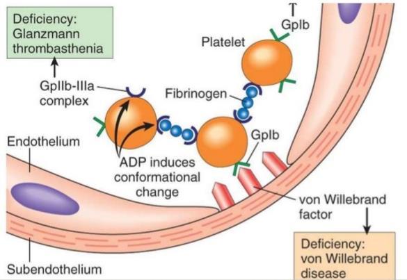
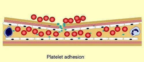
19 Platelet function Platelets do not adhere to themselves / other blood cells / endothelial membranes, as long as the negative surface charge is maintained by the presence of proteoglycans (mainly heparan sulfate). Platelet adhesion occurs in response to: Platelet adhesion - an increase in the shearing force at the surface of platelets or endothelial cells - in response to vessel injury - in response to humoral signals. Platelet adhesion - the binding of platelets to themselves or to other components, is mediated by platelet receptors = glycoproteins (GP) in the platelet membrane. Platelet GP receptors are integrins - integral membrane proteins (a class of matrix receptors) Willebrand factor (vwf) - is a glycoprotein that binds to platelet receptors Ib/Ia (Gp Ib/Ia) - is present in the blood plasma and made by endothelial cells (stored here in the Weibel-Palade bodies) and megakaryocytes (stored in α granules of platelets). - high shear, certain cytokines, and hypoxia trigger the release of vwf from endothelial cells. - a breach of the endothelium exposes platelet receptors to ligands that are components of the subendothelial matrix (collagen, which binds to Gp Ia/IIa, fibronectin and laminin, both of which bind to Gp Ic/Iia).
20 Platelets cell membrane glycoproteins/adhesion proteins: GPIa, GPIb, GPIIb/GPIIIa Platelet Membrane GPIb GPIIb GPIIIa GPIa Von Willebrand Factor (vwf): -released from endothelial cells (ECs) and platelets -its release from ECs is increased in stress, exercise, adrenaline infusion Adhesion Von Willebrand Factor Subendothelial microfibrils Aggregation Exposes Glycoproteins (GP) of the surface coat are important in initial events of platelet plug formation, platelet adhesion and aggregation: GP Ia, GP Ia/IIa - adhesion to collagen GP Ib, IIb/IIIa - attachment to vascular subendothelium through vwf GP IIb/IIIa - receptor for fibrinogen (platelet to platelet aggregation) GP Ic/IIa - binds fibronectin and laminin Adhesion to collagen
21
22
23 Platelet activation The binding on GP receptors of vwf, collagen, fibronectin, laminin, thrombin, etc, triggers a conformational change in the platelet receptors that initiates an intracellular signaling cascade, which leads to an exocytotic event = the release reaction or platelet activation. The signal-transduction cascade involves the activation of phospholipase C and an influx of Ca2+. Activated platelets exocytose the contents of their: - dense storage granules, (ATP, ADP, serotonin, and Ca2+). - α granules (growth factors, vwf, clotting factor V and fibrinogen). Activated platelets use cyclooxygenase (COX) to initiate the breakdown of arachidonic acid (AA) to thromboxane A2 (TXA2), which they release. Platelet activation is also associated with marked cytoskeletal and morphological changes as the platelet extends first a broad lamellipodium and then many finger-like filopodia.


24 Actin filament dynamics and platelet activation Activated platelet after contraction
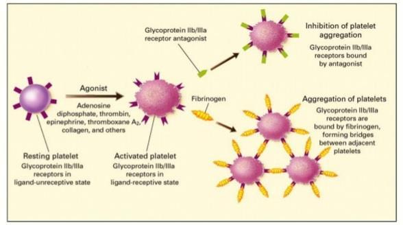
25 Platelet aggregation - consists in irreversible fusion and fibrin embedding of platelets - is induced /amplified by the signalling molecules released by the activated platelets: -ADP (binds to P2Y12 receptors on platelets), -serotonin -thromboxane A2 vwf released by activated platelets binds to the platelet receptor Gp Ib/Ia, activating even more platelets and forming molecular bridges between platelets. Platelet activation also induces a conformational change in the platelet receptor Gp IIb/IIIa, endowing it with the capacity to bind fibrinogen from blood and to form bridges between platelets, to promote the platelet plug formation. Antiaggregant medication: -Aspirin, an inhibitor of cyclooxygenase, inhibits clotting by reducing the release of thromboxane A2. -Clopidogrel (Plavix) is an antiplatelet agent that acts by inhibiting the P2Y12 receptors on the platelet surface.
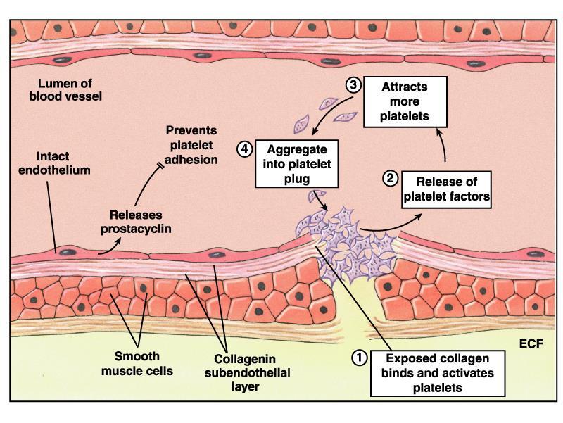
, Ca 2+ adhesion of more platelets Aggregation Formation of platelet plug (primary haemostatic plug) Release of PF 3 - procoagulant action thrombin,")
26 Platelet contact with collagen/damaged wall platelet swelling/shape changes/contraction release of granules Adhesion to vwp/collagen Secretion of TXA 2, ADP (P 2Y12 platelets receptors), Ca 2+ adhesion of more platelets Aggregation Formation of platelet plug (primary haemostatic plug) Release of PF 3 - procoagulant action thrombin, fibrin


27 Platelet plug formation (primary hemostasis)
![Opposing effects of PGI2 and TXA2 on AC/cAMP production/[ca] i Aspirin - inhibitor of cyclooxygenase, inhibits clotting by reducing the](/docs-images/94/119408933/images/28-0.jpg "release of TXA 2. Clopidogrel - antiplatelet agent that acts by inhibiting the P 2Y12 rec. on the platelet surface.")
28 Opposing effects of PGI2 and TXA2 on AC/cAMP production/[ca] i Aspirin - inhibitor of cyclooxygenase, inhibits clotting by reducing the release of TXA 2. Clopidogrel - antiplatelet agent that acts by inhibiting the P 2Y12 rec. on the platelet surface. adhesion & aggregation

29 Thrombocytopenia Thrombocytopenia - spontaneous skin purpura and haemorrhage failure of platelet production: drugs: chloramphenicol, penicillamine, phenylbutazone chemicals: benzene radiotherapy increased consumption of platelets: - immune or autoimmune, drug-induced: phenacetin, rifampicin, penicillin, sulphonamides, diazepam, furosemide, tolbutamide, digitoxin; - disseminated intravascular coagulation (DIC) - splenomegaly (abnormal distribution/destruction of platelets) - platelet aggregation (ristocetin, low MW heparin)
30 Platelet Anti-platelet Antibody (IgG) Macrophage Autoimmune thrombocytopenic purpura Life-span of platelets reduced from 10 days to a few hours Usually idiopathic, but also in HIV infection, etc.
31 Tests for platelet plug formation (primary hemostasis) Bleeding time (global test of platelet role in hemostasis) - time to stop bleeding after skin injury Platelet count and Mean Platelet Volume (MPV) - number and the uniformity of the size of platelet population Platelet granule content - electron microscopy vwf assay - measurement of the amount of vwf and its function (e.g. its interaction with platelet receptors) Platelet membrane receptors/glycoproteins - monoclonal antibodies and flow cytometry
32 Hemostasis and fibrinolysis events (1) vasoconstriction to collapse vessels with an intravascular pressure below the critical closing pressure (2) increased tissue pressure vessel radius decrease, to diminish blood flow, decrease the hemorrhage (3) adhesion, activation an aggregation of platelets resulting in platelet plug formation, in the case of capillary bleeding/vessel rupture stop the hemorrhage, (4) coagulation or clot formation through controlled proteolysis of coagulation proteins fibrin network stabilize the clot (5) anticoagulant processes that prevent excessive hemostasis (6) clot retraction and fibrinolysis that breaks up clots vessel wall repair or fibrous organization of the clot into fibrous tissue
, lipids and ions resulting in the production of fibrin and an insoluble blood clot.")
33 (4) Hemostasis and fibrinolysis: Plasmatic components involved and blood clot formation Coagulation is a cascade process of enzymatic reactions involving several plasma proteins (proenzymes and procofactors which are activated sequentially), lipids and ions resulting in the production of fibrin and an insoluble blood clot. A blood clot is a semisolid mass composed of both platelets and fibrin, including entrapped erythrocytes, leukocytes, and serum. A thrombus is an intravascular blood clot. The relative composition of thrombi varies with the site of thrombosis (i.e., thrombus formation): - a higher proportion of platelets is present in clots of the arterial circulation, - a higher proportion of fibrin is present in clots of the venous circulation.
 that play a role in blood clotting.")
34 There is a molecular crosstalk between the processes involved in platelet plug formation and clot formation that helps coordinate hemostasis Platelet plug formation and blood clotting are related but distinct events that may occur in parallel or in the absence of one other. Activated platelets can release small amounts of some of the factors (e.g., Ca2+) that play a role in blood clotting. Conversely, some clotting factors (e.g., thrombin and fibrinogen) play a role in platelet plug formation. Fluido-coagulant balance is important, because: inadequate clotting would lead to the leakage of blood from the vascular system and, ultimately, to hypovolemia; overactive clotting would lead to thrombosis and cessation of blood flow
35 The cardiovascular system achieves the balance between an antithrombotic (anticoagulant) and a prothrombotic (procoagulant) state by a variety of components of the vascular wall and blood Promoting an antithrombotic state is normal for the endothelial cells in the vascular system. Promoting a prothrombotic state are events associated with: - vascular damage: (1) the failure of endothelial cells to produce the proper antithrombotic factors, (2) the physical removal or injury of endothelial cells, which permits the blood to come into contact with thrombogenic factors that lie beneath the endothelium. - activation of platelets by: (1) ligands that bind to platelet receptors (2) shearing forces that activate the platelets (e.g. platelets flow past artificial mechanical heart valves).
36 Endothelial cells and the anti- and pro-thrombotic state Endothelial cells (EC) produce: -Von Willebrand Factor (stored in Weibel-Palade bodies in EC, also synthesized in megakariocytes and stored in platelet α-granules): involved in platelet adhesion & aggregation, carries Factor VIII -Prostacyclin (PGI 2 ): vasodilation, inhibit platelet adhesion & aggregation -Antithrombin III (AT) & Protein C (PC) activator (thrombomodulin) both of which inhibit coagulation -Tissue plasminogen activator (t-pa) which activates fibrinolysis by activating plasminogen to plasmin. The intact vessel wall has an important role in preventing hemostasis!

37
38 The coagulation cascade According to the classical view, coagulation cascade is divided into the intrinsic, extrinsic and common pathways. This division has been done to facilitate the understanding of in vitro laboratory tests, but in vivo however, the pathways are very closely interlinked. Extrinsic and intrinsic pathways are initiated by distinct mechanisms, and converge on a common pathway that generates thrombin and, ultimately, stable fibrin that leads to clot formation.
39 The coagulation cascade The intrinsic pathway (surface contact activation) becomes activated when blood comes into contact with a negatively charged surface (e.g. in vitro - a glass test tube); occurs mainly at the membrane of activated platelets. The extrinsic pathway is activated when blood comes in contact with damaged cell membranes; occurs mainly at a tissue factor that is membrane bound. In both cases, the precipitating event triggers a chain reaction of controlled proteolysis that converts precursors (zymogens) into activated factors (serine proteases), which in turn catalyze the conversion of other precursors into other activated factors, amplifying the clotting signals. The coagulation cascades do not occur in the fluid phase of the blood, where the concentration of coagulation factors is low.
40 Three essential steps for blood coagulation: (1) rupture of the vessel or damage to the blood itself a complex cascade of chemical reactions occurs in the blood involving coagulation factors formation of a complex of activated substances = prothrombinase / prothrombin activator (2) the prothrombin activator catalyzes conversion of prothrombin into thrombin. Much of the prothrombin first attaches to prothrombin receptors on the platelets already bound to the damaged tissue. (3) thrombin acts as an enzyme to convert fibrinogen into fibrin fibers that enmesh platelets, blood cells, and plasma to form the blood clot. The rate-limiting factor in causing blood coagulation is usually the formation of prothrombin activator and not the subsequent reactions beyond that point.

41 Coagulation is the series of physiological processes resulting in the arrest of bleeding. There are four stages, all closely integrated: 1. Vascular reaction 2. Platelet reaction 3. Clot formation 4. Dissolution of the clot - fibrinolysis.
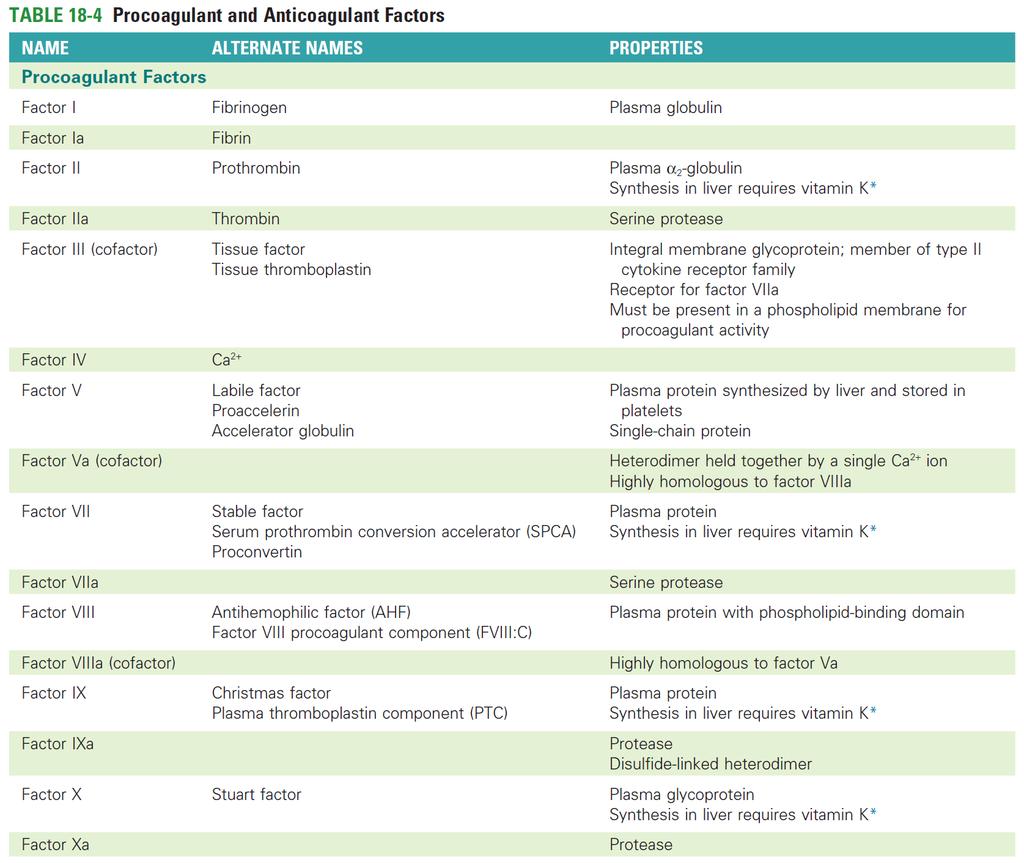
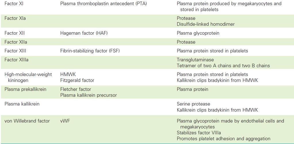
42 The coagulation cascade The domain structure of the proteins of the coagulation cascade -a signal peptide required for the translocation of the polypeptide into the endoplasmic reticulum, where the signal peptide is cleaved. -a propeptide or γ-carboxyglutamic acid rich domain (Gla domain) is rich in glutamic acid residues that undergo γ-carboxylation under the influence of the γ- carboxylase that requires vitamin K; is required for Ca2+ binding. -an epidermal growth factor (EGF)-like domain has a role in forming protein complexes. -a kringle domain is a loop structure created by several disulfide bonds that play a role in forming protein complexes and attaching the protease to its target. -a catalytic domain confers the serine protease function to the coagulation proteins and is homologous to trypsin, chymotrypsin, and other serine proteases -some other domains are variable among these proteins.

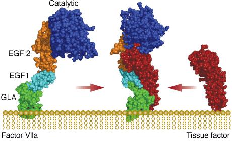
43 Interaction of tissue factor with FVII and FX GLA domain Factor VII embraces tissue factor, contacting the entire length of the molecule. FVII has 4 domains strung together with flexible linkers. At the bottom is the GLA domain, which has 9 modified glutamic acids, labeled CGU. These modified amino acids have an extra carboxylic acid group that traps calcium ions. The ions interact with the membrane surface, helping FVII find tissue factor. The uppermost domain of FVII is a protein-cutting enzyme that will make the break in the factor X. This domain looks very much like other serine proteases such as trypsin and thrombin. In the middle are two small domains that assist with the recognition of tissue factor. The small molecule in green is an inhibitor that blocks the active site and thus acts as an anticoagulant that stops blood clotting (doi: /rcsb_pdb/mom_2006_3).
44
45
46
47
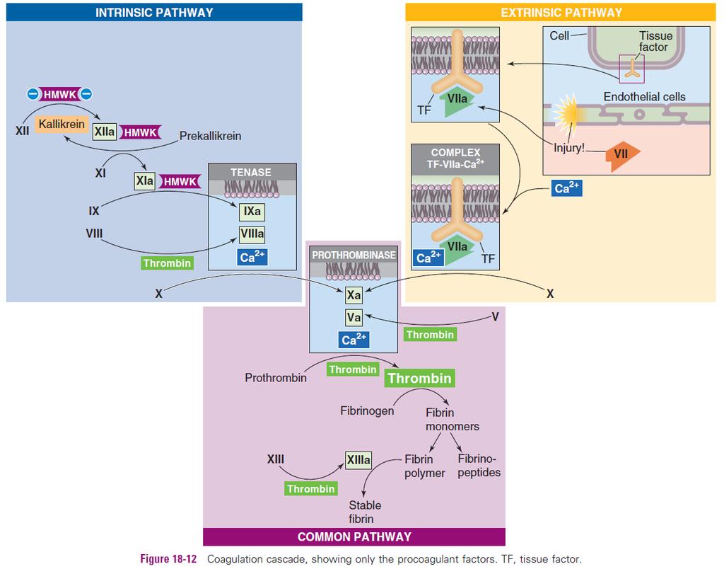
48 Intrinsic Pathway (Surface Contact Activation) is a cascade of protease reactions initiated by factors present within blood Factor XII (Hageman factor) is a plasma protein activated to factor XIIa when comes in contact with a negatively charged surface (the membrane of activated platelets or endothelial cells or glass surface in vitro) in the presence of HMWK. High molecular-weight kininogen (HMWK), a product of platelets that may be attached to the platelet membrane, serves as a cofactor and helps anchor factor XII to the charged surface. The HMWK-assisted conversion of factor XII to factor XIIa is limited in speed. Once a small amount of factor XIIa accumulates, this protease converts prekallikrein to kallikrein, with HMWK as an anchor. In turn, kallikrein accelerates, in a positive feedback, the conversion of factor XII to factor XIIa. Factor XIIa (anchored to HMWK) proteolytically cleaves factor XI to factor XIa. In turn, factor XIa (also bound to the charged surface by HMWK) proteolytically cleaves factor IX (Christmas factor) to factor IXa, which is a protease. Factor IXa (and two downstream products of the cascade, factors Xa and thrombin) proteolytically cleave factor VIII to factor VIIIa, a cofactor in the next reaction. Finally, factors IXa and VIIIa, together with Ca2+ (which may come largely from activated platelets) and negatively charged phospholipids, form a trimolecular complex called tenase. Tenase then converts factor X (Stuart factor) to the active protease factor Xa, where the intrinsic and extrinsic coagulation pathways converge.

49 The intrinsic pathway One of the responses of platelets to activation is the presentation of platelet phospholipid on their surfaces, that allows the tenase complex to form. The role of factor VIII in this process is to act as a receptor, in the form of factor VIIIa, for factor IXa, Ca2+ and factor X. Factor VIIIa is termed a cofactor in the clotting cascade and is formed in the presence of minute quantities of thrombin. As the concentration of thrombin increases, factor VIIIa is ultimately cleaved by thrombin and inactivated. This dual action of thrombin, upon factor VIII, acts to limit the extent of tenase complex formation and thus the extent of the coagulation cascade.
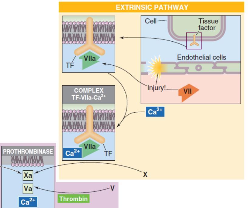
50 Extrinsic Pathway (Tissue Factor Activation) is a cascade of protease reactions initiated by factors that are outside the vascular system Tissue factor (tissue thromboplastin, or factor III) is an integral membrane protein constitutively express by nonvascular/perivascular cells, acting as a receptor for the plasma protein factor VII. In case of an endothelial injury, factor VII come into contact with tissue factor and activates to factor VIIa. The tissue factor, factor VIIa, and Ca2+ form a trimolecular complex analogous to tenase, and this complex proteolytically cleaves the proenzyme factor X to factor Xa. When factor X binds to the trimolecular complex, factor VIIa undergoes a conformational change that prevents it from dissociating from tissue factor. Regardless of whether factor Xa arises by the intrinsic or extrinsic pathway, the coagulation cascade proceeds along the common pathway.
51
52 Common Pathway Factor Xa from either the intrinsic or extrinsic pathway is the first protease of the common pathway. Reminiscent of the conversion of factor VIII to the cofactor VIIIa in the intrinsic pathway, the downstream product thrombin clips factor V to form the cofactor Va. Factor V is highly homologous to factor VIII Factors Xa, Va, Ca2+ and phospholipids (phosphatidylinositol and phosphatidylserine), form the prothrombinase complex. On the surface of activated platelets, prothrombinase acts on plasma protein prothrombin (factor II) to form thrombin (factor IIa). Thrombin is the central protease of the coagulation cascade responsible for proteolysis of fibrinogen (factor I) and releasing of fibrin monomers that further assemble into a fibrin polymer.

53
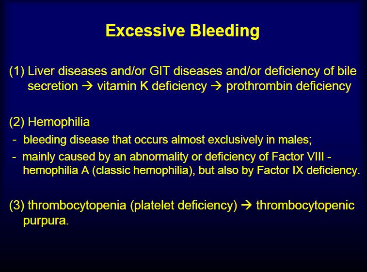
54 Prothrombin = a plasma protein / alpha 2-globulin (68,700 da) - 15 mg/dl in normal plasma - formed continually by the liver in the presence of Vitamin K. - it is an unstable protein splits easily into smaller compounds (thrombin - 33,700 da). Lack of vitamin K or the presence of liver disease prevent normal prothrombin formation, decrease the prothrombin level bleeding tendency Platelets play an important role in prothrombin conversion, as this first attaches to the its platelets receptors (PARs - Proteaseactivated receptors).
55 Thrombin is the central protease of the coagulation cascade, responsible for: 1. Activation of downstream components in the clotting cascade: - catalyze the proteolysis of fibrinogen and the formation of sluble fibrin monomers. Fibrin monomers (α, β, and γ chains) spontaneously polymerize to form a gel of fibrin polymers that traps blood cells. - activates factor XIII to factor XIIIa, which mediates the covalent cross-linking of the α and γ chains of fibrin polymers to form a stable fibrin mesh that is even less soluble than fibrin. 2. Positive feedback at several upstream levels of the cascade, as it catalyzes: - the formation of new thrombin from prothrombin - the formation of the cofactors Va and VIIIa. 3. Paracrine actions that influence hemostasis: - activate platelets through PAR-1, a protease-activated receptor (a G proteincoupled receptor). - causes endothelial cells to release nitric oxide (inhibit platelet aggregation and adhesion), prostaglandin I2 (PGI2), ADP, vwf, and tissue plasminogen activator. - combines with thrombomodulin present on endothelial cell surfaces and form a complex that activates protein C. The cofactor protein S and activated protein C degrade factors Va and VIIIa, thereby limiting their procoagulant activity.
56 Fibrinogen = high-molecular-weight (MW = 340,000) plasmatic protein to 700 mg/dl in plasma - formed in the liver - liver disease can decrease the concentration of circulating fibrinogen - normally does not leak from the blood vessels into the interstitial fluids interstitial fluids ordinarily do not coagulate (exception when capillaries permeability becomes pathologically increased)
57 Coagulation as a Connected Diagram: the intrinsic and extrinsic pathway are strongly interconnected to form a network The classical concept of independent intrinsic and extrinsic branches converging on a common pathway become nowadays obsolete. Coagulation cascade is best conceptualized as a connected diagram in which the branches may interconnect in both the upstream and downstream directions: -thrombin has multiple actions -the trimolecular complex of [tissue factor + factor VIIa + Ca2+] of the extrinsic pathway, activates factors IX and XI of the intrinsic pathway. -factors IXa and Xa of the intrinsic pathway can activate factor VII of the extrinsic pathway. Clinical evidence suggests that coagulation depends largely on the extrinsic pathway. While tissue factor is normally absent from intravascular cells, inflammation can trigger peripheral blood monocytes and endothelial cells to express tissue factor, which increases the risk of coagulation (e.g. during sepsis, the tissue factor produced by circulating monocytes initiates intravascular thrombosis).
58 Hemostasis and fibrinolysis events (1) vasoconstriction to collapse vessels with an intravascular pressure below the critical closing pressure (2) increased tissue pressure vessel radius decrease, to diminish blood flow, decrease the hemorrhage (3) adhesion, activation an aggregation of platelets resulting in platelet plug formation, in the case of capillary bleeding/vessel rupture stop the hemorrhage, (4) coagulation or clot formation through controlled proteolysis of coagulation proteins fibrin network stabilize the clot (5) anticoagulant processes that prevent excessive hemostasis (6) clot retraction and fibrinolysis that breaks up clots vessel wall repair or fibrous organization of the clot into fibrous tissue
 - promotes vasodilation and thus blood flow - inhibits platelet activation and thus clotting.")
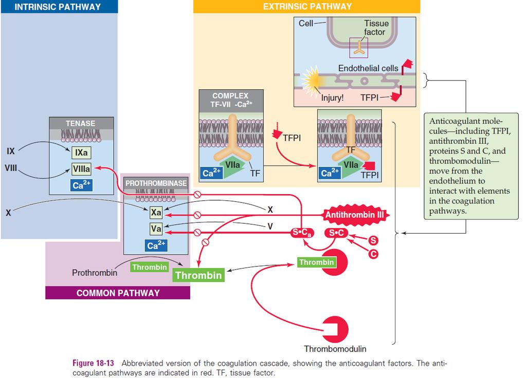
59 Anticoagulants keep the clotting network in check There are important paracrine factors and anticoagulant factors, mainly of endothelial origin, that prevent hemostasis from running out of control Paracrine Factors: - prostacyclin (PGI2) - promotes vasodilation and thus blood flow - inhibits platelet activation and thus clotting. -nitric oxide (NO), that inhibits platelet adhesion and aggregation through cgmp. Anticoagulant Factors generated by endothelial cells interfere with the clotting cascade that generates fibrin.
60 Anticoagulant Factors 1. Tissue factor pathway inhibitor (TFPI) -is a plasma protein that binds to the trimolecular complex [tissue factor + factor VIIa + Ca2+] in the extrinsic pathway and blocks the protease activity of factor VIIa. -is glycosylphosphatidylinositol (GPI) linked to the endothelial cell membrane, where it maintains an antithrombotic surface. 2. Antithrombin III (AT III) binds to and inhibits factor Xa and thrombin. The sulfated glycosaminoglycans heparan sulfate and heparin enhance the binding of AT III to factor Xa or to thrombin, thus inhibiting coagulation. Heparan sulfate is present on the external surface of most cells, including endothelial surfaces. Mast cells and basophils release heparin. 3. Thrombomodulin is a glycosaminoglycan product of endothelial cells, that forms a complex with thrombin, removing thrombin from the circulation and inhibiting coagulation; also binds protein C. 4. Protein C activates by binding to thrombomodulin-thrombin complex. Activated protein C (Ca) is a protease that, together with its cofactor protein S, inactivates the cofactors Va and VIIIa, thus inhibiting coagulation. 5. Protein S is the cofactor of protein C and is thus an anticoagulant. Finally, clearance of activated clotting factors by the Kupffer cells of the liver also keeps hemostasis under control.
61 Heparin - powerful anticoagulant, its concentration in the blood is normally low - highly negatively charged conjugated polysaccharide that increases a 100x to 1000x its anticoagulant potency when it combines with antithrombin III - in the presence of excess heparin, removal of free thrombin from the circulating blood by antithrombin III is almost instantaneous. - the complex of heparin and antithrombin III removes several other activated coagulation factors in addition to thrombin: factors XII, XI, X, and IX. -heparin is produced by many different cells of the body, but especially by the basophilic mast cells in the pericapillary connective tissue (> in the lungs, ~ in the liver) and by the basophil cells of the blood. - used widely as a pharmacological agent in medical practice in much higher concentrations to prevent intravascular clotting (purified animal heparin): mg/kg bw increase rapidly blood-clotting time from 6 min to ~30 min (act for hrs); injected heparin is destroyed in the blood by an enzyme - heparinase
62
63 Hemostasis and fibrinolysis events (1) vasoconstriction to collapse vessels with an intravascular pressure below the critical closing pressure (2) increased tissue pressure vessel radius decrease, to diminish blood flow, decrease the hemorrhage (3) adhesion, activation an aggregation of platelets resulting in platelet plug formation, in the case of capillary bleeding/vessel rupture stop the hemorrhage, (4) coagulation or clot formation through controlled proteolysis of coagulation proteins fibrin network stabilize the clot (5) anticoagulant processes that prevent excessive hemostasis (6) clot retraction and fibrinolysis that breaks up clots vessel wall repair or fibrous organization of the clot into fibrous tissue
64 (6) Clot retraction and fibrinolysis: Clot retraction/contraction Within a few minutes after a clot is formed, it begins to contract through the interaction of actin and myosin in the platelets expresses the fluid from the clot (serum) within 20 to 60 minutes the edges of the broken blood vessel are pulled together Platelets are necessary for clot retraction to occur: - they become attached to the fibrin fibers bond different fibers together - continue to release procoagulant substances -fibrin-stabilizing factor cross-linking bonds between adjacent fibrin fibers - contribute directly to clot contraction by activating platelet contractile proteins (thrombosthenin, actin, and myosin molecules) contraction of the platelet spicules attached to the fibrin Clot contraction is activated and accelerated by thrombin and calcium ions released from calcium stores in the mitochondria, endoplasmic reticulum, and Golgi apparatus of the platelets.
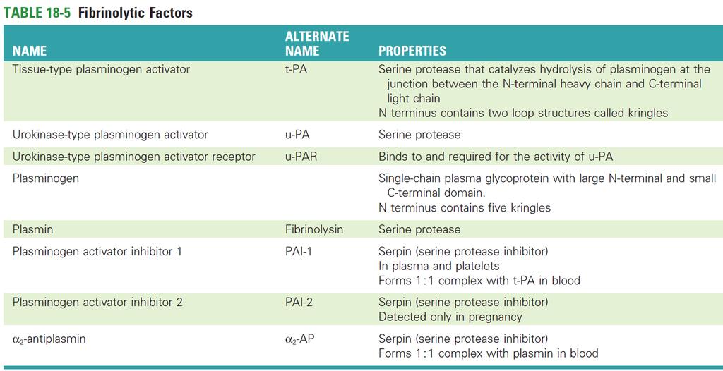
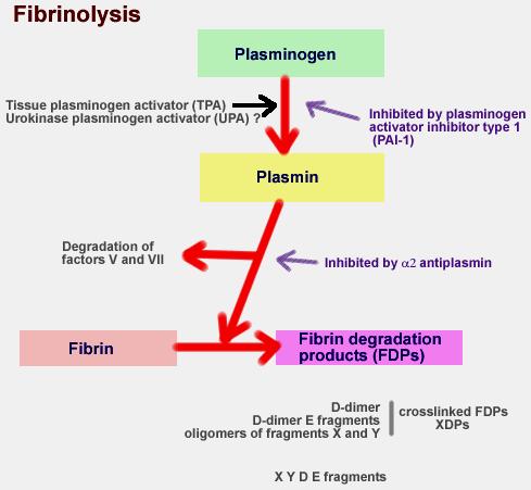
65 (6) Clot retraction and fibrinolysis: Lysis of blood clots Fibrinolysis/thrombolysis When a clot is formed, a large amount of plasminogen is trapped in the clot along with other plasma proteins. This will not become plasmin or cause lysis of the clot until it is activated. The process of fibrinolysis begins with the conversion of plasminogen (profibrinolysin) to plasmin (fibrinolysin, a trypsin-like proteolytic enzyme), catalyzed by one of two activators: - tissue-type plasminogen activator (t-pa) or - urokinase-type plasminogen activator (u-pa).

66 Tissue plasminogen activator (t-pa) -a serine protease of endothelial origin -converts the plasma zymogen plasminogen to the active fibrinolytic protease plasmin. The presence of fibrin greatly accelerates the conversion of plasminogen to plasmin. Urokinase-type plasminogen activator (u-pa) -present in plasma either as a single-chain protein or as the two-chain product of a proteolytic cleavage. -converts plasminogen to the active protease plasmin, an this proteolysis requires that u-pa attach to a receptor on the cell surface called urokinase plasminogen activator receptor (u-par).
67 Plasminogen - is a large, single chain glycoprotein mainly synthetized by the liver - cleaved by t-pa at the junction between its heavy and light chains to form plasmin. Plasmin - is a serine protease that proteolytically cleaves stable fibrin to fibrin breakdown products. - also breaks down fibrinogen, Factor V, Factor VIII, prothrombin, and Factor XII. - also cleaves t-pa - determines lysis of a clot by destroying many of the clotting factors blood hypocoagulability An important function of the plasmin system is to remove minute clots from peripheral vessels that eventually would become occluded.
68
 and plasminogen activator inhibitor 2 (PAI-2) are serine protease inhibitors (serpins) that reduce the activity of the plasminogen activators: -PAI-1 is")
69 The cardiovascular system regulates fibrinolysis at several levels, using both enhancing and inhibitory mechanisms - Catecholamines and bradykinin increase the levels of circulating t-pa. - Plasminogen activator inhibitor 1 (PAI-1) and plasminogen activator inhibitor 2 (PAI-2) are serine protease inhibitors (serpins) that reduce the activity of the plasminogen activators: -PAI-1 is produced mainly by endothelial cells and complexes with and inhibits t-pa and u-pa. -PAI-2 mainly inhibits u-pa; is important in pregnancy because it is produced by the placenta and may contribute to increased risk of thrombosis in pregnancy. - Activated protein C, which inhibits coagulation, also inhibits PAI-1 and PAI-2, thereby facilitating fibrinolysis. α2-antiplasmin (α2-ap) is a serpin that targets plasmin and is made by liver, kidney, and other tissues. -when plasmin is not bound to fibrin (plasmin is in free solution), α2-ap complexes with and thereby readily inactivates plasmin. -when plasmin is attached to fibrin, the inhibition by α2-ap is greatly reduced, and fibrinolysis is promoted.
70
71 Hemostasis overview
 Coagulation is typically initiated by an injury to the vascular ECs, which results in the exposure of TF and collagen from the sub-endothelial tissue to the blood and the release of vwf.")
72 (a) Resting ECs provide natural anticoagulants (TM, AT and TFPI and ADPase) to inhibit coagulation and keep platelet activation and the coagulation cascade in check. (b) Coagulation is typically initiated by an injury to the vascular ECs, which results in the exposure of TF and collagen from the sub-endothelial tissue to the blood and the release of vwf. (c) Platelets are activated when they are exposed to TF, collagen and vwf. Activated platelets release a number of mediators (ADP, vwf) within their granules, leading to further platelet recruitment, activation, aggregation and plug formation, which is a process termed primary hemostasis. (d) The interaction between TF and factor VII initiates the extrinsic pathway. (e) The exposure of collagen to blood starts the intrinsic pathway. (f) Both the extrinsic and intrinsic pathways result in the initiation of a common pathway, which contains the cascades involved in the production of activated Factor X and thrombin and the formation of fibrin strands. (g) Fibrin strands strengthen the platelet plug and lead to the formation of a stable platelet fibrin clot. This process is termed secondary hemostasis. (h) Kallikrein, upa or tpa activate plasminogen to plasmin, which then degrades and reabsorbs the polymerized fibrin strands (fibrinolysis) and favor wounds healing. Cascades of the coagulation system overview AT, antithrombin; ECs, endothelial cells; TF, tissue factor; TFPI, tissue factor pathway inhibitors; TM, thrombomodulin; tpa, tissue plasminogen activator; upa, urokinase plasminogen activator; vwf, von Willebrand factor.
73
74
75
76
77
This slide belongs to iron lecture and it is to clarify the iron cycle in the body and the effect of hypoxia on erythropoitein secretion
 This slide belongs to iron lecture and it is to clarify the iron cycle in the body and the effect of hypoxia on erythropoitein secretion Topics of today lectures: Hemostasis Meaning of hemostasis Mechanisms
Hemostasis. Learning objectives Dr. Mária Dux. Components: blood vessel wall thrombocytes (platelets) plasma proteins
 plasma proteins") Hemostasis Learning objectives 14-16 Dr. Mária Dux Components: blood vessel wall thrombocytes (platelets) plasma proteins Hemostatic balance! procoagulating activity anticoagulating activity 1 Thrombocytes
Hemostasis Learning objectives 14-16 Dr. Mária Dux Components: blood vessel wall thrombocytes (platelets) plasma proteins Hemostatic balance! procoagulating activity anticoagulating activity 1 Thrombocytes
Part IV Antithrombotics, Anticoagulants and Fibrinolytics
 Part IV Antithrombotics, Anticoagulants and Fibrinolytics "The meaning of good and bad, of better and worse, is simply helping or hurting" Emerson Chapter 16: Blood Coagulation and Fibrinolytic System
UNIT VI. Chapter 37: Platelets Hemostasis and Blood Coagulation Presented by Dr. Diksha Yadav. Copyright 2011 by Saunders, an imprint of Elsevier Inc.
 UNIT VI Chapter 37: Platelets Hemostasis and Blood Coagulation Presented by Dr. Diksha Yadav Hemostasis: Prevention of Blood Loss Vascular constriction Formation of a platelet plug Formation of a blood
Hemostasis and. Blood Coagulation
 Hemostasis and Blood Coagulation Events in Hemostasis The term hemostasis means prevention of blood loss. Whenever a vessel is severed or ruptured, hemostasis is achieved by several mechanisms: (1) vascular
Topics of today lectures: Hemostasis
 Topics of today lectures: Hemostasis Meaning of hemostasis Mechanisms of hemostasis - Vascular contraction - Platelets plug - Blood coagulation (clotting) - Structure and functions of platelets - Blood
Blood clotting. Subsequent covalent cross-linking of fibrin by a transglutaminase (factor XIII) further stabilizes the thrombus.
 further stabilizes the thrombus.") Blood clotting It is the conversion, catalyzed by thrombin, of the soluble plasma protein fibrinogen (factor I) into polymeric fibrin, which is deposited as a fibrous network in the primary thrombus. Thrombin
Blood clotting It is the conversion, catalyzed by thrombin, of the soluble plasma protein fibrinogen (factor I) into polymeric fibrin, which is deposited as a fibrous network in the primary thrombus. Thrombin
Ch. 45 Blood Plasma proteins, Coagulation and Fibrinolysis Student Learning Outcomes: Describe basic components of plasma
 Chapt. 45 Ch. 45 Blood Plasma proteins, Coagulation and Fibrinolysis Student Learning Outcomes: Describe basic components of plasma Inheritance of X-linked gene for Factor VIII hemophilia A Explain the
Blood coagulation and fibrinolysis. Blood clotting (HAP unit 5 th )
") Blood coagulation and fibrinolysis Blood clotting (HAP unit 5 th ) Vessel injury Antithrombogenic (Favors fluid blood) Thrombogenic (Favors clotting) 3 Major systems involved Vessel wall Endothelium ECM
Blood coagulation and fibrinolysis Blood clotting (HAP unit 5 th ) Vessel injury Antithrombogenic (Favors fluid blood) Thrombogenic (Favors clotting) 3 Major systems involved Vessel wall Endothelium ECM
Primary Exam Physiology lecture 5. Haemostasis
 Primary Exam Physiology lecture 5 Haemostasis Haemostasis Body s response for the prevention and cessation of bleeding. Broadly consists of: Primary Haemostasis - vascular spasm and platlet plug formation
PHM142 Lecture 4: Platelets + Endothelial Cells
 PHM142 Lecture 4: Platelets + Endothelial Cells 1 Hematopoiesis 2 Platelets Critical in clotting - activated by subendothelial matrix proteins (e.g. collagen, fibronectin, von Willebrand factor) and thrombin
-Hashim ahmed is the one who wrote this sheet. I just edited it according to our record.
 * Subjects of this lecture : - Hemostasis - Platelets, general information, their ultrastructure and role in hemostasis. - Definitions: Thrombus, Embolus, Arteriosclerosis and Atherosclerosis. *NOTE: Prof
Hemostasis and Thrombosis
 Hemostasis Hemostasis and Thrombosis Normal hemostasis is a consequence of tightly regulated processes that maintain blood in a fluid state in normal vessels, yet also permit the rapid formation of a hemostatic
Chapter 19. Hemostasis
 Chapter 19 Hemostasis Hemostasis Hemostasis is the cessation of bleeding stopping potentially fatal leaks important in small blood vessels not effective in hemorrhage excessive bleeding from large blood
Physiology of. The Blood hemostasis. By prof. Israa f. jaafar
 Physiology of The Blood hemostasis By prof. Israa f. jaafar Learning objectives Understand the Platelet structure and function Explane the Platelet production Understand the phases of hemostasis: vascular
Anatomy and Physiology
 Anatomy and Physiology For The First Class 2 nd Semester Thrombocytes = Platelets Thrombocytes = Platelets Blood platelets are non-nucleated disc like cell fragments 2-4 µm in diameter. Platelets are not
Page 1 of 6 THROMBOCYTES
 Page 1 of 6 THROMBOCYTES Platelets are not cells in the strict sense. About one-fourth the diameter of a lymphocyte, they are cytoplasmic fragments of extraordinarily large cells (up to 60 µm in diameter)
Chapter 1. General introduction
 Chapter 1 General introduction 8 Haemostasis All organs and tissues of higher organisms are provided with nutrients and oxygen through the bloodstream. The bloodstream is an extensive vascular system that
Diagnosis of hypercoagulability is by. Molecular markers
 Agenda limitations of clinical laboratories to evaluate hypercoagulability and the underlying cause for thrombosis what is the INR the lupus anticoagulant and the antiphospholipid antibody syndrome hassouna
Oral Anticoagulant Drugs
 Oral Anticoagulant Drugs Spoiled sweet clover caused hemorrhage in cattle(1930s). Substance identified as bishydroxycoumarin. Initially used as rodenticides, still very effective, more than strychnine.
Outline Anti-coagulant and anti-thrombotic drugs Haemostasis and Thrombosis Year 3 Dentistry
 Outline Anti-coagulant and anti-thrombotic drugs Year 3 Dentistry Professor Yotis Senis Cellular Haemostasis y.senis@bham.ac.uk I. Haemostasis and II. Coagulation and anti-coagulants III. Platelets and
Chapter 19 Blood Lecture Outline
 Chapter 19 Blood Lecture Outline Cardiovascular system Circulatory system Blood 1. distribution 2. regulation 3. protection Characteristics: ph 7.4 38 C 4-6 L Composition: Plasma Formed elements Erythrocytes
PHASES OF HAEMOSTASIS
 HAEMOSTASIS Maintains the integrity of a closed, highpressure circulatory system after vascular damage Vessel Wall Injury events in the vessel wall and in the blood which seal breach Delicate balance exists
Chapter 19 Cardiovascular System Blood: Functions. Plasma
 Chapter 19 Cardiovascular System Blood: Functions 19-1 Plasma Liquid part of blood. Colloid: liquid containing suspended substances that don t settle out of solution 91% water. Remainder proteins, ions,
Thrombosis. Jeffrey Jhang, M.D.
 Thrombosis Jeffrey Jhang, M.D. Introduction The human hemostatic system has evolved to maintain blood flow under normal physiologic conditions while remaining primed to rapidly respond to vascular injury
Bleeding and Haemostasis. Saman W.Boskani HDD, FIBMS Maxillofacial Surgeon
 Bleeding and Haemostasis Saman W.Boskani HDD, FIBMS Maxillofacial Surgeon 1 Beeding Its escaping or extravasation of blood contents from blood vessels Types: - Arterial - Venous - Capillary Differences
Hemostasis. Clo)ng factors and Coagula4on NORMAL COAGULATION. Overview of blood coagula4on. The Cascade Theory 5/1/12. Clot
ng factors and Coagula4on NORMAL COAGULATION. Overview of blood coagula4on. The Cascade Theory 5/1/12. Clot") Hemostasis Clo)ng factors and Coagula4on Dr Badri Paudel www.badripaudel.com Hemostasis is defined as a property of circula4on whereby blood is maintained within a vessel and the ability of the system
Hemostasis Clo)ng factors and Coagula4on Dr Badri Paudel www.badripaudel.com Hemostasis is defined as a property of circula4on whereby blood is maintained within a vessel and the ability of the system
Hemostasis Haemostasis means prevention of blood loss from blood vessels.
 ١ Hemostasis Haemostasis means prevention of blood loss from blood vessels. Bleeding is stopped by several mechanisms, which are: 1. Local vasoconstriction 2. Formation of platelet plug 3. Blood coagulation
Chapter 3. Haemostatic abnormalities in patients with liver disease
 Chapter 3 Haemostatic abnormalities in patients with liver disease Ton Lisman, Frank W.G. Leebeek 1, and Philip G. de Groot Thrombosis and Haemostasis Laboratory, Department of Haematology, University
10. Which of the following immune cell is unable to phagocytose (a) neutrophils (b) eosinophils (c) macrophages (d) T-cells (e) monocytes
 neutrophils (b) eosinophils (c) macrophages (d) T-cells (e) monocytes") Chapter 2. Acute and chronic inflammation(6): 1. In acute inflammation, which events occur in the correct chronological order? (Remembered from 2000, 2004 exam.) p50 (a) transient vasoconstriction, stasis
Chapter 2. Acute and chronic inflammation(6): 1. In acute inflammation, which events occur in the correct chronological order? (Remembered from 2000, 2004 exam.) p50 (a) transient vasoconstriction, stasis
Hemodynamic Disorders, Thromboembolic Disease, and Shock
 Hemodynamic Disorders, Thromboembolic Disease, and Shock Kumar et al: Robbins & Cotran Pathologic Basis of Disease 7E Figure 4-1 Factors affecting fluid balance across capillary walls. Capillary hydrostatic
Chapter 1 Introduction
 Chapter 1 Introduction There are several disorders which carry an increased risk of thrombosis, clots that interfere with normal circulation, including: venous thromboembolism (VTE), comprising both deep
Branch of medicine that deals with blood, its formation and disorders is called. Three main functions of cardiovascular system are,, and.
 Chapter 19 The Blood Human body must maintain a balance called. Body fluid inside the cells is called fluid; that outside is called or fluid. Two major fluid networks that help in connecting cells are
Moath Darweesh. Omar Sami. Saleem Khreisha. 1 P a g e
 7 Moath Darweesh Omar Sami Saleem Khreisha 1 P a g e -First of all, I want to give a quick revision to simplify the whole hemostasis mechanism, it will be much easier here with me. Enjoy (you can skip
Hemostasis Haemostasis means prevention of blood loss from blood vessels.
 1 Hemostasis Haemostasis means prevention of blood loss from blood vessels. Bleeding is stopped by several mechanisms, which are: 1. Local vasoconstriction 2. Formation of platelet plug 3. Blood coagulation
WHITE PAPERS PRESENTATION VIDEO DOCUMENTATION EXPERIMENT WO NDCLOT. The WoundClot Principals for Effective Bleeding Control PRESENTATION
 WHITE PAPERS PRESENTATION VIDEO DOCUMENTATION EXPERIMENT ARTICLES OUR STUDY BLEEDING CONTROL 5 POINT MODEL WO NDCLOT The WoundClot Principals for Effective Bleeding Control PRESENTATION Harnessing SCIENCE
Chapter 19: Cardiovascular System: Blood
 Chapter 19: Cardiovascular System: Blood I. Functions of Blood A. List and describe the seven major homeostatic functions of blood: 1. 2. 3. 4. 5. 6. 7. II. Plasma A. Composition 1. It is a fluid consisting
Chapter 11. Lecture and Animation Outline
 Chapter 11 Lecture and Animation Outline To run the animations you must be in Slideshow View. Use the buttons on the animation to play, pause, and turn audio/text on or off. Please Note: Once you have
Coagulation Disorders. Dr. Muhammad Shamim Assistant Professor, BMU
 Coagulation Disorders Dr. Muhammad Shamim Assistant Professor, BMU 1 Introduction Local Vs. General Hematoma & Joint bleed Coagulation Skin/Mucosal Petechiae & Purpura PLT wound / surgical bleeding Immediate
Introduction to coagulation and laboratory tests
 Introduction to coagulation and laboratory tests Marc Jacquemin Special Haemostasis Laboratory Center for Molecular and Vascular Biology University of Leuven Coagulation in a blood vessel: fibrin stabilises
WBCs production(leucopoiesis):
:") WBCs production(leucopoiesis): Note: this sheet contain only extra notes.j - leucopoiesis is the most complicated process in body because many reasons which are : 1- the production of many cells(monocyte,
WBCs production(leucopoiesis): Note: this sheet contain only extra notes.j - leucopoiesis is the most complicated process in body because many reasons which are : 1- the production of many cells(monocyte,
Blood Lecture Test Questions Set 2 Summer 2012
 Blood Lecture Test Questions Set 2 Summer 2012 1. Leukocytes are attracted to a site of injury or disease by: a. diapedesis b. chemotaxis c. leukocytosis d. heparin e. leukomotosis 2. Leukocytes leave
Blood. Plasma. The liquid part of blood is called plasma. 1. Pale yellow fluid; forms more than half the blood volume.
 11 Blood FOCUS: Blood consists of plasma and formed elements. The plasma is 91% water with dissolved or suspended molecules, including albumin, globulins, and fibrinogen. The formed elements include erythrocytes,
Hemodynamic Disorders, Thrombosis, and Shock. Richard A. McPherson, M.D.
 Hemodynamic Disorders, Thrombosis, and Shock Richard A. McPherson, M.D. Edema The accumulation of abnormal amounts of fluid in intercellular spaces of body cavities. Inflammation and release of mediators
The Cardiovascular System: Blood
 C h a p t e r 11 The Cardiovascular System: Blood PowerPoint Lecture Slides prepared by Jason LaPres Lone Star College - North Harris Introduction to the Cardiovascular System A circulating transport system
Blood platelets play important role in coagulation.
 B.N. Bandodkar College of Science, Thane T.Y. B.Sc Paper II Haematology Blood Coagulation / Blood clotting By Dr N.N. Patil Introduction: When blood is shed, it looses its fluidity within few minutes and
HEME 10 Bleeding Disorders
 HEME 10 Bleeding Disorders When injury occurs, three mechanisms occur Blood vessels Primary hemostasis Secondary hemostasis Diseases of the blood vessels Platelet disorders Thrombocytopenia Functional
Coagulation Pathway and Physiology
 Chapter 1 Coagulation Pathway and Physiology Russell A. Higgins, M Introduction Our understanding of blood clotting is intimately tied to the history of civilization. With the advent of writing 5000 years
INFLAMMATION & REPAIR
 INFLAMMATION & REPAIR Lecture 7 Chemical Mediators of Inflammation Winter 2013 Chelsea Martin Special thanks to Drs. Hanna and Forzan Course Outline i. Inflammation: Introduction and generalities (lecture
Y. Helen Zhang, MD Andy Nguyen, MD 10/28/2012
 Y. Helen Zhang, MD Andy Nguyen, MD 10/28/2012 Clinical History Patient: 23-year-old female Clinical course: status-post cholecystectomy, complicated by retained common bile duct stones. Following three
What are blood clots?
 What are blood clots? Dr Matthew Fay GP Principal The Willows Medical Practice- Queensbury GPwSI and Co-Founder Westcliffe Cardiology Service GP Partner Westcliffe Medical Group Created 5/31/18 Dr. Matthew
BIOH122 Human Biological Science 2
 BIOH122 Human Biological Science 2 Session 2 Haematological System Haemostasis and Blood Groups Bioscience Department Endeavour College of Natural Health endeavour.edu.au Session Plan o Platelets Properties
Chapter 14. Blood. Blood Volume. Blood Composition. Blood
 Blood connective tissue transports vital substances maintains stability of interstitial fluid distributes heat Chapter 14 Blood Blood Cells form mostly in red bone marrow red blood cells white blood cells
Bleeding Disorders. Dr. Mazen Fawzi Done by Saja M. Al-Neaumy Noor A Mohammad Noor A Joseph Joseph
 Bleeding Disorders Dr. Mazen Fawzi Done by Saja M. Al-Neaumy Noor A Mohammad Noor A Joseph Joseph Normal hemostasis The normal hemostatic response involves interactions among: The blood vessel wall (endothelium)
Hemostasis. Coagulation vs anticoagulation
 Hemostasis is a process of forming clots in the wall of damaged blood vessels and preventing blood loss while maintaining blood in fluid state within the vascular system. Coagulation vs anticoagulation
Cell Signaling (part 1)
") 15 Cell Signaling (part 1) Introduction Bacteria and unicellular eukaryotes respond to environmental signals and to signaling molecules secreted by other cells for mating and other communication. In multicellular
15 Cell Signaling (part 1) Introduction Bacteria and unicellular eukaryotes respond to environmental signals and to signaling molecules secreted by other cells for mating and other communication. In multicellular
Anticoagulants. Pathological formation of a haemostatic plug Arterial associated with atherosclerosis Venous blood stasis e.g. DVT
 Haemostasis Thrombosis Phases Endogenous anticoagulants Stopping blood loss Pathological formation of a haemostatic plug Arterial associated with atherosclerosis Venous blood stasis e.g. DVT Vascular Platelet
Haemostasis & Coagulation disorders Objectives:
 Haematology Lec. 1 د.ميسم مؤيد علوش Haemostasis & Coagulation disorders Objectives: - Define haemostasis and what are the major components involved in haemostasis? - How to assess the coagulation status?
Disseminated Intravascular Coagulation. M.Bahmanpour MD Assistant professor IUMS
 به نام خدا Disseminated Intravascular Coagulation M.Bahmanpour MD Assistant professor IUMS Algorithm for Diagnosis of DIC DIC Score factor score Presence of known underlying disorder No= 0 yes=2 Coagolation
Thursday, February 26, :00 am. Regulation of Coagulation/Disseminated Intravascular Coagulation HEMOSTASIS/THROMBOSIS III
 REGULATION OF COAGULATION Introduction HEMOSTASIS/THROMBOSIS III Regulation of Coagulation/Disseminated Coagulation necessary for maintenance of vascular integrity Enough fibrinogen to clot all vessels
ACQUIRED COAGULATION ABNORMALITIES
 ACQUIRED COAGULATION ABNORMALITIES ACQUIRED COAGULATION ABNORMALITIES - causes 1. Liver disease 2. Vitamin K deficiency 3. Increased consumption of the clotting factors (disseminated intravascular coagulation
Blood. Biol 105 Lecture 14 Chapter 11
 Blood Biol 105 Lecture 14 Chapter 11 Outline I. Overview of blood II. Functions of blood III. Composition of blood IV. Composition of plasma V. Composition of formed elements VI. Platelets VII. White blood
Coagulation an Overview Dr.Abdolreza Abdolr Afrasiabi Thal assem a & Heamophili hilia G ene i tic R esearc C en er Shiraz Medical Medic University
 In The Name God Coagulation an Overview Dr.Abdolreza Afrasiabi Thalassemia & Heamophilia Genetic Research hcenter Shiraz Medical University Bleeding Clotting Hemostasis Review of platelet function Platelets
Blood. The only fluid tissue in the human body Classified as a connective tissue. Living cells = formed elements Non-living matrix = plasma
 Blood Blood The only fluid tissue in the human body Classified as a connective tissue Living cells = formed elements Non-living matrix = plasma Blood Physical Characteristics of Blood Color range Oxygen-rich
Cover Page. The handle holds various files of this Leiden University dissertation.
 Cover Page The handle http://hdl.handle.net/1887/28736 holds various files of this Leiden University dissertation. Author: Debeij, Jan Title: The effect of thyroid hormone on haemostasis and thrombosis
THROMBOTIC DISORDERS: The Final Frontier
 THROMBOTIC DISORDERS: The Final Frontier Jeffrey I. Weitz, MD, FRCP(C), FACP Professor of Medicine and Biochemistry McMaster University Canada Research Chair in Thrombosis Heart & Stroke Foundation/ J.F.
- Mohammad Sinnokrot. -Ensherah Mokheemer. - Malik Al-Zohlof. 1 P a g e
 -1 - Mohammad Sinnokrot -Ensherah Mokheemer - Malik Al-Zohlof 1 P a g e Introduction Two of the most important problems you will face as a doctor are coagulation and bleeding, normally they are in balance,
L iter diagnostico di laboratorio nelle coagulopatie congenite emorragiche
 L iter diagnostico di laboratorio nelle coagulopatie congenite emorragiche Armando Tripodi Angelo Bianchi Bonomi Hemophilia and Thrombosis Center Dept. of Clinical Sciences and Community Health University
4/5/17. Blood. Blood. Outline. Blood: An Overview. Functions of Blood
 Outline Blood Biol 105 Chapter 11 I. Overview of blood II. Functions of blood III. Composition of blood IV. Composition of plasma V. Composition of formed elements VI. Platelets VII. White blood cells
Reprinted in the IVIS website with the permission of the ACVP and ASVCP
 Proceedings of the Annual Meeting of the American College of Veterinary Pathologists and American Society for Veterinary Clinical Pathology - Tucson, Arizona 2006 - Reprinted in the IVIS website with the
Chapter 19 Blood. Functions of blood:
 Chapter 19 Blood Functions of blood: 1. transportation functions 1. oxygen delivery 2. nutrient delivery 3. transportation of metabolic wastes (urine formation) 4. transportation of hormones (part of the
Coagulation Pathway and Physiology
 CHAPTER 1 Coagulation Pathway and Physiology Jerry B. Lefkowitz, M Introduction Our understanding of blood clotting is intimately tied to the history of civilization. With the advent of writing 5000 years
La Trombosi Arteriosa
 La Trombosi Arteriosa Prof. Giovanni Davì Medicina Interna Chieti Platelet activation and thrombosis Harrison 19 edizione Platelets are essential for primary hemostasis and repair of the endothelium They
Biochemical Reaction Networks and Blood Clotting
 Biochemical Reaction Networks and Blood Clotting Yasmeen Hussain, Aaron Fogelson Abstract Following an injury to the walls of a blood vessel, platelet aggregation and blood coagulation are induced. Through
G. Types of White Blood Cells
 1. White blood cells are also called leukocytes. G. Types of White Blood Cells 2. White blood cells function to protect against diseases. 3. Two hormones that stimulate white blood cell production are
Hemodynamics. Objectives. Filtration across the endothelium 1/17/2017. K[(P cap - P int )-σ(π cap π int )]
![Hemodynamics. Objectives. Filtration across the endothelium 1/17/2017. K[(P cap - P int )-σ(π cap π int )]](/thumbs/90/103064695.jpg "Hemodynamics. Objectives. Filtration across the endothelium 1/17/2017. K[(P cap - P int )-σ(π cap π int )]") Hemodynamics Amir Kol DVM, Ph.D, Dip ACVP (Clinical Pathology) UC Davis, School of Veterinary Medicine akol@ucdavis.edu Objectives Understand the mechanisms that promote extravascular fluid accumulation
Hemodynamics Amir Kol DVM, Ph.D, Dip ACVP (Clinical Pathology) UC Davis, School of Veterinary Medicine akol@ucdavis.edu Objectives Understand the mechanisms that promote extravascular fluid accumulation
Most mammalian cells are located in tissues where they are surrounded by a complex extracellular matrix (ECM) often referred to as connective tissue.
 often referred to as connective tissue.") GLYCOSAMINOGLYCANS Most mammalian cells are located in tissues where they are surrounded by a complex extracellular matrix (ECM) often referred to as connective tissue. The ECM contains three major classes
GLYCOSAMINOGLYCANS Most mammalian cells are located in tissues where they are surrounded by a complex extracellular matrix (ECM) often referred to as connective tissue. The ECM contains three major classes
EDUCATIONAL COMMENTARY PLATELET DISORDERS
 EDUCATIONAL COMMENTARY PLATELET DISORDERS Educational commentary is provided through our affiliation with the American Society for Clinical Pathology (ASCP). To obtain FREE CME/CMLE credits click on Earn
I. Concepts: Fill in the following sections with information from the text and lecture.
 Name: Period: 10 Blood Study Guide I. Concepts: Fill in the following sections with information from the text and lecture. 1. Composition and Function of Blood: 2. Hematopoiesis: 1 Miss School, Miss Out
Blood (II) Huawei Liang, PhD
 Huawei Liang, PhD") Blood (II) Huawei Liang, PhD E-mail: hwliang@zju.edu.cn Physiological hemostasis 生理止血 The arrest of bleeding from a broken blood vessel -- that is, the stoppage of hemorrhage ( 出血 ) Three major events:
Blood (II) Huawei Liang, PhD E-mail: hwliang@zju.edu.cn Physiological hemostasis 生理止血 The arrest of bleeding from a broken blood vessel -- that is, the stoppage of hemorrhage ( 出血 ) Three major events:
Blood ESSENTIALS OF HUMAN ANATOMY & PHYSIOLOGY ELAINE N. MARIEB EIGHTH EDITION
 10 Blood PowerPoint Lecture Slide Presentation by Jerry L. Cook, Sam Houston University ESSENTIALS OF HUMAN ANATOMY & PHYSIOLOGY EIGHTH EDITION ELAINE N. MARIEB Blood The only fluid tissue in the human
Cell-Derived Inflammatory Mediators
 Cell-Derived Inflammatory Mediators Introduction about chemical mediators in inflammation Mediators may be Cellular mediators cell-produced or cell-secreted derived from circulating inactive precursors,
Blood ESSENTIALS OF HUMAN ANATOMY & PHYSIOLOGY ELAINE N. MARIEB EIGHTH EDITION
 10 Blood PowerPoint Lecture Slide Presentation by Jerry L. Cook, Sam Houston University ESSENTIALS OF HUMAN ANATOMY & PHYSIOLOGY EIGHTH EDITION ELAINE N. MARIEB Blood The only fluid tissue in the human
Blood. C h a p t e r. PowerPoint Lecture Slides prepared by Jason LaPres Lone Star College - North Harris
 C h a p t e r 19 Blood PowerPoint Lecture Slides prepared by Jason LaPres Lone Star College - North Harris Copyright 2009 Pearson Education, Inc., publishing as Pearson Benjamin Cummings Introduction to
Pathology note 8 BLEEDING DISORDER
 Pathology note 8 BLEEDING DISORDER Slide75 ( Types of clotting factors deficiency): Today we will talk about public public factor deficiency it could be acquired or inherited, acquired diseases are more
Chapter 4: Haemodynamic disorders, shock
 Chapter 4: Haemodynamic disorders, shock 1. Regarding platelets (2006) (a) They are the main source of thrombin (b) they number 150-300 x10 3 per microlitre (c) They contain a nucleus (d) They are biconcave
Blood = Fluid connective tissue. Formed elements in plasma.
 Blood = Fluid connective tissue Formed elements in plasma. Blood Physical Characteristics Color Viscosity Volume Temperature Blood ph ph = log (1/[H+]) 7 >7
Inflammation. (4 of 5)
") Inflammation (4 of 5) What will we discuss today? Plasma protein derived mediators Anti-inflammatory mediators Morphologic patterns of acute inflammation Plasma protein derived mediators 3 systems: -Complement
Inflammation (4 of 5) What will we discuss today? Plasma protein derived mediators Anti-inflammatory mediators Morphologic patterns of acute inflammation Plasma protein derived mediators 3 systems: -Complement
Blood ESSENTIALS OF HUMAN ANATOMY & PHYSIOLOGY ELAINE N. MARIEB EIGHTH EDITION
 10 Blood PowerPoint Lecture Slide Presentation by Jerry L. Cook, Sam Houston University ESSENTIALS OF HUMAN ANATOMY & PHYSIOLOGY EIGHTH EDITION ELAINE N. MARIEB Blood The only fluid tissue in the human
Unit 10 - Blood The only fluid tissue in the human body. c) Plasma rises to the top (55% of blood)
 Plasma rises to the top (55% of blood)") Unit 10 - Blood 1 I. Unit 10: Blood A. Blood 1. The only fluid tissue in the human body 2. Classified as a connective tissue 3. Components of blood a) Living cells (1) Formed elements b) Non-living matrix
Unit 10 - Blood 1 I. Unit 10: Blood A. Blood 1. The only fluid tissue in the human body 2. Classified as a connective tissue 3. Components of blood a) Living cells (1) Formed elements b) Non-living matrix
4/23/2009. September 15, 2008
 The Current Treatment of Deep Venous Thrombosis: Are We Doing Enough? George H. Meier, MD Professor and Chief Division of Vascular Surgery University of Cincinnati College of Medicine Cincinnati, Ohio
Innate vs Adaptive Response
 General Immunology Innate vs Adaptive Response Innate- non-specific (4 types of barriers) anatomic- ato mechanical ca (skin), ph, mucous, normal flora Physiologic- temperature, ph, chemicals (lysozyme,
Leukocytes (White Blood Cells)
") Leukocytes (White Blood Cells) As we, leukocytes ( white blood cells ) are of two types : 1- Granulocytes :have granules in the cytoplasm and include neutrophils, eosinophils and basophils. 2- Agranulocytes:
Leukocytes (White Blood Cells) As we, leukocytes ( white blood cells ) are of two types : 1- Granulocytes :have granules in the cytoplasm and include neutrophils, eosinophils and basophils. 2- Agranulocytes:
Physiology of Blood. Dr. Hiwa S. Namiq
 Physiology of Blood Dr. Hiwa S. Namiq 7-1-2019 11 January 2019 Introduction Red blood cells (erythrocytes) The major function of RBC is to transport Hb which in turn carries oxygen from lungs to the tissues.
Cell Membranes Valencia college
 6 Cell Membranes Valencia college 6 Cell Membranes Chapter objectives: The Structure of a Biological Membrane The Plasma Membrane Involved in Cell Adhesion and Recognition Passive Processes of Membrane
A. Blood is considered connective tissue. RBC. A. Blood volume and composition 1. Volume varies - average adult has 5 liters
 A. Blood is considered connective tissue. RBC A. Blood volume and composition 1. Volume varies - average adult has 5 liters 2. 45% cells by volume called hematocrit (HCT) a. red blood cells (RBC) mostly
Continuing Medical Education
 Continuing Medical Education WHY DO HEMOPHILIACS BLEED? B.R. Agarwal Z.E. Currimbhoy Deficiency of coagulation factor VIII (antihemophilic factor) and coagulation factor IX (Christmas factor) result in
Approach to bleeding disorders &treatment. by RAJESH.N General medicine post graduate
 Approach to bleeding disorders &treatment by RAJESH.N General medicine post graduate 2 Approach to a patient of bleeding diathesis 1. Clinical evaluation: History, Clinical features 2. Laboratory approach:
The R-subunit would not the able to release the catalytic subunit, so this mutant of protein kinase A would be incapable of being activated.
 1. Explain how one molecule of cyclic AMP can result in activation of thousands of molecules of glycogen phosphorylase. Technically it takes four molecules of cyclic AMP to fully activate one molecule