Page 1 of 6 THROMBOCYTES
|
|
|
- Heather O’Neal’
- 6 years ago
- Views:
Transcription
1 Page 1 of 6 THROMBOCYTES Platelets are not cells in the strict sense. About one-fourth the diameter of a lymphocyte, they are cytoplasmic fragments of extraordinarily large cells (up to 60 µm in diameter) called megakaryocytes. In blood smears, each platelet exhibits a blue-staining outer region and an inner area containing granules that stain purple. The granules contain an impressive array of chemicals that act in the clotting process, including serotonin, Ca 2+, a variety of enzymes, ADP, and platelet-derived growth factor (PDGF) and thromboxane A 2. Platelets are essential for the clotting process that occurs in plasma when blood vessels are ruptured or their lining is injured. By sticking to the damaged site, platelets form a temporary plug that helps seal the break. Because platelets are anucleate, they age quickly and degenerate in about ten days if they are not involved in clotting. In the meantime, they circulate freely, kept mobile but inactive by molecules (nitric oxide, prostacyclin) secreted by endothelial cells lining the blood vessels. Platelet formation is regulated by a hormone called thrombopoietin. Their immediate ancestral cells, the megakaryocytes, are progeny of the hemocytoblast and the myeloid stem cell, but their formation is quite unusual. In this line, repeated mitoses of the megakaryoblast occur, but cytokinesis does not. The final result is the megakaryocyte, a bizarre cell with a huge, multilobed nucleus and a large cytoplasmic mass. When formed, the megakaryocyte presses up against a sinusoid (the specialized type of capillary in the marrow) and sends cytoplasmic extensions through the sinusoid wall into the bloodstream. These extensions rupture, releasing the platelet fragments like stamps being torn from a sheet of postage stamps and seeding the blood with platelets. The plasma membranes associated with each fragment quickly seal around the cytoplasm to form the grainy, roughly disc-shaped platelets, each with a diameter of 2 4 µm. Each cubic millimeter of blood contains between 150,000 and 400,000 of the tiny platelets. Hemostasis Normally, blood flows smoothly past the intact blood vessel lining (endothelium). But if a blood vessel wall breaks, a whole series of reactions is set in motion to accomplish hemostasis, or stoppage of bleeding. Without this plug-the-hole defensive reaction, we would quickly bleed out our entire blood volume from even the smallest cuts. The hemostasis response, which is fast, localized, and carefully controlled, involves many blood coagulation factors normally present in plasma as well as some substances that are released by platelets and injured tissue cells.
2 Page 2 of 6 During hemostasis, three steps occur in rapid sequence: (1) vascular spasms (2) platelet plug formation, and (3) coagulation, or blood clotting Blood loss at the site is permanently prevented when fibrous tissue grows into the clot and seals the hole in the blood vessel. Vascular Spasms The immediate response to blood vessel injury is constriction of the damaged blood vessel (vasoconstriction). Factors that trigger this vascular spasm include direct injury to vascular smooth muscle, chemicals released by endothelial cells and platelets, and reflexes initiated by local pain receptors. The spasm mechanism becomes more and more efficient as the amount of tissue damage increases, and is most effective in the smaller blood vessels. The value of the spasm response is obvious: A strongly constricted artery can significantly reduce blood loss for minutes, allowing time for platelet plug formation and blood clotting to occur.
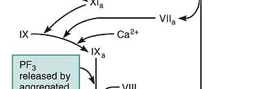
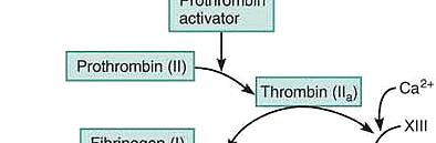
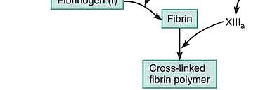
3 Page 3 of 6 In the normal environment the outside of the platelets are positively charged. The lining of the endothelium inside the blood vessel also contains a chemical called prostacyclin which creates a positive charge of the membrane The end result is that the platelets and the lining of the blood vessel all repel one another. However, when an injury occurs and the vessel and surrounding tissue are cut there is a release of collagen, a negatively charged protein that then attracts the platelet. With an initial cut of the vessel, particularly in cross section, the vessel s smooth muscle responds by constricting vascular spasm thus reducing the amount of blood. Platelet Release Reaction Following the vascular spasm a subsequent series of reactions occur called the platelet release reactions. With the injury the platelets drawn to the injury site by the change in charge are expose to N 2 and fracture releasing three groups of substances. Seratonin + thromboxane A 2 - these substances are vasoconstrictors and enhance the constriction of the vessel at the site of the injury thus reducing the overall blood loss. ADP its release triggers a greater attraction to platelets to the sit of injury which subsequently fracture and release more thromboxane and serotonin and ADP attracting more platelets etc.etc. The result is the formation of a water soluble white platelet plug. This you have seen it is the somewhat mucoid, gelatinous material that initially forms over an injury such as a scrape, If washed, it dissolves and the mechanism begins again from scratch. Phospholipids - this release from platelets sets up, activates and establishes the formal clotting mechanism. CLOTTING MECHANISM Coagulation or blood clotting during which blood is transformed from a liquid to a gel, is a multistep process that leads to its critically important last three phases: A complex substance called prothrombin activator is formed. Prothrombin activator converts a plasma protein called prothrombin into thrombin, an enzyme. Thrombin catalyzes the joining of fibrinogen molecules present in plasma to a fibrin mesh, which traps blood cells and effectively seals the hole until the blood vessel can be permanently repaired. The complete coagulation process is much more complicated, however. Over 30 different substances are involved. Factors that enhance clot formation are called clotting factors or procoagulants. Although vitamin K is not directly involved in coagulation, this fat-soluble vitamin is required for the synthesis of four of the procoagulants made by the liver. Factors that inhibit clotting are called anticoagulants. Whether or not blood clots depends on a delicate balance between these two groups of factors. Normally, anticoagulants dominate and clotting is prevented; but when a vessel is ruptured, procoagulant activity in that area increases dramatically and clot formation begins. The procoagulants are numbered I to XIII according to the order of their discovery; hence the numerical order does not reflect the reaction sequence. Tissue factor (III) and Ca2+ (IV) are usually indicated by their names, rather than by numerals. Most of these factors are plasma proteins made by the liver that circulate in an inactive form in blood until mobilized. Phase 1: Two Pathways to Prothrombin Activator Clotting may be initiated by either the intrinsic or the extrinsic pathway, and in the body both pathways are usually triggered by the same tissue-damaging events. Clotting of blood outside the body is initiated only by the intrinsic mechanism.
4 Page 4 of 6 Pivotal components in both mechanisms are negatively charged membranes, particularly those on platelets containing phosphatidylserine, also known as PF3 (platelet factor 3). Many intermediates of both pathways can be activated only in the presence of PF3. In the slower intrinsic pathway, all factors needed for clotting are present in (intrinsic to) the blood. By contrast, when blood is exposed to an additional factor in tissues underneath the damaged endothelium called tissue factor (TF), factor III, or tissue thromboplastin, the shortcut extrinsic mechanism, which bypasses several steps of the intrinsic pathway, is triggered. Each pathway requires calcium and involves the activation of a series of procoagulants, each functioning as an enzyme to activate the next procoagulant in the sequence. The intermediate steps of each pathway cascade toward a common intermediate, factor X. Once factor X has been activated, it complexes with calcium ions, PF3, and factor V to form prothrombin activator. This is usually the slowest step of the blood clotting process, but once prothrombin activator is present, the clot forms in 10 to 15 seconds. Phase 2: Common Pathway to Thrombin Prothrombin activator catalyzes the transformation of the plasma protein prothrombin to the active enzyme thrombin. Phase 3: Common Pathway to the Fibrin Mesh Thrombin catalyzes the polymerization of fibrinogen (another plasma protein made by the liver). As the fibrinogen molecules are aligned into long, hairlike, insoluble fibrin strands, they glue the platelets together and make a web that forms the structural basis of the clot. In the presence of fibrin, plasma becomes gel-like and traps formed elements that try to pass through it. In the presence of calcium ions, thrombin also activates factor XIII (fibrin stabilizing factor), a cross-linking enzyme that binds the fibrin strands tightly together and strengthens and stabilizes the clot. Clot formation is normally complete within 3 to 6 minutes after blood vessel damage. Because the extrinsic pathway involves fewer steps it is more rapid than the intrinsic pathway; in cases of severe tissue trauma it can promote clot formation within 15 seconds.


5 Page 5 of 6
6 Page 6 of 6 Clot Retraction and Repair - Syneresis Within 30 to 60 minutes, the clot is stabilized further by a platelet-induced process called clot retraction. Platelets contain contractile proteins (actin and myosin), and they contract in much the same manner as muscle cells. As the platelets contract, they pull on the surrounding fibrin strands, squeezing serum (plasma minus the clotting proteins) from the mass, compacting the clot and drawing the ruptured edges of the blood vessel more closely together. Even as clot retraction is occurring, vessel healing is taking place. Plateletderived growth factor (PDGF) released by platelet degranulation stimulates smooth muscle cells and fibroblasts to divide and rebuild the wall. As fibroblasts form a connective tissue patch in the injured area, endothelial cells, stimulated by vascular endothelial growth factor (VEGF), multiply and restore the endothelial lining. Fibrinolysis A clot is not a permanent solution to blood vessel injury, and a process called fibrinolysis removes unneeded clots when healing has occurred. Because small clots are formed continually in vessels throughout the body, this cleanup detail is crucial. Without fibrinolysis, blood vessels would gradually become completely blocked. The critical natural clot buster is a fibrin-digesting enzyme called plasmin, which is produced when the plasma protein plasminogen is activated. Large amounts of plasminogen are incorporated into a forming clot, where it remains inactive until appropriate signals reach it. The presence of a clot in and around the blood vessel causes the endothelial cells to secrete tissue plasminogen activator (tpa). Activated factor XII and thrombin released during clotting also serve as plasminogen activators. As a result, most plasmin activity is confined to the clot, and any plasmin that strays into the plasma is quickly destroyed by circulating enzymes. Fibrinolysis begins within two days and continues slowly over several days until the clot is finally dissolved. Factors Limiting Normal Clot Growth Once the clotting cascade has begun, it continues until a clot is formed. Normally, two homeostatic mechanisms prevent clots from becoming unnecessarily large: (1) swift removal of clotting factors, and (2) inhibition of activated clotting factors. For clotting to occur in the first place, the concentration of activated procoagulants must reach certain critical levels. Clot formation in rapidly moving blood is usually curbed because the activated clotting factors are diluted and washed away. For the same reasons, further growth of a forming clot is hindered when it contacts blood flowing normally. Other mechanisms block the final step in which fibrinogen is polymerized into fibrin by restricting thrombin to the clot or by inactivating it if it escapes into the general circulation. As a clot forms, almost all of the thrombin produced is bound onto the fibrin threads. This is an important safeguard because thrombin also exerts positive feedback effects on the coagulation process prior to the common pathway. Not only does it speed up the production of prothrombin activator by acting indirectly through factor V, but it also accelerates the earliest steps of the intrinsic pathway by activating platelets. Thus, fibrin effectively acts as an anticoagulant to prevent enlargement of the clot and prevents thrombin from acting elsewhere. Thrombin not bound to fibrin is quickly inactivated by antithrombin III, a protein present in plasma. Antithrombin III and protein C, another protein produced in the liver, also inhibit the activity of other intrinsic pathway procoagulants. Heparin, the natural anticoagulant contained in basophil and mast cell granules, is also found on the surface of endothelial cells. It inhibits thrombin by enhancing the activity of antithrombin III. Like most other clotting inhibitors, heparin also inhibits the intrinsic pathway.
Chapter 19. Hemostasis
 Chapter 19 Hemostasis Hemostasis Hemostasis is the cessation of bleeding stopping potentially fatal leaks important in small blood vessels not effective in hemorrhage excessive bleeding from large blood
Physiology of. The Blood hemostasis. By prof. Israa f. jaafar
 Physiology of The Blood hemostasis By prof. Israa f. jaafar Learning objectives Understand the Platelet structure and function Explane the Platelet production Understand the phases of hemostasis: vascular
This slide belongs to iron lecture and it is to clarify the iron cycle in the body and the effect of hypoxia on erythropoitein secretion
 This slide belongs to iron lecture and it is to clarify the iron cycle in the body and the effect of hypoxia on erythropoitein secretion Topics of today lectures: Hemostasis Meaning of hemostasis Mechanisms
Anatomy and Physiology
 Anatomy and Physiology For The First Class 2 nd Semester Thrombocytes = Platelets Thrombocytes = Platelets Blood platelets are non-nucleated disc like cell fragments 2-4 µm in diameter. Platelets are not
Topics of today lectures: Hemostasis
 Topics of today lectures: Hemostasis Meaning of hemostasis Mechanisms of hemostasis - Vascular contraction - Platelets plug - Blood coagulation (clotting) - Structure and functions of platelets - Blood
Chapter 19 Blood Lecture Outline
 Chapter 19 Blood Lecture Outline Cardiovascular system Circulatory system Blood 1. distribution 2. regulation 3. protection Characteristics: ph 7.4 38 C 4-6 L Composition: Plasma Formed elements Erythrocytes
UNIT VI. Chapter 37: Platelets Hemostasis and Blood Coagulation Presented by Dr. Diksha Yadav. Copyright 2011 by Saunders, an imprint of Elsevier Inc.
 UNIT VI Chapter 37: Platelets Hemostasis and Blood Coagulation Presented by Dr. Diksha Yadav Hemostasis: Prevention of Blood Loss Vascular constriction Formation of a platelet plug Formation of a blood
Hemostasis and. Blood Coagulation
 Hemostasis and Blood Coagulation Events in Hemostasis The term hemostasis means prevention of blood loss. Whenever a vessel is severed or ruptured, hemostasis is achieved by several mechanisms: (1) vascular
Chapter 19 Cardiovascular System Blood: Functions. Plasma
 Chapter 19 Cardiovascular System Blood: Functions 19-1 Plasma Liquid part of blood. Colloid: liquid containing suspended substances that don t settle out of solution 91% water. Remainder proteins, ions,
Hemostasis Haemostasis means prevention of blood loss from blood vessels.
 ١ Hemostasis Haemostasis means prevention of blood loss from blood vessels. Bleeding is stopped by several mechanisms, which are: 1. Local vasoconstriction 2. Formation of platelet plug 3. Blood coagulation
Blood ESSENTIALS OF HUMAN ANATOMY & PHYSIOLOGY ELAINE N. MARIEB EIGHTH EDITION
 10 Blood PowerPoint Lecture Slide Presentation by Jerry L. Cook, Sam Houston University ESSENTIALS OF HUMAN ANATOMY & PHYSIOLOGY EIGHTH EDITION ELAINE N. MARIEB Blood The only fluid tissue in the human
Chapter 19: Cardiovascular System: Blood
 Chapter 19: Cardiovascular System: Blood I. Functions of Blood A. List and describe the seven major homeostatic functions of blood: 1. 2. 3. 4. 5. 6. 7. II. Plasma A. Composition 1. It is a fluid consisting
Blood ESSENTIALS OF HUMAN ANATOMY & PHYSIOLOGY ELAINE N. MARIEB EIGHTH EDITION
 10 Blood PowerPoint Lecture Slide Presentation by Jerry L. Cook, Sam Houston University ESSENTIALS OF HUMAN ANATOMY & PHYSIOLOGY EIGHTH EDITION ELAINE N. MARIEB Blood The only fluid tissue in the human
Hemostasis Haemostasis means prevention of blood loss from blood vessels.
 1 Hemostasis Haemostasis means prevention of blood loss from blood vessels. Bleeding is stopped by several mechanisms, which are: 1. Local vasoconstriction 2. Formation of platelet plug 3. Blood coagulation
-Hashim ahmed is the one who wrote this sheet. I just edited it according to our record.
 * Subjects of this lecture : - Hemostasis - Platelets, general information, their ultrastructure and role in hemostasis. - Definitions: Thrombus, Embolus, Arteriosclerosis and Atherosclerosis. *NOTE: Prof
Blood ESSENTIALS OF HUMAN ANATOMY & PHYSIOLOGY ELAINE N. MARIEB EIGHTH EDITION
 10 Blood PowerPoint Lecture Slide Presentation by Jerry L. Cook, Sam Houston University ESSENTIALS OF HUMAN ANATOMY & PHYSIOLOGY EIGHTH EDITION ELAINE N. MARIEB Blood The only fluid tissue in the human
Blood. The only fluid tissue in the human body Classified as a connective tissue. Living cells = formed elements Non-living matrix = plasma
 Blood Blood The only fluid tissue in the human body Classified as a connective tissue Living cells = formed elements Non-living matrix = plasma Blood Physical Characteristics of Blood Color range Oxygen-rich
Hemostasis. Learning objectives Dr. Mária Dux. Components: blood vessel wall thrombocytes (platelets) plasma proteins
 plasma proteins") Hemostasis Learning objectives 14-16 Dr. Mária Dux Components: blood vessel wall thrombocytes (platelets) plasma proteins Hemostatic balance! procoagulating activity anticoagulating activity 1 Thrombocytes
Hemostasis Learning objectives 14-16 Dr. Mária Dux Components: blood vessel wall thrombocytes (platelets) plasma proteins Hemostatic balance! procoagulating activity anticoagulating activity 1 Thrombocytes
Chapter 14. Blood. Blood Volume. Blood Composition. Blood
 Blood connective tissue transports vital substances maintains stability of interstitial fluid distributes heat Chapter 14 Blood Blood Cells form mostly in red bone marrow red blood cells white blood cells
Branch of medicine that deals with blood, its formation and disorders is called. Three main functions of cardiovascular system are,, and.
 Chapter 19 The Blood Human body must maintain a balance called. Body fluid inside the cells is called fluid; that outside is called or fluid. Two major fluid networks that help in connecting cells are
The Cardiovascular System: Blood
 C h a p t e r 11 The Cardiovascular System: Blood PowerPoint Lecture Slides prepared by Jason LaPres Lone Star College - North Harris Introduction to the Cardiovascular System A circulating transport system
Chapter 11. Lecture and Animation Outline
 Chapter 11 Lecture and Animation Outline To run the animations you must be in Slideshow View. Use the buttons on the animation to play, pause, and turn audio/text on or off. Please Note: Once you have
Part IV Antithrombotics, Anticoagulants and Fibrinolytics
 Part IV Antithrombotics, Anticoagulants and Fibrinolytics "The meaning of good and bad, of better and worse, is simply helping or hurting" Emerson Chapter 16: Blood Coagulation and Fibrinolytic System
Blood ESSENTIALS OF HUMAN ANATOMY & PHYSIOLOGY ELAINE N. MARIEB EIGHTH EDITION
 10 Blood PowerPoint Lecture Slide Presentation by Jerry L. Cook, Sam Houston University ESSENTIALS OF HUMAN ANATOMY & PHYSIOLOGY EIGHTH EDITION ELAINE N. MARIEB Blood The only fluid tissue in the human
Blood. Plasma. The liquid part of blood is called plasma. 1. Pale yellow fluid; forms more than half the blood volume.
 11 Blood FOCUS: Blood consists of plasma and formed elements. The plasma is 91% water with dissolved or suspended molecules, including albumin, globulins, and fibrinogen. The formed elements include erythrocytes,
Unit 10 - Blood The only fluid tissue in the human body. c) Plasma rises to the top (55% of blood)
 Plasma rises to the top (55% of blood)") Unit 10 - Blood 1 I. Unit 10: Blood A. Blood 1. The only fluid tissue in the human body 2. Classified as a connective tissue 3. Components of blood a) Living cells (1) Formed elements b) Non-living matrix
Unit 10 - Blood 1 I. Unit 10: Blood A. Blood 1. The only fluid tissue in the human body 2. Classified as a connective tissue 3. Components of blood a) Living cells (1) Formed elements b) Non-living matrix
Chapter 19 Blood. Functions of blood:
 Chapter 19 Blood Functions of blood: 1. transportation functions 1. oxygen delivery 2. nutrient delivery 3. transportation of metabolic wastes (urine formation) 4. transportation of hormones (part of the
PHM142 Lecture 4: Platelets + Endothelial Cells
 PHM142 Lecture 4: Platelets + Endothelial Cells 1 Hematopoiesis 2 Platelets Critical in clotting - activated by subendothelial matrix proteins (e.g. collagen, fibronectin, von Willebrand factor) and thrombin
Hemostasis and Thrombosis
 Hemostasis Hemostasis and Thrombosis Normal hemostasis is a consequence of tightly regulated processes that maintain blood in a fluid state in normal vessels, yet also permit the rapid formation of a hemostatic
G. Types of White Blood Cells
 1. White blood cells are also called leukocytes. G. Types of White Blood Cells 2. White blood cells function to protect against diseases. 3. Two hormones that stimulate white blood cell production are
Primary Exam Physiology lecture 5. Haemostasis
 Primary Exam Physiology lecture 5 Haemostasis Haemostasis Body s response for the prevention and cessation of bleeding. Broadly consists of: Primary Haemostasis - vascular spasm and platlet plug formation
BIOH122 Human Biological Science 2
 BIOH122 Human Biological Science 2 Session 2 Haematological System Haemostasis and Blood Groups Bioscience Department Endeavour College of Natural Health endeavour.edu.au Session Plan o Platelets Properties
Composition of Blood
 Blood Blood Blood serves as a vehicle for distributing body heat and for transporting nutrients, respiratory gases, and other substances throughout the body. Composition of Blood Blood is the only fluid
Unit 10: Blood. 2. Buffy coat contains leukocytes and platelets (less than 1% of blood)
") Unit 10: Blood I. Blood A. The only fluid tissue in the human body B. Classified as a connective tissue C. Components of blood 1. Living cells a. Formed elements 2. Non-living matrix a. Plasma D. If blood
Unit 10: Blood I. Blood A. The only fluid tissue in the human body B. Classified as a connective tissue C. Components of blood 1. Living cells a. Formed elements 2. Non-living matrix a. Plasma D. If blood
Blood Outline 17.1 The functions of blood are transport, regulation, and protection (p. 636) A. Transport functions include delivery of oxygen and
 A. Transport functions include delivery of oxygen and") Blood Outline 17.1 The functions of blood are transport, regulation, and protection (p. 636) A. Transport functions include delivery of oxygen and nutrients, transport of metabolic wastes for elimination,
Blood Outline 17.1 The functions of blood are transport, regulation, and protection (p. 636) A. Transport functions include delivery of oxygen and nutrients, transport of metabolic wastes for elimination,
Blood = Fluid connective tissue. Formed elements in plasma.
 Blood = Fluid connective tissue Formed elements in plasma. Blood Physical Characteristics Color Viscosity Volume Temperature Blood ph ph = log (1/[H+]) 7 >7
PHASES OF HAEMOSTASIS
 HAEMOSTASIS Maintains the integrity of a closed, highpressure circulatory system after vascular damage Vessel Wall Injury events in the vessel wall and in the blood which seal breach Delicate balance exists
Chapter 19: The Cardiovascular System: The Blood
 Blood Chapter 9: The Cardiovascular System: The Blood Liquid connective tissue general functions. Transportation Gases, nutrients, hormones, waste products. Regulation ph, body temperature, osmotic pressure.
Chapter 19: The Cardiovascular System: The Blood. Copyright 2009, John Wiley & Sons, Inc.
 Chapter 19: The Cardiovascular System: The Blood Blood Liquid connective tissue 3 general functions 1. Transportation Gases, nutrients, hormones, waste products 2. Regulation ph, body temperature, osmotic
WHITE PAPERS PRESENTATION VIDEO DOCUMENTATION EXPERIMENT WO NDCLOT. The WoundClot Principals for Effective Bleeding Control PRESENTATION
 WHITE PAPERS PRESENTATION VIDEO DOCUMENTATION EXPERIMENT ARTICLES OUR STUDY BLEEDING CONTROL 5 POINT MODEL WO NDCLOT The WoundClot Principals for Effective Bleeding Control PRESENTATION Harnessing SCIENCE
Capillary Action and Blood Components. Biology 20 Unit D: Body Systems Circulation
 Capillary Action and Blood Components Biology 20 Unit D: Body Systems Circulation 1 Remember. Capillaries are so small that blood cells can only pass through single file Important because they are the
Moath Darweesh. Omar Sami. Saleem Khreisha. 1 P a g e
 7 Moath Darweesh Omar Sami Saleem Khreisha 1 P a g e -First of all, I want to give a quick revision to simplify the whole hemostasis mechanism, it will be much easier here with me. Enjoy (you can skip
An Introduction to Blood and the Cardiovascular System
 An Introduction to Blood and the Cardiovascular System The Cardiovascular System consists of: A pump (the heart) A conducting system (blood vessels) A fluid medium (blood) Is specialized fluid of connective
Blood. Physical Characteristics and Volume. Components of Blood
 Blood Functions include Transport Delivering O 2 and nutrients Transporting metabolic wastes (like CO 2 ) and water Transporting hormones Regulation Maintaining body temperature fluid warmed in one area
WBCs production(leucopoiesis):
:") WBCs production(leucopoiesis): Note: this sheet contain only extra notes.j - leucopoiesis is the most complicated process in body because many reasons which are : 1- the production of many cells(monocyte,
WBCs production(leucopoiesis): Note: this sheet contain only extra notes.j - leucopoiesis is the most complicated process in body because many reasons which are : 1- the production of many cells(monocyte,
Blood. C h a p t e r. PowerPoint Lecture Slides prepared by Jason LaPres Lone Star College - North Harris
 C h a p t e r 19 Blood PowerPoint Lecture Slides prepared by Jason LaPres Lone Star College - North Harris Copyright 2009 Pearson Education, Inc., publishing as Pearson Benjamin Cummings Introduction to
Essentials of Anatomy and Physiology, 9e (Marieb) Chapter 10 Blood. Multiple Choice
 Chapter 10 Blood. Multiple Choice") Essentials of Anatomy and Physiology, 9e (Marieb) Chapter 10 Blood Multiple Choice 1) The matrix of blood is called: A) buffy coat B) plasma C) erythrocytes D) lymphocytes E) formed elements Diff: 1 Page
Essentials of Anatomy and Physiology, 9e (Marieb) Chapter 10 Blood Multiple Choice 1) The matrix of blood is called: A) buffy coat B) plasma C) erythrocytes D) lymphocytes E) formed elements Diff: 1 Page
CH 11 Blood OUTLINE: Functions of Blood Composition of Blood Blood Cell Disorders Blood Types Blood Clotting Functions of Blood Transportation
 1 CH 11 Blood OUTLINE: Functions of Blood Composition of Blood Blood Cell Disorders Blood Types Functions of Blood Transportation Protection Regulation ph Temperature Composition of Blood Plasma: liquid
Name: Date: Class: Unit 5 Outline: Blood and the Cardiovascular System
 Name: Date: Class: Unit 5 Outline: Blood and the Cardiovascular System Blood and RBCs Blood The only Classified as a Non-living matrix = Blood Composition tissue in the human body tissue cells = formed
Composition and Functions of Blood. Text p WB 193
 Chapter 10 Blood Composition and Functions of Blood Text p. 337-339 WB 193 Blood Transports everything that must be carried from one place to another. Nutrients, ions, gases, hormones, proteins Urea, waste
Hematology. The Study of blood
 Hematology The Study of blood Average adult = 8-10 pints of blood Composition: PLASMA liquid portion of blood without cellular components Serum plasma after a blood clot is formed Cellular elements are
Ch. 45 Blood Plasma proteins, Coagulation and Fibrinolysis Student Learning Outcomes: Describe basic components of plasma
 Chapt. 45 Ch. 45 Blood Plasma proteins, Coagulation and Fibrinolysis Student Learning Outcomes: Describe basic components of plasma Inheritance of X-linked gene for Factor VIII hemophilia A Explain the
Blood clotting. Subsequent covalent cross-linking of fibrin by a transglutaminase (factor XIII) further stabilizes the thrombus.
 further stabilizes the thrombus.") Blood clotting It is the conversion, catalyzed by thrombin, of the soluble plasma protein fibrinogen (factor I) into polymeric fibrin, which is deposited as a fibrous network in the primary thrombus. Thrombin
Blood clotting It is the conversion, catalyzed by thrombin, of the soluble plasma protein fibrinogen (factor I) into polymeric fibrin, which is deposited as a fibrous network in the primary thrombus. Thrombin
Average adult = 8-10 pints of blood. Functions:
 Average adult = 8-10 pints of blood Functions: Transports nutrients, oxygen, cellular waste products, and hormones Aids in distribution of heat Regulates acid-base balance Helps protect against infection
Chapter 19. Blood. Lecture Presentation by Lee Ann Frederick University of Texas at Arlington Pearson Education, Inc.
 Chapter 19 Blood Lecture Presentation by Lee Ann Frederick University of Texas at Arlington An Introduction to Blood and the Cardiovascular System Learning Outcomes 19-1 Describe the components and major
14.1: Characteristics of Blood. A Centrifuged Blood Sample. Blood Composition. Clinical Application /7/2017. Chapter 14 Lecture Outline
 14.1: Characteristics of Blood Chapter 14 Lecture Outline See separate PowerPoint slides for all figures and tables preinserted into PowerPoint without notes. Blood: A type of connective tissue suspended
TOO MUCH TIME. Cell. Cell. Transport System of the Body: O 2 / Energy. Nutrients Waste O 2 CO 2. Source. External environment: Sink.
 Transport System of the Body: 100 m 1 s 1 mm 100 s 1 cm 10000 s distance = time 2 O 2 / Energy O 2 CO 2 Nutrients Waste Source Cell TOO MUCH TIME External environment: CO 2 / Waste Sink O 2 / Nutrients
TOO MUCH TIME. Cardiovascular System: Blood. Cell. Cell. Transport System of the Body: O 2 / Energy. Nutrients Waste O 2 CO 2.
 Transport System of the Body: 100 m 1 s 1 mm 100 s 1 cm 10000 s distance = time 2 O 2 / Energy O 2 CO 2 Nutrients Waste Source Cell TOO MUCH TIME External environment: CO 2 / Waste Sink O 2 / Nutrients
Blood coagulation and fibrinolysis. Blood clotting (HAP unit 5 th )
") Blood coagulation and fibrinolysis Blood clotting (HAP unit 5 th ) Vessel injury Antithrombogenic (Favors fluid blood) Thrombogenic (Favors clotting) 3 Major systems involved Vessel wall Endothelium ECM
Blood coagulation and fibrinolysis Blood clotting (HAP unit 5 th ) Vessel injury Antithrombogenic (Favors fluid blood) Thrombogenic (Favors clotting) 3 Major systems involved Vessel wall Endothelium ECM
Agenda. Components of blood. Blood is Fluid Connective Tissue. Blood: General functions
 Agenda Chapter 19: Blood Major functions Major Components Structure of RBCs and WBCs ABO Blood Types, and Rh Factor Lab 34.1 and Blood Typing Blood: General functions Transport of dissolved gases, nutrients,
The only fluid tissue in the human body Classified as a connective tissue Living cells = formed elements Non-living matrix = plasma
 The only fluid tissue in the human body Classified as a connective tissue Living cells = formed elements Non-living matrix = plasma Color range Oxygen-rich blood is scarlet red Oxygen-poor blood is dull
Blood. BIOLOGY OF HUMANS Concepts, Applications, and Issues. Judith Goodenough Betty McGuire
 BIOLOGY OF HUMANS Concepts, Applications, and Issues Fifth Edition Judith Goodenough Betty McGuire 11 Blood Lecture Presentation Anne Gasc Hawaii Pacific University and University of Hawaii Honolulu Community
Blood. Biol 105 Lecture 14 Chapter 11
 Blood Biol 105 Lecture 14 Chapter 11 Outline I. Overview of blood II. Functions of blood III. Composition of blood IV. Composition of plasma V. Composition of formed elements VI. Platelets VII. White blood
Blood Lecture Test Questions Set 2 Summer 2012
 Blood Lecture Test Questions Set 2 Summer 2012 1. Leukocytes are attracted to a site of injury or disease by: a. diapedesis b. chemotaxis c. leukocytosis d. heparin e. leukomotosis 2. Leukocytes leave
Blood platelets play important role in coagulation.
 B.N. Bandodkar College of Science, Thane T.Y. B.Sc Paper II Haematology Blood Coagulation / Blood clotting By Dr N.N. Patil Introduction: When blood is shed, it looses its fluidity within few minutes and
I. Concepts: Fill in the following sections with information from the text and lecture.
 Name: Period: 10 Blood Study Guide I. Concepts: Fill in the following sections with information from the text and lecture. 1. Composition and Function of Blood: 2. Hematopoiesis: 1 Miss School, Miss Out
Chapter 06 Lecture Outline. See separate PowerPoint slides for all figures and tables preinserted into PowerPoint without notes.
 Chapter 06 Lecture Outline See separate PowerPoint slides for all figures and tables preinserted into PowerPoint without notes. Copyright 2016 McGraw-Hill Education. 2012 Pearson Permission Education,
A. Blood is considered connective tissue. RBC. A. Blood volume and composition 1. Volume varies - average adult has 5 liters
 A. Blood is considered connective tissue. RBC A. Blood volume and composition 1. Volume varies - average adult has 5 liters 2. 45% cells by volume called hematocrit (HCT) a. red blood cells (RBC) mostly
4/5/17. Blood. Blood. Outline. Blood: An Overview. Functions of Blood
 Outline Blood Biol 105 Chapter 11 I. Overview of blood II. Functions of blood III. Composition of blood IV. Composition of plasma V. Composition of formed elements VI. Platelets VII. White blood cells
Essentials of Human Anatomy and Physiology, 11e (Marieb) Chapter 10 Blood Multiple Choice Part I Questions
 Chapter 10 Blood Multiple Choice Part I Questions") Essentials of Human Anatomy and Physiology, 11e (Marieb) Chapter 10 Blood 10.1 Multiple Choice Part I Questions Using Figure 10.1, identify the following: 1) The neutrophil is indicated by. A) Label A
Essentials of Human Anatomy and Physiology, 11e (Marieb) Chapter 10 Blood 10.1 Multiple Choice Part I Questions Using Figure 10.1, identify the following: 1) The neutrophil is indicated by. A) Label A
Blood. Those made by activated B-lymphocytes are antibodies.
 Blood I. Intro A. Some introductory questions to get you started: what are formed elements? What is plasma? Name a couple of plasma proteins. Where are most blood proteins made? What do Hb, RBC, and WBC
THE CIRCULATORY SYSTEM. Unit 3: Transportation and Respiration
 THE CIRCULATORY SYSTEM Unit 3: Transportation and Respiration Introduction The circulatory system, also called the cardiovascular system, is an organ system that allows blood to flow to all the cells in
What is the composition of blood, including blood cells? What organs and structures control the flow of blood throughout the body?
 3 Chapter 10: Circulatory System and Lymphatic System In this chapter, you will learn about the structure and function of the circulatory system and lymphatic system. What is the composition of blood,
Step 2. Common Blood Tests, and the Coulter Counter Readout
 Step 2. Common Blood Tests, and the Coulter Counter Readout We will be learning about some common blood tests. We will not be preforming most of them in lab. The student should know their names, their
BLOOD & CIRCULATORY SYSTEM. Prepared by Mr.Yeung
 BLOOD & CIRCULATORY SYSTEM Prepared by Mr.Yeung CIRCULATORY SYSTEM Overview Every cell in a living organism must have direct access to its nutrient supply Single celled organisms rely on simple diffusion
Blood Lecture Outline : Fluid Connective Tissue Part I of the Cardiovascular Unit
 Blood Lecture Outline : Fluid Connective Tissue Part I of the Cardiovascular Unit General Characteristics: Extracellular matrix ph Volume Functions of the blood: 1. Transport 2. Regulation 3. Protection
What are blood clots?
 What are blood clots? Dr Matthew Fay GP Principal The Willows Medical Practice- Queensbury GPwSI and Co-Founder Westcliffe Cardiology Service GP Partner Westcliffe Medical Group Created 5/31/18 Dr. Matthew
Principles of Anatomy and Physiology
 Principles of Anatomy and Physiology 14 th Edition CHAPTER 19 The Cardiovascular System: The Blood Functions and Properties of Blood Blood is a liquid connective tissue consisting of cells surrounded by
The Blood Dr. Gary Mumaugh
 The Blood Dr. Gary Mumaugh Overview of Blood Circulation Blood leaves the heart via arteries that branch repeatedly until they become capillaries Oxygen (O 2 ) and nutrients diffuse across capillary walls
Biology 218 Human Anatomy. Adapted form Martini Human Anatomy 7th ed. Chapter 20 The Cardiovascular System: Blood
 Adapted form Martini Human Anatomy 7th ed. Chapter 20 The Cardiovascular System: Blood Introduction The cardiovascular system functions as a system to transport numerous substances throughout the body
Bleeding and Haemostasis. Saman W.Boskani HDD, FIBMS Maxillofacial Surgeon
 Bleeding and Haemostasis Saman W.Boskani HDD, FIBMS Maxillofacial Surgeon 1 Beeding Its escaping or extravasation of blood contents from blood vessels Types: - Arterial - Venous - Capillary Differences
Hemostasis. Clo)ng factors and Coagula4on NORMAL COAGULATION. Overview of blood coagula4on. The Cascade Theory 5/1/12. Clot
ng factors and Coagula4on NORMAL COAGULATION. Overview of blood coagula4on. The Cascade Theory 5/1/12. Clot") Hemostasis Clo)ng factors and Coagula4on Dr Badri Paudel www.badripaudel.com Hemostasis is defined as a property of circula4on whereby blood is maintained within a vessel and the ability of the system
Hemostasis Clo)ng factors and Coagula4on Dr Badri Paudel www.badripaudel.com Hemostasis is defined as a property of circula4on whereby blood is maintained within a vessel and the ability of the system
PHLEBOTOMIST. person trained to draw blood from a patient for clinical or medical testing, transfusions, donations, or research.
 BLOOD PHLEBOTOMIST person trained to draw blood from a patient for clinical or medical testing, transfusions, donations, or research. Blood transports substances and maintains homeostasis in the body Hematophobia
Chapter 19. Openstax: Chapter 18. Blood
 Chapter 19 Blood Openstax: Chapter 18 Chapter 19 Learning Outcomes After completing Chapter 19, you will be able to: 1. Describe the components and major functions of blood and list the physical characteristics
HEART HEALTH WEEK 2 SUPPLEMENT. A Beginner s Guide to Cardiovascular Disease ATHEROSCLEROSIS. Fatty deposits can narrow and harden the artery
 WEEK 2 SUPPLEMENT HEART HEALTH A Beginner s Guide to Cardiovascular Disease ATHEROSCLEROSIS FIGURE 1 Atherosclerosis is an inflammatory process where cholesterol is deposited in the wall of arteries and
Chapter 19: The Cardiovascular System: The Blood. Copyright 2009, John Wiley & Sons, Inc.
 Chapter 19: The Cardiovascular System: The Blood Blood Liquid connective tissue 3 general functions 1. Transportation Gases, nutrients, hormones, waste products 2. Regulation ph, body temperature, osmotic
BLOOD RUNS THROUGH YOUR BODY
 BLOOD RUNS THROUGH YOUR BODY WORKSHEET A Your heart and blood vessels make up your blood system. At the centre of your blood system is your heart. Its job is to pump the blood around your body. The rest
Blood and the Lymphatic System. Lesson Overview. Lesson Overview Blood and the Lymphatic System
 Lesson Overview 33.2 Blood and the Lymphatic System THINK ABOUT IT When you think about body tissues, you probably picture something with a definite shape, like muscle or skin. But blood is a tissue too
Physiology of Blood. Dr. Hiwa S. Namiq
 Physiology of Blood Dr. Hiwa S. Namiq 7-1-2019 11 January 2019 Introduction Red blood cells (erythrocytes) The major function of RBC is to transport Hb which in turn carries oxygen from lungs to the tissues.
Leukocytes (White Blood Cells)
") Leukocytes (White Blood Cells) As we, leukocytes ( white blood cells ) are of two types : 1- Granulocytes :have granules in the cytoplasm and include neutrophils, eosinophils and basophils. 2- Agranulocytes:
Leukocytes (White Blood Cells) As we, leukocytes ( white blood cells ) are of two types : 1- Granulocytes :have granules in the cytoplasm and include neutrophils, eosinophils and basophils. 2- Agranulocytes:
Lab 1 Blood Composition and formed elements
 Lab 1 Blood Composition and formed elements Plasma 55% of whole blood 90% water 8% proteins from liver 2% misc. Nutrients: AA, glucose, lipids vitamins, minerals Wastes: urea, uric acid, creatine, ammonium
10. Which of the following immune cell is unable to phagocytose (a) neutrophils (b) eosinophils (c) macrophages (d) T-cells (e) monocytes
 neutrophils (b) eosinophils (c) macrophages (d) T-cells (e) monocytes") Chapter 2. Acute and chronic inflammation(6): 1. In acute inflammation, which events occur in the correct chronological order? (Remembered from 2000, 2004 exam.) p50 (a) transient vasoconstriction, stasis
Chapter 2. Acute and chronic inflammation(6): 1. In acute inflammation, which events occur in the correct chronological order? (Remembered from 2000, 2004 exam.) p50 (a) transient vasoconstriction, stasis
WHICH OF THE FOLLOWING COMPRISE A
 HEMATOLOGY QUESTION REVIEW WHICH OF THE FOLLOWING COMPRISE A LOGICAL SEQUENCE OF VESSELS AS BLOOD EXITS THE HEART? a. capillaries; arteries; veins b. veins; capillaries; arteries c. arteries; capillaries;
The Blood. Dr. Gary Mumaugh
 The Blood Dr. Gary Mumaugh Blood is the river of life Overview of Blood Circulation Blood leaves the heart via arteries that branch repeatedly until they become capillaries Oxygen (O 2 ) and nutrients
THE BLOOD AND LYMPHATIC SYSTEM. By Group #4
 THE BLOOD AND LYMPHATIC SYSTEM By Group #4 BLOOD CELLS By Mackenzie Lighterink Red Blood Cells White Blood Cells RED BLOOD CELLS Transport Oxygen Produced in red bone marrow Get color from Hemoglobin-a
Functions of Blood. Transport. Transport. Defense. Regulation. Unit 6 Cardiovascular System: Blood
 Unit 6 Cardiovascular System: Blood Functions of Blood With each beat of the heart, approximately 75 ml of blood is pumped On average, the heart beats 70 times per minute Every minute, the heart pumps
Bio& 242 Unit 3 / Lecture 1
 Bio& 242 Unit 3 / Lecture 1 Major Functions of Blood The body contains 4 to 6 liters of blood with an average ph of 7.35 to 7.45. Functions include: Transport Oxygen, Carbon Dioxide, Nutrients, Hormones,
BIOCHEMISTRY OF BLOOD
 BCH 471 BIOCHEMISTRY OF BLOOD Amal Alamri Experiment 1 Separation of Plasma and Serum from Whole Blood Whole Blood It is living tissue that circulates through the heart, arteries, veins, and capillaries
Hematocrit. Hematocrit = using a centrifuge to separate out the parts of blood. Plasma Formed elements:
 Blood Notes Hematocrit Hematocrit = using a centrifuge to separate out the parts of blood Plasma Formed elements: Buffy Coat = Leukocytes and Platelets Erythrocytes General Facts Blood ph = 7.4 Volume