The Management of Humeral Shaft Fractures. David Chapple MSc FRCS
|
|
|
- Coral Logan
- 5 years ago
- Views:
Transcription
1 The Management of Humeral Shaft Fractures David Chapple MSc FRCS
2 SHAFT NOT Proximal Distal
3 Anything New? Anatomy Classifications MOI and Clinical aspects Management options Management indications Management Complications
4 Anatomy three borders Anterior Medial lateral three surfaces anterolateral anteromedial posterior
5 The Humerus Anterior aspect Head necks tuberosities < > & Deltoid sulcus, bicipital groove supracondylar ridges epicondyles Coronoid fossa trochlea/capitulum Supracondylar process
6 Anterior Muscle attachments supraspinatus subscapularis Pectoralis major latissimus dorsi Teres Major triceps medial head deltoid coracobrachialis Brachialis Brachioradialis Extensor Carpi radialis longus Pronator Teres Common Origins
7 The Humerus Posterior aspect Head Necks greater tuberosity Sulcus for radial nerve supracondylar ridges epicondyles Olecranon fossa Trochlear
8 The Humerus Posterior Muscle attachments infraspinatus teres minor Triceps lateral head Deltoid Brachialis triceps Medial Head Anconeus

9 Coracobrachialis Muculocutaneous Biceps Pectoralis major Median nerve Brachial artery Basilic vein Deltoid Ulnar nerve Profunda artery Radial nerve Lateral head of Triceps Long head of Triceps
10 Biceps Median nerve Brachial artery Basilic vein Ulnar nerve Muculocutaneous Brachialis Radial nerve Profunda artery
11 Biceps Median nerve Brachial artery Muculocutaneous Brachialis Radial nerve Basilic vein Ulnar nerve
12 Cross-section Upper section cylindrical Lower section comma shaped flattened AP IM device diameter and length posterior flat surface plates
13 Ossification 8 ossification centres shaft appears at middle of bone and grows towards ends at 8th week of intrauterine life
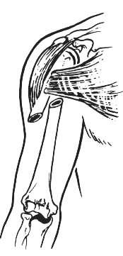
14 Radial Nerve between long and medial heads of triceps Whitson JBJS the radial nerve transversed the triceps at such a depth that it was nowhere in contact with the humerus...as the supracondylar ridge was approached, the radial nerve was
15 Whitson JBJS It was apparent that the separation of the triceps into three heads was artificial and that the medial and lateral heads were in reality a single muscle group traversed by a nerve and an artery. similar to posterior interosseous passes through the supinator. The Spiral Groove in every specimen gave origin to the uppermost fibres of the brachialis,
16
17
18 Whitson JBJS 1954 Admit clinical importance of these observations is not great. Explain that the muscle fibres of triceps and brachialis offer some protection from sharp bone edges.
19 Peripheral Nerve Injury Unit Mr Birch on Whitson s findings.. Not his experience, felt that the nerve had a close relationship to the bone for a considerable distance. possible explanation could be that the cadavers had been lying supine and so compression deformation occurred which distorted the true in vivo anatomical position of the nerve

20 Blood Supply MainNutrient artery to humerus Profunda brachii Gives nutrient deltoid posterior descending radial collateral
21 Blood supply Laing 1956 JBJS 38-A main nutrient artery enters the humerus at the junction of the middle and distal third, or in the lower part of the middle third. Middle third fractures damage this vessel higher rate of delayed union Klenerman JBJS 48-B
22 Humeral Shaft fractures Humeral shaft fractures 3% all all fractures Christensen Acta Chir Scand 1967 Humeral Shaft fractures 1% of all fractures Emmett and Breck 11,000 #
23 Shaft Fractures Classifications anatomical management based comparison useless
24 Classification No universally accepted system for humeral shaft fractures anatomical proximal shaft, middle shaft, or distal shaft relative to muscle attachments pectoralis major, deltoid Character description


25
26 Classification Fracture comminution A-simple B-butterfly fragments C-comminuted
27 Classification Associated soft tissue injury periarticular involvement nerve injury vascular injury intrinsic condition of the bone
28 Mechanism of Injury Klenerman experimental # s Compression proximal or distal # s bending produce transverse # s Torsional forces give spiral # s Bending combined with torsion produces an oblique # with a butterfly fragment
29 Mechanism of Injury Direct and Indirect trauma Falls(FOOSH) RTA s Direct blow to arm Extreme muscle contraction ball and javelin throwing arm wrestling
30 Arm Wrestling Ogawa and Ui 1997 J Trauma 42-2, Tokyo 30 cases all spiral # s 23% radial nerve palsy occurred when trying to change from a static to dynamic phase shoulder rotators:- intense rotational force

31 Andy
32 Signs and Symptoms Pain, swelling and deformity motion and crepitus associated injuries vascular neural secondary injury due to swelling particularly the multiple trauma or unconscious pt.
33 Imaging Plain AP and 90 0 lateral move whole patient not limb include joints associated dislocations, # s into joints traction radiographs for comminuted # s Comparison films for planning Bone scan for pathological # s
34 Goals of treatment Establish union with an acceptable humeral alignment and restore patients to their previous level of function
35 Mal-Union Klenerman JBJS B Concluded: The degree of radiological deformity that can be accepted is far greater than in other long bones anterior bowing of 20 0 varus of 30 0 before clinically obvious
36 Methods of Treatment NUMEROUS OPTIONS Closed Open Good to excellent results have been reported with all methods Patient characteristics Fracture characteristics
37 Management CONSERVATIVE
38 Most humeral shaft fractures can be managed nonoperatively
39 Closed Management Methods Greater than 90% expected union rate Hanging arm cast U-shaped brachial splint Velpeau dressing Abduction humeral splint/shoulder spica cast skeletal traction functional brace
40 Hanging arm cast Gravity traction for reduction arm and cast must be dependant at all times Problems RoM of shoulder and elbow impaired fracture distraction and hinging avoid transverse fractures Indications midshaft spiral or oblique with shortening
41 Hanging arm cast Lightweight elbow at 90 0, forearm in neutral at least 2cm proximal to fracture distal forearm loops dorsal, volar and neutral must hang free regular Follow-up

42 Hanging arm cast Apex anterior angulation shortening of the sling Apex posterior angulation lengthening the sling
43 Hanging arm cast Valgus(Apex medial) angulation using the volar loop Varus(Apex lateral) angulation using the dorsal loop
44 U-shaped splint with C/C Indicated for acute management of # s with minimal shortening slipping of the cast is common poor patient tolerance often exchanged for a functional brace at 2/52
45 Thoracobrachial immmobilization Velpeau shoulder dressing inexpensive, comfortable and easily applied and adjusted minimally displaced # s axillary pad early pendulum exercises
46 Traction rarely indicated, as operative management has same indications
47 Functional bracing Sarmiento 1977JBJS 59-A effects fracture reduction through soft tissue compression allows good shoulder and elbow movement after one week until eight weeks
48 Functional bracing Sarmiento et al 1990, 72-B suggests well proven method for mid shaft # s presents a series of distal shaft # s which had good results from functional bracing after a period of hanging cast treatment
49 Functional bracing Sarmiento et al 1990, 72-B control of angulation showed average of 9 o varus in 81% of patients (n65) high incidence of radial nerve damage (18%) all were resolved or improving residual stiffness of shoulder and elbow minimal loss of RoM and good functional results 96% went onto union
50 Functional bracing Balfour et al 1982 JBJS 64-A, LA California adapted Sarmiento s brace proper fit swelling of the forearm discomfort shoulder flare with sling support
51 Functional bracing Balfour et al 1982 JBJS 64-A Stress that the brace requires the influence of gravity on the dependent arm of an ambulatory patient all except in one patient the fracture united average of 9 0 varus and 6 0 AP bowing RoM elbow and shoulder excellent
52 Functional bracing Camden et al 1992 Injury 23-4 comparison of U-slab with functional brace no difference for healing time and alignment better RoM at elbow Zagorski 1988 JBJS 70-A can be used to treat proximal shaft fractures have less angulation
53 Operative treatment INDICATIONS
54 INTERVENTION INDICATIONS It s Begging for a nail It will be Good fun to plate it? I need the experience. Why don t we try that new nail from? That rep had a delightful, intelligent and generous personality so why don t we use.?
55 Indications for operative management Open fracture associated vascular injury floating elbow segmental fracture pathological fracture Bilateral humeral fractures polytrauma patients radial nerve palsy neurological deficiency after penetrating injury fractures with unacceptable alignment
56 Indications for operative Open fracture management require debridement fracture stabilisation afterwards to reduce infection Not absolute Sarmiento shown cases where no debridement of low velocity gun shot fractures and non operative management of fracture
57 Indications for operative management Associated vascular injury internal or external fixation prior or post repair If repaired then non-operative management is contra-indicated fracture motion jeopardise the repair

58 Associated vascular injury Arteriography controversial clinical assessment can detect 50% time delay Urgent exploration and repair intraluminal shunts end to end or grafts
59 Indications for operative management Floating Elbow Rogers et al 1984 JBJS 66-A, Houston retrospective study higher incidence of non-union of the humerus in injuries without ORIF ORIF of both forearm and humerus indicated
60 Floating Elbow Rogers et al 1984 JBJS 66-A, Houston 19 patients traffic elbow, sideswipe injury severe injury with poor outcome amputation, arthrodesis, non-union and poor elbow function Two groups elbow involvement
61 Floating Elbow Rogers et al 1984 JBJS 66-A, Houston Group I no elbow involvement all mid-shaft humerus 5 open, 6 closed closed did better than open conservatively managed had more nonunions all forearm fractures healed
62 Indications for operative management Segmental fractures Foster et al 1985 JBJS 67-A multi centre trial segmental humeral fractures have a high rate of non-unions if treated nonoperatively at one or both the fracture sites
63 Indications for operative management Pathological fractures internal fixation Enders nails, locked nails, no reaming cement augmentation patient comfort pain relief, regain function daily activities, independence
64 Indications for operative management Bilateral humeral shaft fractures improves patients ability to perform daily tasks and personal toilet
65 Indications for operative management Multiple trauma patient advantages pain relief protect adjacent soft tissues fracture disease help nursing and rehab Brumback et al 1986 JBJS 68-A, Baltimore
66 Multiple trauma patient Brumback et al 1986 JBJS 68-A, Baltimore 58 patients with multiple trauma Shock Trauma Center 2000 patients annually most scooped and run by helicopter retrospective ISS, average 23.5
67 Multiple trauma patient Brumback et al 1986 JBJS 68-A, Baltimore stabilise long bone fractures 95% were stabilised within 1st 24 hrs. Used Rush rods and Enders nails semi-rigid fixation minimal violation of fracture haematoma no reaming
68 Multiple trauma patient Brumback et al 1986 JBJS 68-A Results 5 deaths alignment 98% <15 0 varus RoM dependant on insertion point epicondylar approach had poor results 55% had devices removed
69 Multiple fractures Jensen and Rasmussen 1995 Injury 26(4), Denmark showed poor results for multiple injured patients with bracing Neer score small study
70 Indications for operative Radial nerve palsy management mandatory if occurs after closed manipulation and reduction Packer et al 1972 CORR 88 Shergill and Birch 1997 open wounds arterial injury
71 Radial nerve palsy Commonly middle third # s higher rate in distal third # s Holstein-Lewis fracture oblique, distal third

72 Radial Nerve palsy Triceps sparing Supination lost in the extended elbow flexed allows biceps wrist drop unable to extend MCPj DIP/PIPj s extend via intrinsics
73 Treatment of Radial Neuropathy Associated with Fractures of the Humerus Pollock et al, San Francisco, 1981, JBJS 63-A Retrospective 15 yrs, 23 patients, all with CLOSED treatment of # humerus with a Radial Nerve Palsy 6% of all humeral shaft # s (11% lit) 13 male, 10 female, (1mth-63yrs)
74 Treatment of Radial Neuropathy Associated with Fractures of the Humerus Pollock et al, San Francisco, 1981, JBJS 63-A mainly severe trauma 3 segmental, 5 oblique 4 comminuted 5 transverse 7 spiral
75 Treatment of Radial Neuropathy Associated with Fractures of the Humerus Pollock et al, San Francisco, 1981, JBJS 63-A 3 open, 21 closed 2 prox. 1/3 5 middle 1/3 14 distal 1/3 3 segmental
76 # and Radial palsy Conservative methods of treatment sugar-tong 8 shoulder spica 5 hanging cast 5 palm to axilla cast 3 olecranon traction 2 posterior splint 1
77 # and Radial palsy Extent of palsy complete M & S (n9) partial M (n6) partial M & S (n3) complete M, intact S (n3) isolated S (n1) partial lesions distributed through out length of humerus
78 Treatment of Radial Neuropathy Associated with Fractures of the Humerus Pollock et al, San Francisco, 1981, JBJS 63-A All patients in this series had a complete return of radial nerve function.
79 Treatment of Radial Neuropathy Associated with Fractures of the Humerus Pollock et al, San Francisco, 1981, JBJS 63-A Distal 1/3 fractures have a high incidence of palsies vast majority have a lesion in continuity clinical or EMG improvement should be apparent by 14 to 16 weeks if not then explore and repair
80 Treatment of Radial Neuropathy Associated with Fractures of the Humerus Pollock et al, San Francisco, 1981, JBJS 63-A Time course of recovery complete loss first signs of recovery between 6 days and seven months average seven weeks Full recovery one day to one year, average fifteen weeks
81 Early Exploration Literature review n95 12% found nerve lacerated Nerve recoveries 70% non-recovery 20% lost to follow-up 10%
82 Delayed exploration Literature review n53 3 to 6 months delay divided nerves found 19% entrapped in callus 6% reasonable recovery
83 Delayed exploration Advantages over early time for recovery neurapraxia, axonotmesis evaluation of nerve lesion degree, tinel sign, neurophysiology fracture united results of late repair reported similar to early
84 Indications for operative management Neurological loss after penetrating injury almost an absolute indication similar to other areas of the body primary repair of nerve, requires stabilisation tag and refer after stabilisation
85 Indications for operative management Failure of conservative management failure to maintain acceptable alignment obese, pendulous breasts 20 0 AP 30 0 varus thin individuals, less tolerant 3cm of shortening malrotation well tolerated
86 Obese Failure of conservative Jensen et al 1995 Injury 26-4, Denmark Sarmiento brace compared with nonobese Neer scores lower 45% non-unions pendulous breasts management
87 What Operation? Screws screws and plates cerclage wires External fixation Intra medullary fixation
88 Approaches Anterolateral supine, incision lateral border of biceps, proximal fractures Anterior coracoid to deltoid insertion then lateral border of biceps limited distally Posterior excellent exposure, limited proximally, 8cm from acromium lateral and long heads of triceps, medial head incised
89 Open reduction and internal Disadvantages infection non-union requiring re-operation injury to the radial nerve initially or on removal of metal work prolonged disability fixation
90 Open reduction and internal fixation Advantages early mobilization of limb good joint function good pain relief exploration of radial nerve repair prognosis for recovery bone grafting
91 Open reduction and internal fixation Bell et al 1985 JBJS 67-B, Sunnybrook Griend et al 1986 JBJS 68-A, Mississipi 36 patients had AO plating indications multiple injuries open fractures retrospective
92 AO plating Griend et al 1986 JBJS 68-A, Mississipi..comparisons may not be entirely valid.. multiple methods of fixation uncomplicated fractures cf. Problem fractures anterolateral approach 4.5mm DCP bone grafted if bone loss or comminution
93 AO plating Griend et al 1986 JBJS 68-A, Mississipi One non-union no deep infection, two superficial infections one (transient)post operative radial nerve palsy radial nerve palsy 9 explored, 1 lacerated, 4 contused, 4 normal 6 resolved good RoM, except in severe vascular or neural defect
94 AO plating Griend et al 1986 JBJS 68-A, Mississipi Conclude safe if nerve exposed and protected high rates of union good function only where non-operative management not indicated

95 External fixation Indications open fractures extensive soft tissue injury fractures over burns infected non-unions neurovascular injury

96 External fixation Complications pin tract infections impalement muscle, tendon neurovascular non-union advise direct visual placement of pins
97

98 advise direct visual placement of pins Humerus Musculocutaneous Ulnar nerve Brachial artery Median nerve Brachial veins Radial nerve
99 Intramedullary fixation General advantages mechanical axis less likely to fail by fatigue load-sharing axial gliding osseus alignment less stress shielding less refracture after nail removal biological benefits
100 Intramedullary fixation Flexible intramedullary nails Enders nails, Hackenthal, Rush rods not rigid, # can shorten and rotate entrance point Interlocked nails numerous on the market to ream or not, antegrade insertion can cause impingement

101 Intramedullary fixation Antegrade high rates of shoulder stiffness subacromial impingement Retrograde no shoulder problems can get elbow restriction of extension Epicondylar portal p poor results
102 Locking nails Habernek and Orthner 1991 JBJS 73-B, Austria 19 Seidel nails good results no non-unions, infections, radial nerve palsies only fractures in the middle 60% of the humerus secondary radial palsies lower 5th of shaft # s should not be nailed mal-alignment
103 Locking nails Court-Brown et al 1992 JBJS 74-B, Edinburgh 30 Seidel nails poor results (87% complication rate) technical difficulties failed distal (30%)locking nail protrusion (40%) poor shoulder function did not advocate its use
104 Rehabilitation RoM of hand and wrist started immediately RoM of elbow and shoulder as pain allows shoulder to avoid postfracture stiffness elbow ACTIVE exercises only myositis ossificans post # healing strengthening exercises isometric to isotonic
105 Management of humeral shaft fractures Summary Vast majority can be managed closed There are absolute indications for open management You can find supporting evidence for each type of open method Patient and fracture characteristics dictate management

106 Thank you
Fractures of the shoulder girdle, elbow and fractures of the humerus. H. Sithebe 2012
 Fractures of the shoulder girdle, elbow and fractures of the humerus H. Sithebe 2012 Fractures of the Clavicle (mid-shaft). Fractures of the clavicle Fractures of the clavicle Treatment- conservative.
HUMERAL SHAFT FRACTURES: ORIF, IMN, NONOP What to do?
 HUMERAL SHAFT FRACTURES: ORIF, IMN, NONOP What to do? TRAUMA 101 2018 FRACTURE CARE FOR THE COMMUNITY ORTHOPEDIST William W. Cross III, MD Assistant Professor Division of Orthopaedic Trauma Chair, Division
Fractures and dislocations around elbow in adult
 Lec: 3 Fractures and dislocations around elbow in adult These include fractures of distal humerus, fracture of the capitulum, fracture of the radial head, fracture of the olecranon & dislocation of the
Diaphyseal Humerus Fractures. OTA Course Dallas, TX 1/20/17 Ellen Fitzpatrick MD
 Diaphyseal Humerus Fractures OTA Course Dallas, TX 1/20/17 Ellen Fitzpatrick MD OBJECTIVES TREATMENT OPTIONS SURGICAL INDICATIONS CONTROVERSIES IN MANAGEMENT Humerus Fractures Treatment Goals: Functional
Orthopedics in Motion Tristan Hartzell, MD January 27, 2016
 Orthopedics in Motion 2016 Tristan Hartzell, MD January 27, 2016 Humerus fractures Proximal Shaft Distal Objectives 1) Understand the anatomy 2) Epidemiology and mechanisms of injury 3) Types of fractures
Fascial Compartments of the Upper Arm
 Fascial Compartments of the Upper Arm The upper arm is enclosed in a sheath of deep fascia and has two fascial septa: 1- Medial fascial septum (medial intermuscular septum): attached to the medial supracondylar
1/19/2018. Winter injuries to the shoulder and elbow. Highgate Private Hospital (Whittington Health NHS Trust)
") Winter injuries to the shoulder and elbow Omar Haddo Consultant Orthopaedic Surgeon, Shoulder, Elbow, Hand & Wrist Specialist MBBS, BmedSci, FRCS(Orth) Highgate Private Hospital (Whittington Health NHS
Winter injuries to the shoulder and elbow Omar Haddo Consultant Orthopaedic Surgeon, Shoulder, Elbow, Hand & Wrist Specialist MBBS, BmedSci, FRCS(Orth) Highgate Private Hospital (Whittington Health NHS
region of the upper limb between the shoulder and the elbow Superiorly communicates with the axilla.
 1 region of the upper limb between the shoulder and the elbow Superiorly communicates with the axilla. Inferiorly, a number of important structures pass between arm & forearm through cubital fossa. 2 medial
The Elbow and the cubital fossa. Prof Oluwadiya Kehinde
 The Elbow and the cubital fossa Prof Oluwadiya Kehinde www.oluwadiya.com Elbow and Forearm Anatomy The elbow joint is formed by the humerus, radius, and the ulna Bony anatomy of the elbow Distal Humerus
The study of distal ¼ diaphyseal extra articular fractures of humerus treated with antegrade intramedullary interlocking nailing
 2018; 4(4): 46-50 ISSN: 2395-1958 IJOS 2018; 4(4): 46-50 2018 IJOS www.orthopaper.com Received: 01-08-2018 Accepted: 03-09-2018 Dr. Ankur Parikh Orthopaedics, Jehangir Hospital, Sassoon road, Pune, Dr.
The arm: *For images refer back to the slides
 The arm: *For images refer back to the slides Muscles of the arm: deltoid, triceps (which is located at the back of the arm), biceps and brachialis (it lies under the biceps), brachioradialis (it lies
MEDIAL EPICONDYLE FRACTURES
 MEDIAL EPICONDYLE FRACTURES Demographic 20% of elbow fractures 60% of which are associated with elbow dislocation. 75% in boys between 6-12 years 20% of elbow dislocation with ME fracture, the ME is incarcerated
Upper limb Arm & Cubital region 黃敏銓
 Upper limb Arm & Cubital region 黃敏銓 1 Arm Lateral intermuscular septum Anterior (flexor) compartment: stronger Medial intermuscular septum Posterior (extensor) compartment 2 Coracobrachialis Origin: coracoid
MUSCLES. Anconeus Muscle
 LAB 7 UPPER LIMBS MUSCLES Anconeus Muscle anconeus origin: distal end of dorsal surface of humerus insertion: lateral surface of ulna from distal margin of the semilunar notch to proximal end of the olecranon
1 Humeral fractures 1.13 l Distal humeral fractures Treatment with a splint
 1 Executive Editor: Chris Colton Authors: Mariusz Bonczar, Daniel Rikli, David Ring 1 Humeral fractures 1.13 l Distal humeral fractures Treatment with a splint Indication All 13-A type fractures, excluding
Osteology of the Elbow and Forearm Complex. The ability to perform many activities of daily living (ADL) depends upon the elbow.
 depends upon the elbow.") Osteology of the Elbow and Forearm Complex The ability to perform many activities of daily living (ADL) depends upon the elbow. Activities of Daily Living (ADL) Can you think of anything that you do to
Osteology of the Elbow and Forearm Complex The ability to perform many activities of daily living (ADL) depends upon the elbow. Activities of Daily Living (ADL) Can you think of anything that you do to
The Arm and Cubital Fossa
 The Arm and Cubital Fossa Dr. Andrew Gallagher School of Anatomical Sciences University of the Witwatersrand Introduction The ARM (BRACHIUM) is the most proximal segment of the upper limb musculoskeletal
Elbow Elbow Anatomy. Flexion extension. Pronation Supination. Anatomy. Anatomy. Romina Astifidis, MS., PT., CHT
 Elbow Elbow Anatomy Romina Astifidis, MS., PT., CHT Curtis National Hand Center Baltimore, MD October 6-8, 2017 Link between the arm and forearm to position the hand in space Not just a hinge Elbow = 70%
Human Anatomy Biology 351
 1 Human Anatomy Biology 351 Upper Limb Exam Please place your name on the back of the last page of this exam. You must answer all questions on this exam. Because statistics demonstrate that, on average,
Sports Medicine Unit 16 Elbow
 Sports Medicine Unit 16 Elbow I. Bones a. b. c. II. What movements does the elbow perform? a. Flexion b. c. Pronation d. III. Muscles in motion a. FLEXION (supinated) i Brachialis (pronated) ii (neutral)
The Elbow and Radioulnar Joints Kinesiology. Dr Cüneyt Mirzanli Istanbul Gelisim University
 The Elbow and Radioulnar Joints Kinesiology Dr Cüneyt Mirzanli Istanbul Gelisim University 1 The Elbow & Radioulnar Joints Most upper extremity movements involve the elbow & radioulnar joints. Usually
7/23/2018 DESCRIBING THE FRACTURE. Pattern Open vs closed Location BASIC PRINCIPLES OF FRACTURE MANAGEMENT. Anjan R. Shah MD July 21, 2018.
 BASIC PRINCIPLES OF FRACTURE MANAGEMENT Anjan R. Shah MD July 21, 2018 DESCRIBING THE FRACTURE Pattern Open vs closed Location POLL OPEN HOW WOULD YOU DESCRIBE THIS FRACTURE PATTERN? 1 Spiral 2 Transverse
Axilla and Brachial Region
 L 4 A B O R A T O R Y Axilla and Brachial Region BRACHIAL PLEXUS 5 Roots/Rami (ventral rami C5 T1) 3 Trunks Superior (C5, C6) Middle (C7) Inferior (C8, T1) 3 Cords Lateral Cord (Anterior Superior and Anterior
ARM Brachium Musculature
 ARM Brachium Musculature Coracobrachialis coracoid process of the scapula medial shaft of the humerus at about its middle 1. flexes the humerus 2. assists to adduct the humerus Blood: muscular branches
MUSCLES OF THE ELBOW REGION
 MUSCLES OF THE ELBOW REGION Dr Bronwen Ackermann COMMONWEALTH OF AUSTRALIA Copyright Regulation WARNING This material has been reproduced and communicated to you by or on behalf of the University of Sydney
David G. Simpson, Ph.D.
 David G. Simpson, Ph.D. ARM & CUBITAL FOSSA Revised 7/08 Text References Moores 3 rd ed., p402 408, 436 439, 439 443, 478, 481 LEARNING OBJECTIVES: 1. Describe the humerus, indicating the sites of muscle
STRUCTURAL BASIS OF MEDICAL PRACTICE EXAMINATION 5 October 6, 2006
 STRUCTURAL BASIS OF MEDICAL PRACTICE EXAMINATION 5 October 6, 2006 PART l. Answer in the space provided. (8 pts) 1. Identify the structures. (2 pts) B C A. _pisiform B. _ulnar artery A C. _flexor carpi
THE HUMERUS 20 THE HUMERUS* CROSS SECTION CROSS SECTION SUPERIOR VIEW
 20 THE HUMERUS* CROSS SECTION CROSS SECTION SUPERIOR VIEW The marrow canal of the humerus is funnel-shaped. Its successful pinning is influenced by many factors. With a few exceptions, the entire humerus
Muscles of the Upper Limb
 Muscles of the Upper Limb anterior surface of ribs 3 5 coracoid process Pectoralis minor pectoral nerves protracts / depresses scapula Serratus anterior Subclavius ribs 1-8 long thoracic nerve rib 1 ----------------
Upper limb fractures. Mithun Nambiar Orthopaedic Resident Royal Melbourne Hospital
 Upper limb fractures Mithun Nambiar Orthopaedic Resident Royal Melbourne Hospital http://janeaustensworld.files.wordpress.com/2010/10/17_skeleton.jpg Principles of fracture management Restoration of anatomy
Surgical Care at the District Hospital. EMERGENCY & ESSENTIAL SURGICAL CARE
 Surgical Care at the District Hospital 1 18 Orthopedic Trauma Key Points 2 18.1 Upper Extremity Injuries Clavicle Fractures Diagnose fractures from the history and by physical examination Treat with a
Muscular Nomenclature and Kinesiology - One
 Chapter 16 Muscular Nomenclature and Kinesiology - One Lessons 1-3 (with lesson 4) 1 Introduction 122 major muscles covered in this chapter Chapter divided into nine lessons Kinesiology study of human
Osteology of the Elbow and Forearm Complex
 Osteology of the Elbow and Forearm Complex The ability to perform m any activities of daily living (ADL) d epends upon the elbow. Activities of Daily Living (ADL) Can you think of anything that you do
Netter's Anatomy Flash Cards Section 6 List 4 th Edition
 Netter's Anatomy Flash Cards Section 6 List 4 th Edition https://www.memrise.com/course/1577581/ Section 6 Upper Limb (66 cards) Plate 6-1 Humerus and Scapula: Anterior View 1.1 Acromion 1.2 Greater tubercle
Functional Anatomy of the Elbow
 Functional Anatomy of the Elbow Orthopedic Institute Daryl C. Osbahr, M.D. Chief of Sports Medicine, Orlando Health Chief Medical Officer, Orlando City Soccer Club Orthopedic Consultant, Washington Nationals
*the Arm* -the arm extends from the shoulder joint (proximal), to the elbow joint (distal) - it has one bone ; the humerus which is a long bone
, to the elbow joint (distal) - it has one bone ; the humerus which is a long bone") *the Arm* -the arm extends from the shoulder joint (proximal), to the elbow joint (distal) - it has one bone ; the humerus which is a long bone - muscles in the arm : *brachialis muscle *Biceps brachii
*the Arm* -the arm extends from the shoulder joint (proximal), to the elbow joint (distal) - it has one bone ; the humerus which is a long bone - muscles in the arm : *brachialis muscle *Biceps brachii
陳書佑 / 吳基銓副部長
 陳書佑 / 吳基銓副部長 2012-12-25 1 30 y/o male, TA no radial nerve injury 2 s/p splint arrange functional brace 3 1 month 4 2 months 5 3 months 6 4 months 7 One year 8 Conservative Treatment of Humeral Shaft Fractures
Elbow & Forearm H O W V I T A L I S T H E E L B O W T O O U R D A I L Y L I V E S?
 Elbow & Forearm H O W V I T A L I S T H E E L B O W T O O U R D A I L Y L I V E S? Clarification of Terms The elbow includes: 3 bones (humerus, radius, and ulna) 2 joints (humeroulnar and humeroradial)
Elbow. Chapter 2 LISTEN. Mechanism of Injury (If Applicable) Pain
 Pain") Chapter 2 Elbow LISTEN Mechanism of Injury (If Applicable) Patient usually remembers their position at the time of injury Certain mechanisms of injury result in characteristic patterns Fall on outstretched
Chapter 2 Elbow LISTEN Mechanism of Injury (If Applicable) Patient usually remembers their position at the time of injury Certain mechanisms of injury result in characteristic patterns Fall on outstretched
Lab Activity 11: Group II
 Lab Activity 11: Group II Muscles Martini Chapter 11 Portland Community College BI 231 Origin and Insertion Origin: The place where the fixed end attaches to a bone, cartilage, or connective tissue. Insertion:
Rehabilitation after Total Elbow Arthroplasty
 Rehabilitation after Total Elbow Arthroplasty Total Elbow Atrthroplasty Total elbow arthroplasty (TEA) Replacement of the ulnohumeral articulation with a prosthetic device. Goal of TEA is to provide pain
Proximal Humerus Fractures
 Proximal Humerus Fractures Trafford General Hospital, June 2010 Nehmat Singh, Jawad Sultan Anatomy of the Proximal Humerus Consists of four parts: humeral head, surgical neck and greater and lesser tubercles
Pediatric Fractures. Objectives. Epiphyseal Complex. Anatomy and Physiology. Ligaments. Bony matrix
 1 Pediatric Fractures Nicholas White, MD Assistant Professor of Pediatrics Eastern Virginia Medical School Attending, Pediatric Emergency Department Children s Hospital of The King s Daughters Objectives
HUMERAL SHAFT FRACTURES. Fractures of the shaft of the humerus are common, especially in the elderly.
 HUMERAL SHAFT FRACTURES Introduction Fractures of the shaft of the humerus are common, especially in the elderly. The majority can be treated conservatively but patient coping issues may be significant.
Index. B Backslap technique depth assessment, 82, 83 diaphysis distal trocar, 82 83
 Index A Acromial impingement, 75, 76 Aequalis intramedullary locking avascular necrosis, 95 central humeral head, 78, 80 clinical and functional outcomes, 95, 96 design, 77, 79 perioperative complications,
Dr. Mahir Alhadidi Anatomy Lecture #9 Feb,28 th 2012
 Quick Revision: Upper arm is divided into two compartments: 1. Anterior Compartment: Contains three muscles (Biceps brachii, Coracobrachialis, Brachialis). Innervated by Musculocutaneous nerve. 2. Posterior
Dr. Raja Rathnam Korani
 2018; 2(1): 06-10 ISSN (P): 2521-3466 ISSN (E): 2521-3474 Clinical Orthopaedics www.orthoresearchjournal.com 2018; 2(1): 06-10 Received: 12-11-2017 Accepted: 15-12-2017 Civil Surgeon Specialist, District
REHABILITATION FOR SHOULDER FRACTURES & SURGERIES. Clavicle fractures Proximal head of humerus fractures
 REHABILITATION FOR SHOULDER FRACTURES & SURGERIES Clavicle fractures Proximal head of humerus fractures By Dr. Mohamed Behiry Lecturer Department of physical therapy for Orthopaedic and its surgery. Delta
Posteromedial approach to the distal humerus for fracture fixation
 Acta Orthop. Belg., 2006, 72, 395-399 ORIGINAL STUDY Posteromedial approach to the distal humerus for fracture fixation Cédric LAPORTE, Maurice THIONGO, Dominique JEGOU From the General Hospital of Meaux,
Practical 2 Worksheet
 Practical 2 Worksheet Upper Extremity BONES 1. Which end of the clavicle is on the lateral side (acromial or sternal)? 2. Describe the difference in the appearance of the acromial and sternal ends of the
The Muscular System. Chapter 10 Part C. PowerPoint Lecture Slides prepared by Karen Dunbar Kareiva Ivy Tech Community College
 Chapter 10 Part C The Muscular System Annie Leibovitz/Contact Press Images PowerPoint Lecture Slides prepared by Karen Dunbar Kareiva Ivy Tech Community College Table 10.9: Muscles Crossing the Shoulder
Orthopedics - Dr. Ahmad - Lecture 2 - Injuries of the Upper Limb
 The shoulder and the upper arm Fractures of the clavicle 1. Fall on the shoulder. 2. Fall on outstretched hand. In mid shaft fractures, the outer fragment is pulled down by the weight of the arm and the
Złamania trzonu kości ramiennej kiedy nie operować?
 Złamania trzonu kości ramiennej kiedy nie operować? Jakub Kamiński, Damian Kusz. Humeral shaft fractures when not to operate? Jakub Kamiński, Damian Kusz. Humeral shaft fx general considerations Incidence
Chapter 4: Forearm 4.3 Forearm shaft fractures, transverse (12-D/4)
") AO Manual of ESIN in children s fractures Chapter 4: Forearm 4.3 Forearm shaft fractures, transverse (12-D/4) Title AO Manual of ESIN in children Subtitle Elastic stable intramedullary nailing (ESIN) Author
AO Manual of ESIN in children s fractures Chapter 4: Forearm 4.3 Forearm shaft fractures, transverse (12-D/4) Title AO Manual of ESIN in children Subtitle Elastic stable intramedullary nailing (ESIN) Author
The Shoulder Complex. Anatomy. Articulations 12/11/2017. Oak Ridge High School Conroe, Texas. Clavicle Collar Bone Scapula Shoulder Blade Humerus
 The Shoulder Complex Oak Ridge High School Conroe, Texas Anatomy Clavicle Collar Bone Scapula Shoulder Blade Humerus Articulations Sternoclavicular SC joint. Sternum and Clavicle. Acromioclavicular AC
REFERENCE DIAGRAMS OF UPPER LIMB MUSCLES: NAMES, LOCATIONS, ATTACHMENTS, FUNCTIONS MUSCLES CONNECTING THE UPPER LIMB TO THE AXIAL SKELETON
 REFERENCE DIAGRAMS OF UPPER LIMB MUSCLES: NAMES, LOCATIONS, ATTACHMENTS, FUNCTIONS MUSCLES CONNECTING THE UPPER LIMB TO THE AXIAL SKELETON A25LAB EXERCISES: UPPER LIMB MUSCLES Page 1 MUSCLES CONNECTING
Upper Extremity Trauma.
 Upper Extremity Trauma www.fisiokinesiterapia.biz Topics Clavicle Shoulder Dislocation Humerus Elbow Forearm Distal Radius Clavicle Fractures Clavicle Fractures Mechanism Fall onto shoulder (87%) Direct
CHENNAI SEPTEMBER 2012
 1 A PROSPECTIVE STUDY OF FUNCTIONAL, CLINICAL, AND RADIOLOGICAL OUTCOME OF FRACTURE SHAFT OF HUMERUS MIDDLE & DISTAL ONE THIRD TREATED BY MIPPO TECHNIQUE Dissertation submitted to UNIVERSITY OF SEYCHELLES
Main Menu. Elbow and Radioulnar Joints click here. The Power is in Your Hands
 1 The Elbow and Radioulnar Joints click here Main Menu K.4 http://www.handsonlineeducation.com/classes//k4entry.htm[3/23/18, 1:29:53 PM] Bones Ulna is much larger proximally than radius Radius is much
Connects arm to thorax 3 joints. Glenohumeral joint Acromioclavicular joint Sternoclavicular joint
 Connects arm to thorax 3 joints Glenohumeral joint Acromioclavicular joint Sternoclavicular joint Scapula Elevation Depression Protraction (abduction) Retraction (adduction) Downward Rotation Upward Rotation
Key Relationships in the Upper Limb
 Key Relationships in the Upper Limb This list contains some of the key relationships that will help you identify structures in the lab. They are organized by dissection assignment as defined in the syllabus.
Gross Anatomy Questions That Should be Answerable After October 27, 2017
 Gross Anatomy Questions That Should be Answerable After October 27, 2017 1. The inferior angle of the scapula of a woman who was recently in an automobile accident seems to protrude making a ridge beneath
Other Upper Extremity Trauma. Inje University Sanggye Paik Hospital Yong-Woon Shin
 Other Upper Extremity Trauma Inje University Sanggye Paik Hospital Yong-Woon Shin Forearm Fractures Forearm fractures - the most common orthopaedic injuries in children - 30-50% of all pediatric fractures
NE Nebraska Trauma Conference Tristan Hartzell, MD November 8, 2017
 NE Nebraska Trauma Conference 2017 Tristan Hartzell, MD November 8, 2017 Traumatic arm injuries in the elderly Fractures Hand Wrist Elbow Shoulder Soft tissue injuries Definitions Elderly? old or aging
Fractures of the tibia shaft treated with locked intramedullary nail Retrospective clinical and radiographic assesment
 ARS Medica Tomitana - 2013; 4(75): 197-201 DOI: 10.2478/arsm-2013-0035 Șerban Al., Botnaru V., Turcu R., Obadă B., Anderlik St. Fractures of the tibia shaft treated with locked intramedullary nail Retrospective
Fractures of the Radial and Ulnar Shafts In the Pediatric Patient
 Fractures of the Radial and Ulnar Shafts In the Pediatric Patient Kaye E Wilkins DVM, MD Professor of Orthopedics and Pediatrics Departments of Orthopedics and Pediatrics University of Texas Health Science
Joint G*H. Joint S*C. Joint A*C. Labrum. Humerus. Sternum. Scapula. Clavicle. Thorax. Articulation. Scapulo- Thoracic
 A*C Joint Scapulo- Thoracic Articulation Thorax Sternum Clavicle Scapula Humerus S*C Joint G*H Joint Labrum AC Ligaments SC Ligaments SC JOINT AC Coracoacromial GH GH Ligament Complex Coracoclavicular
Upper Extremity Injury Management. Jonathan Pirie MD, Med, FRCPC, FAAP
 Upper Extremity Injury Management Jonathan Pirie MD, Med, FRCPC, FAAP Learning Objectives At the end of this session, you will be able to manage common fractures of the: 1. Humerus 2. Elbow 3. Forearm
Ligaments of Elbow hinge: sagittal plane so need lateral and medial ligaments
 Ligaments of Elbow hinge: sagittal plane so need lateral and medial ligaments Ulnar Collateral ligament on medial side; arising from medial epicondyle and stops excess valgus movement (lateral movement)
Anatomy Workshop Upper Extremity David Ebaugh, PT, PhD Workshop Leader. Lab Leaders: STATION I BRACHIAL PLEXUS
 Anatomy Workshop Upper Extremity David Ebaugh, PT, PhD Workshop Leader Lab Leaders: STATION I BRACHIAL PLEXUS A. Posterior cervical triangle and axilla B. Formation of plexus 1. Ventral rami C5-T1 2. Trunks
Slides of Anatomy. Spring Dr. Maher Hadidi, University of Jordan
 Slides of Anatomy Please note : These slides are Dr. Maher Hadidi s slides of spring 2016 and were edited by the Premed Academic Team to fit the slides of spring 2019. Spring 2019 Dr. Maher Hadidi, University
Nerves of Upper limb. Dr. Brijendra Singh Professor & Head Department of Anatomy AIIMS Rishikesh
 Nerves of Upper limb Dr. Brijendra Singh Professor & Head Department of Anatomy AIIMS Rishikesh 1 Objectives Origin, course & relation of median & ulnar nerves. Motor & sensory distribution Carpal tunnel
Disclaimer. Evaluation & Treatment of Shoulder and Elbow Pain in the Adult Patient. Objectives. Anatomy
 Evaluation & Treatment of Shoulder and Elbow Pain in the Adult Patient William T. Crowe, RN-C, FNP, MSN, MBA Disclaimer! I, William T Crowe, have relevant financial relationships to be discussed, directly
Levels of the anatomical cuts of the upper extremity RADIUS AND ULNA right
 11 CHAPTER 2 Levels of the anatomical cuts of the upper extremity AND right CUT 1 CUT 4 1 2 3 4 5 6 Isolated fixation of the radius is difficult at this level because of the anterolateral vessels and the
A Patient s Guide to Adult Humerus Shaft Fractures
 A Patient s Guide to Adult Humerus Shaft Fractures Glendale Adventist Medical Center 1509 Wilson Terrace Glendale, CA 91206 Phone: (818) 409-8000 1 DISCLAIMER: The information in this booklet is compiled
Complex fractures of the humeral shaft. Janos Solyom Sahlgrenska University Hospital Gothenburg, Sweden
 Complex fractures of the humeral shaft Janos Solyom Sahlgrenska University Hospital Gothenburg, Sweden Kopenhagen 2018 Complex fracture Changes in the AO/OTA classification system Complex Multifragmentary
Sick Call Screener Course
 Sick Call Screener Course Musculoskeletal System Upper Extremities (2.7) 2.7-2-1 Enabling Objectives 1.46 Utilize the knowledge of musculoskeletal system anatomy while assessing a patient with a musculoskeletal
Biceps Brachii. Muscles of the Arm and Hand 4/4/2017 MR. S. KELLY
 Muscles of the Arm and Hand PSK 4U MR. S. KELLY NORTH GRENVILLE DHS Biceps Brachii Origin: scapula Insertion: radius, fascia of forearm (bicipital aponeurosis) Action: supination and elbow flexion Innervation:
MANAGEMENT OF INTRAARTICULAR FRACTURES OF ELBOW JOINT. By Dr B. Anudeep M. S. orthopaedics Final yr pg
 MANAGEMENT OF INTRAARTICULAR FRACTURES OF ELBOW JOINT By Dr B. Anudeep M. S. orthopaedics Final yr pg INTRAARTICULAR FRACTURES Intercondyar fracture Elbow dislocation Capitellum # Trochlea # Radial head
Goals. Initial management skeletal trauma. Physical Exam ABC OF PRIMARY CARE MEDICINE FRACTURE MANAGEMENT 12/4/2010
 ABC OF PRIMARY CARE MEDICINE FRACTURE MANAGEMENT Brian Feeley, MD UCSF Sports Medicine and Shoulder Surgery Goals Discuss common fractures and initial management, treatment guidelines Let your patients
A Patient s Guide to Adult Forearm Fractures
 A Patient s Guide to Adult Forearm Fractures Orthopedic and Sports Medicine 825 South 8th Street, #550 Minneapolis, MN 55404 Phone: 612-333-5000 Fax: 612-333-6922 1 DISCLAIMER: The information in this
OCCUPATIONAL INJURIES OF THE ELBOW
 PLEASE STAND BY WEBINAR WILL BEGIN AT 12:00 PM PST FOR AUDIO: CALL 866-740-1260 / ACCESS CODE: 764-4915# JAMES VAN DEN BOGAERDE, MD OCCUPATIONAL INJURIES OF THE ELBOW Conflict of Interest Disclosure I,
divided by the bones ( redius and ulna ) and interosseous membrane into :
 and interosseous membrane into :") fossa Cubital Has: * floor. * roof : - Skin - superficial fasica - deep fascia ( include bicipital aponeurosis ) Structures within the roof : -cephalic and basilic veins -and between them median cubital
fossa Cubital Has: * floor. * roof : - Skin - superficial fasica - deep fascia ( include bicipital aponeurosis ) Structures within the roof : -cephalic and basilic veins -and between them median cubital
Nerve Injury. 1) Upper Lesions of the Brachial Plexus called Erb- Duchene Palsy or syndrome.
 Upper Lesions of the Brachial Plexus called Erb- Duchene Palsy or syndrome.") Nerve Injury - Every nerve goes to muscle or skin so if the nerve is injured this will cause paralysis in the muscle supplied from that nerve (paralysis means loss of function) then other muscles and other
Nerve Injury - Every nerve goes to muscle or skin so if the nerve is injured this will cause paralysis in the muscle supplied from that nerve (paralysis means loss of function) then other muscles and other
Upper Extremity Fractures
 Upper Extremity Fractures Ranie Whatley, RN,FNP-C David W. Gray, MD Skeletal Trauma 10 to 15 % of all Childhood Injuries Physeal (Growth Plate) Injuries are ~ 15% of all Skeletal Injuries Orthopaedic Assessment
Chapter 6 The Elbow and Radioulnar Joints
 The Elbow & Radioulnar Chapter 6 The Elbow and Radioulnar Manual of Structural Kinesiology R.T. Floyd, EdD, ATC, CSCS Most upper extremity movements involve the elbow & radioulnar joints Usually grouped
BRACHIAL PLEXUS. DORSAL SCAPULAR NERVE (C5) supraclavicular branch innervates rhomboids (major and minor) and levator scapulae
 supraclavicular branch innervates rhomboids (major and minor) and levator scapulae") THE BRACHIAL PLEXUS DORSAL SCAPULAR NERVE (C5) supraclavicular branch innervates rhomboids (major and minor) and levator scapulae SCHEMA OF THE BRACHIAL PLEXUS THE BRACHIAL PLEXUS PHRENIC NERVE supraclavicular
THE BRACHIAL PLEXUS DORSAL SCAPULAR NERVE (C5) supraclavicular branch innervates rhomboids (major and minor) and levator scapulae SCHEMA OF THE BRACHIAL PLEXUS THE BRACHIAL PLEXUS PHRENIC NERVE supraclavicular
Joints of the upper limb II
 Joints of the upper limb II Prof. Abdulameer Al-Nuaimi E-mail: a.al-nuaimi@sheffield.ac.uk E. mail: abdulameerh@yahoo.com Elbow joint The elbow joint is connecting the upper arm to the forearm. It is classed
The Upper Limb. Elbow Rotation 4/25/18. Dr Peter Friis
 The Upper Limb Dr Peter Friis Elbow Rotation Depending upon the sport, the elbow moves through an arc of approximately 75⁰ to 100⁰ in about 20 to 35 msec. The resultant angular velocity is between 1185
Case Presentation: Comminuted Fractures of the Proximal Ulna 11/28/2017. Disclosures. Surgical Strategy. Implant Choice. Melvin P.
 Current Solutions in Orthopaedic Trauma Case Presentation: Comminuted Fracture of the Proximal Ulna Melvin P. Rosenwasser, MD Robert E. Carroll Professor of Surgery of the Hand Chief, Orthopaedic Hand
Anatomy and Physiology II. Review Shoulder Girdle New Material Upper Extremities - Bones
 Anatomy and Physiology II Review Shoulder Girdle New Material Upper Extremities - Bones Anatomy and Physiology II Shoulder Girdle Review Questions From Last Lecture Can you identify the following muscles?
Hand injuries. The metacarpal bones may fracture through the base, shaft or the neck.
 Hand injuries Metacarpal injuries The metacarpal bones may fracture through the base, shaft or the neck. Shaft fractures; these are caused by direct trauma which may cause transverse # of one or more metacarpal
Fractures of the Hand in Children Which are simple? And Which have pitfalls??
 Fractures of the Hand in Children Which are simple? And Which have pitfalls?? Kaye E Wilkins DVM, MD Professor of Orthopedics and Pediatrics Departments of Orthopedics and Pediatrics University of Texas
Introduction to Fractures. Traumatology RHS 231 Dr. Einas Al-Eisa Lecture 3
 Introduction to Fractures Traumatology RHS 231 Dr. Einas Al-Eisa Lecture 3 Definitions A fracture is an interruption in the continuity of bone Fracture = Break Fracture: mechanical damage produced in a
Basic Care of Common Fractures Utku Kandemir, MD
 Basic Care of Common Fractures Utku Kandemir, MD Assistant Clinical Professor Trauma & Sports Medicine Dept. of Orthopaedic Surgery UCSF / SFGH History Physical Exam Radiology Treatment History Acute trauma
Open reduction; plate fixation 1 Principles
 Executive Editor: Peter Trafton Authors: Martin Jaeger, Frankie Leung, Wilson Li Proximal humerus 11-A2 Open reduction, plate fixation Search search... Shortcuts All Preparations All Approaches All Reductions
Fractures Healing & Management. Traumatology RHS 231 Dr. Einas Al-Eisa Lecture 4
 Fractures Healing & Management Traumatology RHS 231 Dr. Einas Al-Eisa Lecture 4 Fractures Despite their strength, bones are susceptible to fractures. In young people, most fractures result from trauma
Common Limb Fractures. Mr Sheraz Malik MB BS MRCS Instructor Mr Paul Ofori-Atta Mb ChB FRCS President Motc Life UK April 2009
 Common Limb Fractures Mr Sheraz Malik MB BS MRCS Instructor Mr Paul Ofori-Atta Mb ChB FRCS President Motc Life UK April 2009 Objectives To be able to describe all characteristics of a fracture Describe