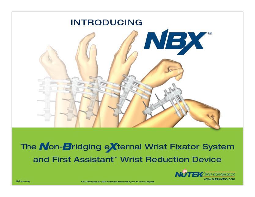
The NBX Non-Bridging External Fixator A Non-Bridging External Fixator/Locking Plate capturing a series of.062mm K-wires and 3mm half-pins that are
|
|
|
- Claud Jefferson
- 5 years ago
- Views:
Transcription
1

2 The NBX Non-Bridging External Fixator A Non-Bridging External Fixator/Locking Plate capturing a series of.062mm K-wires and 3mm half-pins that are inserted in a multiplanar and multi-directional fashion providing an interlocking matrix securing ALL fracture fragments.
3
4 The First Assistant Wrist Reduction Device Two radiolucent plastic paddles or arms (A) that articulate at the wrist joint and allow for ulnar deviation and ligamentotaxis of the wrist, coupled with an ulnar buttress post (B) and volar lift arm (C) that helps recreate and maintain anatomic reduction of the wrist fracture.
5 Patient Positioning The patient can be positioned one of two ways: lateral with the operative arm up and extended off the table or in a beach chair position with the operative side and arm at/over the edge of the table The key is make sure that the forearm is in a neutral position The surgeon can then simply place a sterile drape over the c-arm and use it as the table or use a hand table and bring the c-arm in as needed.

6 Assemble the First Assistant fracture reduction device by threading the center post through the hinge, leaving the connection loose enough to manipulate both paddles. The teeth on the underside and top side of the paddles should engage loosely.

7 Position the hand on the First Assistant with the radial side away from the center post and lift arm beneath the shaft of the distal radius (A) proximal to the fracture for a Colles Fracture and underneath the fracture for a Smith s fracture. Secure the forearm and hand to the two arms of the reduction device with Coban, making sure to capture the base of the thumb (B).

8 Smith s Fracture Colle s Fracture
 and inclination (22-24 degrees).")
9 Reduce the fracture by ligamentotaxis, pulling the hand into ulnar deviation to re-establish radial height (9mm +/- 2mm) and inclination (22-24 degrees).

10 Secure the reduction in ulnar deviation by tightening the TOP knob to lock the arms of the First Assistant in place.

11 Turn the lower knob on the center post clockwise to elevate the distal end of the radial shaft proximal to the fracture for a Colles fracture, and the distal portion of the radius in a Smith s fracture, to restore volar tilt.

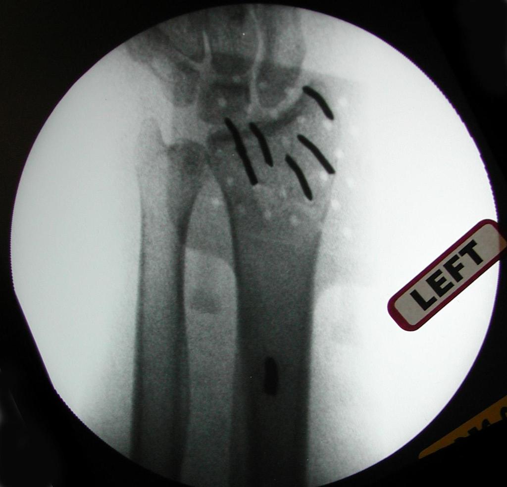
12 Full Reduction AP View

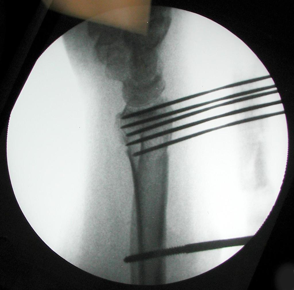
13 Full Reduction Lateral View
14 X-Ray Tips Remember the anatomy and orient the fluoroscopy accordingly: Volar tilt averages degrees Radial inclination averages degrees Radial height averages 9mm +/- 2mm
, making sure that the proximal arm is centered over the shaft of the radius")
15 Position the NBX fixator body over the distal radius to act as a template for the insertion of all K-wires and half-pins, with the distal row of holes over the radial styloid and parallel to the joint covering the fracture site (A), making sure that the proximal arm is centered over the shaft of the radius (B),

16 Place the first.062 k-wire into the ulnar side of the distal radius, taking care not to over penetrate the volar cortex. With the distal row of holes over the radial styloid, this first k-wire will go into either the second or third row of holes.
 at this point to secure the arm of the fixator to the shaft of the radius, and this is strongly")
17 It is critical to ensure that the proximal arm of the fixator is centered over the shaft of the radius (A) before placing the second.062 k-wire into the radial styloid (B). Some surgeons may elect to place the proximal most 3mm halfpin (C) at this point to secure the arm of the fixator to the shaft of the radius, and this is strongly recommended.

18 Place additional.062 k-wires into remaining fracture fragments as desired.
19
20

21 Raise the NBX fixator ½ to ¾ off of the skin to allow clearance for wound and pin site care.

22 Tighten both locking screws on either side of the fixator body to secure the dorsal pins to the NBX fixator. In tightening these 2 screws, the surgeon is pushing the internal plate forward within the fixator.
 Insert two or three.")
23 (A) Slide the side clamp onto the radial side of the NBX fixator. The side clamp can be moved proximally or distally before final locking to the fixator once the radial pins are placed and captured by the pin clamps (B) Insert two or three.062 k-wires from radial to ulnar, with the first parallel to the distal articular surface and the others into fracture fragments as desired to interlock with the dorsal pins and secure the fracture in 3 dimensions. TAKE CARE TO AVOID THE RADIAL NERVE
 Slide each pin clamp into the slot on the side clamp from volar to dorsal and drop it down onto the k-wire.")
24 (A) Loosen each of the appropriate locking screws on the side of the side clamp to allow the pin clamps to slide easily into the side clamp. (B) Slide each pin clamp into the slot on the side clamp from volar to dorsal and drop it down onto the k-wire. (C) Secure each radial k-wire to a pin clamp with the pin clamp nut and wrench, making sure that the shaft of the pin clamp is in either the proximal or distal slot of the side clamp and that the shaft of the pin clamp is not bent in any plane.

25 Secure the pin clamps to the side clamp with the wrench by tightening each locking screw, making sure that the shaft of the pin clamp is in either the proximal or distal portion of the hole in the side clamp and not bent in any plane.

26 Secure the side clamp to the NBX fixator body by tightening the dorsal locking screw in the radial side.
.")
27 Remove the foam piece holding the proximal collet in place and insert the proximal 3mm half-pin into the distraction pin collet, with the pin collet in the distal end of the slot (A) to allow for final distraction of the fracture, and through the radial shaft taking care not to over penetrate the volar cortex (B). DO NOT USE POWER UNTIL THE THREADED PORTION OF THE HALF-PIN IS FULLY THROUGH THE COLLET AND ON BONE AS IT MAY CATCH ON THE COLLET (A small stab incision is normally made and blunt dissection with a small clamp down to bone for all 3mm hal-pins to protect neuro-vascular structures, with a suture on either side of each pin.)
.")
28 Apply the distraction device to the proximal 3mm half-pin, with both arms around the pin above and below the arm of the fixator and distraction screw at the distal end of the fixator (A). Turn the distraction screw clockwise (B) to force the fixator body and secured fracture complex distally (C) to restore normal length.

29 With the final distraction held in place by the distraction device, insert a second 3mm half-pin into the distal radius through one of the two remaining holes on the shaft of the NBX fixator arm. DO NOT USE POWER UNTIL THE THREADED PORTION OF THE HALF- PIN IS FULLY THROUGH THE COLLET AND ON BONE AS IT MAY CATCH ON THE COLLET

30 Tighten the pin collet for the distal 3mm half-pin clockwise to secure the distraction.

31 Remove the distraction device and secure the proximal 3mm halfpin by tightening the pin collet clockwise.

32 Cut the radial k-wires and apply pin covers, then cut the dorsal pins and secure the pin cover by tightening the locking screw on the ulnar dorsal side of the NBX fixator.

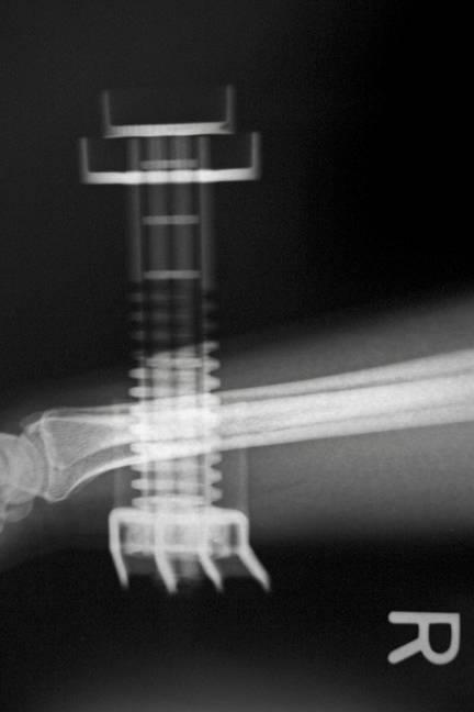
33 Installation of the NBX fixator is complete.

34 Prior to waking the patient up, and after removing the arm from the First Assistant, the surgeon MUST place the wrist through a full range of motion: Volar and dorsal felxion Radial and Ulnar Deviation Pronation and Supination Have the surgeon save and print copies like the 2 below to show to the patient in Recovery to implant the idea that they can move their hand, fingers and wrist!

35 By placing the hand, wrist and fingers through the entire range of motion, the surgeon will free up any extensor tendons that may have been speared by a K-wire. The fibers of the tendons run longitudinally and the range of motion creates a split in line with the fibers that allows full range of motion and heals following removal of the NBX fixator. In well over 140 cases done to date, there has not been a single report of tendon rupture or tenosynovitis.
36 Post-Operative Care Normal closure and dressing will usually consist of a stitch on either side of the proximal half-pins and sterile dressing wrapped under the fixator around all pins and K-wires, with cast padding wrapped around the arm, finally covered with an Ace bandage. Patient is advised to move the fingers and wrist as tolerated. The first post-operative visit usually occurs from 3 to 5 days after surgery, at which time the above dressing and stitches are removed. Patient is instructed in pin tract cleaning and care ( from soap and water to peroxide, etc., based on surgeon preference) and is informed they can shower with the device and resume activities of daily living, with NO heavy lifting or undue stress to be placed on the wrist. Depending on radiographic evidence of healing, the NBX fixator is removed from 6-8 weeks after surgery.
Visualize, stabilize, mobilize. Wristore * Distal Radius Fracture Fixator Abbreviated Surgical Technique
 Visualize, stabilize, mobilize Wristore * Distal Radius Fracture Fixator Abbreviated Surgical Technique Wristore Distal Radius Fracture Fixator 1 Pin Placement Identify anatomy and make a direct (open)
AcUMEDr. FoREARM ROD SYSTEM
 AcUMEDr FoREARM ROD SYSTEM FoREARM ROD SYSTEM Since 1988 Acumed has been designing solutions to the demanding situations facing orthopedic surgeons, hospitals and their patients. Our strategy has been
Distal Radius Plate Instrument and Implant Set. Discontinued December 2017 DSUS/TRM/0916/1063(1)
") Distal Radius Plate Instrument and Implant Set Surgical Technique Discontinued December 2017 DSUS/TRM/0916/1063(1) The Distal Radius Plates Indications For fixation of fractures and osteotomies, including
Distal Radius Plate Instrument and Implant Set Surgical Technique Discontinued December 2017 DSUS/TRM/0916/1063(1) The Distal Radius Plates Indications For fixation of fractures and osteotomies, including
Technique Guide. Adjustable Distal Radius Fixator. Part of the Synthes External Fixation Systems.
 Technique Guide Adjustable Distal Radius Fixator. Part of the Synthes External Fixation Systems. Table of Contents Introduction Adjustable Distal Radius Fixator 2 Indications 3 Surgical Technique Configure
WINSTA-R. Distal Radius System
 Distal Radius System Table of Contents Introduction WINSTA-R System 2 Indication 2 Surgical Technique Palmar Access for Radius Plate 3 Dorsal Access for Radius Plate 3 Positioning of the Radius Plate
Surgical Technique Guide
 Surgical Technique Guide Minimally Invasive, Intramedullary Device For Distal Radius Fragility Fractures The Sonoma WRx Wrist Fracture Repair Device is flexible, inserting easily through a small incision
Acu-Loc Wrist Spanning Plate System. Surgical Technique
 Acu-Loc Wrist Spanning Plate System Surgical Technique Acumed is a global leader of innovative orthopaedic and medical solutions. We are dedicated to developing products, service methods, and approaches
QUICK REFERENCE GUIDE. The Pennig Dynamic Wrist Fixator. Part A: Trans-articular application
 10 The Pennig Dynamic Wrist Fixator Part A: Trans-articular application B1 B2 B3 III IV TRANS-ARTICULAR APPLICATION The fractures that can be treated with this technique include AO type B and C fractures,
Surgical Technique. Forearm Fracture Solutions
 Surgical Technique Forearm Fracture Solutions Acumed is a global leader of innovative orthopaedic and medical solutions. We are dedicated to developing products, service methods, and approaches that improve
System. Humeral Nail. Surgical Technique
 System Humeral Nail Surgical Technique Contents IMPLANT FEATURES 2 1. INDICATIONS 3 2. PRE-OPERATIVE PLANNING 3 3. PATIENT POSITIONING & FRACTURE REDUCTION 3 4. INCISION 4 5. ENTRY POINT 4-6 6. PROXIMAL
3. PATIENT POSITIONING & FRACTURE REDUCTION 3 8. DISTAL GUIDED LOCKING FOR PROXIMAL NAIL PROXIMAL LOCKING FOR LONG NAIL 13
 Contents IMPLANT FEATURES 2 1. INDICATIONS 3 2. PRE-OPERATIVE PLANNING 3 3. PATIENT POSITIONING & FRACTURE REDUCTION 3 4. INCISION 4 5. ENTRY POINT 4-6 6. PROXIMAL NAIL INSERTION 6-7 7. PROXIMAL LOCKING
MICRONAIL. Intramedullary Distal Radius System SURGICAL TECHNIQUE
 MICRONAIL II Intramedullary Distal Radius System SURGICAL TECHNIQUE Contents Introduction 3 4 Chapter 1 5 Chapter 2 6 Appendix A 18 Appendix B 19 Surgeon Design Team Introduction Product Information Surgical
Zimmer MIS Periarticular 3.5mm Proximal Tibial Locking Plate
 Zimmer MIS Periarticular 3.5mm Proximal Tibial Locking Plate Surgical Technique The Science of the Landscape Zimmer MIS Periarticular 3.5mm Proximal Tibial Locking Plate Surgical Technique 1 Zimmer MIS
AcUMEDr. Arc Wrist Tower. For Wrist Arthroscopy And Fracture Reduction
 AcUMEDr Arc Wrist Tower For Wrist Arthroscopy And Fracture Reduction Arc Wrist Tower Since 1988, Acumed has been designing solutions to the demanding situations facing orthopaedic surgeons, hospitals and
Percutaneous Scaphoid Fixation: A Volar Approach
 Percutaneous Scaphoid Fixation: A Volar Approach Surgical Technique N.J. Goddard FRCS, Consultant Orthopaedic Surgeon Royal Free Hospital Pond Street, London NW3 2QG Introduction Scaphoid fractures are
Distal Radius Plate 2.4/2.7 dorsal and volar
 Distal Radius Plate 2.4/2.7 dorsal and volar Surgical Technique This publication is not intended for distribution in the USA. Instruments and implants approved by the AO Foundation. Distal Radius Plate
Wrist Fixation System
 Wrist Fixation System Anatomy / Fracture Implant EXTRA & SIMPLE ARTICULAR Volar Radius Volar Fixed Angle Plate Volar Bearing Plate Radial Peg Plate Volar Hook Plate Volar Buttress Pin Volar Shear Plate
3.5 MM VA-LCP PROXIMAL TIBIA PLATE SYSTEM
 3.5 MM VA-LCP PROXIMAL TIBIA PLATE SYSTEM Part of the DePuy Synthes Variable Angle Periarticular Plating System SURGICAL TECHNIQUE TABLE OF CONTENTS INTRODUCTION 3.5 mm VA-LCP Proximal Tibial Plate 2 AO
Mini External Fixator.
 Mini External Fixator. Assembly and Surgical Technique This publication is not intended for distribution in the USA. Instruments and implants approved by the AO Foundation. Image intensifier control Warning
Acu-Loc Wrist Plating System. Surgical Technique
 Acu-Loc Wrist Plating System Surgical Technique Acumed is a global leader of innovative orthopaedic and medical solutions. We are dedicated to developing products, service methods, and approaches that
Hoffmann II Compact External Fixation System. Modular System for
 Hoffmann II Compact External Fixation System Modular System for Introduction In 1938, Raoul Hoffmann, a surgeon from Geneva, Switzerland, designed a revolutionary External Fixation System. The basic features
Distal Radius and Distal Ulna Plates System Self-Tapping Spherical Locking Screw Self-Tapping Conical Locking Screw Cortex Screw
 DISTAL RADIUS AND ULNA LOCKING PLATE SYSTEM Surgical Technique Distal Radius and Distal Ulna Plates System Self-Tapping Spherical Locking Screw Self-Tapping Conical Locking Screw Cortex Screw Approved
QUICK REFERENCE GUIDE. The PreFix Fixator (92000 Series) ALWAYS INNOVATING
 ALWAYS INNOVATING") 21 The PreFix Fixator (92000 Series) ALWAYS INNOVATING INTRODUCTION The PreFix fixator is designed to provide temporary external fixation. This may be needed when local facilities or the condition of the
21 The PreFix Fixator (92000 Series) ALWAYS INNOVATING INTRODUCTION The PreFix fixator is designed to provide temporary external fixation. This may be needed when local facilities or the condition of the
Technique Guide. 3.5 mm LCP Low Bend Medial Distal Tibia Plate Aiming Instruments. Part of the 3.5 mm LCP Percutaneous Instrument System.
 Technique Guide 3.5 mm LCP Low Bend Medial Distal Tibia Plate Aiming Instruments. Part of the 3.5 mm LCP Percutaneous Instrument System. Table of Contents Introduction 3.5 mm LCP Low Bend Medial Distal
Technique Guide. 2.4 mm Variable Angle LCP Distal Radius System. For fragment-specific fracture fixation with variable angle locking technology.
 Technique Guide 2.4 mm Variable Angle LCP Distal Radius System. For fragment-specific fracture fixation with variable angle locking technology. Table of Contents Introduction 2.4 mm Variable Angle LCP
Surgical Technique. Distal Radius and Foot
 Surgical Technique Distal Radius and Foot JET-X BAR Unilateral Fixator Distal Radius and Foot Surgical Technique Contents Design Features...2 Distal Radius Surgical Technique Indications...10 Surgical
QUICK REFERENCE GUIDE. MiniRail System. Part B: Foot Applications. By Dr. B. Magnan, Dr. E. Rodriguez and Dr. G. Vito ALWAYS INNOVATING
 14 MiniRail System Part B: Foot Applications By Dr. B. Magnan, Dr. E. Rodriguez and Dr. G. Vito ALWAYS INNOVATING ORDERING INFORMATION Sterilization box, empty M190 Can accommodate: M101 Standard MiniRail
Surgical Technique. CONQUEST FN Femoral Neck Fracture System
 Surgical Technique CONQUEST FN Femoral Neck Fracture System Table of Contents Introduction... 3 Indications... 3 Product Overview... 4 Surgical Technique... 5 Patient Positioning... 5 Reduce the Fracture...
AcUMEDr. Locking Proximal Humeral Plate. PoLARUSr PHPt
 AcUMEDr Locking Proximal Humeral Plate PoLARUSr PHPt PoLARUSr PHPt LOCKING PROXIMAL HUMERAL PLATE Since 1988 Acumed has been designing solutions to the demanding situations facing orthopedic surgeons,
Distal Ulnar Locking Plate
 INDEX Indications Patient Position Surgical Technique - Step 1 Approach - Step 2 Plate Contouring - Step 3 Fracture Reduction - Step 4 Distal Plate Fixation - Step 5 Confirm Proper Reconstruction - Step
Modular Ulnar Head surgical technique. Transforming Extremities
 First Choice Modular Ulnar Head surgical technique Transforming Extremities instrumentation Head and Collar Trials Assembly Pad Starter Awl Trial Extractor Osteotomy Guide Stem Trials Implant Impactor
Surgical Technique. Olecranon Locking Plate
 Surgical Technique Olecranon Locking Plate PERI-LOC Locked Plating System Olecranon Locking Plate Surgical Techniquealog Infor Table of Contents Introduction...2 Indications...3 Plate Features...3 Patient
WINSTA-C. Clavicle Plating System
 Clavicle Plating System Clinical Advisor Michael Kurer FRCS FRCS (Orth) Consultant Orthopaedic and Shoulder Surgeon North Middlesex University Hospital NHS Trust Table of Contents Introduction Indication
Low-Profile Wrist Fixator. For stabilization of fractures of the distal radius.
 Low-Profile Wrist Fixator. For stabilization of fractures of the distal radius. Technique Guide Part of the External Fixation System Low-Profile Wrist Fixator Indications Intended for stabilization of
Olecranon Locking Plate II
 INDEX Indications Patient Position Fracture Reduction and Fixation Surgical Technique Step 1 Surgical Approach Step 2 Implantation Step 3 Proximal Locking Screw Insertion Step 4 Distal Screw Insertion
Surgical Technique. Targeter Systems Overview
 Surgical Technique Targeter Systems Overview PERI-LOC Locked Plating System Targeter Systems Overview Table of contents Product overview... 2 Introduction... 2 Indications... 2 Design features and benefits...
Angle Stable Distal Radial Plate System WINSTA-R
 Angle Stable Distal Radial Plate System WINSTA-R Priv.-Doz.Dr.med. Martin Walz Dr. med. Felix Menzinger Prof.Dr.med. Jürgen Rudigier www.marquardt-medizintechnik.de General The problem posed by metaphyseal
Zimmer MIS Periarticular Distal Femoral Locking Plate
 For Clinical Evaluations Zimmer MIS Periarticular Distal Femoral Locking Plate Surgical Technique The Science of the Landscape Zimmer MIS Periarticular Distal Femoral Locking Plate Surgical Technique
Orthopedic Bone Nail System - Distal Femoral Nail Surgical Technique Manual
 Orthopedic Bone Nail System - Distal Femoral Nail Surgical Technique Manual Note: The surgical procedures should be performed under the guidance of qualified skilled orthopedic surgeons, and this surgical
Forearm Fracture Solutions. Surgical Technique
 Forearm Fracture Solutions Surgical Technique Acumed is a global leader of innovative orthopaedic and medical solutions. We are dedicated to developing products, service methods, and approaches that improve
2.4 mm Variable Angle LCP Volar Extra-Articular Distal Radius System. For fragment-specific fracture fixation with variable angle locking technology.
 Technique Guide 2.4 mm Variable Angle LCP Volar Extra-Articular Distal Radius System. For fragment-specific fracture fixation with variable angle locking technology. Table of Contents Introduction 2.4
MiniRail System. Part B: Foot Applications. By Dr. B. Magnan, Dr. E. Rodriguez and Dr. G. Vito
 Q U I C K R E F E R E N C E G U I D E 14 MiniRail System Part B: Foot Applications By Dr. B. Magnan, Dr. E. Rodriguez and Dr. G. Vito ORDERING INFORMATION MiniRail System Kit, M190C Contents: M 101 Standard
Surgical Technique. 3.5mm and 4.5mm Lateral Proximal Tibia Locking Plates
 Surgical Technique 3.5mm and 4.5mm Lateral Proximal Tibia Locking Plates PERI-LOC Periarticular Locked Plating System 3.5mm and 4.5mm Lateral Proximal Tibia Locking Plate Surgical Technique Contents Product
Zimmer Small Fragment Universal Locking System. Surgical Technique
 Zimmer Small Fragment Universal Locking System Surgical Technique Zimmer Small Fragment Universal Locking System 1 Zimmer Small Fragment Universal Locking System Surgical Technique Table of Contents Introduction
Acu-Loc 2 Wrist Plating System
 Surgical Technique Acu-Loc 2 Wrist Plating System Acumed is a global leader of innovative orthopaedic and medical solutions. We are dedicated to developing products, service methods, and approaches that
Surgical Technique. Wrist Plating System
 Surgical Technique Wrist Plating System Acumed is a global leader of innovative orthopaedic and medical solutions. We are dedicated to developing products, service methods, and approaches that improve
LCP Anterolateral Distal Tibia Plate 3.5. The low profile anatomic fixation system with optimal plate placement and angular stability.
 LCP Anterolateral Distal Tibia Plate 3.5. The low profile anatomic fixation system with optimal plate placement and angular stability. Technique Guide LCP Small Fragment System Table of Contents Introduction
Double Engine Orthopedic Bone Nail System Universal Humeral Nail
 Double Engine Orthopedic Bone Nail System ----------- Universal Humeral Nail Surgical Technique Manual Note: The surgical procedures should be performed under the guidance of qualified skilled orthopedic
Technique Guide. 3.5 mm LCP Olecranon Plates. Part of the Synthes locking compression plate (LCP) system.
 system.") Technique Guide 3.5 mm LCP Olecranon Plates. Part of the Synthes locking compression plate (LCP) system. Table of Contents Introduction 3.5 mm LCP Olecranon Plates 2 AO Principles 3 Indications 3 Clinical
Technique Guide 3.5 mm LCP Olecranon Plates. Part of the Synthes locking compression plate (LCP) system. Table of Contents Introduction 3.5 mm LCP Olecranon Plates 2 AO Principles 3 Indications 3 Clinical
Locking Radial Head Plates
 Locking Radial Head Plates Locking Radial Head Plates Since 1988, Acumed has been designing solutions to the demanding situations facing orthopaedic surgeons, hospitals and their patients. Our strategy
Periarticular Aiming Arm Instruments for LCP Proximal Tibial Plate 4.5/5.0. Part of the LCP Periarticular Aiming Arm Instrument System (large).
.") Technique Guide Periarticular Aiming Arm Instruments for LCP Proximal Tibial Plate 4.5/5.0. Part of the LCP Periarticular Aiming Arm Instrument System (large). Image intensifier control Warning This description
Technique Guide Periarticular Aiming Arm Instruments for LCP Proximal Tibial Plate 4.5/5.0. Part of the LCP Periarticular Aiming Arm Instrument System (large). Image intensifier control Warning This description
LCP Anterolateral Distal Tibia Plate 3.5. The low profile anatomic fixation system with optimal plate placement and angular stability.
 LCP Anterolateral Distal Tibia Plate 3.5. The low profile anatomic fixation system with optimal plate placement and angular stability. Technique Guide LCP Small Fragment System Table of Contents Introduction
Conventus CAGE PH Surgical Techniques
 Conventus CAGE PH Surgical Techniques Conventus Orthopaedics The Conventus CAGE PH (PH Cage) is a permanent implant comprised of an expandable scaffold, made from nitinol and titanium, which is deployed
LCP Medial Distal Tibia Plate, without Tab. The Low Profile Anatomic Fixation System with Angular Stability and Optimal Screw Orientation.
 LCP Medial Distal Tibia Plate, without Tab. The Low Profile Anatomic Fixation System with Angular Stability and Optimal Screw Orientation. Technique Guide LCP Small Fragment System Table of Contents Introduction
PROXIMAL TIBIAL PLATE
 SURGICAL NÁSTROJE TECHNIQUE PRO ARTROSKOPII PROXIMAL INSTRUMENTS TIBIAL FOR PLATE ARTHROSCOPY Proximal Tibial Plate Description of medical device The Proximal Tibial Plate is used in epyphyseal and metaphyseal
External Distal Radius Fixator. Supplement to the 8 mm rod fixator system
 External Distal Radius Fixator. Supplement to the 8 mm rod fixator system Surgical technique This publication is not intended for distribution in the USA. Instruments and implants approved by the AO Foundation
A Patient s Guide to Adult Distal Radius (Wrist) Fractures
 Fractures") A Patient s Guide to Adult Distal Radius (Wrist) Fractures Suite 11-13/14/15 Mount Elizabeth Medical Center 3 Mount Elizabeth Singapore, 228510 Phone: (65) 6738 2628 Fax: (65) 6738 2629 1 DISCLAIMER: The
A Patient s Guide to Adult Distal Radius (Wrist) Fractures Suite 11-13/14/15 Mount Elizabeth Medical Center 3 Mount Elizabeth Singapore, 228510 Phone: (65) 6738 2628 Fax: (65) 6738 2629 1 DISCLAIMER: The
TABLE OF CONTENTS. 2 (8144 Rev 2)
") 1 (8144 Rev 2) TABLE OF CONTENTS Introduction Conventus CAGE TM - Proximal Humerus...3 Indications and Contraindications...4 Surgical Summary...5 Patient Positioning & Approach...6 Surgical Technique Plate
1 (8144 Rev 2) TABLE OF CONTENTS Introduction Conventus CAGE TM - Proximal Humerus...3 Indications and Contraindications...4 Surgical Summary...5 Patient Positioning & Approach...6 Surgical Technique Plate
Acu-Loc 2 Wrist Plating System. Surgical Technique
 Acu-Loc 2 Wrist Plating System Surgical Technique Acumed is a global leader of innovative orthopaedic and medical solutions. We are dedicated to developing products, service methods, and approaches that
DFS Hip Distractor. Surgical Technique
 DFS Hip Distractor Surgical Technique Contents Introduction... Page 1 Hip Distractor Associated... Page 2 Components And Instrumentation Radiographs... Page 4 Fixator Orientation... Page 5 Initial Screw
Technique Guide. LCP Proximal Femoral Hook Plate 4.5/5.0. Part of the LCP Periarticular Plating System.
 Technique Guide LCP Proximal Femoral Hook Plate 4.5/5.0. Part of the LCP Periarticular Plating System. Table of Contents Introduction Features and Benefits 2 AO ASIF Principles 4 Indications 5 Surgical
Mark VanDer Kaag 1, Ajmal Ikram 2. Hand Unit, Tygerberg Hospital University of Stellenbosch
 A Prospective, Randomized Controlled Study To Determine The Radiological And Functional Outcomes Of IMN Fixation Of Distal Radius Fractures Using A Novel Device The Sonoma Wrx Distal Radius Nail Compared
Polarus 3 Solution Plates and Nails
 Surgical Technique Polarus 3 Solution Plates and Nails Acumed is a global leader of innovative orthopaedic and medical solutions. We are dedicated to developing products, service methods, and approaches
Small External Fixator Nonspanning Wrist Frame. For the treatment of wrist fractures.
 Small External Fixator Nonspanning Wrist Frame. For the treatment of wrist fractures. Technique Guide Part of the Small External Fixation System Small External Fixator Nonspanning Wrist Frame When to use
SURGICAL TECHNIQUE GUIDE
 SURGICAL TECHNIQUE GUIDE As described by: Jorge L. Orbay, M.D. Miami Hand & Upper Extremity Institute Miami, Florida. 1 ELBOW LANDMARKS With the elbow flexed 90 0, palpate and mark the lateral epicondyle.
Zimmer Trabecular Metal Ankle Interpositional Spacer and Trabecular Metal Ankle Fusion Spacer
 Zimmer Trabecular Metal Ankle Interpositional Spacer and Trabecular Metal Ankle Fusion Spacer Surgical Technique 2 Zimmer Trabecular Metal Ankle Interpositional Spacer and Trabecular Metal Ankle Fusion
M.I.S. MAKE IT SMART IN ONE SYSTEM. Surgical Technique. Hip Knee Spine Navigation
 M.I.S. MAKE IT SMART IN ONE SYSTEM Surgical Technique Hip Knee Spine Navigation M.U.S.T. Mini Open Surgical Technique Hip Knee Spine Navigation 2 C O N T E N T S 1 INTRODUCTION 4 2 SURGICAL TECHNIQUE 5
LCP Proximal Radius Plates 2.4. Plates for radial head rim and for radial head neck address individual fracture patterns of the proximal radius.
 Technique Guide LCP Proximal Radius Plates 2.4. Plates for radial head rim and for radial head neck address individual fracture patterns of the proximal radius. Table of Contents Introduction LCP Proximal
Pre-Operative Planning. Positioning of the Patient
 Surgical Technique Pre-Operative Planning Decide upon the size and angle of the barrel plate to be used from measuring the x-rays. To maximise the sliding action when using shorter lag screws, the Short
Long Volar Plates for Diaphyseal-Metaphyseal Radius Fractures LCP. Dia-Meta Volar Distal Radius Plates. Surgical Technique
 Long Volar Plates for Diaphyseal-Metaphyseal Radius Fractures LCP Dia-Meta Volar Distal Radius Plates Surgical Technique Table of Contents Introduction LCP Dia-Meta Volar Distal Radius Plates 2 AO Principles
Surgical Technique. Proximal Humerus Locking Plate
 Surgical Technique Proximal Humerus Locking Plate PERI-LOC Upper Extremity Locked Plating System 3.5mm & 4.5mm Proximal Humerus Locking PlatesCatalog Infor Table of Contents Introduction.........................................................2
Surgical Technique. Clavicle Locking Plate
 Surgical Technique Clavicle Locking Plate PERI-LOC Locked Plating System Clavicle Locking Plate Surgical Technique Table of Contents Introduction...2 Indications...3 Plate Features...3 Patient Positioning...4
Surgical Technique Carpal Fusion
 Carpal Fusion Patent and Patent Pending CAUTION: Federal Law (USA) restricts this device to sale by or on the order of a physician. INDICATIONS FOR USE The Extremity Medical Lag Screw and X-Post System
Olecranon Osteotomy Nail. For simple fractures and osteotomies of the olecranon.
 Olecranon Osteotomy Nail. For simple fractures and osteotomies of the olecranon. Technique Guide Discontinued June 2016; AVAILABLE FOR IMPLANT REMOVAL PURPOSES ONLY DSEM/TRM/0517/0843 Table of Contents
Early Elbow Motion Protocol Ligament Repair of the elbow
 499 Blossom Hill Rd, San Jose, Ca 95123 Tel: 408-268-8536 Fax: 408-268-8727 www.handsoncaretherapy.com Early Elbow Motion Protocol Ligament Repair of the elbow EARLY MOTION PROTOCOL 1-3 DAYS POST OP LIGAMENT
Instrument and Implant for wrist fracture
 Instrument and Implant for wrist fracture Jansri Janpanya Product specialist The Bangkok Unitrade Co,.ltd. Objectives Type of LCP for distal radius Fx. The new LCP design for distal radius Fx. Have knowledge
VariAx TM Distal Radius Locking Plate System
 Osteosynthesis VariAx TM Distal Radius Locking Plate System Operative Technique Anatomical & Universal Volar Plates Dorsal Plates Fragment Specific Plates Introduction -15 +15 The NEW VariAx Distal Radius
Small External Fixator
 Surgical Technique Original Instruments and Implants of the Association for the Study of Internal Fixation AO/ASIF Table of Contents Description of System 2 Indications/ Contraindications 3 Bridging Surgical
Polarus 3 Solution Plates and Nails. Surgical Technique 4.3. Screws
 Polarus 3 Solution Plates and Nails Surgical Technique 4.3 mm Screws Acumed is a global leader of innovative orthopaedic and medical solutions. We are dedicated to developing products, service methods,
OPERATIVE TECHNIQUE GALAXY FIXATION SHOULDER
 OPERATIVE TECHNIQUE GALAXY FIXATION SHOULDER cop2 OPERATIVE TECHNIQUE INTRODUCTION 1 FEATURES OF SHOULDER COMPONENTS 2 EQUIPMENT REQUIRED 5 PREOPERATIVE PLANNING 6 SURGICAL PROCEDURE 8 POST OPERATIVE MANAGEMENT
3. Insert Tocar Sleeves Insert the NCB tissue protection sleeve assembly 1.6 to 10mm through a skin incision (Fig. 38).
.") NCB Proximal Humerus Plating System Surgical Technique 19 2. Temporary Plate Fixation The plate can be temporary fixed to the bone with 1.6mm K-wire through the proximal cannulated fixation screw of the
NCB Proximal Humerus Plating System Surgical Technique 19 2. Temporary Plate Fixation The plate can be temporary fixed to the bone with 1.6mm K-wire through the proximal cannulated fixation screw of the
Choice, Simplicity, Transition. TransFx External Fixation System Small and Mini Surgical Technique
 Choice, Simplicity, Transition TransFx External Fixation System Small and Mini Surgical Technique TransFx External Fixation System Small and Mini Surgical Technique 1 Surgical Technique For TransFx External
AcUMEDr. LoCKING CLAVICLE PLATE SYSTEM
 AcUMEDr LoCKING CLAVICLE PLATE SYSTEM LoCKING CLAVICLE PLATE SYSTEM Since 1988 Acumed has been designing solutions to the demanding situations facing orthopedic surgeons, hospitals and their patients.
Agee WristJack Multiplanar Ligamentotaxis
 Agee WristJack Multiplanar Ligamentotaxis Fracture Reduction System Fracture reduction and external fixation for treatment of distal radius fractures Surgeon's Manual Hand Biomechanics Lab Restoring Length
Zimmer Natural Nail System
 Zimmer Natural Nail System Antegrade Femoral Nail Surgical Technique (Piriformis Fossa & Greater Trochanteric Approaches) Zimmer Natural Nail System Antegrade Femoral Surgical Technique 1 Zimmer Natural
SURGICAL TECHNIQUE GUIDE
 DANGER indicates an imminently hazardous situation which, if not avoided, will result in death or serious injury. WARNING indicates a potentially hazardous situation which, if not avoided, could result
Technique Guide. *smith&nephew SPEEDSCREW Fully Threaded Knotless Implant
 Technique Guide *smith&nephew SPEEDSCREW Fully Threaded Knotless Implant SPEEDSCREW system Fully threaded knotless implant system The SPEEDSCREW system is specifically designed for rotator cuff repair
Surgical Technique. Calcaneal Locking Plate
 Surgical Technique Calcaneal Locking Plate PERI-LOC Locked Plating System Calcaneal Locking Plate Surgical TechniqueCatalog Infor Table of Contents Introduction...2 Indications...3 Plate Features...3 Patient
Surgical Technique. Distal Humerus Locking Plate
 Surgical Technique Distal Humerus Locking Plate PERI-LOC Locked Plating System Distal Humerus Locking Plate Surgical Technique Table of Contents Introduction...2 Indications...3 Plate Features...3 Patient
Humerus Block. Discontinued December 2016 DSEM/TRM/0115/0296(1) Surgical Technique. This publication is not intended for distribution in the USA.
 Surgical Technique. This publication is not intended for distribution in the USA.") Humerus Block Surgical Technique Discontinued December 2016 DSEM/TRM/0115/0296(1) This publication is not intended for distribution in the USA. Instruments and implants approved by the AO Foundation. Contents
Humerus Block Surgical Technique Discontinued December 2016 DSEM/TRM/0115/0296(1) This publication is not intended for distribution in the USA. Instruments and implants approved by the AO Foundation. Contents
Surgical Technique International Version. Clavicle Locking Plate
 Surgical Technique International Version Clavicle Locking Plate PERI-LOC Upper Extremity Locked Plating System Clavicle Surgical Techniquefor Table of Contents Introduction........................................................2
A N D R E W I R W I N, F R C S E D ( O R T H ) C O N S U L T A N T O R T H O P A E D I C S U R G E O N W E S T H E R T S N H S T R U S T, U K
 C O N S U L T A N T O R T H O P A E D I C S U R G E O N W E S T H E R T S N H S T R U S T, U K") Wrist Fractures A N D R E W I R W I N, F R C S E D ( O R T H ) C O N S U L T A N T O R T H O P A E D I C S U R G E O N W E S T H E R T S N H S T R U S T, U K St Albans Abbey, 1077 onwards Watford General
Wrist Fractures A N D R E W I R W I N, F R C S E D ( O R T H ) C O N S U L T A N T O R T H O P A E D I C S U R G E O N W E S T H E R T S N H S T R U S T, U K St Albans Abbey, 1077 onwards Watford General
Imola Lateral IBF System Surgical Technique
 Imola Lateral IBF System Surgical Technique IMOLA CIRCUIT TABLE OF CONTENTS Design Rationale Instructions for Use Surgical Technique 1. Table Mounting 2. Surgical Planning & Targeting 3. Access and Preparation
Technique Guide. 3.5 mm LCP Periarticular Proximal Humerus Plate. Part of the Synthes locking compression plate (LCP) system.
 system.") Technique Guide 3.5 mm LCP Periarticular Proximal Humerus Plate. Part of the Synthes locking compression plate (LCP) system. Table of Contents Introduction 3.5 mm LCP Proximal Humerus Plate 2 AO Principles
Technique Guide 3.5 mm LCP Periarticular Proximal Humerus Plate. Part of the Synthes locking compression plate (LCP) system. Table of Contents Introduction 3.5 mm LCP Proximal Humerus Plate 2 AO Principles
Complications of Distal Radius Fractures. How to Treat a Distal Radius Fx 11/13/2017. Michael S. Bednar, M.D. Loyola University Chicago
 Complications of Distal Radius Fractures Michael S. Bednar, M.D. Loyola University Chicago How to Treat a Distal Radius Fx Need to restore motion, begin with uninvolved parts Need to reduce an unreduced
Extron External Fixator
 Operative Technique Extron External Fixator The disposable set for a Distal Radius Fracture The EXTRON-External Fixator made by tantum provides you with a new generation of supply engineering for distal
2.7 mm/3.5 mm Variable Angle LCP Elbow System DJ9257-B 1
 2.7 mm/3.5 mm Variable Angle LCP Elbow System DJ9257-B 1 System overview Simply complete: A comprehensive system, consisting of five (5) distal humerus plates and three (3) types of olecranon plates Implant
Elbow Hinge Fixator. Guided Flexion/Extension for Unstable Elbow Fractures.
 Elbow Hinge Fixator. Guided Flexion/Extension for Unstable Elbow Fractures. Surgical Technique MR Safe Radiolucent Table of Contents System Description 3 Indications and Contraindications 4 Fixation Components
Surgical Technique. Anterolateral and Medial Distal Tibia Locking Plates
 Surgical Technique Anterolateral and Medial Distal Tibia Locking Plates PERI-LOC Periarticular Locked Plating System Anterolateral and Medial Distal Tibia Locking Plates Surgical Technique Contents Product
DFS STANDARD FIXATOR DFS ANKLE CLAMP DFS T-CLAMP
 DFS STANDAD FIXATO DFS ANKLE CLAMP DFS T-CLAMP SUGICAL TECHNIQUE Dr. James V. Nepola Professor of Orthopaedics University of Iowa Hospitals and Clinics Iowa City, Iowa Patent No. 5,662,650 C ontents DynaFix