Bone grafting developments used in veterinary orthopaedics part two
|
|
|
- Gilbert Stafford
- 5 years ago
- Views:
Transcription
1 Vet Times The website for the veterinary profession Bone grafting developments used in veterinary orthopaedics part two Author : John Innes, Peter Myint Categories : Vets Date : September 7, 2009 JOHN INNES and PETER MYINT illustrate how advanced technology allows veterinary surgeons to incorporate bone grafting in their treatment of fractures and arthrodeses In the first part (VT 39.31) of this two-part article, the authors outlined the various allografts available for bone grafting in dogs, including demineralised bone matrix (DBM), cancellous chips, cancellous blocks and dowels, and cortical sections and struts. In this second part, case studies describe where these products have been useful in promoting bone healing. Cruciate ligament rupture A three-year old Labrador presented with unilateral cranial cruciate ligament rupture. The surgeon elected to perform tibial tuberosity advancement (TTA). Pre-operative planning An extended 135 mediolateral radiograph of the stifle was taken for implant sizing. The TTA template (Kyon, Switzerland) was then used to calculate the advancement distance to produce a patella tendon/tibial plateau angle of 90 at full extension ( Figure 1). A line was drawn tangentially to the tibial plateau and the template was adjusted until a line perpendicular to the tangent intersected with the cranial border of the patella tendon origin. The distance of advancement was read from the template this equated to the required cage size. 1 / 19
2 The template was also used to calculate the maximum size of the plate that could be attached to the tibial tuberosity. Sequential plate transparencies were offered up to the radiograph until the maximum plate size was determined. Patient preparation A standard surgical clip was performed from the hip to just above the tarsus. The foot was covered with an impermeable barrier, and the dog positioned in dorsolateral recumbency with the affected limb on the lower side. The foot was suspended from a stand that was positioned on the animal s dorsal side. Fourquarter draping with an additional impermeable layer was used and an adhesive, transparent incise drape was placed over the exposed skin. Surgical approach A medial approach to the stifle and proximal tibia was performed, followed by medial arthrotomy to assess meniscal damage. Using the eight-hole drill guide, the required number of holes were predrilled into the tuberosity using a 2.0mm drill bit. A partial curvilinear tibial tuberosity osteotomy was performed using an oscillating saw although the proximal aspect of the tuberosity was not completely cut until the plate was secured in place. Implant placement A fork of appropriate size was inserted into the plate, which was then hammered on to the tuberosity using a fork inserter. Once the osteotomy was complete the fragment was advanced using a spreader. The appropriate size cage was attached to the main body of the tibia using a 2.4mm self-tapping screw (Figure 2). With the stifle in full flexion, the tuberosity was compressed distally against the tibia, using the cage as a fulcrum, and secured with boneholding forceps. The plate was then secured to the tibia with two self-tapping screws (2.7 mm diameter for plates with two to five holes, and 3.5mm for plates with six holes or more). The cranial part of the cage was attached to the tuberosity, above the level of the plate, using a 2.4mm screw. Bone grafting Grafting the osteotomy gap with allograft was performed as recommended for this technique. A 3cc vial of freeze-dried canine cancellous chips (2-4mm;Veterinary Tissue Bank, Wrexham) was opened and rehydrated with sterile Hartmann s solution in a Galli pot. The graft was packed into the void created by the advancement osteotomy to provide an osteoconductive scaffold for bone 2 / 19
3 healing with concomitant mechanical support during the healing process. Closure of the surgical site was routine. Follow-up At a routine eightweek postoperative check, healing of the osteotomy was noted on radiography of the stifle joint. Lameness was improved and cranial tibial thrust in the stifle joint was eliminated. Bilateral palmar ligament rupture This five-year old bearded collie presented with bilateral palmar ligament rupture, having fallen over a 3m high wall. The dog exhibited a bilaterally palmigrade stance and radiographs confirmed rupture of the palmar supporting ligaments of the carpus at all three joint levels, necessitating bilateral pancarpal arthrodesis. Patient preparation Both limbs were clipped from just below the elbow joint to the level of the main carpal pad. An impervious barrier was taped in position over the toes. The patient was positioned in dorsal recumbency with the operative limbs retracted caudally. The limbs were freedraped and a sterile impervious drape wrapped over the foot (Figure 3). Sterilised cohes ive dressing was then tightly wrapped around the foot and progressed proximally to act as an Esmarch bandage;at the level of the proximal antebrachium, the dressing was twisted and used as a tourniquet. Because allograft was to be used, there was no need to clip and prepare the proximal limbs, saving considerable time. Surgical approach The surgical technique was similar for both limbs. A skin incision was made through the cohesive dressing on the dorsal aspect of the carpus from the distal third of the antebrachium to the distal third of the third metacarpal bone. Care was taken to avoid the cephalic vein. The subcuticular tissues were then stapled to the cohesive dressing on each side of the incision to isolate the wound. Subcutaneous tissues were incised and the extensor carpi radialis (ECR) tendon of insertion identified on the medial aspect of the carpus. Lateral to this, the common tendon of the digital extensor (CDE) muscle was identified and protected. Fascia between the ECR tendon and the CDE tendon was incised and the CDE tendon retracted 3 / 19
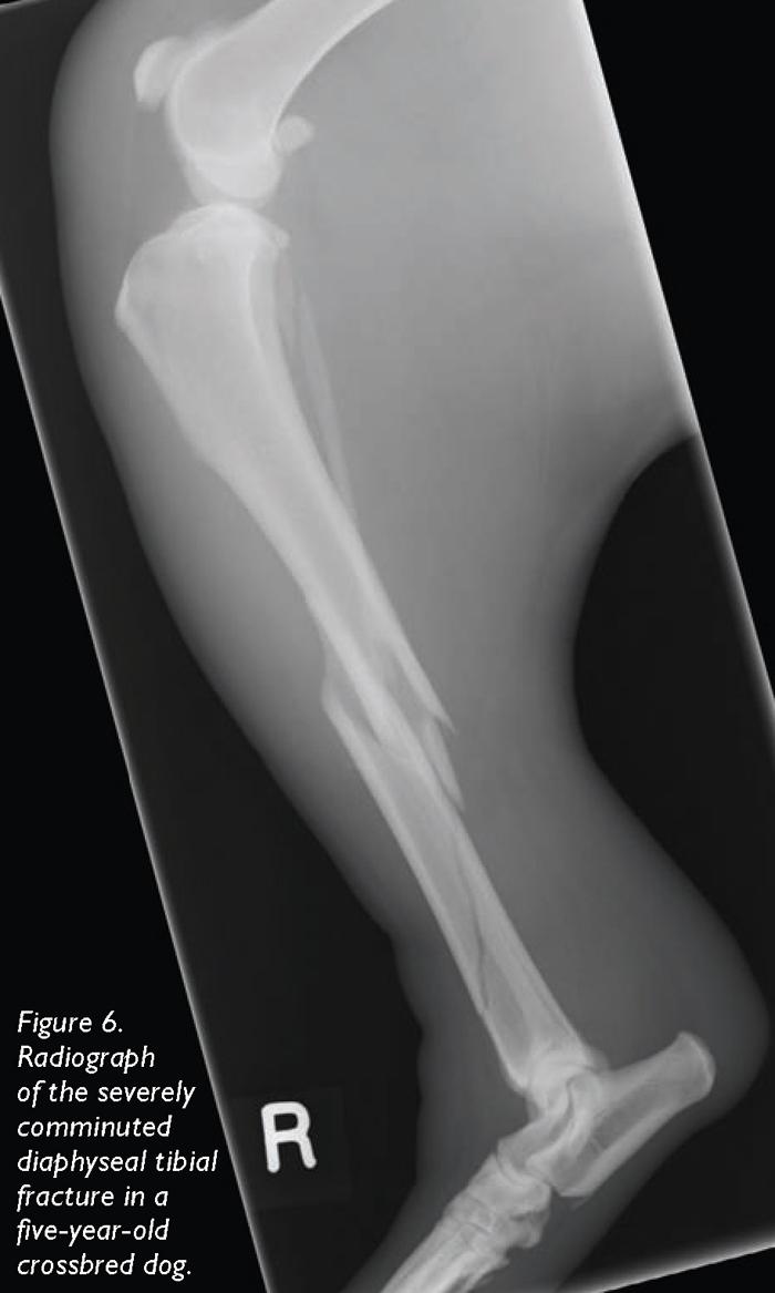
4 laterally. The joint capsule at all three joint levels was incised and the carpus flexed to open the joint space. Articular cartilage was removed using a combination of sharp dissection and curettage with manual and powered instruments. The subchondral plate of the radius was penetrated with two to three small drill holes to encourage mesenchymal cell migration. Bone graft preparation A 3cc vial of demineralized bone matrix (DBM;Veterinary Tissue Bank, Wrexham;Hoffer, Griffon et al, 2008) for osteoinduction, was mixed with a 3cc vial of freeze-dried canine cancellous chips (0.1-1mm;Veterinary Tissue Bank, Wrexham) and rehydrated with sterile Hartmann s solution in a Galli pot. This provided enough graft for both carpi. Implant placement A 3.5mm/2.7mm pancarpal arthrodesis plate (Veterinary Instrumentation, Sheffield) was positioned such that a central screw hole was over the radial carpal bone. The screw was placed and then the most proximal and distal screw holes were filled with appropriate screws. The screws and plate were carefully removed and the allograft, mixed with DBM, was packed in all the joint spaces. The plate was replaced and the screws re-inserted before inserting all remaining screws. Closure was routine and postoperative radiography revealed satisfactory placement of all implants (Figure 4). Each distal limb was then placed in Robert Jones dressings for 48 hours prior to external coaptation for six weeks. Radiographic union of the arthrodesis was noted at routine radiography eight weeks postoperatively (Figure 5). Comminuted tibial fracture The five-year-old German shepherd crossbreed dog was involved in a road traffic accident and sustained a closed, comminuted fracture of the right tibial diaphysis with severe bruising of the soft tissues (Figure 6). There were no other significant injuries. The fracture was considered reconstructable and neutralisation plate and screw fixation was planned. 4 / 19
5 As is policy for all diaphyseal fractures in skeletally mature dogs, bone grafting of the fracture site was planned. Patient preparation Following the trauma, the dog was stabilised overnight and was then prepared for surgery to take place the next day. Following induction of anaesthesia, an epidural anaesthetic was administered using a combination of morphine and bupivicaine. The right pelvic limb was clipped and prepared from the level of the proximal femur to the main pad of the pes. The dog was placed in right lateral recumbency for surgery via a medial approach to the right tibia. Surgical approach A craniomedial incision over the tibia was made and the underlying fascia incised. The saphenous vein was preserved. A combination of lag screws and cerclage wire were used to reconstruct the distal fragment. A 14-hole broad 3.5 DCP was contoured to the craniomedial aspect of the tibia and applied to the bone to fixate the proximal and distal fragments. Bone grafting A 3cc vial of Veterinary Tissue Bank DBM was opened and rehydrated. The graft was packed around the fracture site (Figure 7) prior to routine closure. Postoperative care Radiography revealed good fracture reduction and reconstruction with satisfactory placement of all implants (Figure 8). The dog was hospitalised for a further 24 hours for analgesia and nursing care prior to discharge with analgesics for the next eight weeks. Initially, this involved a combination of carprofen (Rimadyl, Pfizer) and paracetamol/codeine (Pardale V) for five days, with continued analgesia on carprofen only. Follow-up Fracture healing progressed and at eight weeks, this severe comminuted fracture exhibited 5 / 19
6 radiographic union. Summary Advances in bone grafting technology now make allografts available to the wider community, and allow veterinary surgeons to incorporate bone grafting in their everyday treatment of fractures and arthrodeses, while maximising efficiency and maintaining standards in the operating room. Further reading Hoffer M J, Griffon D J and Schaeffer D J et al (2008). Clinical applications of demineralized bone matrix: A retrospective and case-matched study of seventy-five dogs, Veterinary Surgery 37(7): / 19

7 7 / 19
8 Figure 1 (left). Templating the cage size for the tuberosity advancement. 8 / 19

9 Figure 2 (above). Securing the TTA cage to the tibia. The plate can be seen attached to the tuberosity, but has yet to be secured to the tibial diaphysis. 9 / 19

10 Figure 3. Dog prepared for bilateral carpal arthrodesis incorporating allografting;there is no need to prepare for autogenous graft retrieval, saving preparation and anaesthetic time as well as patient morbidity. 10 / 19

11 11 / 19
12 Figure 4 (far left). Immediate postoperative radiographs indicating suitable implant placement and anatomical alignment. 12 / 19

13 13 / 19
14 Figure 5 (left). Eight-week postoperative radiograph confirming union of the arthrodesis. 14 / 19
15 15 / 19

16 Figure 6. Radiograph of the severely comminuted diaphyseal tibial fracture in a five-year-old crossbred dog. 16 / 19
17 Figure 7 (above). The rehydrated demineralised bone matrix (DBM) was packed around the fracture site. 17 / 19

18 18 / 19
19 Figure 8 (right). Postoperative radiograph of the reconstructed tibial fracture showing good fracture reduction and satisfactory placement of all implants. Fracture healing progressed well. 19 / 19 Powered by TCPDF (
Developments in bone grafting in veterinary orthopaedics part one
 Vet Times The website for the veterinary profession https://www.vettimes.co.uk Developments in bone grafting in veterinary orthopaedics part one Author : John Innes, Peter Myint Categories : Vets Date
Robert Botte, DVM, Diplomate ACVS Veterinary Surgical Service San Diego, California. Kyon Symposium 2010 Zurich
 Robert Botte, DVM, Diplomate ACVS Veterinary Surgical Service San Diego, California Kyon Symposium 2010 Zurich ! Special Considerations " Anatomic variation " Precise implant placement " Factors affecting
TTA Wedge System INSTRUCTIONS FOR USE
 TTA Wedge System INSTRUCTIONS FOR USE 1 INSTRUCTIONS FOR USE The OssAbility TTA Wedge System consists of the following products: Wedge Implants Osteotomy Guide Advancement Levers Osteotomy Planning Overlay
Fracture and Dislocation of the Carpus ( 1-Jan-1985 )
") In: Textbook of Small Animal Orthopaedics, C. D. Newton and D. M. Nunamaker (Eds.) Publisher: International Veterinary Information Service (www.ivis.org), Ithaca, New York, USA. Fracture and Dislocation
In: Textbook of Small Animal Orthopaedics, C. D. Newton and D. M. Nunamaker (Eds.) Publisher: International Veterinary Information Service (www.ivis.org), Ithaca, New York, USA. Fracture and Dislocation
TTA Rapid with Patellar Luxation
 TTA Rapid with Patellar Luxation The dog is placed in a dorsal recumbency with the affected limb suspended from a stand. Make sure that the dog s paws are not fixed too tightly, since the affected limb
Fracture and Dislocation of Metacarpal Bones, Metacarpophalangeal Joints, Phalanges, and Interphalangeal Joints ( 1-Jan-1985 )
") In: Textbook of Small Animal Orthopaedics, C. D. Newton and D. M. Nunamaker (Eds.) Publisher: International Veterinary Information Service (www.ivis.org), Ithaca, New York, USA. Fracture and Dislocation
In: Textbook of Small Animal Orthopaedics, C. D. Newton and D. M. Nunamaker (Eds.) Publisher: International Veterinary Information Service (www.ivis.org), Ithaca, New York, USA. Fracture and Dislocation
TTA-Rapid Protocol. i. Where possible, calibrate the radiograph on the screen to real size.
 Legeweg 157 i 8020 Oostkamp Tel: 050/31.18.76 Fax: 050/31.58.86 www.instrulife.be TTA-Rapid Protocol The dog is placed in a dorsal recumbency with the affected limb suspended from a stand. Make sure that
Surgical Technique. Cannulated Angled Blade Plate 3.5 and 4.5, 90
 Surgical Technique Cannulated Angled Blade Plate 3.5 and 4.5, 90 Cannulated Angled Blade Plate 3.5 and 4.5, 90 Table of contents Indications/Contraindications 2 Implants 3 Surgical technique 5 Implant
Extracapsular Repair Monofilament Nylon Suture
 Extracapsular Repair Monofilament Nylon Suture Management of the ruptured Cranial Cruciate Ligament (CCL) by placing a non-absorbable suture between the lateral fabella and the proximal, cranial tibia
TTA. Common Tangent Method
 TTA Common Tangent Method This document is derived from a presentation by Dr. Randy Boudrieau DVM, Dipl. ACVS, ECVS, Prof. of Surgery, Cummings School of Veterinary Medicine, Tufts University IVET DESIG
Cranial cruciate ligament rupture and tibial tuberosity advancement
 Vet Times The website for the veterinary profession https://www.vettimes.co.uk Cranial cruciate ligament rupture and tibial tuberosity advancement Author : Nick Wiliams Categories : Vets Date : September
Physeal fractures in immature cats and dogs: part 1 forelimbs
 Vet Times The website for the veterinary profession https://www.vettimes.co.uk Physeal fractures in immature cats and dogs: part 1 forelimbs Author : Lee Meakin, Sorrel Langley-Hobbs Categories : Canine,
2.4 mm Variable Angle LCP Volar Extra-Articular Distal Radius System. For fragment-specific fracture fixation with variable angle locking technology.
 Technique Guide 2.4 mm Variable Angle LCP Volar Extra-Articular Distal Radius System. For fragment-specific fracture fixation with variable angle locking technology. Table of Contents Introduction 2.4
UvA-DARE (Digital Academic Repository) Treatment of osteochondral defects of the talus van Bergen, C.J.A. Link to publication
 Treatment of osteochondral defects of the talus van Bergen, C.J.A. Link to publication") UvA-DARE (Digital Academic Repository) Treatment of osteochondral defects of the talus van Bergen, C.J.A. Link to publication Citation for published version (APA): van Bergen, C. J. A. (2014). Treatment
UvA-DARE (Digital Academic Repository) Treatment of osteochondral defects of the talus van Bergen, C.J.A. Link to publication Citation for published version (APA): van Bergen, C. J. A. (2014). Treatment
SIMITRI STABLE IN STRIDE SURGICAL PROCEDURE
 Copyright 2016 NGD. All rights reserved Neil Embleton, B.Sc., DVM and Veronica Barkowski, DVM Helivet Mobile Surgical Services, Sundre, AB, Canada July 2016 SIMITRI STABLE IN STRIDE SURGICAL PROCEDURE
Surgical Technique. Distal Humerus Locking Plate
 Surgical Technique Distal Humerus Locking Plate PERI-LOC Locked Plating System Distal Humerus Locking Plate Surgical Technique Table of Contents Introduction...2 Indications...3 Plate Features...3 Patient
Cranial Cruciate Ligament Disease
 24- hour Emergency Service 01635 47170 The Tibial Tuberosity Advancement (TTA) procedure is one of the advanced procedures for the treatment of cranial cruciate ligament disease in dogs. TTA is now available
Locking Ankle Plating System. Surgical Technique
 Locking Ankle Plating System Surgical Technique Acumed is a global leader of innovative orthopaedic and medical solutions. We are dedicated to developing products, service methods, and approaches that
Surgical Technique. Olecranon Locking Plate
 Surgical Technique Olecranon Locking Plate PERI-LOC Locked Plating System Olecranon Locking Plate Surgical Techniquealog Infor Table of Contents Introduction...2 Indications...3 Plate Features...3 Patient
Technique Guide. 3.5 mm LCP Proximal Tibia Plate. Part of the Synthes Small Fragment LCP System.
 Technique Guide 3.5 mm LCP Proximal Tibia Plate. Part of the Synthes Small Fragment LCP System. Table of Contents AO ASIF Principles of Internal Fixation 4 Indications/Contraindications 5 Surgical Technique
LCP Distal Tibia Plate
 Surgical Technique LCP Locking Compression Plate Original Instruments and Implants of the Association for the Study of Internal Fixation AO/ASIF Table of contents Indications 3 Implants/Instruments 5 Surgical
LCP Distal Humerus Plates
 The anatomic fixation system for the distal humerus with angular stability Surgical technique LCP Locking Compression Plate Contents Indications and contraindications 2 Implants 3 Instruments 5 Preparation
LCP Medial Distal Tibia Plate, without Tab. The Low Profile Anatomic Fixation System with Angular Stability and Optimal Screw Orientation.
 LCP Medial Distal Tibia Plate, without Tab. The Low Profile Anatomic Fixation System with Angular Stability and Optimal Screw Orientation. Technique Guide LCP Small Fragment System Table of Contents Introduction
Proceedings of the World Small Animal Veterinary Association Sydney, Australia 2007
 Proceedings of the World Small Animal Sydney, Australia 2007 Hosted by: Next WSAVA Congress CRANIAL CRUCIATE LIGAMENT INJURIES SURGICAL MANAGEMENT Warrick J. Bruce BVSc(dist), MVM, DSAS(orthopaedics),
Surgical Technique. Ankle Plating System
 Surgical Technique Ankle Plating System Acumed is a global leader of innovative orthopaedic and medical solutions. We are dedicated to developing products, service methods, and approaches that improve
The nature, incidence and response to treatment of injuries to the distal limbs in the racing Greyhound. Mike Guilliard MA VetMB CertSAO MRCVS
 The nature, incidence and response to treatment of injuries to the distal limbs in the racing Greyhound Mike Guilliard MA VetMB CertSAO MRCVS Objectives: To determine the nature, incidence and response
Complex angular and torsional deformities (distal femoral malunions)
") Online Supplementary Material to: Complex angular and torsional deformities (distal femoral malunions) Preoperative planning using stereolithography and surgical correction with locking plate fixation
Online Supplementary Material to: Complex angular and torsional deformities (distal femoral malunions) Preoperative planning using stereolithography and surgical correction with locking plate fixation
Surgical Technique Carpal Fusion
 Carpal Fusion Patent and Patent Pending CAUTION: Federal Law (USA) restricts this device to sale by or on the order of a physician. INDICATIONS FOR USE The Extremity Medical Lag Screw and X-Post System
The Flower Four Corner Fusion Plate
 The Flower Four Corner Fusion Plate PROCEDURE GUIDE www.flowerortho.com The Flower Upper Extremity Application PROXIMAL HUMERUS PLATE SMALL BONE PLATES FOUR CORNER FUSION PLATE ANATOMIC DISTAL RADIUS PLATE
Distal Radius Plate Instrument and Implant Set. Discontinued December 2017 DSUS/TRM/0916/1063(1)
") Distal Radius Plate Instrument and Implant Set Surgical Technique Discontinued December 2017 DSUS/TRM/0916/1063(1) The Distal Radius Plates Indications For fixation of fractures and osteotomies, including
Distal Radius Plate Instrument and Implant Set Surgical Technique Discontinued December 2017 DSUS/TRM/0916/1063(1) The Distal Radius Plates Indications For fixation of fractures and osteotomies, including
MINI TIBIAL PLATEAU LEVELING OSTEOTOMY (TPLO) SYSTEM
 SYSTEM") MINI TIBIAL PLATEAU LEVELING OSTEOTOMY (TPLO) SYSTEM For stabilizing osteotomies of the canine and feline proximal tibia SURGICAL TECHNIQUE TABLE OF CONTENTS INTRODUCTION Mini Tibial Plateau Leveling
MINI TIBIAL PLATEAU LEVELING OSTEOTOMY (TPLO) SYSTEM For stabilizing osteotomies of the canine and feline proximal tibia SURGICAL TECHNIQUE TABLE OF CONTENTS INTRODUCTION Mini Tibial Plateau Leveling
Integra. Spider and Mini Spider Limited Wrist Fusion System SURGICAL TECHNIQUE
 Integra Spider and Mini Spider Limited Wrist Fusion System SURGICAL TECHNIQUE Table of contents Description... 02 Indications... 02 Contraindications... 02 Surgical Technique... 03 Spider Introduction-Four
Cruciate ligament injury
 Cruciate ligament injury This is an extremely common injury in dogs, less so in cats. Let s start by looking at the anatomy of the stifle (knee) joint of the dog. The important differences between the
TIBIAXYS ANKLE FUSION
 TIBIAXYS ANKLE FUSION SURGICAL TECHNIQUE TIBIAXYS Ankle Fusion Plate features Anatomically contoured plates The plates are designed to approximate the patient s bony and soft tissue anatomy The plate designs
Triple Tibial Osteotomy (TTO)
") Triple Tibial Osteotomy (TTO) Objective: This operation is based on the biomechanical analysis performed by Dr Slobodan Tepic, which revealed that in order to remove the shear strain from the cranial cruciate
Triple Tibial Osteotomy (TTO) Objective: This operation is based on the biomechanical analysis performed by Dr Slobodan Tepic, which revealed that in order to remove the shear strain from the cranial cruciate
GASTROCNEMIUS TENDON REPAIR VETLIG USING THE STIF CAT 30 SOFT TISSUE INTERNAL FIXATION VETLIG
 VETLIG SOFT TISSUE INTERNAL FIXATION GASTROCNEMIUS TENDON REPAIR USING THE STIF CAT 30 VETLIG A R T I F I C I A L L I G A M E N T S F O R V E T E R I N A R Y U S E VETLIG MANAGEMENT OF CHRONIC GASTROCNEMIUS
Locking Radial Head Plates
 Locking Radial Head Plates Locking Radial Head Plates Since 1988, Acumed has been designing solutions to the demanding situations facing orthopaedic surgeons, hospitals and their patients. Our strategy
Cannulated Angled Blade Plate 3.5 and 4.5, 90.
 Cannulated Angled Blade Plate 3.5 and 4.5, 90. Technique Guide This publication is not intended for distribution in the USA. Instruments and implants approved by the AO Foundation. Table of Contents Introduction
TIBIAL PLATEAU LEVELING OSTEOTOMY (TPLO)
") TIBIAL PLATEAU LEVELING OSTEOTOMY (TPLO) Cruciate disease in the dog Cranial cruciate ligament (CCL) disease is the most common cause of hindlimb lameness in the dog. It affects the stifle joint, the equivalent
TIBIAL PLATEAU LEVELING OSTEOTOMY (TPLO) Cruciate disease in the dog Cranial cruciate ligament (CCL) disease is the most common cause of hindlimb lameness in the dog. It affects the stifle joint, the equivalent
Cranial Cruciate disease
 Cranial Cruciate disease Anatomy The Cranial cruciate ligament is located in the stifle joint (or knee). It is a thick fibrous band that runs from the distal femur to the proximal tibia. It is designed
AcUMEDr. Locking Proximal Humeral Plate. PoLARUSr PHPt
 AcUMEDr Locking Proximal Humeral Plate PoLARUSr PHPt PoLARUSr PHPt LOCKING PROXIMAL HUMERAL PLATE Since 1988 Acumed has been designing solutions to the demanding situations facing orthopedic surgeons,
Surgical Technique. Anterolateral and Medial Distal Tibia Locking Plates
 Surgical Technique Anterolateral and Medial Distal Tibia Locking Plates PERI-LOC Periarticular Locked Plating System Anterolateral and Medial Distal Tibia Locking Plates Surgical Technique Contents Product
Surgical Technique. 3.5mm and 4.5mm Lateral Proximal Tibia Locking Plates
 Surgical Technique 3.5mm and 4.5mm Lateral Proximal Tibia Locking Plates PERI-LOC Periarticular Locked Plating System 3.5mm and 4.5mm Lateral Proximal Tibia Locking Plate Surgical Technique Contents Product
Technique Guide. 2.4 mm Variable Angle LCP Distal Radius System. For fragment-specific fracture fixation with variable angle locking technology.
 Technique Guide 2.4 mm Variable Angle LCP Distal Radius System. For fragment-specific fracture fixation with variable angle locking technology. Table of Contents Introduction 2.4 mm Variable Angle LCP
The Flower Proximal Humerus Plate
 The Flower Proximal Humerus Plate PROCEDURE GUIDE www.flowerortho.com The Flower Upper Extremity Application PROXIMAL HUMERUS PLATE SMALL BONE PLATES FOUR CORNER FUSION PLATE ANATOMIC DISTAL RADIUS PLATE
2.7 mm/3.5 mm Variable Angle LCP Elbow System DJ9257-B 1
 2.7 mm/3.5 mm Variable Angle LCP Elbow System DJ9257-B 1 System overview Simply complete: A comprehensive system, consisting of five (5) distal humerus plates and three (3) types of olecranon plates Implant
Acu-Loc Wrist Spanning Plate System. Surgical Technique
 Acu-Loc Wrist Spanning Plate System Surgical Technique Acumed is a global leader of innovative orthopaedic and medical solutions. We are dedicated to developing products, service methods, and approaches
Surgical Technique. Calcaneal Locking Plate
 Surgical Technique Calcaneal Locking Plate PERI-LOC Locked Plating System Calcaneal Locking Plate Surgical TechniqueCatalog Infor Table of Contents Introduction...2 Indications...3 Plate Features...3 Patient
LCP Medial Proximal Tibial Plate 3.5. Part of the Synthes small fragment Locking Compression Plate (LCP) system.
 system.") LCP Medial Proximal Tibial Plate 3.5. Part of the Synthes small fragment Locking Compression Plate (LCP) system. Technique Guide This publication is not intended for distribution in the USA. Instruments
LCP Medial Proximal Tibial Plate 3.5. Part of the Synthes small fragment Locking Compression Plate (LCP) system. Technique Guide This publication is not intended for distribution in the USA. Instruments
early return to full function 7 Esmarch's bandage 28 exercise schedules 33 external fixation 7, 32
 General References Hickman, J, Walker, RG: An Atlas of Veterinary Surgery. JB Lippincott Co., Philadelphia-Toronto 1973 Leonard, EP: Orthopedic Surgery of the Dog and Cat. WB Saunders Co., Philadelphia-London-Toronto
WINSTA-R. Distal Radius System
 Distal Radius System Table of Contents Introduction WINSTA-R System 2 Indication 2 Surgical Technique Palmar Access for Radius Plate 3 Dorsal Access for Radius Plate 3 Positioning of the Radius Plate
Technique Guide. 3.5 mm LCP Periarticular Proximal Humerus Plate. Part of the Synthes locking compression plate (LCP) system.
 system.") Technique Guide 3.5 mm LCP Periarticular Proximal Humerus Plate. Part of the Synthes locking compression plate (LCP) system. Table of Contents Introduction 3.5 mm LCP Proximal Humerus Plate 2 AO Principles
Technique Guide 3.5 mm LCP Periarticular Proximal Humerus Plate. Part of the Synthes locking compression plate (LCP) system. Table of Contents Introduction 3.5 mm LCP Proximal Humerus Plate 2 AO Principles
Technique Guide. LCP Proximal Femoral Hook Plate 4.5/5.0. Part of the LCP Periarticular Plating System.
 Technique Guide LCP Proximal Femoral Hook Plate 4.5/5.0. Part of the LCP Periarticular Plating System. Table of Contents Introduction Features and Benefits 2 AO ASIF Principles 4 Indications 5 Surgical
A locking plate system that expands a surgeon s options in trauma surgery. Zimmer NCB Plating System
 A locking plate system that expands a surgeon s options in trauma surgery Zimmer NCB Plating System The Power of Choice The power of having true intraoperative options is at your fingertips. Using standard
Surgical Technique. Clavicle Locking Plate
 Surgical Technique Clavicle Locking Plate PERI-LOC Locked Plating System Clavicle Locking Plate Surgical Technique Table of Contents Introduction...2 Indications...3 Plate Features...3 Patient Positioning...4
Surgical Technique. Proximal Humerus Locking Plate
 Surgical Technique Proximal Humerus Locking Plate PERI-LOC Upper Extremity Locked Plating System 3.5mm & 4.5mm Proximal Humerus Locking PlatesCatalog Infor Table of Contents Introduction.........................................................2
Olecranon Osteotomy Nail. For simple fractures and osteotomies of the olecranon.
 Olecranon Osteotomy Nail. For simple fractures and osteotomies of the olecranon. Technique Guide Discontinued June 2016; AVAILABLE FOR IMPLANT REMOVAL PURPOSES ONLY DSEM/TRM/0517/0843 Table of Contents
EVOS MINI with IM Nailing
 Case Series Dr. John A. Scolaro EVOS MINI with IM Nailing A series of studies Introduction Intramedullary nailing has become the standard for many long bone fractures. Fracture reduction prior to nail
SpeedTip CCS 2.2, 3.0
 PRODUCT INFORMATION SpeedTip CCS 2.2, 3.0 Cannulated Compression Screws APTUS 2 SpeedTip CCS 2.2, 3.0 Cannulated Compression Screws SpeedTip CCS * 2.2, 3.0 Cannulated Compression Screws A new generation
LCP Wrist Fusion Set. Anatomic plates for total wrist fusion.
 LCP Wrist Fusion Set. Anatomic plates for total wrist fusion. Technique Guide This publication is not intended for distribution in the USA. Instruments and implants approved by the AO Foundation. Table
Technique Guide. 3.5 mm LCP Low Bend Medial Distal Tibia Plates. Part of the Synthes locking compression plate (LCP) system.
 system.") Technique Guide 3.5 mm LCP Low Bend Medial Distal Tibia Plates. Part of the Synthes locking compression plate (LCP) system. Table of Contents Introduction 3.5 mm LCP Low Bend Medial Distal Tibia Plates
Technique Guide 3.5 mm LCP Low Bend Medial Distal Tibia Plates. Part of the Synthes locking compression plate (LCP) system. Table of Contents Introduction 3.5 mm LCP Low Bend Medial Distal Tibia Plates
SURGICAL TECHNIQUE GUIDE: JONES FRACTURE USING THE PRECISION JONES FRACTURE SCREW SYSTEM
 PRODUCT DESCRIPTION The PRECISION Jones Fracture Screw System offers extensive options of Type II Anodized Titanium screws. System-specific instrumentation is designed to address procedural challenges
Cruciate Ligament. Summary of the Doctoral Thesis
 Study of the Effect of Excessive Tibial Plateau Angle on Degenerative Changes of Canine Cranial Cruciate Ligament Summary of the Doctoral Thesis Tom Ichinohe Graduate School of Veterinary Medicine and
Total Knee Original System Primary Surgical Technique
 Surgical Procedure Total Knee Original System Primary Surgical Technique Where as a total hip replacement is primarily a bony operation, a total knee replacement is primarily a soft tissue operation. Excellent
Joop Hopmans, Small animal orthopedic surgeon. Wednesday, April 24, 13
 Joop Hopmans, Small animal orthopedic surgeon 1 Cranial Cruciate Trauma What to do????? TightRope OTT TPLO MMT FHT MRIT TTA TCC TTO TTArap TTA-2 2 The biggest ones TPLO TTA TTO TCC 3 Why changing/modifying?
Carpal hyperextension injuries
 Vet Times The website for the veterinary profession https://www.vettimes.co.uk Carpal hyperextension injuries Author : Matt Matiasovic, Mark Bush Categories : Companion animal, Vets Date : June 13, 2016
The Flower Medial Column Fusion Plate
 The Flower Medial Column Fusion Plate PROCEDURE GUIDE www.flowerortho.com The Flower Foot & Ankle Application NC FUSION PLATE 2-HOLE COMPRESSION PLATE TMT FUSION PLATE LAPIDUS FUSION PLATE COMPRESSION
OBSOLETED. LCP Medial Distal Tibia Plate, without Tab. The Low Profile Anatomic Fixation System with Angular Stability and Optimal Screw Orientation.
 LCP Medial Distal Tibia Plate, without Tab. The Low Profile Anatomic Fixation System with Angular Stability and Optimal Screw Orientation. Surgical Technique LCP Small Fragment System This publication
Iso-toggle LigaFiba. Academy Step by Step. Issue 3
 Issue 3 t +44 (0)114 258 8530 info@vetinst.com www.vetinst.com Academy Step by Step Iso-toggle LigaFiba Fig. 1 Introduction Stabilisation of the canine stifle using extra-articular sutures is a well established
Technique Guide. TomoFix Osteotomy System. A comprehensive plating system for stable fixation of osteotomies around the knee.
 Technique Guide TomoFix Osteotomy System. A comprehensive plating system for stable fixation of osteotomies around the knee. Table of Contents Introduction TomoFix Osteotomy System 2 AO Principles 4 Indications
Outline. Extracapsular Repair !"#!"$!% COMPARISON OF SURGICAL METHODS FOR CRUCIATE DISEASE. Ursula Krotscheck, DVM DACVS Cornell University
 COMPARISON OF SURGICAL METHODS FOR CRUCIATE DISEASE Ursula Krotscheck, DVM DACVS Cornell University Outline! Basic concepts behind the 3 major surgical procedures! Prospective study! Expected outcomes
Angle-stable Foot plate system Pedus-O and Pedus-U
 www.marquardt-medizintechnik.de Angle-stable Foot plate system Pedus-O and Pedus-U Angle-stable Pedus-O and Pedus-U foot plate system > Angle-stable Pedus-O foot plate system 1. Product characteristics
Flower Opening Wedge Plate
 Flower Opening Wedge Plate PROCEDURE GUIDE www.flowerortho.com The Flower Foot & Ankle Application NC FUSION PLATE 2-HOLE COMPRESSION PLATE TMT FUSION PLATE LAPIDUS FUSION PLATE COMPRESSION T-PLATE, OBLIQUE
Long Volar Plates for Diaphyseal-Metaphyseal Radius Fractures LCP. Dia-Meta Volar Distal Radius Plates. Surgical Technique
 Long Volar Plates for Diaphyseal-Metaphyseal Radius Fractures LCP Dia-Meta Volar Distal Radius Plates Surgical Technique Table of Contents Introduction LCP Dia-Meta Volar Distal Radius Plates 2 AO Principles
JOINT RULER. Surgical Technique For Knee Joint JRReplacement
 JR JOINT RULER Surgical Technique For Knee Joint JRReplacement INTRODUCTION The Joint Ruler * is designed to help reduce the incidence of flexion, extension, and patellofemoral joint problems by allowing
Zimmer Small Fragment Universal Locking System. Surgical Technique
 Zimmer Small Fragment Universal Locking System Surgical Technique Zimmer Small Fragment Universal Locking System 1 Zimmer Small Fragment Universal Locking System Surgical Technique Table of Contents Introduction
Technique Guide. LCP Distal Fibula Plates. Part of the Synthes locking compression plate (LCP) system.
 system.") Technique Guide LCP Distal Fibula Plates. Part of the Synthes locking compression plate (LCP) system. Table of Contents Introduction LCP Distal Fibula Plates 2 AO Principles 4 Indications 5 Surgical Technique
Technique Guide LCP Distal Fibula Plates. Part of the Synthes locking compression plate (LCP) system. Table of Contents Introduction LCP Distal Fibula Plates 2 AO Principles 4 Indications 5 Surgical Technique
Distal Radius Plate 2.4/2.7 dorsal and volar
 Distal Radius Plate 2.4/2.7 dorsal and volar Surgical Technique This publication is not intended for distribution in the USA. Instruments and implants approved by the AO Foundation. Distal Radius Plate
LCP Medial Proximal Tibial Plate 4.5/5.0. Part of the Synthes LCP periarticular plating system.
 LCP Medial Proximal Tibial Plate 4.5/5.0. Part of the Synthes LCP periarticular plating system. Technique Guide This publication is not intended for distribution in the USA. Instruments and implants approved
Knee spanning solutions
 Knee spanning solutions System features Indications Intended to be used on adults or pediatric patients as required for fracture fixation (open or closed); post-traumatic joint contracture which has resulted
ANATOMIC LOCKED PLATING SYSTEM
 ANATOMIC LOCKED PLATING SYSTEM There is only one...dvr Anatomic. There is only one... ANATOMIC LOCKED PLATING SYSTEM Distal Tibia TiMAX for strength, biocompatibility and enhanced imaging capabilities
Building A Future In Orthopaedics. Course Programme Autumn/Winter
 Building A Future In Orthopaedics Course Programme Autumn/Winter 2016 08450 702498 info@securos.co.uk www.securos.co.uk Speakers He is a Director at Rutland House Referrals, St Helens Dr Kinley Smith where
Calcium Phosphate Cement
 Calcium Phosphate Cement Fast-Setting Bone Graft and AutoGraft Extender. * Ossilix is a high performance next generation calcium phosphate cement indicated for filling bony defects in cancellous bone.
Surgical Technique. Targeter Systems Overview
 Surgical Technique Targeter Systems Overview PERI-LOC Locked Plating System Targeter Systems Overview Table of contents Product overview... 2 Introduction... 2 Indications... 2 Design features and benefits...
Technique Guide. 2.7 mm/3.5 mm LCP Distal Fibula Plates. Part of the Synthes locking compression plate (LCP) system.
 system.") Technique Guide 2.7 mm/3.5 mm LCP Distal Fibula Plates. Part of the Synthes locking compression plate (LCP) system. Table of Contents Introduction 2.7 mm/3.5 mm LCP Distal Fibula Plates 2 AO Principles
Technique Guide 2.7 mm/3.5 mm LCP Distal Fibula Plates. Part of the Synthes locking compression plate (LCP) system. Table of Contents Introduction 2.7 mm/3.5 mm LCP Distal Fibula Plates 2 AO Principles
Triple Tibial Osteotomy (TTO)
") Objective: This operation is based on the biomechanical analysis performed by Dr Slobodan Tepic, which revealed that in order to remove the shear strain from the cranial cruciate ligament the tibial plateaux
Objective: This operation is based on the biomechanical analysis performed by Dr Slobodan Tepic, which revealed that in order to remove the shear strain from the cranial cruciate ligament the tibial plateaux
Pre-operative evaluation
 Pre-operative evaluation Andrea Meyer-Lindenberg Clinic of Small Animal Surgery and eproduction Ludwig-Maximilians-University Munich Importance of pre-operative planning Evaluate patient before selecting
A locking plate system that expands a surgeon s options in trauma surgery. Zimmer NCB Plating System
 A locking plate system that expands a surgeon s options in trauma surgery Zimmer NCB Plating System The Power of Choice The power of having true intraoperative options is at your fingertips. Using standard
LCP Anterior Ankle Arthrodesis Plates. Part of the Synthes Locking Compression Plate (LCP) System.
 System.") LCP Anterior Ankle Arthrodesis Plates. Part of the Synthes Locking Compression Plate (LCP) System. Technique Guide Instruments and implants approved by the AO Foundation Table of Contents Introduction
LCP Anterior Ankle Arthrodesis Plates. Part of the Synthes Locking Compression Plate (LCP) System. Technique Guide Instruments and implants approved by the AO Foundation Table of Contents Introduction
Surgical Care at the District Hospital. EMERGENCY & ESSENTIAL SURGICAL CARE
 Surgical Care at the District Hospital 1 18 Orthopedic Trauma Key Points 2 18.1 Upper Extremity Injuries Clavicle Fractures Diagnose fractures from the history and by physical examination Treat with a
Simitri Stable in Stride
 Simitri Stable in Stride Surgical Technique Copyright 2016 NGD. All rights reserved. Simitri Stable in Stride Note: Although this technique contains descriptions of a particular surgical procedure, it
INnate. Surgical Technique Guide. Anatomic Fixation in Your Hands. The INnate Implant
 INnate Surgical Technique Guide The INnate Implant Anatomic Fixation in Your Hands www.exsomed.com INDICATIONS FOR USE The ExsoMed INnate Cannulated Screw System is intended for fixation of intra-articular
Merete PlantarMAX Lapidus Plate Surgical Technique. Description of Plate
 Merete PlantarMAX Lapidus Plate Surgical Technique Description of Plate Merete Medical has designed the PlantarMax; a special Plantar/Medial Locking Lapidus plate which places the plate in the most biomechanically
Zimmer MIS Periarticular 3.5mm Proximal Tibial Locking Plate
 Zimmer MIS Periarticular 3.5mm Proximal Tibial Locking Plate Surgical Technique The Science of the Landscape Zimmer MIS Periarticular 3.5mm Proximal Tibial Locking Plate Surgical Technique 1 Zimmer MIS
Flower Medium Headless & Cannulated Screws
 Flower Medium Headless & Cannulated Screws PROCEDURE GUIDE www.flowerortho.com The Flower Foot & Ankle Application NC FUSION PLATE 2-HOLE COMPRESSION PLATE TMT FUSION PLATE LAPIDUS FUSION PLATE COMPRESSION
FLT105 12/02.
 FLT105 12/02 www.biometmerck.co.uk Disclaimer Biomet Merck Ltd, as the manufacturer of this device, does not practice medicine and does not recommend this or any other surgical technique for use on a
Foot & Ankle. Smart Toe II. Intramedullary Implant. Operative Technique. Foot & Ankle
 Foot & Ankle Smart Toe II Intramedullary Implant Operative Technique Foot & Ankle Smart Toe This publication sets forth detailed recommended procedures for using Stryker Osteosynthesis devices and instruments.