Outline. Extracapsular Repair !"#!"$!% COMPARISON OF SURGICAL METHODS FOR CRUCIATE DISEASE. Ursula Krotscheck, DVM DACVS Cornell University
|
|
|
- Francis Summers
- 5 years ago
- Views:
Transcription

1 COMPARISON OF SURGICAL METHODS FOR CRUCIATE DISEASE Ursula Krotscheck, DVM DACVS Cornell University Outline! Basic concepts behind the 3 major surgical procedures! Prospective study! Expected outcomes per procedure Extracapsular Repair $%

2 Extracapsular Repair!Traditional! Fabello-tibial suture cycles! Uses nylon (monofilament)! Expect loosening! Dogs still function well Fixation Techniques!Knots! Square knots! Sliding half hitch! Self-locking knots!crimps! One person tensioning! Able to put through range of motion before committing Ethibond?! Ethibond and clamped square knots! Does not allow for restoration of physiological stifle stability! After few cycles of passive joint motion further destabilizes the joint! One of the reasons:! Clamped square knots do not allow for conservation of initial loop tension.! Clinical relevance: lateral suture stabilization using a multi strand Ethibond loop and clamped square knots should be avoided Böttcher P 2010 #%
 Order of go:! Craniolateral incision from proximal patella to mid tibial tuberosity!")
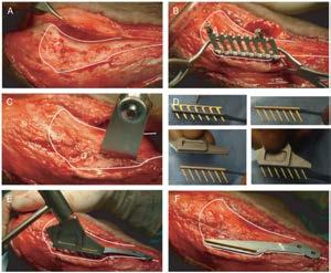
3 Materials required! Standard surgical pack! Hohmann retractor, stifle distractor! Gross hook and spoon and Meniscal knife! Securos needle! Sterile Leader line (40, 80, 100 lb test)! Steinmann pin and hand chuck! +/- Crimping system (securos) Order of go:! Craniolateral incision from proximal patella to mid tibial tuberosity! Sharp dissection through lateral retinaculum and reflection until access to fabella! Move skin incision to medial side! Small medial arthrotomy, joint exploration, closure! Placement of lateral fabellar suture! Pass suture under cranial tibial muscle! Closure: tensor fascia, SQ, skin &%


4 Approach Approach!SQ!Lateral retinaculum! ID lateral fabella Arthrotomy! Pull skin incision to medial side! Perform standard craniomedial stifle arthrotomy '%


5 Arthrotomy! Distally to tibial plateau and proximal to patella if desired! Can be smaller arthrotomy than craniolateral! Important structures are easily accessed through medial incision! Can still use Hohman, stifle distractor, etc! Close in standard fashion when done! Let skin return to lateral side Medial Arthrotomy Evaluation of the CCL and medial meniscus Intact CCL Ruptured CCL (%


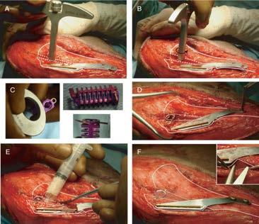
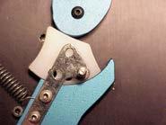
6 Arthrotomy medial meniscus!+/- Cruciate debridement!+/- Meniscal debridement! Debride if there is a tear!closure of the joint capsule Tibial Tuberosity Hole! At level of PT insertion! Exposure:! Incision along cranial border of cranial tibial muscle! Periosteal elevator to push muscle caudally on bone! Use hand chuck or drill for hole! Most common mistake:! Too distal! Too cranial! Goal:! Minimize cycling of implant Tibial Tuberosity Hole!Perpendicular to long axis of tibia!make small incision and reflect back cranial tibial muscle!%


7 Tibial Tuberosity Hole Pass Suture! Good pass AROUND fabella! Goals:! Entry point:! In valley between fabella and femur! Exit point:! Usually well distal to fabella! Fabella should move slightly when suture pulled up tensioned! Should NOT be able to feel suture! Should be able to lift up dog slightly Pass Suture! Repeat pass if:! Can feel suture either superficial or caudal/lateral to fabella! Grabbing too much soft tissue! Will loosen later! May incorporate nerve:! Sciatic! Common peroneal! Tibial )%


8 Pass Suture! Good pass AROUND fabella! If repeat too often, soft tissues will be macerated! Pass UNDER cranial tibial muscle! Come out through dissected area created for tibial tuberosity hole Pass Suture! What to do if tibial tuberosity hole not large enough to pass suture and needle?! Usually in small dogs! Equipment! Large-ish needle! 18 g or so! Large-ish suture! 0, #1 Pass Suture!Insert needle through tibial tuberosity hole! Medial to lateral *%


9 Pass Suture!Loop suture through leaderline! Pass both ends through needle Pass Suture!Remove needle!use suture loop to pull leaderline through TT hole Pass Suture! Repeat! Under patellar tendon! EXTRA- SYNOVIAL +%


10 Ta-Da!!! Now crimp or tie Crimp! Great if no surgical assistant! Most secure fixation! Allows testing of ROM before commitment Sliding Half-Hitch $,%


11 Closure! Place stifle through ROM! Minimal drawer! Minimal limitation of ROM! Close lateral retinaculum! 2-0 PDS! Close SQ! 3-0 Monocryl! Close Skin! Intradermal or skin sutures Outcome! Why this procedure works! Short term stability! Provided by the lateral suture! Decrease in instability = decrease in inflammation and increase in comfort! Long term! Stability provided by fibrous tissue created by the body! The suture will ultimately fail! Healing is a race between build up of fibrous tissue around the joint and when the suture breaks Outcome! Most animals do very well with procedure! Return to near normal function! Complications! Implant failure prior to the development of periarticular fibrosis leading to instability! Infection! Late meniscal damage! Incisional complications! Nerve damage! Tearing of the fabello-femoral ligament $$%

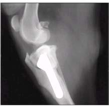
12 TTA TTA! Aim is to rearrange biomechanics of stifle joint and minimize/eliminate the need for the cruciate ligament Tibial Tuberosity Advancement $#%
13 Tibial Tuberosity Advancement $&%

!")
14 Post-Operative TTA Post-Op Care and Rehab! Incisional drainage! Bandaging! Expected function Post Operative Care! Modified Robert Jones bandage! 3 layers! Ice hours post op! ROM can begin within 1 week post op (usually while still in hospital)! Recheck 2 week suture removal! Leash walks only 8 weeks! Rads if TPLO or TTA $'%
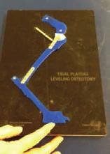
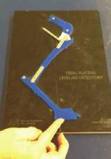
15 6/26/16 Prognosis Good to Excellent Determined by: Amount of pre-existing OA Presence of meniscal damage Surgical complications TPLO TPLO Tibial plateau leveling osteotomy Take away the need for the cruciate 15


16 So how does it do this? $!%

17 TPLO Femu r Cd Cr Tibia $)%

18 Pre-Operative Radiographs! Radiographs for OA, dx, presence of fabellae, etc! CC and lateral, including stifle and tarsus! Must include measuring bar/ball! Tibial Plateau Angle (TPA):! Describes the angle of the medial tibial plateau relative to the long axis of the tibia! The greater the TPA, the greater the tibial thrust and the resultant stress placed on the CCL! Accurate after 90 days of age Measure Radiographs A C B! A: Intercondylar eminences to center of talus! B: Medial tibial plateau (CCL insertion to caudal plateau)! C: perpendicular to A where A and B intersect Rotation Amount Saw Radius Starting TPA Rotation on amount (mm) $*%
!")
19 Tibial Plateau Angle! All TPLOs aim to correct the TPA to ~5 degrees! Follow-up: no clinical difference in post-op TPA 0-14 degrees! New data suggests slight overcorrection to ~3 degrees Surgical Procedure! Joint:! Craniomedial approach! Arthroscopy! TPLO:! Tibial Exposure! Jig Placement (optional)! Osteotomy, rotation and temporary fixation! Plate application! Closure Arthrotomy rotomy! Remove fat pad for visualization! Evaluate cranial and caudal cruciate ligaments! Evaluate menisci for any damage! Close with absorbable suture (PDS) $+%


20 Medial approach to tibia! Extend incision over proximalmedial tibia! Reflect back caudal head of sartorius mm! Visualize medial collateral Attach jig! Functions of the jig:! Maintain medial-lateral alignment of proximal vs. distal piece of tibia! Functions as point of rotation for proximal piece! Saw template attachment Tibial Osteotomy! MUST be perpendicular in all planes! Saw should intersect caudal tibial cortex perpendicularly! MUST maintain enough tibial crest to prevent later fracture! MOST IMPORTANT PART OF PROCEDURE #,%


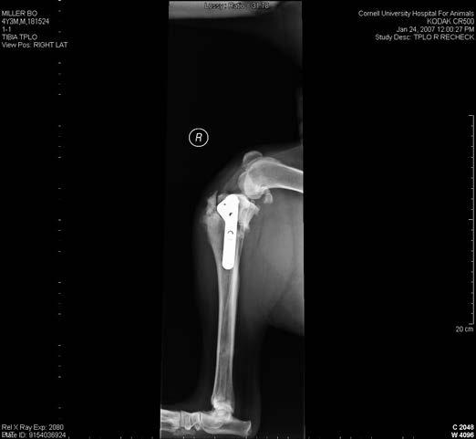
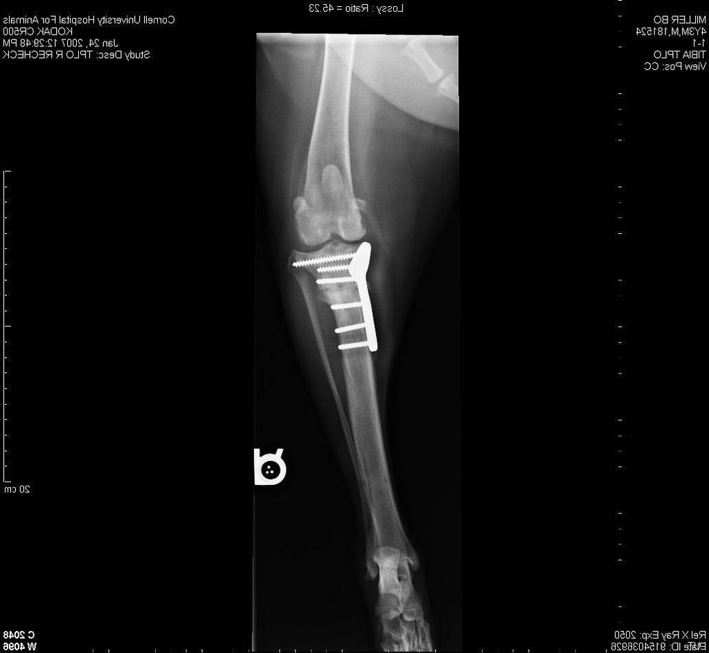
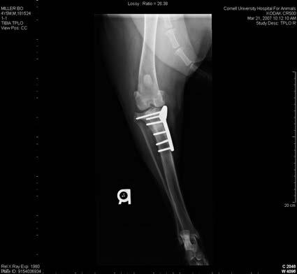
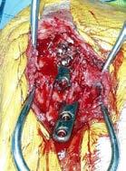
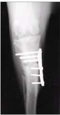
21 Apply TPLO Plate! All sizes from cat to giant breed! R and L Pre and Post-Op rads #$%
22 6/26/16 Prospective Study Materials and Methods Study population: Dogs with unilateral cranial cruciate rupture, >15 kg Normal radiographs of the contralateral stifle and hips Surgical intervention determined by owner Required recheck schedule: 2 weeks, 8 weeks, 6 months, 12 months Gait analysis at all visits Radiographs at 8 week visit for TPLO and TTA groups Radiographs for all at 12 month visit Materials and Methods Surgical procedure Craniomedial (TPLO, TTA) or craniolateral (ECR) approach Partial or caudal horn meniscectomy as indicated No meniscal release 22
 fabellotibial suture, secured with crimps (Securos ) or self-locking knot Materials and Methods! Gait analysis!")
23 Materials and Methods! Implants! Synthes locking TPLO plate! Kyon TTA implants! Monofilament nylon (80 or 100 lb test) fabellotibial suture, secured with crimps (Securos ) or self-locking knot Materials and Methods! Gait analysis! Two serial force platforms 1 embedded in a 10 m walkway! Performed at walk and trot! Minimum of 5 acceptable trials! Good paw strikes, no distractions or pulling! Controlled velocity with dual-photocell system:! Walk ( m/s)! Trot ( m/s)! Real-time processing with video and custom software /01%23%41/5067%87631%9-:;%<%=./172>?@%9A76B1%C$%BD13%C#;% -./01%23%41/5067%87631%9-:;%<%D23E72>?@% 9A76B1%C$%BD13%C#;% F67G% Time (seconds) #&%
24 6/26/16 Materials and Methods Ground reaction forces: Peak vertical force (PVF) normalized to weight (N/N) Contact time (CT) Vertical impulse (VI) - normalized to weight (N/N) Symmetry indexes calculated SI = 1 n SI = symmetry index n R k n = number of trials Lk w k k=1 R k = mean of the right (or operated) limb measurement L k = mean of the left (or unoperated) limb measurement w k = weighing factor (=1 in normal level locomotion) Materials and Methods Control population: Mean (±SD) of the population s mean SI for acceptable trials for each GRF were calculated Single session Treatment population: Mean (±SD) for acceptable trials for each GRF at each recheck time period were calculated Normal function defined as: SI of a GRF within one SD of the mean of the control population. Materials and Methods Statistical analysis: Repeated measures ANOVA/General linear model Variables: age weight sex side Statistical significance: P <0.05 tear type meniscectomy interaction between treatment and time period after surgery 24
25 Time (seconds)!"#!"$!% Materials and Methods! Pearson correlation coefficients!! All ground reaction forces at the walk and trot Rear limbs only Time (seconds) Results! 80 control dogs! 38 treated dogs! TPLO n=15! TTA n=14! ECR n=23 Results! Recheck time periods for operated group:! 1-49 days! days! days!!300 days ECR TPLO TTA #(%
26 6/26/16 Results Control (n=80) TPLO (n=15) TTA (n=14) ECR (n=23) Weight (kg) Age (yr) Sex Male Female Results Control (n=80) TPLO (n=15) TTA (n=14) ECR (n=23) Weight (kg) 30.2 ± ± ± ± 7.2 Age (yr) 3.3 ± ± ± ± 2.6 Sex Male Female Results Tear type Meniscal status TPLO (n=15) TTA (n=14) ECR (n=23) Partial Complete No meniscectomy Meniscectomy P-value
27 6/26/16 Results Reasons for case decrease: Lost to follow-up (7 TPLO, 4 TTA, 7 ECR) Meniscal tear (1 TPLO, 1 ECR) Tibial tuberosity fracture (1 TPLO) Contralateral cranial cruciate rupture (3 TTA, 3 ECR) TPLO (n=15) TTA (n=14) ECR (n=23) 2 days weeks weeks months months Results Control Population Symmetry Indexes PVF VI CT Walk 1.00 (0.050) 1.01 (0.050) 1.01 (0.031) Trot 1.00 (0.045) 1.01 (0.074) 1.02 (0.053) Continuous data reported as mean (SD) Results Repeated measures ANOVA/General linear model Variables: age weight sex side tear type meniscectomy interaction between treatment and time period after surgery 27
28 6/26/16 Results Repeated measures ANOVA/General linear model Variables: age weight sex side tear type meniscectomy interaction between treatment and time period after surgery Example 1.2 Symmetry Index = Significant difference between treatment groups R-square = X F-value = X P-value = X TPLO ECR TTA Control 0 days 1-49 days days days >300 days Time Period after surgery Results,, and indicate no significant difference between TPLO, ECR, and TTA group and the control group, respectively 28
29 Results!,, and indicate no significant difference between TPLO, ECR, and TTA group and the control group, respectively Results!,, and indicate no significant difference between TPLO, ECR, and TTA group and the control group, respectively!! Post-op TTA PTA CT = 91.9 degrees No correlation between cage size or post-op PTA CT and function Walk PVF TPLO ECR TTA Control days 1-49 days days days 300 days and over #+%
30 Walk VI TPLO ECR TTA Control B 0 days 1-49 days days days 300 days and over Trot PVF TPLO ECR TTA Control days 1-49 days days days 300 days and over Trot VI TPLO ECR TTA Control B 0 days 1-49 days days days 300 days and over &,%


31 Discussion! TPLO achieves normal function at walk and trot 6-12 months after surgery! TTA has most rapid recovery in early post-operative stage at the walk, achieves normal function at walk 12 months after surgery, but not at trot! Function did not correlate with amount of advancement! Function did not correlate with post-op PTA CT! ECR never achieves normal function Limitations! Small sample size! All R 2 are! 0.63! Not randomized! Owner determined Complication Rates - TPLO! % overall!5% Re-op rate!bilateral simultaneous surgery? &$%

32 Avulsion of Tibial Crest &#%


33 Implant Failure Implant Loosening Implant/Soft Tissue Infection &&%
 573-586.")

34 Implant/Soft t Tissue Infection Patellar Fracture TTA Complications! 31.5% overall! 12.3% major, 19.3% minor Lafaver S. et al. Vet Surg 2007 (36) ! Subsequent meniscal tear, tibial fracture, implant failure, infection, lick granuloma, incisional trauma, and MPL! All major complications were treated and resolved, all but 2 minor complications resolved &'%
 How to Choose:! Considerations:! How strong does the implant need to be for the dog s personality?")
35 Tibial Fracture/Implant Failure Other Complications! Septic joint! Patellar tendon desmitis! Nerve damage! Meniscal injury/re-injury! OA progression! Injury of contralateral CCL (~54% will rupture contralateral within a year) How to Choose:! Considerations:! How strong does the implant need to be for the dog s personality?! What is the tibial plateau angle?! What is the intended use/job?! Is the caudal cruciate intact?! How quickly does the dog need to be able to use the limb?! Financial considerations &(%
36 6/26/16 Summary Pick appropriate procedure for client-patient pair Only do a procedure if you re able to deal with the complications Any Questions? 36
Extracapsular Repair Monofilament Nylon Suture
 Extracapsular Repair Monofilament Nylon Suture Management of the ruptured Cranial Cruciate Ligament (CCL) by placing a non-absorbable suture between the lateral fabella and the proximal, cranial tibia
Robert Botte, DVM, Diplomate ACVS Veterinary Surgical Service San Diego, California. Kyon Symposium 2010 Zurich
 Robert Botte, DVM, Diplomate ACVS Veterinary Surgical Service San Diego, California Kyon Symposium 2010 Zurich ! Special Considerations " Anatomic variation " Precise implant placement " Factors affecting
Proceeding of the NAVC North American Veterinary Conference Jan. 8-12, 2005, Orlando, Florida
 Proceeding of the NAVC North American Veterinary Conference Jan. 8-12, 2005, Orlando, Florida Reprinted in the IVIS website with the permission of the NAVC http:/// The North American Veterinary Conference
Cranial Cruciate disease
 Cranial Cruciate disease Anatomy The Cranial cruciate ligament is located in the stifle joint (or knee). It is a thick fibrous band that runs from the distal femur to the proximal tibia. It is designed
Iso-toggle LigaFiba. Academy Step by Step. Issue 3
 Issue 3 t +44 (0)114 258 8530 info@vetinst.com www.vetinst.com Academy Step by Step Iso-toggle LigaFiba Fig. 1 Introduction Stabilisation of the canine stifle using extra-articular sutures is a well established
Cranial Cruciate Ligament Disease
 24- hour Emergency Service 01635 47170 The Tibial Tuberosity Advancement (TTA) procedure is one of the advanced procedures for the treatment of cranial cruciate ligament disease in dogs. TTA is now available
SIMITRI STABLE IN STRIDE SURGICAL PROCEDURE
 Copyright 2016 NGD. All rights reserved Neil Embleton, B.Sc., DVM and Veronica Barkowski, DVM Helivet Mobile Surgical Services, Sundre, AB, Canada July 2016 SIMITRI STABLE IN STRIDE SURGICAL PROCEDURE
TTA Rapid with Patellar Luxation
 TTA Rapid with Patellar Luxation The dog is placed in a dorsal recumbency with the affected limb suspended from a stand. Make sure that the dog s paws are not fixed too tightly, since the affected limb
Cruciate ligament injury
 Cruciate ligament injury This is an extremely common injury in dogs, less so in cats. Let s start by looking at the anatomy of the stifle (knee) joint of the dog. The important differences between the
TTA-Rapid Protocol. i. Where possible, calibrate the radiograph on the screen to real size.
 Legeweg 157 i 8020 Oostkamp Tel: 050/31.18.76 Fax: 050/31.58.86 www.instrulife.be TTA-Rapid Protocol The dog is placed in a dorsal recumbency with the affected limb suspended from a stand. Make sure that
Pre-operative evaluation
 Pre-operative evaluation Andrea Meyer-Lindenberg Clinic of Small Animal Surgery and eproduction Ludwig-Maximilians-University Munich Importance of pre-operative planning Evaluate patient before selecting
Transfemoral Amputation
 Transfemoral Amputation Pre-Op: 42 year old male who sustained severe injuries in a motorcycle accident. Note: he is a previous renal transplant recipient and is on immunosuppressive treatments. His injuries
Cranial cruciate ligament rupture and tibial tuberosity advancement
 Vet Times The website for the veterinary profession https://www.vettimes.co.uk Cranial cruciate ligament rupture and tibial tuberosity advancement Author : Nick Wiliams Categories : Vets Date : September
Triple Tibial Osteotomy (TTO)
") Triple Tibial Osteotomy (TTO) Objective: This operation is based on the biomechanical analysis performed by Dr Slobodan Tepic, which revealed that in order to remove the shear strain from the cranial cruciate
Triple Tibial Osteotomy (TTO) Objective: This operation is based on the biomechanical analysis performed by Dr Slobodan Tepic, which revealed that in order to remove the shear strain from the cranial cruciate
Ruptured Anterior (Cranial) Cruciate Ligament
 Cruciate Ligament") THE PET HEALTH LIBRARY By Wendy C. Brooks, DVM, DipABVP Educational Director, VeterinaryPartner.com Ruptured Anterior (Cranial) Cruciate Ligament First, the Basics The knee is a fairly complicated joint.
THE PET HEALTH LIBRARY By Wendy C. Brooks, DVM, DipABVP Educational Director, VeterinaryPartner.com Ruptured Anterior (Cranial) Cruciate Ligament First, the Basics The knee is a fairly complicated joint.
Proceedings of the World Small Animal Veterinary Association Sydney, Australia 2007
 Proceedings of the World Small Animal Sydney, Australia 2007 Hosted by: Next WSAVA Congress CRANIAL CRUCIATE LIGAMENT INJURIES SURGICAL MANAGEMENT Warrick J. Bruce BVSc(dist), MVM, DSAS(orthopaedics),
THE PET HEALTH LIBRARY By Wendy C. Brooks, DVM, DipABVP Educational Director, VeterinaryPartner.com. Ruptured Anterior (Cranial) Cruciate Ligament
 Cruciate Ligament") THE PET HEALTH LIBRARY By Wendy C. Brooks, DVM, DipABVP Educational Director, VeterinaryPartner.com Ruptured Anterior (Cranial) Cruciate Ligament First, the Basics There are two cruciate ligaments that
THE PET HEALTH LIBRARY By Wendy C. Brooks, DVM, DipABVP Educational Director, VeterinaryPartner.com Ruptured Anterior (Cranial) Cruciate Ligament First, the Basics There are two cruciate ligaments that
Joop Hopmans, Small animal orthopedic surgeon. Wednesday, April 24, 13
 Joop Hopmans, Small animal orthopedic surgeon 1 Cranial Cruciate Trauma What to do????? TightRope OTT TPLO MMT FHT MRIT TTA TCC TTO TTArap TTA-2 2 The biggest ones TPLO TTA TTO TCC 3 Why changing/modifying?
Cranial Cruciate Ligament Rupture
 6910 Carpenter Fire Station Road, Cary NC 27519 Phone (919) 545-1001 www.quartetvet.com Cranial Cruciate Ligament Rupture This information is provided to help you understand the condition that has been
Cruciate Ligament Disease
 The Cranial Cruciate Ligament Cruciate Ligament Disease The cranial cruciate ligament (CrCL, aka anterior cruciate ligament or ACL) is one of several structures in the stifle (equivalent to our knee) that
Cranial cruciate ligament rupture in Dogs
 Clinical sheet - Surgery Cranial cruciate ligament rupture in Dogs Cranial cruciate ligament rupture is one of the most common orthopedic conditions in dogs. Rupture of the cranial cruciate ligament is
TTA. Common Tangent Method
 TTA Common Tangent Method This document is derived from a presentation by Dr. Randy Boudrieau DVM, Dipl. ACVS, ECVS, Prof. of Surgery, Cummings School of Veterinary Medicine, Tufts University IVET DESIG
Meniscus cartilage replacement with cadaveric
 Technical Note Meniscal Allografting: The Three-Tunnel Technique Kevin R. Stone, M.D., and Ann W. Walgenbach, R.N.N.P., M.S.N. Abstract: This technical note describes an improved arthroscopic technique
TTA Wedge System INSTRUCTIONS FOR USE
 TTA Wedge System INSTRUCTIONS FOR USE 1 INSTRUCTIONS FOR USE The OssAbility TTA Wedge System consists of the following products: Wedge Implants Osteotomy Guide Advancement Levers Osteotomy Planning Overlay
Cruciate Ligament Disease
 The Cranial Cruciate Ligament Cruciate Ligament Disease The cranial cruciate ligament (CrCL, aka in humans anterior cruciate ligament or ACL) is one of several structures in the stifle (equivalent to our
Triple Tibial Osteotomy (TTO)
") Objective: This operation is based on the biomechanical analysis performed by Dr Slobodan Tepic, which revealed that in order to remove the shear strain from the cranial cruciate ligament the tibial plateaux
Objective: This operation is based on the biomechanical analysis performed by Dr Slobodan Tepic, which revealed that in order to remove the shear strain from the cranial cruciate ligament the tibial plateaux
Coxofemoral Luxation System
 Coxofemoral Luxation System Tech Sheet The Securos Coxofemoral Luxation Management System Traumatic dislocation of the coxofemoral joint disrupts the joint capsule as well as the round ligament of the
veterinarian recommendation
 Brace Yourself: The Role of Orthotics in Cruciate Disease David Dycus, DVM, MS, CCRP, DACVS-SA Orthopedic Staff Surgeon Veterinary Orthopedic and Sports Medicine Group (VOSM) Annapolis Junction, MD Cranial
Tibial Tuberosity Advancement For the Treatment of Cranial Cruciate Deficiency
 Tibial Tuberosity Advancement For the Treatment of Cranial Cruciate Deficiency Cranial cruciate ligament deficiency in the dog is the most common orthopedic lameness seen in practice today. Many reasons
Ruptured cranial cruciate ligament (CCL) Ruptured cruciate, Ruptured ligament, Ruptured anterior cruciate ligament (ACL), Torn ACL, Torn ligament
 Ruptured cruciate, Ruptured ligament, Ruptured anterior cruciate ligament (ACL), Torn ACL, Torn ligament") 1333 Plaza Blvd, Suite E, Central Point, OR 97502 * www.mountainviewvet.net Category: Canine Ruptured cranial cruciate ligament (CCL) Ruptured cruciate, Ruptured ligament, Ruptured anterior cruciate ligament
1333 Plaza Blvd, Suite E, Central Point, OR 97502 * www.mountainviewvet.net Category: Canine Ruptured cranial cruciate ligament (CCL) Ruptured cruciate, Ruptured ligament, Ruptured anterior cruciate ligament
This page is intentionally blank
 This page is intentionally blank 1 Focus on Canine Sports Medicine Cranial Cruciate Ligament Injury in Agility Dogs Part 1 By Sherman O. Canapp, Jr., DVM, MS, Diplomate ACVS TIEN TRAN PHOTOGRAPHY Kili,
Canine cranial cruciate ligament rupture (CrCLR) has
 has") Peer Reviewed Canine Cranial Cruciate Disease An Evidence-Based Look at Current Treatment Modalities James K. Roush, DVM, MS, Diplomate ACVS Kansas State University This is the second article in a 2-part
Peer Reviewed Canine Cranial Cruciate Disease An Evidence-Based Look at Current Treatment Modalities James K. Roush, DVM, MS, Diplomate ACVS Kansas State University This is the second article in a 2-part
1 Anatomy. 2 Pathophysiology
 Cranial cruciate ligaments in dogs: why do they rupture and how can we fix it Daniel Koch Dr. med. vet. ECVS, Diessenhofen/Switzerland, www.dkoch.ch 1 Anatomy The cranial cruciate ligament (CrCL) runs
Bone grafting developments used in veterinary orthopaedics part two
 Vet Times The website for the veterinary profession https://www.vettimes.co.uk Bone grafting developments used in veterinary orthopaedics part two Author : John Innes, Peter Myint Categories : Vets Date
Zoran Lončar. CONGRESS AMVAC/RoSAVA September, 2014
 Zoran Lončar Veterinary Clinic www.vetnovak.com loncarzor@yahoo.co.uk Belgrade, Serbia CONGRESS AMVAC/RoSAVA 11-13 September, 2014 ALL ABOUT THE KNEEE To become familiar with the EXAM To recognize most
Knee Disarticulation Amputation
 Knee Disarticulation Amputation Pre-Op 64 year old man, previous spinal cord injury, diabetes, renal failure, and a history of spasticity with dynamic knee flexion contracture. He had an open left ankle
TIBIAL PLATEAU LEVELING OSTEOTOMY (TPLO)
") TIBIAL PLATEAU LEVELING OSTEOTOMY (TPLO) Cruciate disease in the dog Cranial cruciate ligament (CCL) disease is the most common cause of hindlimb lameness in the dog. It affects the stifle joint, the equivalent
TIBIAL PLATEAU LEVELING OSTEOTOMY (TPLO) Cruciate disease in the dog Cranial cruciate ligament (CCL) disease is the most common cause of hindlimb lameness in the dog. It affects the stifle joint, the equivalent
Zurich Open Repository and Archive
 University of Zurich Zurich Open Repository and Archive Winterthurerstr. 190 CH-8057 Zurich http://www.zora.uzh.ch Year: 2008 Force plate gait analysis to assess limb function after tibial tuberosity advancement
Technique Guide. *smith&nephew N8TIVE ACL Anatomic ACL Reconstruction System
 Technique Guide *smith&nephew N8TIVE ACL Anatomic ACL Reconstruction System N8TIVE ACL System The N8TIVE ACL Anatomic Reconstruction System provides a novel and simple approach to ACL repair. The N8TIVE
Simitri Stable in Stride
 Simitri Stable in Stride Surgical Technique Copyright 2016 NGD. All rights reserved. Simitri Stable in Stride Note: Although this technique contains descriptions of a particular surgical procedure, it
Treatment of cranial cruciate ligament rupture with the ligament augmentation and reconstruction system (LARS) in dogs: An in vitro study
 in dogs: An in vitro study") Treatment of cranial cruciate ligament rupture with the ligament augmentation and reconstruction system (LARS) in dogs: Master research project E.J.C. van den Brink 3754200 Project tutor: Dr. L.F. H. Theyse
Treatment of cranial cruciate ligament rupture with the ligament augmentation and reconstruction system (LARS) in dogs: Master research project E.J.C. van den Brink 3754200 Project tutor: Dr. L.F. H. Theyse
Development of a canine stifle computer model to investigate cranial cruciate ligament deficiency.
 University of Louisville ThinkIR: The University of Louisville's Institutional Repository Electronic Theses and Dissertations 8-2009 Development of a canine stifle computer model to investigate cranial
MINI TIBIAL PLATEAU LEVELING OSTEOTOMY (TPLO) SYSTEM
 SYSTEM") MINI TIBIAL PLATEAU LEVELING OSTEOTOMY (TPLO) SYSTEM For stabilizing osteotomies of the canine and feline proximal tibia SURGICAL TECHNIQUE TABLE OF CONTENTS INTRODUCTION Mini Tibial Plateau Leveling
MINI TIBIAL PLATEAU LEVELING OSTEOTOMY (TPLO) SYSTEM For stabilizing osteotomies of the canine and feline proximal tibia SURGICAL TECHNIQUE TABLE OF CONTENTS INTRODUCTION Mini Tibial Plateau Leveling
JOINT RULER. Surgical Technique For Knee Joint JRReplacement
 JR JOINT RULER Surgical Technique For Knee Joint JRReplacement INTRODUCTION The Joint Ruler * is designed to help reduce the incidence of flexion, extension, and patellofemoral joint problems by allowing
Cruciate Disease in Dogs
 Cruciate Disease in Dogs Improving your skills in diagnosis and treatment Chris Preston FACVSc DACVS Pet Emergency & Specialist Centre 1103 Dandenong Rd, Malvern East VIC 3145 Tel: (03) 9569 3677 Fax:
SIMITRI STABLE IN STRIDE
 Copyright 2016 NGD. All rights reserved Neil Embleton, B.Sc., DVM and Veronica Barkowski, DVM Helivet Mobile Surgical Services, Sundre, AB, Canada July 2016 SIMITRI STABLE IN STRIDE 1. SIMITRI STABLE IN
How I manage. Combined CCL rupture and patella luxation. in small dogs and cats 1/27/2017 NOVOS FORUM 28 JANUARY
 How I manage Combined CCL rupture and patella luxation in small dogs and cats SUNE JERRE DVM,SPECIALIST IN SURGERY FOR DOGS AND CATS 1/27/2017 NOVOS FORUM 28 JANUARY 2017 1 Clinicl examination of dogs
Effect of 9 mm Tibial Tuberosity Advancement on Cranial Tibial Translation in the Canine Cranial Cruciate Ligament Deficient Stifle
 Effect of 9 mm Tibial Tuberosity Advancement on Cranial Tibial Translation in the Canine Cranial Cruciate Ligament Deficient Stifle By Jonathan Mark Miller Thesis submitted to the Faculty of the Virginia
THE INCIDENCE of medial meniscal tears in association. A Novel Pin Distraction Device for Arthroscopic Assessment of the Medial Meniscus in Dogs
 Veterinary Surgery 38:595 600, 2009 BRIEF COMMUNICATION A Novel Pin Distraction Device for Arthroscopic Assessment of the Medial Meniscus in Dogs PETER BÖTTCHER, Dr. med. vet., Diplomate ECVS, PHILIPP
GREENS SURGICALS. Redefining Excellence INSTRUMENT SYSTEM PREPARED BY: DR. VINAY KUMAR
 GREENS SURGICALS Redefining Excellence TIBIA AND FEMUR INSTRUMENT SYSTEM PREPARED BY: DR. VINAY KUMAR OPERATIVE TECHNIQUES INDEX SR.NO CONTENTS 1 LIST OF INSTRUMENT FOR TIBIA AND FEMUR. 2 RADIO GRAPH OF
Tibial tuberosity fracture as a complication of tibial tuberosity advancement
 Clinical Communication 148 Tibial tuberosity fracture as a complication of tibial tuberosity advancement I. Calvo 1 ; J. Aisa 2 ; D. Chase 3 ; P. Garcia-Fernandez 4 ; F. San Roman 4 ; D. Bennett 1 1 Glasgow
Lateral Meniscus Transplant
 Lateral Meniscus Transplant Using the CONMED Meniscus Allograft Transplant (MAT) Instruments A complete guide to Lateral Meniscus Transplant using the CONMED meniscus allograft transplant Instruments.
Anterior Cruciate Ligament Surgery
 Anatomy Anterior Cruciate Ligament Surgery Roger Ostrander, MD Andrews Institute Anatomy Anatomy Function Primary restraint to anterior tibial translation Secondary restraint to internal tibial rotation
MULTIPLE SURGICAL techniques have been
 Effect of Tibial Plateau Leveling on Stability of the Canine Cranial Cruciate Deficient Stifle Joint: An In Vitro Study Veterinary Surgery 31:147-154, 2002 ULLRICH REIF, DVM, DONALD A. HULSE, DVM, Diplomate
ACL Athletic Career. ACL Rupture - Warning Features Intensive pain Immediate swelling Locking Feel a Pop Dead leg Cannot continue to play
 FIMS Ambassador Tour to Eastern Europe, 2004 Belgrade, Serbia Montenegro Acute Knee Injuries - Controversies and Challenges Professor KM Chan OBE, JP President of FIMS Belgrade ACL Athletic Career ACL
GASTROCNEMIUS TENDON REPAIR VETLIG USING THE STIF CAT 30 SOFT TISSUE INTERNAL FIXATION VETLIG
 VETLIG SOFT TISSUE INTERNAL FIXATION GASTROCNEMIUS TENDON REPAIR USING THE STIF CAT 30 VETLIG A R T I F I C I A L L I G A M E N T S F O R V E T E R I N A R Y U S E VETLIG MANAGEMENT OF CHRONIC GASTROCNEMIUS
Patellar Ligament Disease.
 Patellar Ligament Disease. The patellar ligament disease is a condition of the stifle where the cartilage keeping the patella in place over knee joint is weakened or damaged. The patella is held in place
ACL Reconstruction Cross-Pin Technique
 ACL Reconstruction Cross-Pin Technique Surgical Technique Lonnie E. Paulos, MD Salt Lake City, Utah 325 Corporate Drive Mahwah, NJ 07430 t: 201 831 5000 www.stryker.com A surgeon should always rely on
Minimally Invasive ACL Surgery
 Minimally Invasive ACL Surgery KOCO EATON, M.D. T A M P A B A Y R A Y S ( 1 9 9 5 P R E S E N T ) T A M P A B A Y B U C C A N E E R S ( 2 0 1 5 2 0 1 6 ) T A M P A B A Y R O W D I E S ( 2 0 1 4 2 0 1 7
Transfemoral Amputation
 Transfemoral Amputation Preop This 26 year old male sustained a gunshot wound to the left thigh. He was treated emergently with revascularization and fasciotomies. He was transferred to our regional trauma
Joints of the Lower Limb II
 Joints of the Lower Limb II Lecture Objectives Describe the components of the knee and ankle joint. List the ligaments associated with these joints and their attachments. List the muscles acting on these
Figure 3 Figure 4 Figure 5
 Figure 1 Figure 2 Begin the operation with examination under anesthesia to confirm whether there are any ligamentous instabilities in addition to the posterior cruciate ligament insufficiency. In particular
CORRECTIVE OSTEOTOMY BRINGING THE PLAN TO THE BONE (TRIGONOMETERY, GUIDE WIRES, SLA MODELING AND ART)
") CORRECTIVE OSTEOTOMY BRINGING THE PLAN TO THE BONE (TRIGONOMETERY, GUIDE WIRES, SLA MODELING AND ART) Randy J. Boudrieau, DVM, DACVS, DECVS Cummings School of Veterinary Medicine at Tufts University, North
CORRECTIVE OSTEOTOMY BRINGING THE PLAN TO THE BONE (TRIGONOMETERY, GUIDE WIRES, SLA MODELING AND ART) Randy J. Boudrieau, DVM, DACVS, DECVS Cummings School of Veterinary Medicine at Tufts University, North
Surgical Technique. VISIONAIRE FastPak Instruments for the LEGION Total Knee System
 Surgical Technique VISIONAIRE FastPak Instruments for the LEGION Total Knee System VISIONAIRE FastPak for LEGION Instrument Technique* Nota Bene The technique description herein is made available to the
UvA-DARE (Digital Academic Repository) Treatment of osteochondral defects of the talus van Bergen, C.J.A. Link to publication
 Treatment of osteochondral defects of the talus van Bergen, C.J.A. Link to publication") UvA-DARE (Digital Academic Repository) Treatment of osteochondral defects of the talus van Bergen, C.J.A. Link to publication Citation for published version (APA): van Bergen, C. J. A. (2014). Treatment
UvA-DARE (Digital Academic Repository) Treatment of osteochondral defects of the talus van Bergen, C.J.A. Link to publication Citation for published version (APA): van Bergen, C. J. A. (2014). Treatment
Measurement of Tibial Translation in Dogs with Anterior Cruciate Ligament Rupture
 Measurement of Tibial Translation in Dogs with Anterior Cruciate Ligament Rupture October 17, 2009 Team: Graham Bousley: Team Leader Alex Bloomquist: Communicator James Madsen: BSAC Mike Nonte: BWIG Client:
May 2011, Issue 31. In addition to our regular ER hours, AMVS is providing emergency and critical care services to your patients: Fridays, all day
 Page 1 of 5 Having Trouble Viewing this Email? Click Here You're receiving this email because of your relationship with Aspen Meadow Veterinary Specialists. Please confirm your continued interest in receiving
Total Knee Original System Primary Surgical Technique
 Surgical Procedure Total Knee Original System Primary Surgical Technique Where as a total hip replacement is primarily a bony operation, a total knee replacement is primarily a soft tissue operation. Excellent
Surgical Care at the District Hospital. EMERGENCY & ESSENTIAL SURGICAL CARE
 Surgical Care at the District Hospital 1 18 Orthopedic Trauma Key Points 2 18.1 Upper Extremity Injuries Clavicle Fractures Diagnose fractures from the history and by physical examination Treat with a
Veronica J. Barkowski and Neil A. Embleton
 Surgical Technique and Initial Clinical Experience with a Novel Extracapsular Articulating Implant for Treatment of the Canine Cruciate Ligament Deficient Stifle Joint Veronica J. Barkowski and Neil A.
Revolution. Unicompartmental Knee System
 Revolution Unicompartmental Knee System While Total Knee Arthroplasty (TKA) is one of the most predictable procedures in orthopedic surgery, many patients undergoing TKA are in fact excellent candidates
Biceps Femoris Muscle in Dogs Diana Powell 11/25/2016
 Biceps Femoris Muscle in Dogs Diana Powell 11/25/2016 The Biceps Femoris is the largest muscle in the muscle group that makes up the hamstring. The Biceps Femoris is covered only by fascia and skin and
Arthrex Open Wedge Osteotomy Technique Designed in conjunction with:
 Arthrex Open Wedge Osteotomy Technique Designed in conjunction with: Dr. Giancarlo Puddu, M.D. Dr. Peter Fowler, M.D. Dr. Ned Amendola, M.D. To treat pain and instability associated with lower extremity
The Impact of Age on Knee Injury Treatment
 The Impact of Age on Knee Injury Treatment Focus on the Meniscus Dr. Alvin J. Detterline, MD Sports Medicine and Orthopaedic Surgery Towson Orthopaedic Associates University of Maryland St. Joseph Medical
The NBX Non-Bridging External Fixator A Non-Bridging External Fixator/Locking Plate capturing a series of.062mm K-wires and 3mm half-pins that are
 The NBX Non-Bridging External Fixator A Non-Bridging External Fixator/Locking Plate capturing a series of.062mm K-wires and 3mm half-pins that are inserted in a multiplanar and multi-directional fashion
CRUCIATE DISEASE - LATERAL SUTURE STABILISATION (LSS)
") CRUCIATE DISEASE - LATERAL SUTURE STABILISATION (LSS) Cruciate disease in the dog Cranial cruciate ligament (CCL) disease is the most common cause of hindlimb lameness in the dog. It affects the stifle
CRUCIATE DISEASE - LATERAL SUTURE STABILISATION (LSS) Cruciate disease in the dog Cranial cruciate ligament (CCL) disease is the most common cause of hindlimb lameness in the dog. It affects the stifle
Non Surgical Management of Soft Tissue Injuries. Megan LeFave, DVM cvma
 Non Surgical Management of Soft Tissue Injuries Megan LeFave, DVM cvma Non Surgical Management of Soft Tissue Injuries Biomechanical Principles Common front limb and hind limb injuries In hospital treatments
MCL Reconstruction Surgical Protocol by Tarek Fahl, M.D.
 MCL Reconstruction Surgical Protocol by Tarek Fahl, M.D. Features A unique weave in which a single strand of braided polyethylene is woven through itself twice in opposite directions This construct allows
The Knee Joint By Prof. Dr. Muhammad Imran Qureshi
 The Knee Joint By Prof. Dr. Muhammad Imran Qureshi Structurally, it is the Largest and the most complex joint in the body because of the functions that it performs: Allows mobility (flexion/extension)
EFFECTS OF TIBIAL PLATEAU LEVELING OSTEOTOMY AND TIBIAL TUBEROSITY ADVANCEMENT ON STIFLE CONTACT MECHANICS AND KINEMATICS
 EFFECTS OF TIBIAL PLATEAU LEVELING OSTEOTOMY AND TIBIAL TUBEROSITY ADVANCEMENT ON STIFLE CONTACT MECHANICS AND KINEMATICS By STANLEY EUNWOO KIM A THESIS PRESENTED TO THE GRADUATE SCHOOL OF THE UNIVERSITY
ACL RECONSTRUCTION HAMSTRING METHOD. Presents ACL RECONSTRUCTION HAMSTRING METHOD. Multimedia Health Education
 HAMSTRING METHOD Presents HAMSTRING METHOD Multimedia Health Education Disclaimer Stephen J. Incavo MD This movie is an educational resource only and should not be used to make a decision on Anterior Cruciate
Triathlon Knee System
 Triathlon Knee System Express Instruments Surgical Protocol Posterior Stabilized & Cruciate Retaining TriathlonKneeSystem Express Instruments Surgical Protocol Acknowledgments..........................................................2
3.5 MM VA-LCP PROXIMAL TIBIA PLATE SYSTEM
 3.5 MM VA-LCP PROXIMAL TIBIA PLATE SYSTEM Part of the DePuy Synthes Variable Angle Periarticular Plating System SURGICAL TECHNIQUE TABLE OF CONTENTS INTRODUCTION 3.5 mm VA-LCP Proximal Tibial Plate 2 AO
Patellar Luxation. Anatomy, Function, and Dysfunction
 6910 Carpenter Fire Station Road, Cary NC 27519 Phone (919) 545-1001 Patellar Luxation This information is provided to help you understand the condition that has been diagnosed in your pet. We find that
Canine Cranial Cruciate Ligament Repair Anchor System
 SURGICAL TECHNIQUE Canine Cranial Cruciate Ligament Repair Anchor System Surgical Technique The patient is positioned in lateral or dorsal recumbency under general anesthetic. A hanging limb technique
LCP Medial Distal Tibia Plate, without Tab. The Low Profile Anatomic Fixation System with Angular Stability and Optimal Screw Orientation.
 LCP Medial Distal Tibia Plate, without Tab. The Low Profile Anatomic Fixation System with Angular Stability and Optimal Screw Orientation. Technique Guide LCP Small Fragment System Table of Contents Introduction
The Knee. Prof. Oluwadiya Kehinde
 The Knee Prof. Oluwadiya Kehinde www.oluwadiya.sitesled.com The Knee: Introduction 3 bones: femur, tibia and patella 2 separate joints: tibiofemoral and patellofemoral. Function: i. Primarily a hinge joint,
Dates and images Intrauma S.p.A. reserves the right to modify the design and finishing of the products shown and described in the present catalog
 Product catalog Dates and images Intrauma S.p.A. reserves the right to modify the design and finishing of the products shown and described in the present catalog without any notice. The images are informative
Medical Practice for Sports Injuries and Disorders of the Knee
 Sports-Related Injuries and Disorders Medical Practice for Sports Injuries and Disorders of the Knee JMAJ 48(1): 20 24, 2005 Hirotsugu MURATSU*, Masahiro KUROSAKA**, Tetsuji YAMAMOTO***, and Shinichi YOSHIDA****
Large Distractor Femur
 Fracture Reduction and Provisional Stabilization Large Distractor Femur Surgical Technique Table of Contents Introduction Standard Femoral Distraction 2 Large Distractor System 4 Surgical Technique Prepare
ANATOMIC. Navigated Surgical Technique 4 in 1 TO.G.GB.016/1.0
 ANATOMIC Navigated Surgical Technique 4 in 1 TO.G.GB.016/1.0 SCREEN LAYOUT Take screenshot Surgical step Dynamic navigation zone Information area and buttons 2 SCREEN LAYOUT Indicates action when yellow
Building A Future In Orthopaedics. Course Programme Autumn/Winter
 Building A Future In Orthopaedics Course Programme Autumn/Winter 2016 08450 702498 info@securos.co.uk www.securos.co.uk Speakers He is a Director at Rutland House Referrals, St Helens Dr Kinley Smith where
Tibial & Femoral Opening Wedge Osteotomy System. Surgical Technique
 Tibial & Femoral Opening Wedge Osteotomy System Surgical Technique Opening Wedge Osteotomy Tibial & Femoral Opening Wedge Osteotomy 2 Prior to the osteotomy, a diagnostic arthroscopy is performed to verify
Knee Joint Anatomy 101
 Knee Joint Anatomy 101 Bone Basics There are three bones at the knee joint femur, tibia and patella commonly referred to as the thighbone, shinbone and kneecap. The fibula is not typically associated with
TRK REVISION KNEE Surgical Technique
 1 TRK REVISION KNEE Surgical Technique 1. 2. 3. 4. 5. 6. 7. 8. 9. 10. INTERCONDYLAR RESECTION...... page FEMORAL STEM...... page NON CEMENTED FEMORAL STEM...... page TRIAL FEMORAL COMPONENTS...... page
MCL Injuries: When and How to Repair Scott D. Mair, MD
 MCL Injuries: When and How to Repair Scott D. Mair, MD Professor and Team Physician: Orthopaedic Surgery University of Kentucky School of Medicine Disclosure Institution: Research/Education Smith-Nephew
Aesculap Orthopaedics Columbus MIOS
 Aesculap Orthopaedics Columbus MIOS Minimally Invasive Orthopaedic Solutions Manual TKA Surgical Technique MIOS 4-in-1 Cutting Block MIOS Distal Femoral Cutting Block MIOS Tibial Left and Right Cutting
Patellar Luxation. The Patella. Dr. PJ Rocheleau, DVM and Associates 138 Tudhope St, Espanola ON, P5E 1S6
 Patellar Luxation The Patella The patella (equivalent to the knee cap ) is one of several structures in the stifle (equivalent to our knee) that provide joint stability and allow normal function. The stifle
Fracture and Dislocation of Metacarpal Bones, Metacarpophalangeal Joints, Phalanges, and Interphalangeal Joints ( 1-Jan-1985 )
") In: Textbook of Small Animal Orthopaedics, C. D. Newton and D. M. Nunamaker (Eds.) Publisher: International Veterinary Information Service (www.ivis.org), Ithaca, New York, USA. Fracture and Dislocation
In: Textbook of Small Animal Orthopaedics, C. D. Newton and D. M. Nunamaker (Eds.) Publisher: International Veterinary Information Service (www.ivis.org), Ithaca, New York, USA. Fracture and Dislocation