Cranial cruciate ligament rupture and tibial tuberosity advancement
|
|
|
- Dora Reeves
- 5 years ago
- Views:
Transcription
1 Vet Times The website for the veterinary profession Cranial cruciate ligament rupture and tibial tuberosity advancement Author : Nick Wiliams Categories : Vets Date : September 14, 2009 Nick Wiliams provides an overview of the diagnosis and treatment of cranial cruciate ligament deficient patients and summarises a new surgical method IT is vital that the general practitioner has a good understanding of how to diagnose and, in many cases, treat the cranial cruciate ligament (CCL) deficient patient, as it is the most common cause of hindlimb lameness in the dog. This article provides a refresher in the diagnosis of CCL, and summarises a relatively new method of stifle stabilisation surgery known as tibial tuberosity advancement (TTA). Anatomy The cruciate ligaments are paired ligaments within the stifle and are named after their relative insertions into the tibial plateau (that is, the cranial ligament inserts cranially and vice versa). Together with the collateral ligaments, they provide stability to the joint. The paired action of the cranial and caudal cruciate ligaments act like a four-joint chain to allow both rolling and sliding of the femoral condyles over the tibial plateau (see Figure 1). The CCL has four main functions within the joint: to oppose translation of the tibia with regard to the femur ( cranial draw ); to oppose internal rotation of the tibia; 1 / 16
2 to oppose overextension of the femur; and to provide a limited degree of varus-valgus support during stifle flexion. The CCL is further divided into cranio-medial and caudolateral bands, which, during stifle flexion, twist around each other, thereby limiting the amount of internal rotation of the tibia. The craniomedial band is taut in all phases of stifle motion, but the caudo-lateral band is only taut during stifle extension, becoming relaxed during flexion. For this reason, stifle joints should be examined for cranial draw during both flexion and extension as partial tears of the ligament may otherwise be missed if performed only in extension. Mechanoreceptors within the ligament itself provide feedback to prevent excessive flexion and extension of the joint. Nutrition to both cruciate ligaments comes primarily from the synovium. The natural healing potential of the ligament is very limited. Another important structure to be aware of within the stifle is the meniscus a fibrocartilaginous pad that acts as a shock absorber and compensates for incongruity between the tibial plateau and the femoral condyles. This pad is composed of two semilunar pads anchored to the tibia and femur by five ligaments, and to each other by the intermeniscal ligament. The medial menisci is further stabilised by an attachment to the medial collateral ligament this makes it more immobile and hence more vulnerable to injury by the pinching and internal rotational forces of the CCL deficient stifle. Blood supply to the menisci is also poor, especially to the inner portions, meaning the chance of healing is also slim. The menisci have a good nerve supply to aid with proprioception but this, in turn, can cause an extremely painful stifle when damaged. Aetiology Many different causes and theories exist to explain CCL rupture, the discussion of which are beyond the scope of this article. Possible causes are: Trauma. Trauma to an otherwise healthy ligament is probably a fairly uncommon cause of CCL rupture. This may come about when the CCL is tested beyond its normal breaking strain during stifle overextension or overrotation; a classic example being a foot down a rabbit hole. CCL disease. Studies suggest this is one of the major aetiologies for CCL deficient stifles in adult dogs1.this occurs due to gradual degenerative and adaptive changes to the cells and matrix of the ligament associated with synovial inflammation, meaning only a small amount of trauma (or even normal force) is needed to elicit a rupture1. 2 / 16
3 Inflammatory arthropathies. Either immune mediated or infectious. Anatomical. Certain breeds, notably the West Highland white terrier, are often found to have steep tibial plateau angle (TPA), altering stifle mechanics and predisposing to CCL rupture. This condition can also occur with premature closure of the caudal aspect of the proximal tibial growth plate. In these cases, corrective osteotomy is often the treatment of choice. Various authors have investigated the effects of tibial plateau angle on the incidence of cruciate disease with varying results some groups have shown no significant difference between TPA angles of CCL deficient and normal stifles of the same breed2, while others have shown significant differences between greyhounds, and the stifles of affected Labradors3. The mean TPA in dogs is around 23 4,5. Clinical signs Although CCL rupture is more common in large breed dogs such as Labradors, Rottweilers and mastiffs, it should be noted that any breed of dog can suffer CCL damage. Clinical signs include: Lameness. This can vary from non-weight bearing to mild and intermittent, depending on the nature and the chronicity of the rupture. Acute ruptures are generally (but not exclusively) accompanied by severe non-weight bearing lameness. Chronic ruptures can vary in severity of lameness, but are often presented as weightbearing or intermittently weight-bearing lame. Owners, on being questioned, often mention a period of acute lameness sometime previously. This is often accompanied by a difficulty in rising, increasing lameness post-exercise and after rest, and a characteristic CCL rupture posture when sitting, where the affected limb is held away from the body and extended to minimise the degree of flexion of the stifle and, therefore, pain. The instability in the stifle leads to the development of osteoarthritis6,7,8, which is associated with chronic lameness. Partial tears often show mild weight-bearing lameness associated with exercise, but often improving with rest; this can continue for many months before eventually completely rupturing. All tears can be accompanied by meniscal injury, which will reduce the response rate to conservative management. Muscle atrophy. Over the affected limb, especially noted with chronic disease. Stifle effusion. This is best noted by a lack of definition of the patella tendon. It is often helpful in the unilaterally affected dog to feel the normal stifle in the contra lateral limb. Pain on flexion and extension. Medial buttress. This is a thickening on the medial aspect of the stifle as a result of osteoarthritis 3 / 16
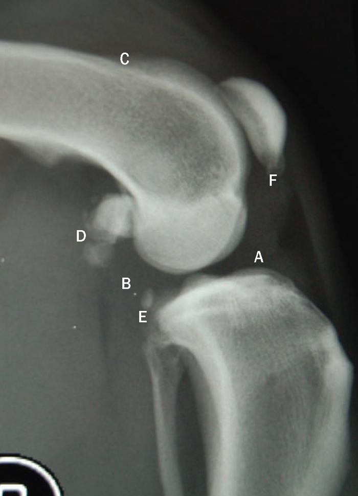
4 and soft tissue proliferation. It is best felt in the chronically affected stifle. Clicking or popping. These can often be heard or felt during stifle flexion and extension, and are due to the rolling and movement of a torn meniscus. Crepitus. This is due to osteophytes formed in the osteoarthritic joint. Joint instability. An assessment can be made while the animal is conscious, but it is far easier in the sedated or anaesthetised dog. When the animal is conscious, muscle contracture due to apprehension and soft tissue proliferation from chronic injury can often lead to false negatives. Two main methods are employed for this assessment and are best performed in lateral recumbency and from the rear of the dog: Cranial draw test (Figure 2): This must be done in both flexion and extension of the stifle. It is performed by positioning the index finger of the proximally placed hand over the dorsal femur (A) and the thumb on the caudal femur over the fabella (B). The distally placed hand has the index finger over the tibial crest (C) and the thumb over the caudal tibia (D). Force is then applied through the thumb (D) of the distally placed hand, pushing the tibial plateau forward. A movement of 0-2mm is expected in a normal stifle9. If excessive cranial draw is noted, it indicates CCL rupture. A falsepositive result can be seen in young dogs due to an inherent laxity in the ligament that can give a normal draw movement of 4-5mm9. In these young animals, a distinct endpoint to the drawing action is noted. Tibial compression test (Figure 3): This is best achieved with the limb in moderate flexion. It is performed by grasping the distal quadriceps in the proximally placed hand and laying the tip of one finger over the patella tendon and on to the tibial crest (A). The distally placed hand then grasps the metatarsal region and flexes the hock (B). If cranial movement of the tibial crest is noted, this indicates cruciate damage. It should be noted that both methods can show false-negative results in partial tears, particularly if performed only in extension. Diagnostic imaging A high level of suspicion of CCL rupture should be followed by further investigation. Many imaging modalities exist for the diagnosis of CCL disease, including ultrasound, MRI, and arthroscopy, but for the general practitioner, radiography continues to be the modality of choice. For this reason, it is the only method discussed in this article. Radiography is always helpful in confirming a diagnosis of CCL rupture and should be used to rule out other possible causes of stifle lameness. It is especially useful with chronic and partial tears where other clinical signs may have been lacking or missed. It is also necessary in the planning phase of correction methods including TTA, tibial plateau levelling osteotomy (TPLO) and other corrective osteotomy techniques. 4 / 16
5 Medio-lateral and caudocranial radiographs should always be obtained, but it is the mediolateral view that is often the most helpful. Normal limb views can be used for comparison. Radiographic features that are often seen are shown in Figure 4, and include: cranial fat pad effusion and loss of definition noted by an increase in opacity of the cranial joint space (A); caudal joint capsule extension due to joint effusion (B); distal displacement of the popliteal sesamoid, noted as a relative change during tibial compression and particularly useful in the diagnosis of partial CCL rupture; and osteophyte formation, which is seen particularly in cases of chronic joint instability and partial tears. Osteophytes can be seen over the trochlear ridges of the femur (C), around the fabellae (D), the caudal surface of the tibial plateau (E), and the distal patella (F). Treatment Treatment of CCL disease can be loosely divided into medical and surgical methods. It is beyond the scope of this article to discuss all possible surgical techniques, but it should be noted that success rates of CCL surgery are reported to be more than 90 per cent, regardless of the surgical technique used10. This article focuses only on the relatively new method (five to six years in general use) of tibial tuberosity advancement (TTA). TTA works by altering the angle between the pull of the quadriceps (patella) tendon, and the tibial plateau. To understand this, we first need to under s t and some of the forces acting on the stifle: TTA uses the proven theory that in the stifle there is a large tibiofemoral compressive force, approximately the same magnitude and direction as the patella tendon, pulling the tibia cranially (Figure 5). This force is normally counteracted by the CCL (the absence of a functional CCL allows the cranial draw action), and is best seen during stifle extension (A). As the stifle flexes, the force on the patella tendon becomes perpendicular to the tibial plateau and is, therefore, neutralised (B). At full flexion, the direction of the force changes and causes a caudal draw action (opposed by the caudal cruciate ligament; C). TTA works by advancing the attachment of the quadriceps mechanism cranially, thereby making the angle between the patella tendon and the tibial plateau perpendicular, or at an angle less than 5 / 16
6 90 in all phases of motion. This means there is no cranial draw force on the tibial plateau. A caudal draw force still exists. TTA requires specific equipment and implants, namely an oscillating saw, TTA jig, TTA plates and forks and TTA cages (Figure 6). Implant selection is made after medio-lateral radiography of the limb in a standing (approximately 135 ) stance. The procedure is performed via a medial approach to the stifle and proximal tibia, which also enables the surgeon to make a limited medial arthrotomy to assess the condition of the menisci. Using a specific jig, a number of holes are drilled into the proximal tibial crest, where the plate holding fork will eventually sit. The number of holes needed will depend on the size of plate required, which is determined by the size of the limb. Once all holes are complete, an osteotomy of the tibial crest is made using an oscillating saw. The mobile tibial crest is then rotated forward a set distance, bringing the pull of the quadriceps perpendicular to the tibial crest. The crest is held away from the tibial body using a cage with two screws. The maximum distance of advancement and, therefore, the largest cage size is 12mm. The centre of the cage is then packed with a cancellous bone graft to aid rapid osetogenesis. Aftercare is aimed at exercise restriction postoperatively until bone healing can be observed radiographically (Figure 7); one study put the mean time for this at 11.3 weeks11, while others have reported advanced healing in 94 per cent of cases reviewed at six weeks postop12. Major complications of this procedure include postoperative meniscal trauma, tibial tuberosity fracture, tibial fracture and implant failure, and occur at a similar rate to those reported with treatment by tibial plateau levelling osteotomy (TPLO)14. As with any technique, there are limitations that will affect case selection. The main limitation with TTA is that in dogs with excessive tibial plateau angles (TPA), a TTA in excess of 12mm would be required in order to create the desired 90 angle. In such dogs, other techniques such as TPLO may be better suited. This cut-off angle is still up for debate and no data has been published regarding the range of TPA in dogs with TTA. However, it has been presented that successful procedures have been performed in dogs with TPA of approximately Short-term reports of this procedure are promising, with a good to excellent outcome in 85 to 97 per cent of cases assessed subjectively by owners11,12,13. Overall, TTA is showing extremely good short-term results and provides a good alternative to other 6 / 16
7 stifle stabilisation techniques. It may also have an advantage over other osteotomy techniques as technical aspects are fewer and it appears to have a shorter learning curve. References 1. Hayashi K, Manley P A and Muir P (2004). Cranial cruciate ligament pathophysiology in dogs with cruciate disease: a review, J Am Anim Hosp Assoc Sep-Oct 40(5): Reif U, Probst C W (2003). Comparison of tibial plateau angles in normal and cranial cruciate deficient stifles of Labrador retrievers, Vet Surg Jul-Aug 32(4): Wilke V L, Conzemius M G, Besancon M F, Evans R B and Ritter M (2002). Comparison of tibial plateau angle between clinically normal greyhounds and Labrador retrievers with and without rupture of the cranial cruciate ligament, J Am Vet Med Assoc 221(10): 1,426-1, Grierson J, Sanders M, Guitan J and Pead M (2005). Comparison of anatomical tibial plateau angle versus observer measurement from lateral radiographs in dogs, Vet Comp Orthop Traumatol 18(4): Morris E and Lipowitz A J (2001). Comparison of tibial plateau angles in dogs with and without cranial cruciate ligament injuries, J Am Vet Med Assoc 218(3): Elkins A D (1991). A retrospective study evaluating the degree of degenerative joint disease in the stifle of dogs following surgical repair of anterior cruciate ligament rupture, J Am Anim Hosp Assoc 27: Vasseur P B and Berry C R (1992). Progression of stifle osteoarthritis following reconstruction of the cranial cruciate ligament in 21 dogs, J Am Anim Hosp Assoc 28: Arnoczky S P and Marshall J L (1977). The cruciate ligaments of the canine stifle: an anatomical and functional analysis, Am J Vet Res 38: 1,807-1, Fossum T W, Hedlund C, Johnson A L, Schulz K S, Seim H B, Willard M D, Bahr A and Carrol G L (2007). Small Animal Surgery (3rd edn). 10. Moore K W and Read R A (1995). Cranial cruciate ligament rupture in the dog-a retrospective study comparing surgical techniques. Aus Vet J 72(8): Lafaver S, Miller N A, Stubbs W P, Taylor R A and Boudrieau R J (2007). Tibial tuberosity advancement for stabilization of the canine cranial cruciate ligament-deficient stifle joint: surgical technique, early results, and complications in 101 dogs, Vet Surg August 36(6): Stein S and Schmoekel H (2008). Short-term and eight to 12 months results of a tibial tuberosity advancement as treatment of canine cruciate ligament damage, JSAP 49(8): Hoffman D E, Miller J M, Ober C P, Lanz O I, Martin R A and Shires P K (2006). Tibial tuberosity advancement in 65 canine stifles, Vet Comp Orthop Traumatol 19(4): Boudrieau R J (2008). Tibial plateau levelling osteotomy or tibial tuberosity advancement, Vet Surg 38: Boudrieau R J (2008) Veterinary symposium the surgical summit: pre- 7 / 16
 and caudal (blue) cruciate ligaments.")
8 symposium laboratories: TTA Lab. Figure 1. Diagram showing rolling motion of the femoral condyle over the tibial plateau during stifle flexion and the relative positions of the cranial (orange) and caudal (blue) cruciate ligaments. Based on human stifle joints. Diagram adapted from lecture material by Dr H. Schmokel, course notes in cruciate repair, Improve International Ltd. 8 / 16

9 Figure 2. Cranial draw test. 9 / 16

10 Figure 3. Tibial compression test. 10 / 16
11 11 / 16
12 12 / 16

13 Figure 4. Radiographic features often seen include: cranial fat pad effusion and loss of definition; caudal joint capsule extension; distal displacement of the popliteal sesamoid; and osteophyte formation. 13 / 16
14 Figure 5. (A) Stifle in full extension showing an overall cranial draw force (orange arrow) when the angle of the patella tendon (white arrow) to the tibial plateau (blue) is more than 90. (B) Stifle in mid flexion showing no draw force when the patella tendon is perpendicular to the tibial plateau. (C) Stifle in flexion showing an overall caudal draw force (orange arrow) when the patella tendon is at an angle less than 90 to the tibial plateau. 14 / 16

15 Figure 6. (left to right). 2 x 6mm cages, largest eight hole plate and fork, six hole plate and fork, three hole plate and fork, and two hole plate. 15 / 16

16 Figure 7. Postoperative radiographs. 16 / 16 Powered by TCPDF (
Cranial Cruciate disease
 Cranial Cruciate disease Anatomy The Cranial cruciate ligament is located in the stifle joint (or knee). It is a thick fibrous band that runs from the distal femur to the proximal tibia. It is designed
Cranial Cruciate Ligament Disease
 24- hour Emergency Service 01635 47170 The Tibial Tuberosity Advancement (TTA) procedure is one of the advanced procedures for the treatment of cranial cruciate ligament disease in dogs. TTA is now available
Ruptured cranial cruciate ligament (CCL) Ruptured cruciate, Ruptured ligament, Ruptured anterior cruciate ligament (ACL), Torn ACL, Torn ligament
 Ruptured cruciate, Ruptured ligament, Ruptured anterior cruciate ligament (ACL), Torn ACL, Torn ligament") 1333 Plaza Blvd, Suite E, Central Point, OR 97502 * www.mountainviewvet.net Category: Canine Ruptured cranial cruciate ligament (CCL) Ruptured cruciate, Ruptured ligament, Ruptured anterior cruciate ligament
1333 Plaza Blvd, Suite E, Central Point, OR 97502 * www.mountainviewvet.net Category: Canine Ruptured cranial cruciate ligament (CCL) Ruptured cruciate, Ruptured ligament, Ruptured anterior cruciate ligament
Proceeding of the NAVC North American Veterinary Conference Jan. 8-12, 2005, Orlando, Florida
 Proceeding of the NAVC North American Veterinary Conference Jan. 8-12, 2005, Orlando, Florida Reprinted in the IVIS website with the permission of the NAVC http:/// The North American Veterinary Conference
Proceedings of the World Small Animal Veterinary Association Sydney, Australia 2007
 Proceedings of the World Small Animal Sydney, Australia 2007 Hosted by: Next WSAVA Congress CRANIAL CRUCIATE LIGAMENT INJURIES SURGICAL MANAGEMENT Warrick J. Bruce BVSc(dist), MVM, DSAS(orthopaedics),
Cruciate ligament injury
 Cruciate ligament injury This is an extremely common injury in dogs, less so in cats. Let s start by looking at the anatomy of the stifle (knee) joint of the dog. The important differences between the
1 Anatomy. 2 Pathophysiology
 Cranial cruciate ligaments in dogs: why do they rupture and how can we fix it Daniel Koch Dr. med. vet. ECVS, Diessenhofen/Switzerland, www.dkoch.ch 1 Anatomy The cranial cruciate ligament (CrCL) runs
Pre-operative evaluation
 Pre-operative evaluation Andrea Meyer-Lindenberg Clinic of Small Animal Surgery and eproduction Ludwig-Maximilians-University Munich Importance of pre-operative planning Evaluate patient before selecting
THE PET HEALTH LIBRARY By Wendy C. Brooks, DVM, DipABVP Educational Director, VeterinaryPartner.com. Ruptured Anterior (Cranial) Cruciate Ligament
 Cruciate Ligament") THE PET HEALTH LIBRARY By Wendy C. Brooks, DVM, DipABVP Educational Director, VeterinaryPartner.com Ruptured Anterior (Cranial) Cruciate Ligament First, the Basics There are two cruciate ligaments that
THE PET HEALTH LIBRARY By Wendy C. Brooks, DVM, DipABVP Educational Director, VeterinaryPartner.com Ruptured Anterior (Cranial) Cruciate Ligament First, the Basics There are two cruciate ligaments that
Ruptured Anterior (Cranial) Cruciate Ligament
 Cruciate Ligament") THE PET HEALTH LIBRARY By Wendy C. Brooks, DVM, DipABVP Educational Director, VeterinaryPartner.com Ruptured Anterior (Cranial) Cruciate Ligament First, the Basics The knee is a fairly complicated joint.
THE PET HEALTH LIBRARY By Wendy C. Brooks, DVM, DipABVP Educational Director, VeterinaryPartner.com Ruptured Anterior (Cranial) Cruciate Ligament First, the Basics The knee is a fairly complicated joint.
This page is intentionally blank
 This page is intentionally blank 1 Focus on Canine Sports Medicine Cranial Cruciate Ligament Injury in Agility Dogs Part 1 By Sherman O. Canapp, Jr., DVM, MS, Diplomate ACVS TIEN TRAN PHOTOGRAPHY Kili,
HOW DO WE DIAGNOSE LAMENESS IN YOUR HORSE?
 HOW DO WE DIAGNOSE LAMENESS IN YOUR HORSE? To help horse owners better understand the tools we routinely use at VetweRx to evaluate their horse s soundness, the following section of this website reviews
Bone grafting developments used in veterinary orthopaedics part two
 Vet Times The website for the veterinary profession https://www.vettimes.co.uk Bone grafting developments used in veterinary orthopaedics part two Author : John Innes, Peter Myint Categories : Vets Date
ACTA VET. BRNO 2013, 82: ; doi: /avb
 ACTA VET. BRNO 2013, 82: 215 218; doi:10.2754/avb201382020215 Radiographic changes of the patellar ligament in dogs after tibial tuberosity advancement Ladislav Stehlík 1, Pavel Proks 1, Petra Fedorová
RN(EC) ENC(C) GNC(C) MN ACNP *** MECHANISM OF INJURY.. MOST IMPORTANT *** - Useful in determining mechanism of injury / overuse
 ENC(C) GNC(C) MN ACNP *** MECHANISM OF INJURY.. MOST IMPORTANT *** - Useful in determining mechanism of injury / overuse") HISTORY *** MECHANISM OF INJURY.. MOST IMPORTANT *** Age of patient Sport / Occupation - Certain conditions are more prevalent in particular age groups (Osgood Schlaters in youth / Degenerative Joint Disease
HISTORY *** MECHANISM OF INJURY.. MOST IMPORTANT *** Age of patient Sport / Occupation - Certain conditions are more prevalent in particular age groups (Osgood Schlaters in youth / Degenerative Joint Disease
May 2011, Issue 31. In addition to our regular ER hours, AMVS is providing emergency and critical care services to your patients: Fridays, all day
 Page 1 of 5 Having Trouble Viewing this Email? Click Here You're receiving this email because of your relationship with Aspen Meadow Veterinary Specialists. Please confirm your continued interest in receiving
TTA Rapid with Patellar Luxation
 TTA Rapid with Patellar Luxation The dog is placed in a dorsal recumbency with the affected limb suspended from a stand. Make sure that the dog s paws are not fixed too tightly, since the affected limb
Concepts in managing canine medial patellar luxation cases
 Vet Times The website for the veterinary profession https://www.vettimes.co.uk Concepts in managing canine medial patellar luxation cases Author : Toby Gemmill, Bill Oxley Categories : Companion animal,
ACL Athletic Career. ACL Rupture - Warning Features Intensive pain Immediate swelling Locking Feel a Pop Dead leg Cannot continue to play
 FIMS Ambassador Tour to Eastern Europe, 2004 Belgrade, Serbia Montenegro Acute Knee Injuries - Controversies and Challenges Professor KM Chan OBE, JP President of FIMS Belgrade ACL Athletic Career ACL
Robert Botte, DVM, Diplomate ACVS Veterinary Surgical Service San Diego, California. Kyon Symposium 2010 Zurich
 Robert Botte, DVM, Diplomate ACVS Veterinary Surgical Service San Diego, California Kyon Symposium 2010 Zurich ! Special Considerations " Anatomic variation " Precise implant placement " Factors affecting
Outline. Extracapsular Repair !"#!"$!% COMPARISON OF SURGICAL METHODS FOR CRUCIATE DISEASE. Ursula Krotscheck, DVM DACVS Cornell University
 COMPARISON OF SURGICAL METHODS FOR CRUCIATE DISEASE Ursula Krotscheck, DVM DACVS Cornell University Outline! Basic concepts behind the 3 major surgical procedures! Prospective study! Expected outcomes
veterinarian recommendation
 Brace Yourself: The Role of Orthotics in Cruciate Disease David Dycus, DVM, MS, CCRP, DACVS-SA Orthopedic Staff Surgeon Veterinary Orthopedic and Sports Medicine Group (VOSM) Annapolis Junction, MD Cranial
TTA. Common Tangent Method
 TTA Common Tangent Method This document is derived from a presentation by Dr. Randy Boudrieau DVM, Dipl. ACVS, ECVS, Prof. of Surgery, Cummings School of Veterinary Medicine, Tufts University IVET DESIG
Ben 5 year old M mixed breed dog. Dr. Norman Ackerman Memorial Radiography Case Challenge
 February 2014 Dr. Norman Ackerman served the University of Florida, College of Veterinary Medicine with distinction as Professor of Radiology from 1979 to 1994. A concerned teacher of veterinary students
The Knee. Two Joints: Tibiofemoral. Patellofemoral
 Evaluating the Knee The Knee Two Joints: Tibiofemoral Patellofemoral HISTORY Remember the questions from lecture #2? Girth OBSERVATION TibioFemoral Alignment What are the consequences of faulty alignment?
SIMITRI STABLE IN STRIDE SURGICAL PROCEDURE
 Copyright 2016 NGD. All rights reserved Neil Embleton, B.Sc., DVM and Veronica Barkowski, DVM Helivet Mobile Surgical Services, Sundre, AB, Canada July 2016 SIMITRI STABLE IN STRIDE SURGICAL PROCEDURE
Cranial cruciate ligament rupture in Dogs
 Clinical sheet - Surgery Cranial cruciate ligament rupture in Dogs Cranial cruciate ligament rupture is one of the most common orthopedic conditions in dogs. Rupture of the cranial cruciate ligament is
Extracapsular Repair Monofilament Nylon Suture
 Extracapsular Repair Monofilament Nylon Suture Management of the ruptured Cranial Cruciate Ligament (CCL) by placing a non-absorbable suture between the lateral fabella and the proximal, cranial tibia
TTA Wedge System INSTRUCTIONS FOR USE
 TTA Wedge System INSTRUCTIONS FOR USE 1 INSTRUCTIONS FOR USE The OssAbility TTA Wedge System consists of the following products: Wedge Implants Osteotomy Guide Advancement Levers Osteotomy Planning Overlay
Cruciate Ligament. Summary of the Doctoral Thesis
 Study of the Effect of Excessive Tibial Plateau Angle on Degenerative Changes of Canine Cranial Cruciate Ligament Summary of the Doctoral Thesis Tom Ichinohe Graduate School of Veterinary Medicine and
Tibial tuberosity fracture as a complication of tibial tuberosity advancement
 Clinical Communication 148 Tibial tuberosity fracture as a complication of tibial tuberosity advancement I. Calvo 1 ; J. Aisa 2 ; D. Chase 3 ; P. Garcia-Fernandez 4 ; F. San Roman 4 ; D. Bennett 1 1 Glasgow
TTA-Rapid Protocol. i. Where possible, calibrate the radiograph on the screen to real size.
 Legeweg 157 i 8020 Oostkamp Tel: 050/31.18.76 Fax: 050/31.58.86 www.instrulife.be TTA-Rapid Protocol The dog is placed in a dorsal recumbency with the affected limb suspended from a stand. Make sure that
Medical Practice for Sports Injuries and Disorders of the Knee
 Sports-Related Injuries and Disorders Medical Practice for Sports Injuries and Disorders of the Knee JMAJ 48(1): 20 24, 2005 Hirotsugu MURATSU*, Masahiro KUROSAKA**, Tetsuji YAMAMOTO***, and Shinichi YOSHIDA****
STATE OF THE ART OF ACL SURGERY (Advancements that have had an impact)
") STATE OF THE ART OF ACL SURGERY (Advancements that have had an impact) David Drez, Jr., M.D. Clinical Professor of Orthopaedics LSU School of Medicine Financial Disclosure Dr. David Drez has no relevant
STATE OF THE ART OF ACL SURGERY (Advancements that have had an impact) David Drez, Jr., M.D. Clinical Professor of Orthopaedics LSU School of Medicine Financial Disclosure Dr. David Drez has no relevant
TIBIAL PLATEAU LEVELING OSTEOTOMY (TPLO)
") TIBIAL PLATEAU LEVELING OSTEOTOMY (TPLO) Cruciate disease in the dog Cranial cruciate ligament (CCL) disease is the most common cause of hindlimb lameness in the dog. It affects the stifle joint, the equivalent
TIBIAL PLATEAU LEVELING OSTEOTOMY (TPLO) Cruciate disease in the dog Cranial cruciate ligament (CCL) disease is the most common cause of hindlimb lameness in the dog. It affects the stifle joint, the equivalent
ASSESSMENT AND MANAGEMENT OF THE KNEE AND LOWER LIMB.
 ASSESSMENT AND MANAGEMENT OF THE KNEE AND LOWER LIMB www.fisiokinesiterapia.biz Overview History Examination X-rays Fractures and Dislocations. Soft Tissue Injuries Other Knee/Lower limb Problems Anatomy
Knee Injury Assessment
 Knee Injury Assessment Clinical Anatomy p. 186 Femur Medial condyle Lateral condyle Femoral trochlea Tibia Intercondylar notch Tibial tuberosity Tibial plateau Fibula Fibular head Patella Clinical Anatomy
Patellar Ligament Disease.
 Patellar Ligament Disease. The patellar ligament disease is a condition of the stifle where the cartilage keeping the patella in place over knee joint is weakened or damaged. The patella is held in place
Inclination of the patellar ligament in relation to flexion angle in stifle joints of dogs without degenerative joint disease
 10/16/2006 10:53 AM Page 1849 Inclination of the patellar ligament in relation to flexion angle in stifle joints of dogs without degenerative joint disease Renate Dennler, DVM; Nicolas M. Kipfer, DVM;
Supplementary Material to this article is available online at
 Clinical Communication 536 Combined tibial plateau levelling osteotomy and tibial tuberosity transposition for treatment of cranial cruciate ligament insufficiency with concomitant medial patellar luxation
Development of a canine stifle computer model to investigate cranial cruciate ligament deficiency.
 University of Louisville ThinkIR: The University of Louisville's Institutional Repository Electronic Theses and Dissertations 8-2009 Development of a canine stifle computer model to investigate cranial
Knee Joint Assessment and General View
 Knee Joint Assessment and General View Done by; Mshari S. Alghadier BSc Physical Therapy RHPT 366 m.alghadier@sau.edu.sa http://faculty.sau.edu.sa/m.alghadier/ Functional anatomy The knee is the largest
Effect of 9 mm Tibial Tuberosity Advancement on Cranial Tibial Translation in the Canine Cranial Cruciate Ligament Deficient Stifle
 Effect of 9 mm Tibial Tuberosity Advancement on Cranial Tibial Translation in the Canine Cranial Cruciate Ligament Deficient Stifle By Jonathan Mark Miller Thesis submitted to the Faculty of the Virginia
The Knee. Prof. Oluwadiya Kehinde
 The Knee Prof. Oluwadiya Kehinde www.oluwadiya.sitesled.com The Knee: Introduction 3 bones: femur, tibia and patella 2 separate joints: tibiofemoral and patellofemoral. Function: i. Primarily a hinge joint,
Small Animal radiography Stifle Joint and CruS
 Peer reviewed ImagIng EssEnTIals Small Animal radiography Stifle Joint and CruS Danielle Mauragis, CVT, and Clifford R. erry, DVM, Diplomate ACVR This is the fourth article in our Imaging Essentials series,
Cranial Cruciate Ligament Rupture
 6910 Carpenter Fire Station Road, Cary NC 27519 Phone (919) 545-1001 www.quartetvet.com Cranial Cruciate Ligament Rupture This information is provided to help you understand the condition that has been
Prevention and Treatment of Injuries. Anatomy. Anatomy. Chapter 20 The Knee Westfield High School Houston, Texas
 Prevention and Treatment of Injuries Chapter 20 The Knee Westfield High School Houston, Texas Anatomy MCL, Medial Collateral Ligament LCL, Lateral Collateral Ligament PCL, Posterior Cruciate Ligament ACL,
MULTIPLE SURGICAL techniques have been
 Effect of Tibial Plateau Leveling on Stability of the Canine Cranial Cruciate Deficient Stifle Joint: An In Vitro Study Veterinary Surgery 31:147-154, 2002 ULLRICH REIF, DVM, DONALD A. HULSE, DVM, Diplomate
Triple Tibial Osteotomy (TTO)
") Triple Tibial Osteotomy (TTO) Objective: This operation is based on the biomechanical analysis performed by Dr Slobodan Tepic, which revealed that in order to remove the shear strain from the cranial cruciate
Triple Tibial Osteotomy (TTO) Objective: This operation is based on the biomechanical analysis performed by Dr Slobodan Tepic, which revealed that in order to remove the shear strain from the cranial cruciate
KNEE EXAMINATION. Tips & Tricks from an Emergency Physician Perspective. EM Physicians Less Exposed to MSK Medicine
 KNEE EXAMINATION Tips & Tricks from an Emergency Physician Perspective Dr P O CONNOR Emergency Medicine Physician EUSEM 10/09/2018 EM Physicians Less Exposed to MSK Medicine Musculoskeletal Medicine becoming
Tibial Tuberosity Advancement For the Treatment of Cranial Cruciate Deficiency
 Tibial Tuberosity Advancement For the Treatment of Cranial Cruciate Deficiency Cranial cruciate ligament deficiency in the dog is the most common orthopedic lameness seen in practice today. Many reasons
Physeal fractures in immature cats and dogs: part 1 forelimbs
 Vet Times The website for the veterinary profession https://www.vettimes.co.uk Physeal fractures in immature cats and dogs: part 1 forelimbs Author : Lee Meakin, Sorrel Langley-Hobbs Categories : Canine,
Analysis of Plate Bone Construct Failure Following Tibial Tuberosity Advancment
 Analysis of Plate Bone Construct Failure Following Tibial Tuberosity Advancment W. T. McCartney 1,2 E. Galvin 2 B Mac Donald D Comiskey 3 1 Marie Louise Veterinary Hospital, Baldoyle, Dublin 13, Ireland
Medial Patella Luxation
 Medial Patella Luxation Anatomy The Patella is the large sesamoid bone (Kneecap) in the stifle joint. It forms part of the quadriceps muscle mechanism which is the main muscle group responsible for extension
Zoran Lončar. CONGRESS AMVAC/RoSAVA September, 2014
 Zoran Lončar Veterinary Clinic www.vetnovak.com loncarzor@yahoo.co.uk Belgrade, Serbia CONGRESS AMVAC/RoSAVA 11-13 September, 2014 ALL ABOUT THE KNEEE To become familiar with the EXAM To recognize most
Torn ACL - Anatomic Footprint ACL Reconstruction
 Torn ACL - Anatomic Footprint ACL Reconstruction The anterior cruciate ligament (ACL) is one of four ligaments that are crucial to the stability of your knee. It is a strong fibrous tissue that connects
The Knee. Tibio-Femoral
 The Knee Tibio-Femoral Osteology Distal Femur with Proximal Tibia Largest Joint Cavity in the Body A modified hinge joint with significant passive rotation Technically, one degree of freedom (Flexion/Extension)
A NEW CONCEPT IN FUNCTIONAL ORTHOSES
 A NEW CONCEPT IN FUNCTIONAL ORTHOSES THE KNEE in movement! Climbing stairs, walking and running are everyday actions that we can perform thanks to our knees. The knee joint is one of the most exposed and
TOTAL KNEE ARTHROPLASTY (TKA)
") TOTAL KNEE ARTHROPLASTY (TKA) 1 Anatomy, Biomechanics, and Design 2 Femur Medial and lateral condyles Convex, asymmetric Medial larger than lateral 3 Tibia Tibial plateau Medial tibial condyle: concave
TOTAL KNEE ARTHROPLASTY (TKA) 1 Anatomy, Biomechanics, and Design 2 Femur Medial and lateral condyles Convex, asymmetric Medial larger than lateral 3 Tibia Tibial plateau Medial tibial condyle: concave
Fracture and Dislocation of Metacarpal Bones, Metacarpophalangeal Joints, Phalanges, and Interphalangeal Joints ( 1-Jan-1985 )
") In: Textbook of Small Animal Orthopaedics, C. D. Newton and D. M. Nunamaker (Eds.) Publisher: International Veterinary Information Service (www.ivis.org), Ithaca, New York, USA. Fracture and Dislocation
In: Textbook of Small Animal Orthopaedics, C. D. Newton and D. M. Nunamaker (Eds.) Publisher: International Veterinary Information Service (www.ivis.org), Ithaca, New York, USA. Fracture and Dislocation
Please answer the following questions by responding with a score of 0 to 10. Please answer for how your dog is doing NOW.
 Online Supplementary Material to: Distal femoral lateral closing wedge osteotomy as a component of comprehensive treatment of medial patellar luxation and distal femoral varus in dogs Barry E. Brower;
EFFECTS OF TIBIAL PLATEAU LEVELING OSTEOTOMY AND TIBIAL TUBEROSITY ADVANCEMENT ON STIFLE CONTACT MECHANICS AND KINEMATICS
 EFFECTS OF TIBIAL PLATEAU LEVELING OSTEOTOMY AND TIBIAL TUBEROSITY ADVANCEMENT ON STIFLE CONTACT MECHANICS AND KINEMATICS By STANLEY EUNWOO KIM A THESIS PRESENTED TO THE GRADUATE SCHOOL OF THE UNIVERSITY
CORRECTIVE OSTEOTOMY BRINGING THE PLAN TO THE BONE (TRIGONOMETERY, GUIDE WIRES, SLA MODELING AND ART)
") CORRECTIVE OSTEOTOMY BRINGING THE PLAN TO THE BONE (TRIGONOMETERY, GUIDE WIRES, SLA MODELING AND ART) Randy J. Boudrieau, DVM, DACVS, DECVS Cummings School of Veterinary Medicine at Tufts University, North
CORRECTIVE OSTEOTOMY BRINGING THE PLAN TO THE BONE (TRIGONOMETERY, GUIDE WIRES, SLA MODELING AND ART) Randy J. Boudrieau, DVM, DACVS, DECVS Cummings School of Veterinary Medicine at Tufts University, North
Knee Injuries. PSK 4U Mr. S. Kelly North Grenville DHS. Medial Collateral Ligament Sprain
 Knee Injuries PSK 4U Mr. S. Kelly North Grenville DHS Medial Collateral Ligament Sprain Result from either a direct blow from the lateral side in a medial direction or a severe outward twist Greater injury
Medical Diagnosis for Michael s Knee
 Medical Diagnosis for Michael s Knee Introduction The following report mainly concerns the diagnosis and treatment of the patient, Michael. Given that Michael s clinical problem surrounds an injury about
Musculoskeletal Examination Benchmarks
 Musculoskeletal Examination Benchmarks _ The approach to examining the musculoskeletal system is the same no matter what joint or limb is being examined. The affected and contralateral region should both
Recognizing common injuries to the lower extremity
 Recognizing common injuries to the lower extremity Bones Femur Patella Tibia Tibial Tuberosity Medial Malleolus Fibula Lateral Malleolus Bones Tarsals Talus Calcaneus Metatarsals Phalanges Joints - Knee
ACL RECONSTRUCTION HAMSTRING METHOD. Presents ACL RECONSTRUCTION HAMSTRING METHOD. Multimedia Health Education
 HAMSTRING METHOD Presents HAMSTRING METHOD Multimedia Health Education Disclaimer Stephen J. Incavo MD This movie is an educational resource only and should not be used to make a decision on Anterior Cruciate
Lateral knee injuries
 Created as a free resource by Clinical Edge Based on Physio Edge podcast episode 051 with Matt Konopinski Get your free trial of online Physio education at Orthopaedic timeframes Traditionally Orthopaedic
What is Medial Plica Syndrome?
 What is Medial Plica Syndrome? It is a congenital disorder in which the thin wall of fibrous tissue extends from the synovial capsule of the knee. Pain usually occurs when the synovial capsule becomes
In the name of god. Knee. By: Tofigh Bahraminia Graduate Student of the Pathology Sports and corrective actions. Heat: Dr. Babakhani. Nov.
 In the name of god Knee By: Tofigh Bahraminia Graduate Student of the Pathology Sports and corrective actions Heat: Dr. Babakhani Nov. 2014 1 Anatomy-Bones Bones Femur Medial/lateral femoral condyles articulate
Overview Ligament Injuries. Anatomy. Epidemiology Very commonly injured joint. ACL Injury 20/06/2016. Meniscus Tears. Patellofemoral Problems
 Overview Ligament Injuries Meniscus Tears Pankaj Sharma MBBS, FRCS (Tr & Orth) Consultant Orthopaedic Surgeon Manchester Royal Infirmary Patellofemoral Problems Knee Examination Anatomy Epidemiology Very
Rehabilitation Guidelines for Anterior Cruciate Ligament (ACL) Reconstruction
 Reconstruction") Rehabilitation Guidelines for Anterior Cruciate Ligament (ACL) Reconstruction The knee is the body's largest joint, and the place where the femur, tibia, and patella meet to form a hinge-like joint. These
Rehabilitation Guidelines for Anterior Cruciate Ligament (ACL) Reconstruction The knee is the body's largest joint, and the place where the femur, tibia, and patella meet to form a hinge-like joint. These
ACL AND PCL INJURIES OF THE KNEE JOINT
 ACL AND PCL INJURIES OF THE KNEE JOINT Dr.KN Subramanian M.Ch Orth., FRCS (Tr & Orth), CCT Orth(UK) Consultant Orthopaedic Surgeon, Special interest: Orthopaedic Sports Injury, Shoulder and Knee Surgery,
Priorities Forum Statement GUIDANCE
 Priorities Forum Statement Number 21 Subject Knee Arthroscopy including arthroscopic knee washouts Date of decision November 2016 Date refreshed March 2017 Date of review November 2018 Osteoarthritis of
Influence of Limb Positioning and Measurement Method on the Magnitude of the Tibial Plateau Angle
 Veterinary Surgery 33:368 375, 2004 Influence of Limb Positioning and Measurement Method on the Magnitude of the Tibial Plateau Angle ULLRICH REIF, DVM, Diplomate ACVS & ECVS, LOIC M. DEJARDIN, DVM, MS,
A Patient s Guide to Knee Anatomy
 A Patient s Guide to Knee Anatomy 15195 Heathcote Blvd Suite 334 Haymarket, VA 20169 Phone: 703-369-9070 Fax: 703-369-9240 DISCLAIMER: The information in this booklet is compiled from a variety of sources.
Joop Hopmans, Small animal orthopedic surgeon. Wednesday, April 24, 13
 Joop Hopmans, Small animal orthopedic surgeon 1 Cranial Cruciate Trauma What to do????? TightRope OTT TPLO MMT FHT MRIT TTA TCC TTO TTArap TTA-2 2 The biggest ones TPLO TTA TTO TCC 3 Why changing/modifying?
Minimally Invasive ACL Surgery
 Minimally Invasive ACL Surgery KOCO EATON, M.D. T A M P A B A Y R A Y S ( 1 9 9 5 P R E S E N T ) T A M P A B A Y B U C C A N E E R S ( 2 0 1 5 2 0 1 6 ) T A M P A B A Y R O W D I E S ( 2 0 1 4 2 0 1 7
As for the forelimb, treatment of condition of the hindlimb may be treated by both localised therapy, applying the laser
 MLS Master Class - Veterinary Imaging Presented by CelticSMR Ltd Free Phone (UK): 0800 279 9050 International: +44 (0) 1646 603150 AUTHOR DETAILS Carl Gorman BVSc MRCVS PUBLISHER DETAILS Mike Howe B Vet
Arthritic history is similar to that of the hip. Add history of give way and locking, swelling
 KNEE VASU PAI Arthritic history is similar to that of the hip. Add history of give way and locking, swelling INJURY MECHANISM When How Sequence Progress Disability IKDC Activity I - Strenuous activity
Diagnosing Forelimb Lameness in Canine Patients
 OCTOBER 2018 Diagnosing Forelimb Lameness in Canine Patients DR. SEVIMA AKTAY, VMD, DACVS Diagnosing and treating forelimb lameness in dogs can often be challenging. Our patients rarely demonstrate overt
Meniscus Problems - Torn Meniscus Repair
 Meniscus Problems - Torn Meniscus Repair The two crescent-shaped menisci in each knee absorb shock, disperse weight, and reduce friction when the knee moves. Activities such as walking or jumping transfer
Freedom and safety in treatment
 medi GmbH & Co. KG Medicusstraße 1 95448 Bayreuth Germany T +49 921 912-0 F +49 921 912 780 ortho@medi.de www.medi.de 97E37/09.2012 Freedom and safety in treatment An informative brochure with an individual
The Knee Joint By Prof. Dr. Muhammad Imran Qureshi
 The Knee Joint By Prof. Dr. Muhammad Imran Qureshi Structurally, it is the Largest and the most complex joint in the body because of the functions that it performs: Allows mobility (flexion/extension)
UNUSUAL ACL CASE: Tibial Eminence Fracture in a Female Collegiate Basketball Player
 UNUSUAL ACL CASE: Tibial Eminence Fracture in a Female Collegiate Basketball Player Cheri Drysdale, MEd,, ATC Margot Putukian,, MD Jeffery Bechler,, MD Princeton University How many of you have done an
Clinical examination of the dog with thoracic limb lameness
 Clinical examination of the dog with thoracic limb lameness Examination of the patient Examination of the patient with musculoskeletal disease should start with a general physical examination. Particular
Feasibility of utilizing the patellar ligament angle for assessing cranial cruciate ligament rupture in dogs
 pissn 1229-845X, eissn 1976-555X J. Vet. Sci. (2014), 15(4), 563-568 http://dx.doi.org/10.4142/jvs.2014.15.4.563 Received: 6 Jan. 2014, Revised: 17 Jun. 2014, Accepted: 19 Jun. 2014 Original Article JOURNAL
Canine cranial cruciate ligament disease part 1 pathophysiology
 Vet Times The website for the veterinary profession https://www.vettimes.co.uk Canine cranial cruciate ligament disease part 1 pathophysiology Author : Albane Fauron, Karen Perry Categories : Companion
Tibial tuberosity conformation as a risk factor for cranial cruciate ligament rupture in the dog
 16 Original Research 2009 Schattauer GmbH Tibial tuberosity conformation as a risk factor for cranial cruciate ligament rupture in the dog R. Inauen 1 ; D. Koch 1 ; M. Bass 1 ; M. Haessig 2 1 Koch & Bass
Zurich Open Repository and Archive
 University of Zurich Zurich Open Repository and Archive Winterthurerstr. 190 CH-8057 Zurich http://www.zora.uzh.ch Year: 2008 Force plate gait analysis to assess limb function after tibial tuberosity advancement
Triple Tibial Osteotomy (TTO)
") Objective: This operation is based on the biomechanical analysis performed by Dr Slobodan Tepic, which revealed that in order to remove the shear strain from the cranial cruciate ligament the tibial plateaux
Objective: This operation is based on the biomechanical analysis performed by Dr Slobodan Tepic, which revealed that in order to remove the shear strain from the cranial cruciate ligament the tibial plateaux
Knee Joint Anatomy 101
 Knee Joint Anatomy 101 Bone Basics There are three bones at the knee joint femur, tibia and patella commonly referred to as the thighbone, shinbone and kneecap. The fibula is not typically associated with
Original Report. The Reverse Segond Fracture: Association with a Tear of the Posterior Cruciate Ligament and Medial Meniscus
 Eva M. Escobedo 1 William J. Mills 2 John. Hunter 1 Received July 10, 2001; accepted after revision October 1, 2001. 1 Department of Radiology, University of Washington Harborview Medical enter, 325 Ninth
Treatment of cranial cruciate ligament rupture with the ligament augmentation and reconstruction system (LARS) in dogs: An in vitro study
 in dogs: An in vitro study") Treatment of cranial cruciate ligament rupture with the ligament augmentation and reconstruction system (LARS) in dogs: Master research project E.J.C. van den Brink 3754200 Project tutor: Dr. L.F. H. Theyse
Treatment of cranial cruciate ligament rupture with the ligament augmentation and reconstruction system (LARS) in dogs: Master research project E.J.C. van den Brink 3754200 Project tutor: Dr. L.F. H. Theyse
Cruciate Ligament Disease
 The Cranial Cruciate Ligament Cruciate Ligament Disease The cranial cruciate ligament (CrCL, aka anterior cruciate ligament or ACL) is one of several structures in the stifle (equivalent to our knee) that
Your Practice Online
 Your Practice Online Disclaimer P R E S E N T S - PATELLAR TENDON This movie is an educational resource only and should not be used to make a decision on Anterior Cruciate Ligament (ACL) Reconstruction.
Femoral Shaft Fracture
 Femoral Shaft Fracture The femoral shaft is well padded with muscles(an advantage in protecting the bone from all but the most powerful forces)but the disadvantage is that fractures are often severely