Simitri Stable in Stride
|
|
|
- Eileen Mills
- 5 years ago
- Views:
Transcription


1 Simitri Stable in Stride Surgical Technique Copyright 2016 NGD. All rights reserved.
2 Simitri Stable in Stride Note: Although this technique contains descriptions of a particular surgical procedure, it is only to be used as a tool for licensed educated medical professionals. Ultimately, the surgeon should be guided by their own professional judgment in making any final determinations regarding product usage and technique.
3 Introduction: The New Generation Devices (NGD) Simitri Stable in Stride internal stifle stabilization System is a patented three part modular system, designed to provide the surgeon with an option for the management of any form of canine stifle instability. Simitri may be of benefit in speeding up the recovery period by providing immediate stabilization upon repair. Laboratory and clinical research have shown Simitri to be an effective method of managing canine stifle instability. Primary surgical indications: (The damaged ligaments do not require a primary repair prior to Simitri implantation). Primary repair method for cranial cruciate ligament (CrCL) disruption. Either partial or complete. Primary repair method for caudal cruciate ligament (CaCL) disruption. Either partial or complete. Primary repair method for any combination of cranial and or caudal cruciate ligament disruption. Secondary surgical indications: (The damaged ligaments must undergo a primary repair using standard accepted surgical techniques prior to Simitri implantation). Medial collateral ligament (MCL) disruption. Either partial or complete. Lateral collateral ligament (LCL) disruption. Either partial or complete. Design Features: Simitri can address instability of the diseased canine stifle, while allowing for normal range of motion, normal internal and external tibial rotation, normal joint compression and expansion and for normal varus and valgus tibial movement. Simitri is comprised of implant quality stainless steel femoral and tibial plates which are interconnected with an ultra high molecular weight polyethylene (UHMWPE) sliding articulation. Simitri s femoral and tibial plates each have two, 2mm holding pin holes. These are designed to accept a 5/64 pin and are used to hold the plates in place prior to final implantation. The plates can be independently contoured to allow for variations in anatomy and are designed to accept either cortical screws or NGD locking screws. Plates are available in a variety of sizes to accommodate varying anatomies and breeds. Concurrent luxating patella. Contraindications: Simitri is contraindicated in cases with neoplastic or infectious diseases of the stifle joint. As with all bone plates, Simitri is contraindicated when active infections are present at the implant site.
.")
4 Simitri Stable in Stride Background: The optimal surgical treatment for the cranial cruciate ligament (CrCL) deficient stifle should be to provide immediate stabilization to the unstable stifle joint while allowing normal joint contact mechanics and movement in all planes (1,2). Many studies have shown that the efficacy of current surgical techniques for treatment of the CrCL deficient stifle are similar and that progressive osteoarthritis is a common consequence regardless of the surgical procedure chosen (3-6). Current evidence suggests that there is no one surgical procedure that can claim superiority and consistently return dogs to normal function after CrCL injury and based on the goals of treatment described above, none of these current surgical techniques could be considered optimal (5,6). Simitri, which can be utilized as a completely extracapsular articulated stifle stabilizing implant, has been in development since The implant provides immediate and continuous cranial caudal and rotational stability, regardless of the phase of stride or position of the stifle. 1. Kim SE, Pozzi A, Banks S, Conrad B, et al. Effect of tibial plateau leveling osteotomy on femorotibial contact mechanics and stifle kinematics. J Vet Surg 2009; 38: Johnston KA. The effect of stifle angle on stifle kinematic following TPLO: An in vitro experimental analysis, Masters Thesis Virginia Polytechnic Institute and State University April 2010 Blacksburg VA. 3. Morgan JP, Voss K, Damur D, Guerrero T, Haessig M, Montavon P. Correlation of radiographic changes after tibial tuberosity advancement in dogs with cranial cruciate deficient stifles with functional outcome. J Vet Surg 2010; 39: Lineberger JA, Allen DA, Wilson ER, et al. Comparison of radiographic arthritic changes associated with two variations of tibial plateau leveling osteotomy. Vet Comp Orthop Traumatol 2005; 1: Budsberg SC, Aragon LA. Application of evidence-based medicine: cranial cruciate ligament injury repair in the dog. J Vet Surg 2005; 34: Conzemius MG, Evans RB et al. Effect of surgical technique on limb function after surgery for rupture of the cranial cruciate ligament in dogs. J Am Vet Med Assoc 2005; 226: Simitri Implant Design Rationale and Proposed Advantages: Provide immediate and continuous cranial caudal and rotational stifle stability regardless of the phase of stride or position of the stifle Have minimal impact on normal stifle kinematics Can be used completely extracapsular Less invasive (no osteotomy required) Does not require any specialized implantation equipment Designed by Neil Embleton, B.Sc., DVM, Veronica Barkowski, DVM, Helivet Mobile Surgical Services, Sundre, Alberta, Canada


5 1. Preparation Preoperative planning is important for implant selection and determining plate positioning. Simitri has been designed to fit a wide weight range of patients, however, due to great variability in femoral condyle sizes between breeds, selection of the appropriate sized implant is primarily determined by the size and shape of the medial femoral condyle. Two pre-surgical radiographs of the stifle are required; an extended lateral view with the femoral condyles superimposed and a cranial caudal view. Measurements made on the lateral view include: Femoral diaphysis width and dimensions of the medial femoral condyle. These measurements aid in the selection of an appropriately sized femoral implant. The cranial caudal view is used to assess tibial component size (offset), approximate the length of screws required and assess any anatomical abnormalities that may adversely affect the procedure. The extended lateral radiograph is also used to predetermine the ideal placement of the implant with the goal of placing the femoral ball at the theoretical center point of the medial condyle. Measurements made on this radiograph will be used during surgery to find this location on the patient. It is important to remember that all radiographic images are magnified (range of 5% to 25%) and therefore all measurements made on a radiograph must be corrected for this magnification. To determine magnification, radiograph a metal object of a known length or diameter at the same height as the patient s stifle joint, determine the percentage of magnification and correct all measurements. 2. Implant Size Selection It is important to select an appropriately sized implant to fit the medial femoral condyle of each patient. With the screw segment of the femoral plate parallel to the diaphysis of the femur, the ball on the plate will be centered over the medial condyle where the medial collateral ligament attaches, which is in a position caudal to Blumensaat s line. If the distal portion of the femoral plate is wider or deeper that the medial condyle, then the plate is too large. When the ball is in the correct location, the screw segment of the femoral shaft will fit within the limits of the femoral diaphysis. The tibial plate offset size is selected based upon the difference between measurements of the width of the proximal tibial epiphysis and the diaphysis. 3. Patient Preparation and Positioning The patient is induced and maintained using a balanced anesthetic protocol. Epidural and preoperative antibiotics are administered. The surgical limb is prepped from the greater trochanter to the distal tibia using aseptic technique. The patient is positioned in dorsal recumbency and the limb is draped in a standard fashion. Surgical tip: Placing the patient s back in an appropriately sized V trough will aid in positioning.
. Arthroscopy or arthrotomy is performed as required.")

.")


6 Simitri Stable in Stride 4. Surgical Approach A. A 15cm curvilinear incision is made on the medial aspect of the surgical limb. The incision is centered over the stifle and extends equidistant both proximally and distally (Figure A). Arthroscopy or arthrotomy is performed as required. Meniscal release is not required as further meniscal damage has not been associated with Simitri cases. Exposure to the proximal tibia is made with a 6 cm incision through the conjoined tendons of the Sartorius, Gracilis and Semitendinosus. The incision begins at the level of the proximal tibia and extends distally while remaining parallel to the tibial crest. Caudal reflection of the incised tendons exposes the proximal tibia and medial collateral ligament. This ligament will be used as a landmark for plate location (Figure B). For exposure of the distal femur, extend the facial incision proximally between the bellies of the cranial and caudal Sartorius muscles, exposing the Vastus musculature (Figure C). B. C. Using palpation, locate the tendon of the Pectineus, place a pair of scissors cranial to this landmark and bluntly dissect and expose the Vastus Medialis. Be cautious to preserve the descending Genicular artery and medical articular nerve. Cranial reflection of this muscle will expose the medial femur (Figure D). In order to place the femoral plate in close contact with the medial femoral diaphysis, a periarticular soft tissue tunnel must be created. Using a heavy pair of curved Mayo scissors, they are introduced at the level of the femoral exposure, deep to the descending genicular artery and medical articular nerve. While remaining superficial to the joint capsule, the scissors exit distally at the level of the distal patella, cranial to the medial collateral ligament. The tunnel remains extracapsular and allows for the femoral plate to be implanted in close contact with the condyle (Figure E). D. E.
 to ensure that the screw segment of each plate is implanted in close contact with the diaphysis. The femoral plate comes precontoured.")


7 5. Femoral Plate Contouring The goal of the contouring is two fold: (1) to ensure that throughout the full range of motion, the two plates remain as parallel as possible where they are in contact at the articulation and then (2) to ensure that the screw segment of each plate is implanted in close contact with the diaphysis. The femoral plate comes precontoured. However, depending on the position of the screw segment on the femoral diaphysis, adjustments may be required to maintain the correct position of the ball relative to the long axis of the femur. Contouring does not need to be exact, as the plate uses locking screws. Place locking plugs in the screw holes prior to significant contouring. This will prevent damage to the screw holes which may affect screw locking. Bend Generally a 1-2 mm concave bend located between the screw segment and the ball segment is needed to accommodate varying shaped distal femora. The goal of this bend is to ensure that the femoral plate follows the contour of the distal femur and that the ball and stem are as perpendicular as possible to the long axis of the femur. Be conservative with the amount of bend induced as it can greatly affect the proximal distal orientation of the femoral ball. Twist Femoral plates come pre-contoured such that the screw segment and ball segment are essentially parallel. The degree of twist adjustment is determined by the position of the screw segment relative to the midpoint of the femoral diaphysis. If the screw segment is located cranial to the midpoint the twist is decreased. If the screw segment is located caudal to the midpoint then the degree of twist is increased. This ensures that the screw segment is in close contact with the femur and the ball and stem are neither deflected cranially or caudally relative to the femur.

.")
.")
8 Simitri Stable in Stride 6. Femoral Plate Positioning It is important that the stifle is held in full extension during femoral plate positioning. To determine the distal extent of the femoral plate, locate the most proximal aspect of the tibial plateau at the level of the MCL. Using a 25 ga. needle, walk along the cranial edge of the MCL until it enters the joint. Using the measurement obtained during preoperative planning, measure proximal to the needle with a caliper/ruler and place a stay suture centered over the MCL (Figure F). The screw segment of the femoral plate is slide into the soft tissue tunnel, aligned parallel to the long axis of the femur and positioned as predetermined during the preoperative planning. The femoral plate is positioned such that the distal edge of the plate is in contact with the stay suture with the ball located over the center of the MCL (Figure G). With the limb in full extension, place a k-wire in the proximal pin hole and engage through the first cortex. Flex and extend the leg and visualize the stay suture following along the distal portion of the plate. If the stay suture s position remains constant, then the ball is in an isometric position. If the suture moves away during flexion, the ball is in a cranial position and should be moved caudally. If the suture moves under the implant during flexion, the ball is in a caudal position and should be moved cranially. If after position adjustment the ball is located either too far caudal or cranial to the MCL, re-evaluate and adjust the proximal distal position of the femoral plate. Once acceptable placement is confirmed, place a k-wire in the distal pin hole and engage both cortices. F. G. 7. Tibial Component Positioning With the limb held in full extension and the tibia in a neutral position, place the tibial plate by sliding the ball from the femoral plate into the articulation. The shaft of the tibial plate is aligned over the thickest part of the tibia. Ideally, the ball should be located roughly in the middle of the sliding articulation and the tibial and femoral plates should be as parallel as possible. If the screw segment is not in contact with the tibial diaphysis, a contouring adjustment to one or both of the tibial bends may be needed. Correct placement is confirmed when there is no resistance felt within the stifle and the ball movement is minimal and does not come within 1-2mm of either the top or the bottom of the articulation (Figure H). H. Once placement is correct, place a k-wire in the distal hole, reduce the cranially subluxated tibia and then place an additional k-wire in the proximal hole. At this point, both plates have been provisionally fixed in the proper location.
.")


.")
9 8. Confirmation of Placement Fully flex and extend the stifle with the components temporarily fixed in place. There must be absolutely no interference with full flexion and full extension. The ball should not travel closer than 1-2mm to either end of the travel channel. Any interference with normal stifle flexion or extension suggests incorrect placement of the implant (Figure I). The femoral and tibial plates around the area of the articulation section should remain as parallel as possible throughout the full range of motion. If the articulation does not remain parallel, adjustments must be made to either the position of the components or the tibial offset. Typically, the femoral plate positioning or contouring is the likely cause. I. J. 9. Permanent Implantation The femoral component is implanted first. Three bicortically placed 3.5 or 4.0 mm cortical locking screws are used in the femoral plate. The screw holes are numbered 1, 2 and 3. Screw hole #1 is closest to the joint. A 3.5 mm non locking cortical screw is initially used to compress the plate to the femur. Screw holes 2 and 3 are then drilled and 3.5 or 4.0 mm cortical locking screws are then inserted. The cortical screw in hole #1 is then exchanged for a 4.0 mm cortical locking screw. Holding pins are removed once two of the three screws have been placed (Figure J). The process is repeated for the tibial component where three locking screws are to be used (Figure K). The tibial plate is generally not compressed prior to locking screw insertion. As with the femoral plate, hole #1 always uses a 4.0 mm cortical locking screw. Prior to closure, reconfirm correct positioning by placing the stifle through full flexion and extension. The stifle should move freely with no interference felt within the joint. K. L. 10. Closure Routine closure of the soft tissues is performed using a continuous pattern of an appropriate suture. The skin is closed with surgical staples or as per surgeon s preference (Figure L).

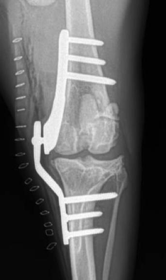
10 Simitri Stable in Stride Post-operative Radiographs
11 Simitri Plate Ordering Information Plates Cat# SIMI2.7L SIMI2.7R SIMI3.5-10L SIMI3.5-10R SIMI3.5-13L SIMI3.5-13R Description 2.7mm Simitri Plate Left 2.7mm Simitri Plate Right 3.5mm Simitri Plate with 10mm offset Left 3.5mm Simitri Plate with 10mm offset Right 3.5mm Simitri Plate with 13mm offset Left 3.5mm Simitri Plate with 13mm offset Right Screws Cat# Description Length LSTCBS2.7-xx 2.7mm Locking Self-Tapping Screws 10mm-34mm in 2mm increments LSTCBS3.5-xx 3.5mm Locking Self-Tapping Screws 12mm-40mm in 2mm increments LSTCBS3.5-xx 3.5mm Locking Self-Tapping Screws 40mm-60mm in 5mm increments LSTCBS4.0-xx 4.0mm Locking Self-Tapping Screws 20mm-40mm in 2mm increments LSTCBS4.0-xx 4.0mm Locking Self-Tapping Screws 40mm-60mm in 5mm increments Instruments Cat# LDG2.7S LDG3.5S LDG4.0S DBQC2.0 DB2.0 DBQC2.5 DB2.5 DBQC3.2 DB3.2 Description Locking Drill Guide for 2.7mm Screws Locking Drill Guide for 3.5mm Screws Locking Drill Guide for 4.0mm Screws 2.0mm Drill Bit with AO Quick Coupling 2.0mm Drill Bit 2.5mm Drill Bit with AO Quick Coupling 2.5mm Drill Bit 3.2mm Drill Bit with AO Quick Coupling 3.2mm Drill Bit US Pat. D735, 861S Additional Patents Pending
12 Simitri Surgical Equipment List Scalpel handles: Standard with a # 10 blade. Beaver handle with a # 64 blade Hemostats: 6 curved, 1 straight Mayo scissors: 1 straight, 1 curved (heavy) Towel clamps: 6-8 small Periosteal elevator Meniscal probe Retractors: Weitlaner, Senn, US Army, Hohmann, Gelpi Needle driver Tissue forceps: 1 Addison, 1 tissue (rat tooth), 1 Allis IM pins. 5/64th Three trocar / trocar cut in half Metal ruler or calipers Assortment of appropriate drills, taps, hex head screwdrivers and drill guides appropriate for 2.7 mm, 3.5 mm and 4.0 mm cortical locking screws Quick connect chuck Drill, key Plate bender, twisting irons 2.7, 3.5 and 4.0 mm cortical, cancellous and cortical locking screws 22, 25 gauge needle (3/4-1 inch) 35 cc syringe and 22 gauge needle (1.5 in.) for drill cooling Simitri S.T. -- February, 2016
SIMITRI STABLE IN STRIDE SURGICAL PROCEDURE
 Copyright 2016 NGD. All rights reserved Neil Embleton, B.Sc., DVM and Veronica Barkowski, DVM Helivet Mobile Surgical Services, Sundre, AB, Canada July 2016 SIMITRI STABLE IN STRIDE SURGICAL PROCEDURE
SIMITRI STABLE IN STRIDE
 Copyright 2016 NGD. All rights reserved Neil Embleton, B.Sc., DVM and Veronica Barkowski, DVM Helivet Mobile Surgical Services, Sundre, AB, Canada July 2016 SIMITRI STABLE IN STRIDE 1. SIMITRI STABLE IN
Extracapsular Repair Monofilament Nylon Suture
 Extracapsular Repair Monofilament Nylon Suture Management of the ruptured Cranial Cruciate Ligament (CCL) by placing a non-absorbable suture between the lateral fabella and the proximal, cranial tibia
PediLoc 3.5mm and 4.5mm Contour Femur Plate Surgical Technique
 PediLoc 3.5mm and 4.5mm Contour Femur Plate Surgical Technique Surgical Technique Contour Femur Plate The technique description herein is made available to the healthcare professional to illustrate the
MINI TIBIAL PLATEAU LEVELING OSTEOTOMY (TPLO) SYSTEM
 SYSTEM") MINI TIBIAL PLATEAU LEVELING OSTEOTOMY (TPLO) SYSTEM For stabilizing osteotomies of the canine and feline proximal tibia SURGICAL TECHNIQUE TABLE OF CONTENTS INTRODUCTION Mini Tibial Plateau Leveling
MINI TIBIAL PLATEAU LEVELING OSTEOTOMY (TPLO) SYSTEM For stabilizing osteotomies of the canine and feline proximal tibia SURGICAL TECHNIQUE TABLE OF CONTENTS INTRODUCTION Mini Tibial Plateau Leveling
Technique Guide. 3.5 mm LCP Low Bend Medial Distal Tibia Plate Aiming Instruments. Part of the 3.5 mm LCP Percutaneous Instrument System.
 Technique Guide 3.5 mm LCP Low Bend Medial Distal Tibia Plate Aiming Instruments. Part of the 3.5 mm LCP Percutaneous Instrument System. Table of Contents Introduction 3.5 mm LCP Low Bend Medial Distal
3.5 MM VA-LCP PROXIMAL TIBIA PLATE SYSTEM
 3.5 MM VA-LCP PROXIMAL TIBIA PLATE SYSTEM Part of the DePuy Synthes Variable Angle Periarticular Plating System SURGICAL TECHNIQUE TABLE OF CONTENTS INTRODUCTION 3.5 mm VA-LCP Proximal Tibial Plate 2 AO
Veronica J. Barkowski and Neil A. Embleton
 Surgical Technique and Initial Clinical Experience with a Novel Extracapsular Articulating Implant for Treatment of the Canine Cruciate Ligament Deficient Stifle Joint Veronica J. Barkowski and Neil A.
LCP Medial Distal Tibia Plate, without Tab. The Low Profile Anatomic Fixation System with Angular Stability and Optimal Screw Orientation.
 LCP Medial Distal Tibia Plate, without Tab. The Low Profile Anatomic Fixation System with Angular Stability and Optimal Screw Orientation. Technique Guide LCP Small Fragment System Table of Contents Introduction
Surgical Technique. Lower Extremity Plates and Straight Plates
 Surgical Technique Lower Extremity Plates and Straight Plates 2 Table of contents Overview...4 Indications... 4 Contraindications... 4 Screw Options... 5 Straight Plate Options... 6 Proximal Tibia Plate
KineSpring Knee Implant System Surgical Technique Guide
 KineSpring Knee Implant System Surgical Technique Guide Copyright Trademarks Patents Requests For Information 2014 Moximed, Inc. All rights reserved. No part of this publication may be reproduced, transmitted,
Surgical Technique. Proximal Humerus Locking Plate
 Surgical Technique Proximal Humerus Locking Plate PERI-LOC Upper Extremity Locked Plating System 3.5mm & 4.5mm Proximal Humerus Locking PlatesCatalog Infor Table of Contents Introduction.........................................................2
PediLoc 3.5mm and 4.5mm Bowed Femur Plate Surgical Technique
 PediLoc 3.5mm and 4.5mm Bowed Femur Plate Surgical Technique 2957 Bow Broch_REV_B.indd 1 2/10/11 12:47 PM Surgical Technique Bowed Femur Plate The technique description herein is made available to the
Surgical Technique. Distal Humerus Locking Plate
 Surgical Technique Distal Humerus Locking Plate PERI-LOC Locked Plating System Distal Humerus Locking Plate Surgical Technique Table of Contents Introduction...2 Indications...3 Plate Features...3 Patient
Surgical Technique. Olecranon Locking Plate
 Surgical Technique Olecranon Locking Plate PERI-LOC Locked Plating System Olecranon Locking Plate Surgical Techniquealog Infor Table of Contents Introduction...2 Indications...3 Plate Features...3 Patient
Surgical Technique. Targeter Systems Overview
 Surgical Technique Targeter Systems Overview PERI-LOC Locked Plating System Targeter Systems Overview Table of contents Product overview... 2 Introduction... 2 Indications... 2 Design features and benefits...
Surgical Technique. Clavicle Locking Plate
 Surgical Technique Clavicle Locking Plate PERI-LOC Locked Plating System Clavicle Locking Plate Surgical Technique Table of Contents Introduction...2 Indications...3 Plate Features...3 Patient Positioning...4
TTA Rapid with Patellar Luxation
 TTA Rapid with Patellar Luxation The dog is placed in a dorsal recumbency with the affected limb suspended from a stand. Make sure that the dog s paws are not fixed too tightly, since the affected limb
SMV Scientific Bone Plate and Screw System Surgical Technique
 SMV Scientific Bone Plate and Screw System Surgical Technique Description: The SMV Scientific Bone Plate and Screw System consists of non-locking plates and bone screw fasteners in a variety of lengths,
Surgical Technique. Anterolateral and Medial Distal Tibia Locking Plates
 Surgical Technique Anterolateral and Medial Distal Tibia Locking Plates PERI-LOC Periarticular Locked Plating System Anterolateral and Medial Distal Tibia Locking Plates Surgical Technique Contents Product
Surgical Technique. 3.5mm and 4.5mm Lateral Proximal Tibia Locking Plates
 Surgical Technique 3.5mm and 4.5mm Lateral Proximal Tibia Locking Plates PERI-LOC Periarticular Locked Plating System 3.5mm and 4.5mm Lateral Proximal Tibia Locking Plate Surgical Technique Contents Product
LCP Anterolateral Distal Tibia Plate 3.5. The low profile anatomic fixation system with optimal plate placement and angular stability.
 LCP Anterolateral Distal Tibia Plate 3.5. The low profile anatomic fixation system with optimal plate placement and angular stability. Technique Guide LCP Small Fragment System Table of Contents Introduction
Technique Guide. 3.5 mm LCP Low Bend Medial Distal Tibia Plates. Part of the Synthes locking compression plate (LCP) system.
 system.") Technique Guide 3.5 mm LCP Low Bend Medial Distal Tibia Plates. Part of the Synthes locking compression plate (LCP) system. Table of Contents Introduction 3.5 mm LCP Low Bend Medial Distal Tibia Plates
Technique Guide 3.5 mm LCP Low Bend Medial Distal Tibia Plates. Part of the Synthes locking compression plate (LCP) system. Table of Contents Introduction 3.5 mm LCP Low Bend Medial Distal Tibia Plates
Technique Guide. LCP Proximal Femoral Hook Plate 4.5/5.0. Part of the LCP Periarticular Plating System.
 Technique Guide LCP Proximal Femoral Hook Plate 4.5/5.0. Part of the LCP Periarticular Plating System. Table of Contents Introduction Features and Benefits 2 AO ASIF Principles 4 Indications 5 Surgical
Technique Guide. TomoFix Osteotomy System. A comprehensive plating system for stable fixation of osteotomies around the knee.
 Technique Guide TomoFix Osteotomy System. A comprehensive plating system for stable fixation of osteotomies around the knee. Table of Contents Introduction TomoFix Osteotomy System 2 AO Principles 4 Indications
LCP Anterolateral Distal Tibia Plate 3.5. The low profile anatomic fixation system with optimal plate placement and angular stability.
 LCP Anterolateral Distal Tibia Plate 3.5. The low profile anatomic fixation system with optimal plate placement and angular stability. Technique Guide LCP Small Fragment System Table of Contents Introduction
Surgical Technique. CONQUEST FN Femoral Neck Fracture System
 Surgical Technique CONQUEST FN Femoral Neck Fracture System Table of Contents Introduction... 3 Indications... 3 Product Overview... 4 Surgical Technique... 5 Patient Positioning... 5 Reduce the Fracture...
Surgical Technique International Version. Clavicle Locking Plate
 Surgical Technique International Version Clavicle Locking Plate PERI-LOC Upper Extremity Locked Plating System Clavicle Surgical Techniquefor Table of Contents Introduction........................................................2
3.5 mm LCP Low Bend Medial Distal Tibia Plate Aiming Instruments
 Part of the 3.5 mm LCP 3.5 mm LCP Low Bend Medial Distal Tibia Plate Aiming Instruments Surgical Technique TABLE OF CONTENTS INTRODUCTION 3.5 mm LCP Low Bend Medial Distal Tibia Plate 2 Aiming Instruments
LCP Anterior Ankle Arthrodesis Plates. Part of the Synthes Locking Compression Plate (LCP) System.
 System.") LCP Anterior Ankle Arthrodesis Plates. Part of the Synthes Locking Compression Plate (LCP) System. Technique Guide Instruments and implants approved by the AO Foundation Table of Contents Introduction
LCP Anterior Ankle Arthrodesis Plates. Part of the Synthes Locking Compression Plate (LCP) System. Technique Guide Instruments and implants approved by the AO Foundation Table of Contents Introduction
WINSTA-C. Clavicle Plating System
 Clavicle Plating System Clinical Advisor Michael Kurer FRCS FRCS (Orth) Consultant Orthopaedic and Shoulder Surgeon North Middlesex University Hospital NHS Trust Table of Contents Introduction Indication
Zimmer Small Fragment Universal Locking System. Surgical Technique
 Zimmer Small Fragment Universal Locking System Surgical Technique Zimmer Small Fragment Universal Locking System 1 Zimmer Small Fragment Universal Locking System Surgical Technique Table of Contents Introduction
Locking Ankle Plating System. Surgical Technique
 Locking Ankle Plating System Surgical Technique Acumed is a global leader of innovative orthopaedic and medical solutions. We are dedicated to developing products, service methods, and approaches that
Surgical Technique. Calcaneal Locking Plate
 Surgical Technique Calcaneal Locking Plate PERI-LOC Locked Plating System Calcaneal Locking Plate Surgical TechniqueCatalog Infor Table of Contents Introduction...2 Indications...3 Plate Features...3 Patient
AcUMEDr. LoCKING CLAVICLE PLATE SYSTEM
 AcUMEDr LoCKING CLAVICLE PLATE SYSTEM LoCKING CLAVICLE PLATE SYSTEM Since 1988 Acumed has been designing solutions to the demanding situations facing orthopedic surgeons, hospitals and their patients.
Zimmer MIS Periarticular 3.5mm Proximal Tibial Locking Plate
 Zimmer MIS Periarticular 3.5mm Proximal Tibial Locking Plate Surgical Technique The Science of the Landscape Zimmer MIS Periarticular 3.5mm Proximal Tibial Locking Plate Surgical Technique 1 Zimmer MIS
Technique Guide. 3.5 mm LCP Periarticular Proximal Humerus Plate. Part of the Synthes locking compression plate (LCP) system.
 system.") Technique Guide 3.5 mm LCP Periarticular Proximal Humerus Plate. Part of the Synthes locking compression plate (LCP) system. Table of Contents Introduction 3.5 mm LCP Proximal Humerus Plate 2 AO Principles
Technique Guide 3.5 mm LCP Periarticular Proximal Humerus Plate. Part of the Synthes locking compression plate (LCP) system. Table of Contents Introduction 3.5 mm LCP Proximal Humerus Plate 2 AO Principles
TOMOFIX Medial Distal Femur Plate
 For Closed-Wedge Varus Femoral Osteotomies TOMOFIX Medial Distal Femur Plate Surgical Techniquee TABLE OF CONTENTS INTRODUCTION TOMOFIX Medial Distal Femur Plate 2 AO Principles 4 Indications 5 SURGICAL
LCP Distal Humerus Plates
 The anatomic fixation system for the distal humerus with angular stability Surgical technique LCP Locking Compression Plate Contents Indications and contraindications 2 Implants 3 Instruments 5 Preparation
TTA. Common Tangent Method
 TTA Common Tangent Method This document is derived from a presentation by Dr. Randy Boudrieau DVM, Dipl. ACVS, ECVS, Prof. of Surgery, Cummings School of Veterinary Medicine, Tufts University IVET DESIG
PROXIMAL TIBIAL PLATE
 SURGICAL NÁSTROJE TECHNIQUE PRO ARTROSKOPII PROXIMAL INSTRUMENTS TIBIAL FOR PLATE ARTHROSCOPY Proximal Tibial Plate Description of medical device The Proximal Tibial Plate is used in epyphyseal and metaphyseal
LCP Medial Proximal Tibial Plate 3.5. Part of the Synthes small fragment Locking Compression Plate (LCP) system.
 system.") LCP Medial Proximal Tibial Plate 3.5. Part of the Synthes small fragment Locking Compression Plate (LCP) system. Technique Guide This publication is not intended for distribution in the USA. Instruments
LCP Medial Proximal Tibial Plate 3.5. Part of the Synthes small fragment Locking Compression Plate (LCP) system. Technique Guide This publication is not intended for distribution in the USA. Instruments
JOINT RULER. Surgical Technique For Knee Joint JRReplacement
 JR JOINT RULER Surgical Technique For Knee Joint JRReplacement INTRODUCTION The Joint Ruler * is designed to help reduce the incidence of flexion, extension, and patellofemoral joint problems by allowing
LCP Medial Proximal Tibial Plate 4.5/5.0. Part of the Synthes LCP periarticular plating system.
 LCP Medial Proximal Tibial Plate 4.5/5.0. Part of the Synthes LCP periarticular plating system. Technique Guide This publication is not intended for distribution in the USA. Instruments and implants approved
Periarticular Aiming Arm Instruments for LCP Proximal Tibial Plate 4.5/5.0. Part of the LCP Periarticular Aiming Arm Instrument System (large).
.") Technique Guide Periarticular Aiming Arm Instruments for LCP Proximal Tibial Plate 4.5/5.0. Part of the LCP Periarticular Aiming Arm Instrument System (large). Image intensifier control Warning This description
Technique Guide Periarticular Aiming Arm Instruments for LCP Proximal Tibial Plate 4.5/5.0. Part of the LCP Periarticular Aiming Arm Instrument System (large). Image intensifier control Warning This description
Distal Radius Plate Instrument and Implant Set. Discontinued December 2017 DSUS/TRM/0916/1063(1)
") Distal Radius Plate Instrument and Implant Set Surgical Technique Discontinued December 2017 DSUS/TRM/0916/1063(1) The Distal Radius Plates Indications For fixation of fractures and osteotomies, including
Distal Radius Plate Instrument and Implant Set Surgical Technique Discontinued December 2017 DSUS/TRM/0916/1063(1) The Distal Radius Plates Indications For fixation of fractures and osteotomies, including
Zimmer NexGen MIS Tibial Component. Cemented Surgical Technique IMAGE TO COME
 Zimmer NexGen MIS Tibial Component Cemented Surgical Technique IMAGE TO COME Zimmer NexGen MIS Tibial Component Cemented Surgical Technique 1 Zimmer NexGen MIS Tibial Component Cemented Surgical Technique
Surgical Technique. Locking Small Fragment Overview
 Surgical Technique Locking Small Fragment Overview PERI-LOC Locked Plating System Locking Small Fragment Overview Surgical Technique Table of contents Product overview... 2 Introduction... 2 Indications...
TTA Wedge System INSTRUCTIONS FOR USE
 TTA Wedge System INSTRUCTIONS FOR USE 1 INSTRUCTIONS FOR USE The OssAbility TTA Wedge System consists of the following products: Wedge Implants Osteotomy Guide Advancement Levers Osteotomy Planning Overlay
LCP Low Bend Medial Distal Tibia Plates 3.5 mm. Anatomic plates with low profile head for intra- and extraarticular fractures.
 LCP Low Bend Medial Distal Tibia Plates 3.5 mm. Anatomic plates with low profile head for intra- and extraarticular fractures. Surgical Technique This publication is not intended for distribution in the
3.5 mm Locking Attachment Plate
 For Treatment of Periprosthetic Fractures 3.5 mm Locking Attachment Plate Surgical Technique Table of Contents Introduction 3.5 mm Locking Attachment Plate 2 Indications 4 Surgical Technique Preparation
VA-LCP Condylar Plate 4.5/5.0. Part of the Synthes Variable Angle Periarticular Plating System.
 VA-LCP Condylar Plate 4.5/5.0. Part of the Synthes Variable Angle Periarticular Plating System. Technique Guide This publication is not intended for distribution in the USA. Instruments and implants approved
Technique Guide. VA-Locking Intercarpal Fusion System. Variable angle locking technology for mediocarpal partial arthrodesis.
 Technique Guide VA-Locking Intercarpal Fusion System. Variable angle locking technology for mediocarpal partial arthrodesis. Table of Contents Introduction VA-Locking Intercarpal Fusion System 2 Indications
Opening Wedge Osteotomy
 Tibial Opening Wedge Osteotomy System with Titanium Plates and Screws and OSferion B-TCP Osteotomy Wedge Surgical Technique Opening Wedge Osteotomy Tibial Opening Wedge Osteotomy System w/titanium Plates
AFX. Femoral Implant. System. The AperFix. AM Portal Surgical Technique Guide. with the. The AperFix System with the AFX Femoral Implant
 The AperFix System AFX with the Femoral Implant AM Portal Surgical Technique Guide The Cayenne Medical AperFix system with the AFX Femoral Implant is the only anatomic system for soft tissue ACL reconstruction
Knee Surgical Technique
 Knee Surgical Technique COMPASS Universal Hinge by Jimmy Tucker, M.D. Orthopaedic Surgeon Director, Arkansas Sports Medicine, P.A. Little Rock, Arkansas Table of contents Design features 3 Indications
4.5 mm VA-LCP. Part of the Variable Angle Periarticular Plating System
 4.5 mm VA-LCP Curved Condylar Plate Part of the Variable Angle Periarticular Plating System Surgical Technique Table of Contents Introduction 4.5 mm VA-LCP Curved Condylar Plates 2 4.5 mm VA-LCP Curved
Proceedings of the World Small Animal Veterinary Association Sydney, Australia 2007
 Proceedings of the World Small Animal Sydney, Australia 2007 Hosted by: Next WSAVA Congress CRANIAL CRUCIATE LIGAMENT INJURIES SURGICAL MANAGEMENT Warrick J. Bruce BVSc(dist), MVM, DSAS(orthopaedics),
NCB Proximal Humerus Plating System
 NCB Proximal Humerus Plating System Surgical Technique The right locking option for tough fractures Disclaimer This document is intended exclusively for experts in the field, i.e. physicians in particular,
Locking Radial Head Plates
 Locking Radial Head Plates Locking Radial Head Plates Since 1988, Acumed has been designing solutions to the demanding situations facing orthopaedic surgeons, hospitals and their patients. Our strategy
Surgical Technique. Cannulated Angled Blade Plate 3.5 and 4.5, 90
 Surgical Technique Cannulated Angled Blade Plate 3.5 and 4.5, 90 Cannulated Angled Blade Plate 3.5 and 4.5, 90 Table of contents Indications/Contraindications 2 Implants 3 Surgical technique 5 Implant
GASTROCNEMIUS TENDON REPAIR VETLIG USING THE STIF CAT 30 SOFT TISSUE INTERNAL FIXATION VETLIG
 VETLIG SOFT TISSUE INTERNAL FIXATION GASTROCNEMIUS TENDON REPAIR USING THE STIF CAT 30 VETLIG A R T I F I C I A L L I G A M E N T S F O R V E T E R I N A R Y U S E VETLIG MANAGEMENT OF CHRONIC GASTROCNEMIUS
Low Bend Distal Tibia Plates
 Part of the DePuy Synthes Locking Compression Plate (LCP ) System 3.5 mm LCP Low Bend Medial Distal Tibia Plates Surgical Technique Table of Contents Introduction 3.5 mm LCP Low Bend Medial Distal Tibia
TRK REVISION KNEE Surgical Technique
 1 TRK REVISION KNEE Surgical Technique 1. 2. 3. 4. 5. 6. 7. 8. 9. 10. INTERCONDYLAR RESECTION...... page FEMORAL STEM...... page NON CEMENTED FEMORAL STEM...... page TRIAL FEMORAL COMPONENTS...... page
LCP Superior Clavicle Plate. The anatomically precontoured fixation system with angular stability for clavicle shaft and lateral clavicle.
 Technique Guide LCP Superior Clavicle Plate. The anatomically precontoured fixation system with angular stability for clavicle shaft and lateral clavicle. Table of Contents Introduction LCP Superior Clavicle
TTA-Rapid Protocol. i. Where possible, calibrate the radiograph on the screen to real size.
 Legeweg 157 i 8020 Oostkamp Tel: 050/31.18.76 Fax: 050/31.58.86 www.instrulife.be TTA-Rapid Protocol The dog is placed in a dorsal recumbency with the affected limb suspended from a stand. Make sure that
Figure 3 Figure 4 Figure 5
 Figure 1 Figure 2 Begin the operation with examination under anesthesia to confirm whether there are any ligamentous instabilities in addition to the posterior cruciate ligament insufficiency. In particular
NeoGen Femoral Nail System
 NeoGen Femoral Nail System LESS IS MORE TE-2070-04 Surgical Technique BLE OF CONTENT Preface Standard Femoral Mode Recon Mode Post-Operative Management Appendix Products Information Indication Patient
LCP Distal Tibia Plate
 Surgical Technique LCP Locking Compression Plate Original Instruments and Implants of the Association for the Study of Internal Fixation AO/ASIF Table of contents Indications 3 Implants/Instruments 5 Surgical
A locking plate system that expands a surgeon s options in trauma surgery. Zimmer NCB Plating System
 A locking plate system that expands a surgeon s options in trauma surgery Zimmer NCB Plating System The Power of Choice The power of having true intraoperative options is at your fingertips. Using standard
Conventus CAGE PH Surgical Techniques
 Conventus CAGE PH Surgical Techniques Conventus Orthopaedics The Conventus CAGE PH (PH Cage) is a permanent implant comprised of an expandable scaffold, made from nitinol and titanium, which is deployed
Zimmer FuZion Instruments. Surgical Technique (Beta Version)
") Zimmer FuZion Surgical Technique (Beta Version) INTRO Surgical Technique Introduction Surgical goals during total knee arthroplasty (TKA) include establishment of normal leg alignment, secure implant fixation,
Zimmer FuZion Surgical Technique (Beta Version) INTRO Surgical Technique Introduction Surgical goals during total knee arthroplasty (TKA) include establishment of normal leg alignment, secure implant fixation,
Triple Tibial Osteotomy (TTO)
") Triple Tibial Osteotomy (TTO) Objective: This operation is based on the biomechanical analysis performed by Dr Slobodan Tepic, which revealed that in order to remove the shear strain from the cranial cruciate
Triple Tibial Osteotomy (TTO) Objective: This operation is based on the biomechanical analysis performed by Dr Slobodan Tepic, which revealed that in order to remove the shear strain from the cranial cruciate
Orthopedic Bone Nail System - Distal Femoral Nail Surgical Technique Manual
 Orthopedic Bone Nail System - Distal Femoral Nail Surgical Technique Manual Note: The surgical procedures should be performed under the guidance of qualified skilled orthopedic surgeons, and this surgical
ACL Reconstruction for BTB Grafts
 Transtibial ACL Reconstruction System for BTB Grafts Surgical Technique Designed in conjunction with John C. Garrett, M.D., Atlanta, GA ACL Reconstruction for BTB Grafts Reference Anatomical Constants
Total Knee Replacement
 Total Knee Replacement A total knee replacement, also known as total knee arthroplasty, involves removing damaged portions of the knee, and capping the bony surfaces with man-made prosthetic implants.
Case Report. Antegrade Femur Lengthening with the PRECICE Limb Lengthening Technology
 Case Report Antegrade Femur Lengthening with the PRECICE Limb Lengthening Technology S. Robert Rozbruch, MD Hospital for Special Surgery New York, NY, USA ABSTRACT This is a case illustrating a 4.5 cm
DOUBLE/TRIPLE PELVIC OSTEOTOMY PLATES For Treating Coxofemoral Joint Instability and Subluxation in Immature Dogs
 DOUBLE/TRIPLE PELVIC OSTEOTOMY PLATES For Treating Coxofemoral Joint Instability and Subluxation in Immature Dogs Instruments and implants approved by the AO Foundation. This publication is not intended
2.7 mm/3.5 mm Variable Angle LCP Elbow System DJ9257-B 1
 2.7 mm/3.5 mm Variable Angle LCP Elbow System DJ9257-B 1 System overview Simply complete: A comprehensive system, consisting of five (5) distal humerus plates and three (3) types of olecranon plates Implant
Technique Guide. TomoFix Medial Distal Femur (MDF). For closed-wedge varus femoral osteotomies.
. For closed-wedge varus femoral osteotomies.") Technique Guide TomoFix Medial Distal Femur (MDF). For closed-wedge varus femoral osteotomies. Table of Contents Introduction Features and Benefits of the TomoFix Knee 2 Osteotomy System Osteotomy Principles
Technique Guide TomoFix Medial Distal Femur (MDF). For closed-wedge varus femoral osteotomies. Table of Contents Introduction Features and Benefits of the TomoFix Knee 2 Osteotomy System Osteotomy Principles
Surgical Technique. VISIONAIRE FastPak Instruments for the LEGION Total Knee System
 Surgical Technique VISIONAIRE FastPak Instruments for the LEGION Total Knee System VISIONAIRE FastPak for LEGION Instrument Technique* Nota Bene The technique description herein is made available to the
Instrument and Implant for wrist fracture
 Instrument and Implant for wrist fracture Jansri Janpanya Product specialist The Bangkok Unitrade Co,.ltd. Objectives Type of LCP for distal radius Fx. The new LCP design for distal radius Fx. Have knowledge
Technique Guide. *smith&nephew N8TIVE ACL Anatomic ACL Reconstruction System
 Technique Guide *smith&nephew N8TIVE ACL Anatomic ACL Reconstruction System N8TIVE ACL System The N8TIVE ACL Anatomic Reconstruction System provides a novel and simple approach to ACL repair. The N8TIVE
Tibial & Femoral Opening Wedge Osteotomy System. Surgical Technique
 Tibial & Femoral Opening Wedge Osteotomy System Surgical Technique Opening Wedge Osteotomy Tibial & Femoral Opening Wedge Osteotomy 2 Prior to the osteotomy, a diagnostic arthroscopy is performed to verify
PediLoc Extension Osteotomy Plate (PLEO)
") PediLoc Extension Osteotomy Plate (PLEO) Left PLEO Plates Sizes: 6, 8 and 10 hole plates Right PLEO Plates Sizes: 6, 8 and 10 hole plates PediLoc Extension Osteotomy Plate The technique description herein
PediLoc Extension Osteotomy Plate (PLEO) Left PLEO Plates Sizes: 6, 8 and 10 hole plates Right PLEO Plates Sizes: 6, 8 and 10 hole plates PediLoc Extension Osteotomy Plate The technique description herein
Pre-Operative Planning. Positioning of the Patient
 Surgical Technique Pre-Operative Planning Decide upon the size and angle of the barrel plate to be used from measuring the x-rays. To maximise the sliding action when using shorter lag screws, the Short
Total Knee Original System Primary Surgical Technique
 Surgical Procedure Total Knee Original System Primary Surgical Technique Where as a total hip replacement is primarily a bony operation, a total knee replacement is primarily a soft tissue operation. Excellent
3.5 mm LCP Extra-articular Distal Humerus Plate
 Part of the DePuy Synthes Locking Compression Plate (LCP ) System 3.5 mm LCP Extra-articular Distal Humerus Plate Surgical Technique Table of Contents Introduction 3.5 mm LCP Extra-articular Distal Humerus
Medial Patellofemoral Ligament (MPFL) Surgical Technique
 Surgical Technique") Medial Patellofemoral Ligament (MPFL) Surgical Technique Medial Patellofemoral Ligament The medial patellofemoral complex, consisting of the medial patellofemoral ligament (MPFL) and the medial patellotibial
Medial Patellofemoral Ligament (MPFL) Surgical Technique Medial Patellofemoral Ligament The medial patellofemoral complex, consisting of the medial patellofemoral ligament (MPFL) and the medial patellotibial
Femur. Monoaxial Locking Plate System. Operative Technique. Distal Lateral Femur Universal Holes Targeting Instrumentation.
 Femur AxSOS 3 Titanium Monoaxial Locking Plate System Femur Fractures Operative Technique Distal Lateral Femur Universal Holes Targeting Instrumentation This publication sets forth detailed recommended
Revolution. Unicompartmental Knee System
 Revolution Unicompartmental Knee System While Total Knee Arthroplasty (TKA) is one of the most predictable procedures in orthopedic surgery, many patients undergoing TKA are in fact excellent candidates
Olecranon Locking Plate II
 INDEX Indications Patient Position Fracture Reduction and Fixation Surgical Technique Step 1 Surgical Approach Step 2 Implantation Step 3 Proximal Locking Screw Insertion Step 4 Distal Screw Insertion
NCB Distal Femur System. Surgical Technique
 NCB Distal Femur System Surgical Technique NCB Distal Femur System Surgical Technique 3 Surgical Technique NCB Distal Femur System Table of Contents Introduction 4 Indications 8 Preoperative Planning
Cannulated Pediatric Osteotomy System (CAPOS). A single system of osteotomy blade plates and cannulated instrumentation.
. A single system of osteotomy blade plates and cannulated instrumentation.") Cannulated Pediatric Osteotomy System (CAPOS). A single system of osteotomy blade plates and cannulated instrumentation. Technique Guide This publication is not intended for distribution in the USA. Instruments
Cannulated Pediatric Osteotomy System (CAPOS). A single system of osteotomy blade plates and cannulated instrumentation. Technique Guide This publication is not intended for distribution in the USA. Instruments
Technique Guide. 2.4 mm Variable Angle LCP Distal Radius System. For fragment-specific fracture fixation with variable angle locking technology.
 Technique Guide 2.4 mm Variable Angle LCP Distal Radius System. For fragment-specific fracture fixation with variable angle locking technology. Table of Contents Introduction 2.4 mm Variable Angle LCP
CONTRIBUTING SURGEON. Barry Waldman, MD Director, Center for Joint Preservation and Replacement Sinai Hospital of Baltimore Baltimore, MD
 CONTRIBUTING SURGEON Barry Waldman, MD Director, Center for Joint Preservation and Replacement Sinai Hospital of Baltimore Baltimore, MD System Overview The EPIK Uni is designed to ease the use of the
LCP Superior Clavicle Plate. The anatomically precontoured fixation system with angular stability for clavicle shaft and lateral clavicle.
 LCP Superior Clavicle Plate. The anatomically precontoured fixation system with angular stability for clavicle shaft and lateral clavicle. Surgical Technique This publication is not intended for distribution
MIS Cemented Tibial Component
 MIS Cemented Tibial Component NexGen Complete Knee Solution Surgical Technique Table of Contents Surgical Exposure... 2 Finish the Tibia... 2 Position Based on Anatomic Landmarks... 3 Lateral Posterior