Operative Technique Resurfacing Humeral Head
|
|
|
- Milo Allen
- 6 years ago
- Views:
Transcription


1 EXACTECH SHOULDER Operative Technique Resurfacing Humeral Head

2 TABLE OF CONTENTS INTRODUCTION...1 RESURFACING HUMERAL HEAD...1 SYSTEM SPECIFICATIONS...1 DETAILED OPERATIVE TECHNIQUE...2 PATIENT POSITIONING...2 SURGICAL APPROACH...2 SIZING THE HUMERUS FOR A RESURFACING IMPLANT...5 K-WIRE...6 REAMING THE HUMERUS...6 IMPLANTATION OF GLENOID...7 TRIALING...7 DRILLING FOR THE HUMERAL CAGE...8 TAMPING FOR THE HUMERAL CAGE...8 IMPLANTING THE HUMERAL CAGE...9 IMPLANTING THE RESURFACING HUMERAL HEAD...10 BACK TABLE ASSEMBLY (OPTIONAL)...10 EXPLANTING THE DEVICES CLOSURE...12 POST-OPERATIVE REHABILITATION...13 IMPLANT SCOPE...14 INSTRUMENT SCOPE...14 INTRODUCTION The Equinoxe Shoulder System redefines anatomical. The design goal of the resurfacing humeral head is to offer a bone preserving treatment option for the skeletally mature patient with early-stage arthritis. The platform primary stem is designed to allow independent adjustability of all four anatomic parameters in situ. The reverse shoulder is an optimized design that is designed to minimize both scapular notching and torque on the glenoid while seamlessly integrating with the primary and platform fracture stems. The platform fracture stem s offset anterior-lateral fin and asymmetric tuberosity beds define the next generation in complex fracture reconstruction. The platform nature of the Equinoxe primary and fracture stem allows the surgeon to have intra-operative flexibility to choose between a surface replacement, humeral hemiarthroplasty, primary total shoulder or reverse total shoulder, with seamless conversion to a reverse should a revision become necessary. Thank you for considering the Equinoxe Shoulder System. We began the Equinoxe product development process by identifying concerns our team had with current shoulder replacement systems. Our goal was to develop solutions to those concerns, and we sought the following improvements: RESURFACING HUMERAL HEAD Modularity: Facilitates implantation through a cuff-preserving approach (if desired), leaving the subscapularis essentially intact. Anatomic Sizing: Prevents overstuffing of the joint and aids in restoring the patient s own unique humeral head anatomy with anatomic sized implants. Low Profile Instrumentation: Cannulated system facilitates a seamless transition between surgical steps. We hope that you come to agree, based on your experiences with the Equinoxe Shoulder System in the O.R., that we have accomplished our goals. Finally, while we have taken a comprehensive approach to this operative technique, we would be remiss if we failed to make it clear that shoulder replacements are challenging procedures and should be performed by surgeons with significant experience. If you are new to shoulder arthroplasty, please consider observing a shoulder specialist, watching a shoulder surgical DVD, performing a sawbone and/or implanting in a cadaver to ensure you are comfortable with the surgical technique. We would be happy to facilitate any aspect of this training to ensure A Great Day in the O.R. for the surgeon and the staff. Respectfully, Pierre-Henri Flurin, MDCurt Noel, MD Felix Buddy Savoie, MD Ryan Simovitch, MD Thomas W. Wright, MD Joseph D. Zuckerman, MD SYSTEM SPECIFICATIONS Cage Length Min. Diameter Max. Diameter 25mm 9.5mm 14.8mm 30mm 9.3mm 14.8mm Resurfacing Head Head Diameter Head Height 38 38mm 14mm 41 41mm 14mm 44 44mm 15mm 47 47mm 16mm 50 50mm 17mm 53 53mm 18mm Cage Length Max Diameter Min Diameter Head Height Head Diameter 1


3 DETAILED OPERATIVE TECHNIQUE As part of the pre-operative assessment, the surgeon must ensure that no biological, biomechanical, or other factors exist that might adversely affect the surgery and/or postoperative period. Bone quality must be considered. Inadequate or poor quality bone stock could result in disassociation of the modular components, aseptic loosening, or implant migration. It is recommended that bone quality be assessed via CT or other equivalent means. PATIENT POSITIONING The patient should be placed on an operating table in a supine position. The head of the operating table should be elevated approximately 30 degrees in a modified beach chair position. A small bolster should be placed laterally behind the involved shoulder. The patient should be moved to the side of the table so that the upper extremity can be placed into maximum extension without obstruction by the operating table. Alternatively, a Captain s chair or similar positioning device can be used for proper patient positioning. The patient should be secured to the operating table to minimize any changes in position intra-operatively. Once the patient is secure, the extremity is examined to assess the range of motion, with particular attention to external rotation with the arm at the side. If external rotation is restricted (i.e., internal rotation contracture) the need for more extensive subscapularis mobilization or lengthening procedures may be necessary. The entire upper extremity should be prepped and draped to allow complete access to the operative area and full mobility during the procedure. SURGICAL APPROACH An anterior deltopectoral incision is made beginning inferior to the clavicle and passing over the coracoid process and extending distally toward the deltoid insertion. Medial and lateral subcutaneous flaps are created, and the deltopectoral interval is identified. A thin fat stripe is usually located over the cephalic vein. The interval is usually developed medial to the cephalic vein; the interval can also be developed laterally depending on the surgeon s preference. Branches of the cephalic vein on the approach side are tied off or cauterized, and the interval is developed distal to proximal to expose the clavipectoral fascia. In cases where the cephalic vein is not easily identified distally, it may be more easily identified by exposing the coracoid proximally. Figure 1 Use of the Chandler Anterior Humeral Head Retractor Figure 2 Use of the Small Darrach Anterior Humeral Head Retractor The advantage of retracting the cephalic vein with the deltoid is that the majority of the branches come from the deltoid. The disadvantage is the vein is more exposed to injury from retractors as they crosses the superior aspect of the interval. The subdeltoid space is mobilized with a blunt elevator or curved Mayo scissors. Progressive internal rotation of the arm will allow further posterior release. The clavipectoral fascia is incised longitudinally up to the coracoacromial ligament and the conjoined tendon is mobilized. A self-retaining retractor is placed with care to avoid excessive traction on the conjoined tendon and musculocutaneous nerve. The coracoacromial ligament is identified and the subacromial space is mobilized with a blunt elevator or curved Mayo scissors. The coracoacromial ligament can be released based on pathology, surgeon preference or need for additional exposure. Releases of the subdeltoid and subacromial space must extend posteriorly to allow adequate visualization, especially of the glenoid. The subscapularis tendon insertion on the lesser tuberosity is identified along with the rotator interval. The anterior humeral circumflex vessels along the inferior border of the subscapularis muscle, the three sisters, are cauterized extensively or tied off, and the biceps tendon is palpated in its groove. The subscapularis tendon and the capsule are tenotomized 1cm medial to the lesser tuberosity and tagged with #1 sutures. One alternative approach is to elevate the subscapularis directly off of bone or elevate its insertion with a thin wafer of bone (1-2mm thick) using an osteotome. The choice is based primarily on surgeon preference. The rotator interval is divided in a lateral to medial direction up to the superior glenoid rim. With the humerus extended, adducted and externally rotated, the capsule is carefully dissected off the inferior humeral neck, protecting the axillary nerve inferiorly with a small blunt retractor placed just inferior to the capsule. The capsular releases should be performed to allow 90 degrees of external rotation. The selfretaining retractor is then repositioned to retract the subscapularis. At this point, the humeral head can be dislocated and the humeral head can be sized. Anterior humeral head retractors and a Browne deltoid retractor are supplied with the system to facilitate this exposure (Figures 1 and 2). 2 3


.")
.")
4 Another alternative approach is to retain the superior portion of the subscapularis and only reflect the bottom third to half. A full-thickness L-shaped incision is made in the inferior half of the subscapularis with the shoulder held in full external rotation. This incision should be made through both the subscapularis and the underlying joint capsule and extended laterally to within 1 cm of the bicipital groove. The vertical limb of the incision is then extended inferiorly parallel to the biceps tendon past the inferior edge of the subscapularis tendon insertion and is stopped at the pectoralis major insertion. The L-shaped flap of subscapularis and capsule is then completed and elevated with increasing shoulder external rotation until adequate visualization is obtained. The superior 50% of the subscapularis insertion is left intact. (Figures 3 and 4). Once this flap is completely developed, the humeral head is then delivered through the capsulotomy underneath the subscapularis (Figure 5). The head passes inferior to the remaining intact subscapularis tendon. After dislocation, the humeral head can be sized (Figure 6). Figure 3 Alternative Subscapularis Preserving Technique: L-Shaped Incision Figure 4 Alternative Subscapularis Preserving Technique: Elevation of the L-shaped Flap Figure 7 Sizing the Humeral Head SIZING THE HUMERUS FOR A RESURFACING IMPLANT The Resurfacing Humeral Head Sizer is placed onto the Impactor Handle. The Sizer is used to find the center of the humeral head and to determine the proper Resurfacing Humeral Head size (Figure 7). If there are osteophytes present around the humeral head, it is important to remove with a rongeur or osteotome, as they could lead to placing the Sizer incorrectly. The tendency is to place the Sizer in varus which must be avoided. The anatomic neck and rotator cuff should be used for reference of native anatomy. Each sizer is color coded to streamline measurement and selection of each instrument (Table 1). Once the Sizer is positioned properly, the 3mm x 250mm K-wire is placed through the cannulation in the handle and inserted into the humerus until the far cortex is penetrated (Figure 8). Penetration of the far cortex will ensure pin retention throughout the remaining steps of preparation and implant insertion. It is important not to plunge with the pin to avoid Axillary nerve trauma. Note: The K-wire should be positioned in the center of the humeral head to ensure concentric reaming. If the K-wire is not positioned in the center of the humeral head, eccentric reaming could result in removal of an insufficient amount of bone. Table 1 Colors are used to streamline selecting the surgical instruments Figure 5 Alternative Subscapularis Preserving Technique: Humeral Head Dislocation Under Upper Portion of the Subscapularis Size 38mm 41mm 44mm 47mm 50mm 53mm Color Black Blue Brown Green Orange Purple Figure 6 Alternative Subscapularis Preserving Technique: Humeral Head Exposure Figure 8 Placing K-Wire Through the Cannulated Handle 4 5
5 K-WIRE The remaining steps of the humeral preparation are performed over the K-wire. Selection of the Humeral Cage Implant length can be done by preoperative templating or determined intraoperatively. Note: The K-wire should be inserted to the depth of the lateral cortex but not penetrated. REAMING THE HUMERUS The handle is pulled off the K-wire leaving the K-wire in the humeral head (Figure 9). The initial Sizer chosen will dictate the Resurfacing Reamer size that is to be used. The Reamer is placed onto the Modular Reamer Handle and a powered handpiece is used to facilitate reaming (Figure 10). Reaming is done to remove the articular cartilage of the humeral head in preparation for the Resurfacing Humeral Head Implant. There is a stop on the Reamer body to help avoid excessive reaming. However, care must be exercised with soft bone, collapsed bone or in cases of significant head deformity not to over ream. Subchondral bone should be preserved. Note: The humeral head should be reamed fully as determined by complete seating of the reamer on the bone surface. Failure to fully ream the humeral head could result in incorrect implant placement, instability, bone fracture and excessive joint tissue tensioning. There are two laser marked lines on the K-wire. If the first line is below the surface of the Impactor Handle, then the K-wire has been positioned deep enough to prepare for the 25mm Humeral Cage. If both lines are below the surface of the Impactor Handle, then the K-wire has been positioned deep enough to prepare for the 30mm Humeral Cage. Figure 9 K-wire Placement in the Humeral Head Figure 11 Trialing the Resurfacing Humeral Head IMPLANTATION OF GLENOID After reaming, if a prosthetic glenoid component will be implanted, refer to the Equinoxe Platform Shoulder System Surgical Technique or the Equinoxe Posterior Augment Glenoid Surgical Technique Addendum. Glenoid exposure is the challenging portion of shoulder resurfacing. Generally, it is easier to accomplish in patients with smaller body habitus and who maintain a good degree of their shoulder motion preoperatively. Exposure depends on adequate osteophyte resection and soft tissue release during the initial exposure. Care must be exercised not to compress the humeral head with retractors while exposing the glenoid because this bone is necessary to support the humeral head prosthesis. During the initial approach as described above, an aggressive subscapularis mobilization was done. This should have included release of the inferior capsule (protect axillary nerve) and coracohumeral ligament. To provide additional exposure the upper 1/3 to 1/2 of the Pectoralis Major tendon can be released (repaired at completion of the case). Next, to release the posterior and superior capsule, a laminar spreader is inserted and used to distract the humeral head and glenoid. With the forearm in neutral rotation, the capsule is released just off of the glenoid rim until the muscles of the posterior and superior rotator cuff are visualized. Release of this capsule creates a pocket in which to displace the humeral head. A 360 degree release of the labrum is done to help with exposure. A Fukuda retractor or other humeral head retractor can be used to retract the humeral head posteriorly and inferiorly. It is important to make sure the bed or arm positioner is not preventing displacement of the humeral head posteriorly. TRIALING After humeral head reaming, the Trial is placed over the K-wire to verify that adequate reaming has occurred for placement of the Resurfacing Head (Figure 11). The windows of the Trial provide visibility of the reamed bone. The crown (most proximal) portion of the trial should be resting on the prepared bone surface. Figure 10 Reaming the Humeral Head Over the K-wire 6 7


6 DRILLING FOR THE HUMERAL CAGE After trialing, the appropriately sized Cannulated Drill is then attached to the Modular Reamer Handle and placed over the K-wire (Figure 12). The 25mm Drill is used to prepare for the 25mm Humeral Cage, and the 30mm Drill is used to prepare for the 30mm Humeral Cage. The drill has a stop to prevent over/under drilling. TAMPING FOR THE HUMERAL CAGE The Tamp is used to prepare the humerus for the fin pattern of the Humeral Cage Peg. The Tamp should be connected to the Impactor Handle and Impacted with a mallet. To enable axial control, the impactor handle has a retention feature: by twisting the tamp on the inserter handle, the tamp may be axially extracted during/after impaction. Additionally, the tamp has a stop to prevent over/under tamping (Figure 13). This step should be done over the K-wire. IMPLANTING THE HUMERAL CAGE The Cage Impactor Tip is placed onto the Impactor Handle, and is used to impact the Humeral Cage into the humerus. Insertion of the humeral cage may be performed over the K-wire (Figure 14). Note that the fins of the Humeral Cage will be held in place with a press-fit on the Impactor Tip. The Impactor Handle with the connected Impactor Tip is then impacted with a mallet (Figure 15). The Impactor Tip has a positive stop so that the Humeral Cage is left approximately 1mm proud after Impaction (Figure 16). This is done to ensure that the Resurfacing Head taper engages the female taper of the Humeral Cage. Note: Care should be taken to ensure that the fins of the Humeral Cage are properly aligned. Note: If the bone is soft and the surgeon is concerned about the ability to engage the morse taper between the humeral head and cage insitu, a back table assembly can be done. (See following section.) Figure 12 Drilling the Humeral Cage Peg Figure 14 Inserting the Humeral Cage Peg Over K-wire Figure 16 Humeral Cage Peg Left Approximately 1mm Proud After Impaction Figure 13 Tamping the Humeral Cage Peg Figure 15 Impacting the Humeral Cage Peg 8 9
.")


.")

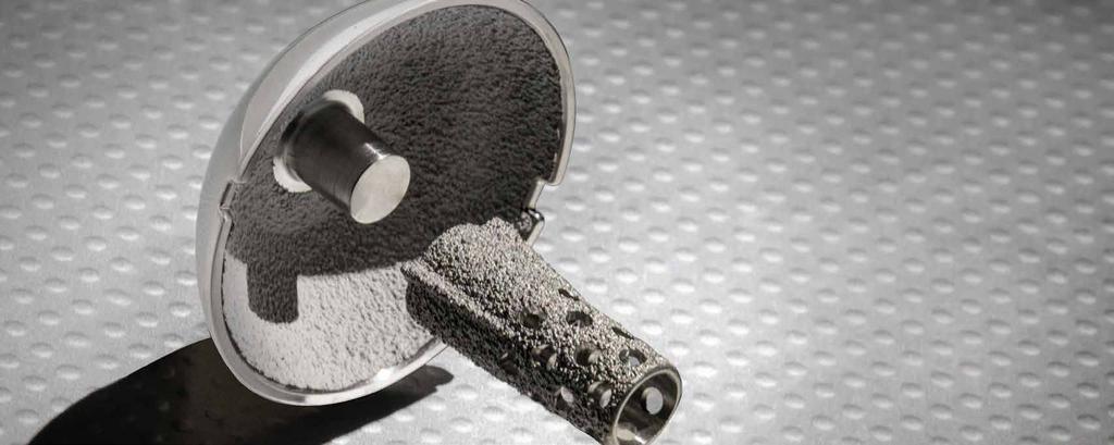
7 IMPLANTING THE RESURFACING HUMERAL HEAD The Head Impactor is placed onto the Impactor Handle, and is used to impact the Resurfacing Humeral Head onto the Humeral Cage. The first impaction will engage the tapers of the implants. Subsequent impaction will fully seat the cage and the humeral component (Figure 17). Care should be taken to leave a gap of approximately 1mm between the inferior reamed surface of the humerus and the bottom rim of the Resurfacing Humeral Head to prevent stress shielding. If there is no gap, a rongeur can be used to create it. BACK TABLE ASSEMBLY (OPTIONAL) Depending on surgeon preference, the Humeral Cage and Resurfacing Head can be assembled on the back table prior to implantation. Care should be taken to align the notches of the Resurfacing Head with fins of the Humeral Cage. The Back Table Assembly device is marked for visualization of the fins. Proper alignment at back-table assembly will ensure that the fins of the Humeral Cage can be visualized while implanting in the humerus, which will be necessary if the Tamp was used to prepare the humerus. Once the Resurfacing Head is aligned, use the Impactor Handle and Resurfacing Head Impactor to assemble the devices together (Figure 18). Figure 19 Final Resurfacing Humeral Head The final assembled resurfacing humeral head is implanted into the humerus as depicted in Figure 19. EXPLANTING THE DEVICES Two extractors are provided to assist in removing the Resurfacing Humeral Head and Resurfacing Humeral Cage. The Humeral Head Extractor is placed along the notches of the Resurfacing Humeral Head while the thumb screw tightens the jaws of the device. A Slap Hammer may be attached for more leverage to extract the implant (Figure 20). Figure 17 Impaction of the Resurfacing Humeral Head Figure 20 Extracting the Humeral Head Figure 18 Back-Table Assembly of the Humeral Head and Cage Peg 10 11

8 Prior to removing the Humeral Cage, a 3.2mm drill ( ) may be used to drill through the cage to remove any bony in-growth. A small flexible osteotome can be passed around the proximal portion of the cage to release any bone ongrowth if necessary. The Humeral Cage Extractor is threaded to the Humeral Cage and a Slap Hammer may be attached to assist in removing the implant (Figure 21). CLOSURE Closure is performed beginning with the subscapularis. The repair of the subscapularis will depend on the type of exposure used: tenotomy, elevation off bone or elevation with a wafer of bone. In general, #2 non-absorbable braided suture, or its equivalent, is used for either a tendon-to-tendon, tendon-to-bone or bone-tobone repair. The rotator interval is then closed, though it may be left partially open medially to avoid excessive tension of the closure. External rotation is checked at this point to define the parameters for post-operative rehabilitation. A drain may be used, placing it deep into the deltopectoral interval. The deltopectoral interval is closed followed by closure of the subcutaneous tissue and the skin. The upper extremity is then placed in a sling and swathe. After the implantation of the resurfacing humeral head prosthesis, if the subscapularis preserving technique was used then the humeral head is then reduced with internal rotation and flexion of the shoulder. The subscapularis capsule flap is repaired anatomically using a doublerow suture anchor technique and a reinforcing stitch. One suture anchor is placed along the medial aspect of the subscapularis insertion into the lesser tuberosity at the apex of the flap. Two full-thickness mattress sutures are then placed through the subscapularis capsule flap to reduce the medial aspect of the flap to its Figure 21 Extracting the Cage Peg Figure 22 Alternative Subscapularis Preserving Technique Closing the Inferior Subscapularis L-shaped Flap Figure 23 Alternative Subscapularis Preserving Technique: Complete Suture Repair of the Subscapularis original footprint (Figure 22). After tying, these sutures are left at full length and the remaining portion of the suture is used to reinforce the horizontal limb of the flap. Care is taken to avoid over-tightening of the tissues. The vertical limb is also oversewn with the extended length of the other suture. This vertical repair includes the biceps tendon to restore the lateral footprint. A final absorbable suture is used to reinforce and streamline the repair (Figure 23). The arm is then externally rotated to ensure a good closure with no gapping of the repair. POST-OPERATIVE REHABILITATION It is recommended to initiate the rehabilitation program on the same day as surgery and certainly by post-operative day one. All patients begin active range of motion of the elbow, wrist and hand. Range of motion of the shoulder consists of passive forward elevation, external rotation based on the assessment following subscapularis repair and internal rotation to the chest wall. External rotation should be limited for four weeks to the range without tension on the subscapularis repair as determined intraoperatively. If there is concern about the security of the subscapularis repair, external rotation should be limited to 0 degrees for four weeks. Isometric deltoid strengthening can also be performed. Patients should be instructed to perform these exercises five to six times per day for short periods of up to 10 minutes each session. The sling is discontinued after four weeks. The sling should be used longer, if there is a concern about the soft tissue repair. When the sling is discontinued, active range of motion should begin. Internal rotation behind the back can also be started at this time. Isometric internal and external rotation is added at six weeks and gentle resistive strengthening of the deltoid and rotator cuff begins weeks post-operatively. When the sling is removed, the patient is instructed to increase use of the upper extremity for activities of daily living. More vigorous strengthening can be initiated 12 weeks after surgery. Note that this document is a general guideline for postoperative rehabilitation but should not substitute for each surgeon s preference and experience


9 IMPLANT SCOPE Catalog Number Part Description INSTRUMENT SCOPE Resurfacing Head Impactor Tip INSTRUMENT SCOPE Resurfacing Cage, 25mm Resurfacing Cage, 30mm Resurfacing Humeral Head, 38mm Resurfacing Humeral Head, 41mm Resurfacing Humeral Head, 44mm Resurfacing Humeral Head, 47mm Resurfacing Humeral Head, 50mm Resurfacing Humeral Head, 53mm Resurfacing Cage Impactor Tip Resurfacing Head Sizer, 38mm Resurfacing Head Sizer, 41mm Resurfacing Head Sizer, 44mm Resurfacing Head Sizer, 47mm Resurfacing Head Sizer, 50mm Resurfacing Head Sizer, 53mm Reamer Handle Resurfacing Humeral Reamer, 38mm Resurfacing Humeral Reamer, 41mm Resurfacing Humeral Reamer, 44mm Resurfacing Humeral Reamer, 47mm Resurfacing Humeral Reamer, 50mm Resurfacing Humeral Reamer, 53mm mm x 250mm K-Wire Chandler Retractor Catalog Number Part Description Resurfacing Back Table Assembly Small Darrach Retractor Resurfacing Head Trial, 38mm Resurfacing Head Trial, 41mm Resurfacing Head Trial, 44mm Resurfacing Head Trial, 47mm Resurfacing Head Trial, 50mm Resurfacing Head Trial, 53mm Resurfacing Humeral Head Instrument Tray Cage Tamp Resurfacing Cage Drill (short) Resurfacing Cage Drill (long) Resurfacing Humeral Head Instrument Upper Tray Resurfacing Head Extractor Resurfacing Cage Extractor Slap Hammer Resurfacing Impactor Handle 14 15
10 16 17 NOTES NOTES
11 Exactech is proud to have offices and distributors around the globe. For more information about Exactech products available in your country, please visit For additional device information, refer to the Equinoxe Shoulder System Instructions for Use for a device description, indications, contraindications, precautions and warnings. For further product information, please contact Customer Service, Exactech, Inc., 2320 NW 66th Court, Gainesville, Florida , USA. (352) , (800) or FAX (352) Exactech, as the manufacturer of this device, does not practice medicine, and is not responsible for recommending the appropriate surgical technique for use on a particular patient. These guidelines are intended to be solely informational and each surgeon must evaluate the appropriateness of these guidelines based on his or her personal medical training and experience. Prior to use of this system, the surgeon should refer to the product package insert for comprehensive warnings, precautions, indications for use, contraindications and adverse effects. The products discussed herein may be available under different trademarks in different countries. All copyrights, and pending and registered trademarks, are property of Exactech, Inc. This material is intended for the sole use and benefit of the Exactech sales force and physicians. It should not be redistributed, duplicated or disclosed without the express written consent of Exactech, Inc Exactech, Inc Rev. C 0317 GLOBAL HEADQUARTERS: 2320 NW 66TH COURT GAINESVILLE, FL USA EXACTECH
Operative Technique Resurfacing Humeral Head
 EXACTECH SHOULDER Operative Technique Resurfacing Humeral Head TABLE OF CONTENTS INTRODUCTION... 1 SYSTEM SPECIFICATIONS... 1 PATIENT POSITIONING... 2 SURGICAL APPROACH... 2 SIZING THE HUMERUS FOR A RESURFACING
ANATOMICAL. REDEFINED.
 Operative Technique Fracture Stem Equinoxe Fracture Stem Operative Technique ANATOMICAL. REDEFINED. 352-377-1140 1-800-EXACTECH www.exac.com #718-00-33 REV A 0705 2005 EXACTECH, INC. ISO 13485 CERTIFIED
EXACTECH EXTREMITIES. Operative Technique. Preserve Stem. Surgeon focused. Patient driven. TM
 EXACTECH EXTREMITIES Operative Technique Preserve Stem Surgeon focused. Patient driven. TM TABLE OF CONTENTS INTRODUCTION...1 DETAILED OPERATIVE TECHNIQUE...2 PREOPERATIVE PLANNING/PATIENT POSITIONING...2
Technique. Aequalis Resurfacing Humeral Head
 S u r g i c a l Technique Aequalis Resurfacing Humeral Head 1 The Aequalis Resurfacing Humeral Head has been developed in conjunction with Drew Miller, MD - Atlanta, GA. The Aequalis Resurfacing Humeral
URSA HEMI-SHOULDER ARTHROPLASTY B I O T E K
 URSA HEMI-SHOULDER ARTHROPLASTY SURGICAL TECHNIQUE B I O T E K 2 Surgical Position Once general anesthesia has been satisfactorily induced, or a supraclavicular nerve block has been given, the patient
TABLE OF CONTENTS SURGICAL TECHNIQUE 1 POST-OPERATIVE REHABILITATION 18. pages 1 RADIOLOGICAL ASSESSMENT 1 2 PATIENT POSITIONING 1
 TABLE OF CONTENTS SURGICAL TECHNIQUE 1 1 RADIOLOGICAL ASSESSMENT 1 2 PATIENT POSITIONING 1 3 DELTO-PECTORAL APPROACH 2 4 HUMERAL HEAD OSTEOTOMY 6 5 CHOICE OF HUMERAL INCLINATION AND RETROVERSION 7 pages
EXACTECH EXTREMITIES. Operative Technique. Platform Shoulder System. Surgeon focused. Patient driven. TM
 EXACTECH EXTREMITIES Operative Technique Platform Shoulder System Surgeon focused. Patient driven. TM TABLE OF CONTENTS EQUINOXE PLATFORM SHOULDER SYSTEM INTRODUCTION... 3 Primary Shoulder... 3 Reverse
Operative Technique CTA Head
 EXACTECH SHOULDER Operative Technique CTA Head TABLE OF CONTENTS INTRODUCTION... 1 SYSTEM SPECIFICATIONS... 2 PRIMARY SHOULDER OPERATIVE TECHNIQUE OVERVIEW... 3 PRIMARY SHOULDER... 5 INDICATIONS FOR USE...
TORNIER BIO-RSA. Bony Increased Offset - Reversed Shoulder Arthroplasty SURGICAL TECHNIQUE
 TORNIER BIO-RSA Bony Increased Offset - Reversed Shoulder Arthroplasty SURGICAL TECHNIQUE 2 Table of Contents: Concept...4 Bony Increased Offset Reversed Shoulder Arthroplasty (BIO-RSA ) Concept...4 Surgical
The Bio-Modular Choice Shoulder System,
 Surgical Technique The Bio-Modular Choice Shoulder System, designed for both total and hemiarthroplasty of the shoulder, has enjoyed nearly two decades of clinical success. The variety of head types and
Operative Technique. Press-Fit. Anatomical. Redefined.
 Operative Technique Press-Fit Anatomical. Redefined. Table of Contents Introduction... 1 System Specifications... 2 Overview Technique... 3 detailed operative technique... 6 indications... 6 Pre-operative
EXACTECH KNEE. Operative Technique ADDENDUM. Metaphyseal Cones. Surgeon focused. Patient driven. TM
 EXACTECH KNEE Operative Technique ADDENDUM Metaphyseal Cones Surgeon focused. Patient driven. TM TABLE OF CONTENTS INTRODUCTION...1 DESCRIPTION...1 DETAILED OPERATIVE TECHNIQUE Initial Preparation and
3. PATIENT POSITIONING & FRACTURE REDUCTION 3 8. DISTAL GUIDED LOCKING FOR PROXIMAL NAIL PROXIMAL LOCKING FOR LONG NAIL 13
 Contents IMPLANT FEATURES 2 1. INDICATIONS 3 2. PRE-OPERATIVE PLANNING 3 3. PATIENT POSITIONING & FRACTURE REDUCTION 3 4. INCISION 4 5. ENTRY POINT 4-6 6. PROXIMAL NAIL INSERTION 6-7 7. PROXIMAL LOCKING
System Overview 3. Anatomic Sizing And Design Features 4. Instrumentation 6. Indications And Contraindications 7. Surgical Technique 8
 Surgical Technique Table of Contents System Overview 3 Anatomic Sizing And Design Features 4 Instrumentation 6 Indications And Contraindications 7 Surgical Technique 8 Implant Information 22 Instrumentation
Surgical Technique. Proximal Humerus Locking Plate
 Surgical Technique Proximal Humerus Locking Plate PERI-LOC Upper Extremity Locked Plating System 3.5mm & 4.5mm Proximal Humerus Locking PlatesCatalog Infor Table of Contents Introduction.........................................................2
RESURFACING HUMERAL HEAD IMPLANT TRAUMA & EXTREMITIES GROUP
 S U R G I C A L T E C H N I Q U E RESURFACING HUMERAL HEAD IMPLANT TRAUMA & EXTREMITIES GROUP TABLE OF CONTENTS SYSTEM OVERVIEW ANATOMIC SIZING AND DESIGN FEATURES INSTRUMENTATION INDICATIONS AND CONTRAINDICATIONS
EXACTECH SHOULDER. Operative Technique. Reverse Superolateral Approach. Surgeon focused. Patient driven.
 EXACTECH SHOULDER Operative Technique Addendum to the Equinoxe Reverse Operative Technique Reverse Superolateral Approach Surgeon focused. Patient driven. TABLE OF CONTENTS INTRODUCTION... 1 OPERATIVE
System. Humeral Nail. Surgical Technique
 System Humeral Nail Surgical Technique Contents IMPLANT FEATURES 2 1. INDICATIONS 3 2. PRE-OPERATIVE PLANNING 3 3. PATIENT POSITIONING & FRACTURE REDUCTION 3 4. INCISION 4 5. ENTRY POINT 4-6 6. PROXIMAL
S H O U L D E R Solutions by Tornier. BIO-RSA TM ANGled SURGICAL TECHNIQUE. BIO-RSA Angled. surgical technique
 S H O U L D E R Solutions by Tornier BIO-RSA TM ANGled SURGICAL TECHNIQUE BIO-RSA Angled Bony increased offset - reversed shoulder arthroplasty surgical technique BIO-RSA TM ANGled SURGICAL TECHNIQUE BIO-RSA
Arthrex ECLIPSE Stemless Shoulder Prosthesis. Surgical Technique
 Arthrex ECLIPSE Stemless Shoulder Prosthesis Surgical Technique Content Introduction and Glenoid Options... 03 Humeral Preparation... 05 Humeral Implantation... 07 Wound Closure... 09 Postoperative Management...
SURGICAL TECHNIQUE. Global Fx SHOULDER FRACTURE SYSTEM
 Global Fx SHOULDER FRACTURE SYSTEM TABLE OF CONTENTS Introduction................................................................. 2 System Highlights.....................................................
TORNIER SIMPLICITI. Shoulder System SURGICAL TECHNIQUE
 TORNIER SIMPLICITI Shoulder System SURGICAL TECHNIQUE Table of Contents: Indications & Contraindications... 4 System Compatibility & Pre-operative Planning... 4 Humeral Head Resection... 6 Freehand Resection
Solutions by Tornier. surgical technique
 S H O U L D E R Solutions by Tornier S I M P L I C I T I S U R G I C A L T E C H N I Q U E S H O U L D E R S Y S T E M surgical technique S I M P L I C I T I S H O U L D E R S Y S T E M S U R G I C A L
Open reduction; plate fixation 1 Principles
 Executive Editor: Peter Trafton Authors: Martin Jaeger, Frankie Leung, Wilson Li Proximal humerus 11-A2 Open reduction, plate fixation Search search... Shortcuts All Preparations All Approaches All Reductions
Aequalis Press-Fit. Shoulder Prosthesis. Surgical Technique
 Aequalis Press-Fit Shoulder Prosthesis Surgical Technique SURGICAL TECHNIQUE RATIONALE OF THE AEQUALIS PRESS-FIT PROSTHESIS p. 1 THE AEQUALIS PRESS-FIT PROSTHESIS p. 2 SURGICAL TECHNIQUE p. 3-21 1. A detailed
BICEPTOR Tenodesis System
 BICEPTOR Tenodesis System Sub-Pectoral Biceps Tenodesis A Shoulder Series Technique Guide As described by: Nikhil N. Verma, MD As described by: Nikhil N. Verma, MD Midwest Orthopedics at Rush Chicago,
AcUMEDr. Locking Proximal Humeral Plate. PoLARUSr PHPt
 AcUMEDr Locking Proximal Humeral Plate PoLARUSr PHPt PoLARUSr PHPt LOCKING PROXIMAL HUMERAL PLATE Since 1988 Acumed has been designing solutions to the demanding situations facing orthopedic surgeons,
SURGICAL TECHNIQUE. Global Fx SHOULDER FRACTURE SYSTEM
 SURGICAL TECHNIQUE Global Fx SHOULDER FRACTURE SYSTEM TABLE OF CONTENTS Introduction................................................................. 2 System Highlights.....................................................
DEVELOPED BY MEDSHAPE, INC. IN CONJUNCTION WITH PATRICK ST. PIERRE, M.D. BICEPS TENODESIS ARTHROSCOPIC AND SUBPECTORAL SURGICAL TECHNIQUE
 ! SURGICAL TECHNIQUE! DEVELOPED BY MEDSHAPE, INC. IN CONJUNCTION WITH PATRICK ST. PIERRE, M.D. ARTHROSCOPIC AND SUBPECTORAL BICEPS TENODESIS SURGICAL TECHNIQUE BICEPS TENODESIS Indications Tenodesis of
SURGICAL TECHNIQUE. Global Fx SHOULDER FRACTURE SYSTEM
 Global Fx SHOULDER FRACTURE SYSTEM TABLE OF CONTENTS Introduction................................................................. 2 System Highlights.....................................................
Operative Technique. Platform Fracture Stem shoulder ANATOMICAL. REDEFINED.
 Operative Technique Platform Fracture Stem shoulder System ANATOMICAL. REDEFINED. TABLE OF CONTENTS INTRODUCTION... 1 SYSTEM SPECIFICATIONS... 2 hemiarthroplasty OVERVIEW TECHNIQUE... 4 Hemiarthroplasty
D Degenerative joint disease, rotator cuff deficiency with, 149 Deltopectoral approach component removal with, 128
 Index A Abduction exercise, outpatient with, 193, 194 Acromioclavicular arthritis, with, 80 Acromiohumeral articulation, with, 149 Acromio-humeral interval (AHI), physical examination with, 9, 10 Active
Design Rationale. The Design
 2 Table of Contents Design Rational...4 Introduction...6 System Highlights...7 Surgical Technique...10 Patient Positioning...10 Deltopectoral Incision...11 Releasing the Pectoralis Major Tendon and Clavipectoral
This surgical technique describes how to perform an anatomic total shoulder arthroplasty implanting a short stem.
 INTRODUCTION This surgical technique describes how to perform an anatomic total shoulder arthroplasty implanting a short stem. CAUTION Federal law (USA) restricts this device to sale distribution and use
Solar Humeral Fracture System. Surgical Protocol
 Solar Humeral Fracture System Surgical Protocol Surgical Protocol Table of Contents Table of Contents Step By Step Procedure... 1 Patient Positioning... 3 Surgical Exposure... 4 Preparation of Humeral
Surgical. Technique. Aequalis Press-Fit. Shoulder Prosthesis.
 Surgical Technique Shoulder Prosthesis www.tornier.com TABLE OF CONTENTS 1. Preoperative planning 2. Patient positioning 3. Delto-pectoral approach 4. Humeral head osteotomy 5. Reaming the humeral shaft
Surgical. Technique. AEQUALIS Spherical Base Glenoid. Shoulder Prosthesis.
 Surgical Technique Shoulder Prosthesis AEQUALIS Spherical Base Glenoid www.tornier.com CONTENTS CONTENTS 1. Subscapularis 2. Anterior capsule 3. Humeral protector 4. Inserting retractors 1. DESIGN FEATURES
Modular Ulnar Head surgical technique. Transforming Extremities
 First Choice Modular Ulnar Head surgical technique Transforming Extremities instrumentation Head and Collar Trials Assembly Pad Starter Awl Trial Extractor Osteotomy Guide Stem Trials Implant Impactor
Rotator Cuff Repair using JuggerKnot Soft Anchor 2.9mm Surgical Technique
 Rotator Cuff Repair using JuggerKnot Soft Anchor 2.9mm Surgical Technique It s small. It s strong. And it's all suture. The JuggerKnot Soft Anchor represents the next generation of suture anchor technology.
Anatomical Shoulder Glenoid. Surgical Technique
 Anatomical Shoulder Glenoid Surgical Technique Anatomical Shoulder Glenoid Surgical Technique 3 Table of Contents Glenoid Preparation Surgical Steps 4 Anatomical Shoulder Glenoid 4 Glenoid Components
Bigliani/Flatow The Complete Shoulder Solution TSA
 Bigliani/Flatow The Complete Shoulder Solution TSA Surgical Technique Replicates natural shoulder mobility, balance, and stability Bigliani/Flatow The Complete Shoulder Solution Surgical Technique 1 Bigliani/Flatow
Operative Technique. Reverse Shoulder System ANATOMICAL. REDEFINED.
 Operative Technique Reverse Shoulder System ANATOMICAL. REDEFINED. Table Of CONTeNTs INTRODUCTION... 1 PRODUCT HIGHLIGHTS...2 SYSTEM SPECIFICATIONS...3 OVERVIEW TECHNIQUE...4 DETAILED OPERATIVE TECHNIQUE...7
Technique Guide. Primary Stemless Shoulder System
 Technique Guide TM Primary Stemless Shoulder System Anterior Deltopectoral Approach 1. Beachchair position (tilt back to 45 degree angle). 2. Short deltopectoral incision (from coracoid tip to pectoralis
Operative Technique. Press-Fit. Redefined. # EXACTECH ISO CERTIFIED
 Operative Technique Press-Fit Anatomical. Redefined. 352-377-1140 1-800-EXACTECH www.exac.com #718-01-30 1007 2007 Exactech, Inc. ISO 13485 CERTIFIED Table of Contents Introduction... 1 System Specifications...
Twin Tail TightRope System
 Open Stabilization of Acute Acromioclavicular Joint Dislocation using the Twin Tail TightRope System Surgical Technique Twin Tail TightRope System Open Stabilization of Acute Acromioclavicular Joint Dislocation
TORNIER AFFINITI EH 2. Extended Humeral Head SURGICAL TECHNIQUE
 TORNIER AFFINITI EH 2 Extended Humeral Head SURGICAL TECHNIQUE The AFFINITI Extended Humeral Head System was developed in collaboration with: John Brems, MD Cleveland, OH R. Sean Churchill, MD Milwaukee,
TORNIER APPROACH. Shoulder Arthroplasty Program SURGICAL TECHNIQUE. Delivering efficiency & repeatability throughout the continuum of patient care
 TORNIER APPROACH Shoulder Arthroplasty Program SURGICAL TECHNIQUE Delivering efficiency & repeatability throughout the continuum of patient care 2 Table of Contents: APPROACH Program Overview...4 Instrument
Anatomic and Reverse Shoulder. Surgical Technique
 Anatomic and Reverse Shoulder Surgical Technique CONTENTS KEY SURGICAL STEPS GLOBAL UNITE Platform Shoulder System Key Surgical Steps: Anatomic 4 GLOBAL UNITE Platform Shoulder System Key Surgical Steps:
Copeland/ Copeland EAS
 Copeland/ Copeland EAS Humeral Resurfacing Head Surgical Technique Table of Contents Patient Positioning and Approaches... 2 Copeland/Copeland EAS Humeral Resurfacing Head Surgical Technique Copeland
Technique Guide. 3.5 mm LCP Periarticular Proximal Humerus Plate. Part of the Synthes locking compression plate (LCP) system.
 system.") Technique Guide 3.5 mm LCP Periarticular Proximal Humerus Plate. Part of the Synthes locking compression plate (LCP) system. Table of Contents Introduction 3.5 mm LCP Proximal Humerus Plate 2 AO Principles
Technique Guide 3.5 mm LCP Periarticular Proximal Humerus Plate. Part of the Synthes locking compression plate (LCP) system. Table of Contents Introduction 3.5 mm LCP Proximal Humerus Plate 2 AO Principles
Technique Guide Glenoid Latarjet System
 Technique Guide Glenoid Latarjet System Glenoid Grafting without the Grief The Glenojet Allograft System was created to replace and augment anterior glenoid bone loss associated with trauma, recurrent
Conventus CAGE PH Surgical Techniques
 Conventus CAGE PH Surgical Techniques Conventus Orthopaedics The Conventus CAGE PH (PH Cage) is a permanent implant comprised of an expandable scaffold, made from nitinol and titanium, which is deployed
Signature Personalized Patient Care
 Surgical Technique Acetabular Guide System Contents One Surgeon. One Patient. Over 1 million times per year, Biomet helps one surgeon provide personalized care to one patient. The science and art of medical
U2 PSA. Revision Knee. Surgical Protocol
 U2 PSA TM Revision Knee Surgical Protocol Table of Contents 1 Component Removal... 1 2 Tibial Preparation... 1 2.1 Tibial Canal Preparation... 1 2.2 Proximal Tibial Resection... 2 2.3 Non Offset Tibial
EXACTECH SHOULDER. Operative Technique. ExactechGPS Shoulder Application. Surgeon focused. Patient driven. TM
 EXACTECH SHOULDER Operative Technique ExactechGPS Shoulder Application Surgeon focused. Patient driven. TM TABLE OF CONTENTS SYSTEM OVERVIEW...1 PREOPERATIVE PLANNING...4...4 Patient Positioning...14 Station
Comprehensive Fracture System. Surgical Technique
 Comprehensive Fracture System Surgical Technique 3 Comprehensive Fracture System Surgical Technique INDICATIONS 1. Non-inflammatory degenerative joint disease including osteoarthritis and avascular necrosis.
Shoulder and Elbow ORTHOPAEDIC SYPMPOSIUM APRIL 8, 2017 DANIEL DOTY MD
 Shoulder and Elbow ORTHOPAEDIC SYPMPOSIUM APRIL 8, 2017 DANIEL DOTY MD Shoulder Articulations Glenohumeral Joint 2/3 total arc of motion Shallow Ball and Socket Joint Allows for excellent ROM Requires
SHOULDER ARTHROPLASTY SYSTEM
 SURGICAL TECHNIQUE SHOULDER ARTHROPLASTY SYSTEM COMBINING SCIENCE, SIMPLICITY AND CLINICAL SUCCESS Table of Contents Design Rationale................................1 The Glenoid.................................1
Operative Technique PLATFORM SHOULDER SYSTEM ANATOMICAL. REDEFINED.
 Operative Technique LATFORM SHOULDER SYSTEM ANATOMICAL. REDEFINED. TABLE OF CONTENTS INTRODUCTION... 1 1REVERSE REVERSE SHOULDER... 25 25 SYSTEM SECIFICATIONS... 2 2REVERSE REVERSE SHOULDER SHOULDER OERATIVE
TORNIER AEQUALIS ASCEND. Shoulder System SURGICAL TECHNIQUE
 TORNIER AEQUALIS ASCEND Shoulder System SURGICAL TECHNIQUE 2 Table of Contents: Implant/Instrument Rationale...4 Indications & Contraindications...5 Surgical Technique... 6-14 Preoperative Planning and
Integra. Modular Radial Head System SURGICAL TECHNIQUE
 Integra Modular Radial Head System SURGICAL TECHNIQUE Table of Contents System Overview...2 Indications and Contraindications... 3 Modular Radial Head Implant Technique...4 Component Dimensions...8 Implant
Anatomical Shoulder Fracture. Surgical Technique
 Anatomical Shoulder Fracture Surgical Technique Anatomical Shoulder Fracture Surgical Technique 3 Surgical Technique Anatomical Shoulder Fracture Table of Contents Indications 4 Preoperative Planning
CANNULINK. Intraossous Fixation System SURGICAL TECHNIQUE
 CANNULINK Intraossous Fixation System SURGICAL TECHNIQUE Contents Chapter 1 4 Introduction The CANNULINK Advantage Indications for Use Preoperative Planning Chapter 2 5 Surgical Technique CANNULINK Standard
EXACTECH SPINE. Operative Technique. Cervical Spacer System. Surgeon focused. Patient driven. TM
 EXACTECH SPINE Operative Technique Cervical Spacer System Surgeon focused. Patient driven. TM ACAPELLA ONE Acapella One Cervical Spacer System is an anterior cervical discectomy and fusion device with
AEQUALIS PERFORM + REVERSED
 TO R N I E R AEQUALIS PERFORM + REVERSED Glenoid WEDGED AUGMENT SURGIC AL TECHNIQUE Table of Contents: AEQUALIS PERFORM + REVERSED Glenoid...3 Overview...3 Indications/Contraindications...4 Pre-Operative
FUNCTIONAL ANATOMY OF SHOULDER JOINT
 FUNCTIONAL ANATOMY OF SHOULDER JOINT ARTICULATION Articulation is between: The rounded head of the Glenoid cavity humerus and The shallow, pear-shaped glenoid cavity of the scapula. 2 The articular surfaces
Comprehensive Fracture System. Surgical Technique
 Comprehensive Fracture System Surgical Technique 1 Comprehensive Fracture System Surgical Technique INDICATIONS 1. Non-inflammatory degenerative joint disease including osteoarthritis and avascular necrosis.
TEM TED S GRA INTE SHOULDER SY
 INTEGRATED S H O U L D E R S Y S T E M Humeral Stems Ream and trial surgical technique Cobalt chrome (with proximal porous coating available) Fixed head (Neer II and K-II-C) and modular options (Mod-II-C,
Mini Open Latarjet Technique. Surgical Technique
 Mini Open Latarjet Technique Surgical Technique Mini Open Latarjet Technique A 5 cm skin incision is made starting at the tip of the coracoid process and extending inferiorly, through the deltopectoral
GLENOID SURGICAL TECHNIQUE
 UP. EXTREMITY Dual-Platform Shoulder Prosthesis GLENOID SURGICAL TECHNIQUE ANATOMICAL REVERSE SURGICAL TECHNIQUE REFERENCE NUMBERS HUMERAL STEM REFERENCE DIAMETER HEIGHT 267 360 Ø 06 100 265 102 Ø 08 120
ANATOMIC SHOULDER ARTHROPLASTY
 ANATOMIC SHOULDER ARTHROPLASTY INTRODUCTION This surgical technique describes how to perform an anatomic shoulder arthroplasty implanting a cemented pegged glenoid baseplate. CAUTION Federal law (USA)
Percutaneous Humeral Fracture Repair Surgical Technique
 Percutaneous Humeral Fracture Repair Surgical Technique Percutaneous Pinning Percutaneous Humeral Fracture Repair Closed reduction followed by percutaneous fixation reduces risk from soft tissue dissection
Surgical Technique. DISCLOSURE: This device is not approved for sale in the U.S.A. Customer Service:
 DISCLOSURE: This device is not approved for sale in the U.S.A. INDICATIONS FOR USE The KinematX Modular Wrist Arthroplasty System is indicated for the replacement of a wrist joints disabled by pain, deformity,
The suction cup mechanism is enhanced by the slightly negative intra articular pressure within the joint.
 SHOULDER INSTABILITY Stability A. The stability of the shoulder is improved by depth of the glenoid. This is determined by: 1. Osseous glenoid, 2. Articular cartilage of the glenoid, which is thicker at
Polarus 3 Solution Plates and Nails
 Surgical Technique Polarus 3 Solution Plates and Nails Acumed is a global leader of innovative orthopaedic and medical solutions. We are dedicated to developing products, service methods, and approaches
Bipolar Radial Head System
 Bipolar Radial Head System Katalyst Surgical Technique DESCRIPTION The Katalyst Telescoping Bipolar Radial Head implant restores the support and bearing surface of the radial head in the face of fracture,
DK7215-Levine-ch12_R2_211106
 12 Arthroscopic Rotator Interval Closure Andreas H. Gomoll Department of Orthopedic Surgery, Brigham and Women s Hospital, Harvard Medical School, Boston, Massachusetts, U.S.A. Brian J. Cole Departments
TrueSight Personalized Planning & Targeting System. Operative technique
 TrueSight Personalized Planning & Targeting System Operative technique TrueSight Personalized Planning & Targeting System Contents Introduction.... 2 Indications and contraindications... 3 Design rationale...
Polarus 3 Solution Plates and Nails. Surgical Technique 4.3. Screws
 Polarus 3 Solution Plates and Nails Surgical Technique 4.3 mm Screws Acumed is a global leader of innovative orthopaedic and medical solutions. We are dedicated to developing products, service methods,
S h o u l d e r Solutions by Tornier C o n v e r T i b l e S h o u l d e r S y S T e m
 S h o u l d e r Solutions by Tornier C o n v e r t i b l e s h o u l d e r s y s t e m C o n v e r t i b l e s h o u l d e r s y s t e m A n a t o m i c Aequalis Ascend Flex - UDZF131 One System. Two Solutions.
Index. B Backslap technique depth assessment, 82, 83 diaphysis distal trocar, 82 83
 Index A Acromial impingement, 75, 76 Aequalis intramedullary locking avascular necrosis, 95 central humeral head, 78, 80 clinical and functional outcomes, 95, 96 design, 77, 79 perioperative complications,
Biomechanical Impact of Posterior Glenoid Wear on Anatomic Total Shoulder Arthroplasty
 S5 Biomechanical Impact of Posterior Glenoid Wear on Anatomic Total Shoulder Arthroplasty Christopher P. Roche, M.S., M.B.A., Phong Diep, B.S., Sean G. Grey, M.D., and Pierre-Henri Flurin, M.D. Abstract
This publication is not intended for distribution in the USA. PRODUCT RATIONALE & SURGICAL TECHNIQUE
 This publication is not intended for distribution in the USA. PRODUCT RATIONALE & SURGICAL TECHNIQUE INTRODUCTION Introducing the GLOBAL UNITE Platform Shoulder Arthroplasty System, a modular shoulder
MIS Cemented Tibial Component
 MIS Cemented Tibial Component NexGen Complete Knee Solution Surgical Technique Table of Contents Surgical Exposure... 2 Finish the Tibia... 2 Position Based on Anatomic Landmarks... 3 Lateral Posterior
Glenoid Resurfacing Technique Guide
 Glenoid Resurfacing Technique Guide Restoring the Freedom of Motion 2 Description The HemiCAP Contoured Articular Prosthetic humeral component incorporates an articular resurfacing component and a taper
Fixed and Variable Geometry Total Shoulder Arthroplasty
 Fixed and Variable Geometry Total Shoulder Arthroplasty RECOVERY FUNCTION SURVIVORSHIP DePuy believes in an approach to total shoulder replacement that places equal importance on recovery, function and
HUMELOCK II. Cemented + Graft ANATOMICAL and REVERSIBLE if REVISION SURGICAL TECHNIQUE
 TM HUMELOCK II Cemented + Graft ANATOMICAL and REVERSIBLE if REVISION SURGICAL TECHNIQUE CONTENTS - Device descriptionpage 02 - Intended use / indicationspage 03 - Warnings and precautions page 03 - Using
Lesser MPJ Hemi Implant
 Lesser MPJ Hemi Implant Surgical Technique Contents Product The BioPro Lesser MPJ Hemi Implant is a simple, durable, metallic hemiarthroplasty resurfacing prosthesis for the treatment of arthritis, Freiberg
Aequalis -Glenoid. Keeled and Pegged. Surgical Technique
 Aequalis -Glenoid Keeled and Pegged Surgical Technique TABLE OF CONTENTS COMMON OPERATIVE TECHNIQUES FOR THE KEELED AND PEGGED AEQUALIS-GLENOIDS p. 1-3 IMPLANTATION OF THE AEQUALIS KEELED GLENOID p. 4-5
Trilogy Acetabular System
 Trilogy Acetabular System Surgical Technique Versatility in a proven design Trilogy Acetabular System 1 Trilogy Acetabular System Surgical Technique Table of Contents Acetabular Reaming 2 Component Sizing
Zimmer NexGen MIS Tibial Component. Cemented Surgical Technique IMAGE TO COME
 Zimmer NexGen MIS Tibial Component Cemented Surgical Technique IMAGE TO COME Zimmer NexGen MIS Tibial Component Cemented Surgical Technique 1 Zimmer NexGen MIS Tibial Component Cemented Surgical Technique
Total Shoulder System. Anatomic adaptability... simplified SURGICAL TECHNIQUE
 Total Shoulder System Anatomic adaptability... simplified SURGICAL TECHNIQUE Inclination Version Offset 125-140 +/- 10 3.5 mm A of Possibilities I I The Univers II Total Shoulder System was designed in
AcUMEDr. LoCKING CLAVICLE PLATE SYSTEM
 AcUMEDr LoCKING CLAVICLE PLATE SYSTEM LoCKING CLAVICLE PLATE SYSTEM Since 1988 Acumed has been designing solutions to the demanding situations facing orthopedic surgeons, hospitals and their patients.
Shoulder Arthroscopy Lab Manual
 Shoulder Arthroscopy Lab Manual Dalhousie University Orthopaedic Program May 5, 2017 Skills Centre OBJECTIVES 1. Demonstrate a competent understanding of the arthroscopic anatomy and biomechanics of the
Augmented Glenoid Component for Bone Deficiency in Shoulder Arthroplasty
 Clin Orthop Relat Res (2008) 466:579 583 DOI 10.1007/s11999-007-0104-4 SYMPOSIUM: NEW APPROACHES TO SHOULDER SURGERY Augmented Glenoid Component for Bone Deficiency in Shoulder Arthroplasty Robert S. Rice
Surgical Technique Guide PANTERA. Proximal Humerus Fracture Fixation Plate System
 Surgical Technique Guide PANTERA Proximal Humerus Fracture Fixation Plate System Installing the PANTERA is a 4-Step Process: The following technique is designed to optimize the surgical exercise. Step
Silicone PIP, MCP & MCP-X (PreFlex)
") Silicone PIP, MCP & MCP-X (PreFlex) Finger Joint Arthroplasty Operative Technique Silicone PIP Silicone MCP Silicone PreFlex (MCP-X) Stryker Disclaimer This publication sets forth detailed recommended
Silicone PIP, MCP & MCP-X (PreFlex) Finger Joint Arthroplasty Operative Technique Silicone PIP Silicone MCP Silicone PreFlex (MCP-X) Stryker Disclaimer This publication sets forth detailed recommended
ENDOBUTTON Fixation Device
 ENDOBUTTON Fixation Device Distal Biceps Repair A Shoulder Series Technique Guide As described by: Felix Buddy Savoie, MD PRELIMINARY - NOT FOR DISTRIBUTION As described by: Felix Buddy Savoie, MD Chief
REVERSE SHOULDER ARTHROPLASTY
 REVERSE SHOULDER ARTHROPLASTY 1 INTRODUCTION REVERSE SHOULDER ARTHROPLASTY This surgical technique describes how to perform a reverse total shoulder arthroplasty implanting a pegged glenoid baseplate.
SHOULDER INSTABILITY
 SHOULDER INSTABILITY Dr.KN Subramanian M.Ch Orth., FRCS (Tr & Orth), CCT Orth(UK) Consultant Orthopaedic Surgeon, Special interest: Orthopaedic Sports Injury, Shoulder and Knee Surgery, SPARSH Hospital