WAGNER CONE PROSTHESIS. Surgical Technique
|
|
|
- Phillip Gallagher
- 6 years ago
- Views:
Transcription


1 WAGNER CONE TM PROSTHESIS Surgical Technique
2 1
3 WAGNER CONE PROSTHESIS SURGICAL TECHNIQUE CONTENTS INDICATIONS... 3 CONTRAINDICATIONS... 3 BIOMECHANICAL CONCEPT... 4 THE IMPLANT... 5 INSTRUMENTS... 6 PREOPERATIVE PLANNING... 7 SURGICAL TECHNIQUE... 9 POSTOPERATIVE TREATMENT AND CASE EXAMPLES...18 ORDERING INFORMATION
4 Indications The Cone Prosthesis is designed for cylindrical proximal femoral bone that could fracture when conventional flared stems are used. The Cone Prosthesis is also designed for deformities of the femur where fixation of conventional stems is problematic. Specifically, the Cone Prosthesis is indicated when the following factors are present: A cylindrical configuration of the proximal medullary cavity, as found with congenital dysplasia of the hip (CDH), which causes problems for conventional flared stems. Cylindrical femoral bone is usually delicate and found in combination with coxa valga. Contraindications The most important contraindication is the trumpet-shaped proximal widening of the medullary cavity where there is no support for the prosthetic stem in its middle and proximal thirds. In addition, a Cone Prosthesis should not be considered when there is considerable weakening of the bone structure at the proximal end of the femur; implants with a greater length of stem will provide more stable fixation. 49-year-old woman with dysplastic arthritis of the left hip Increased anteversion of the femoral neck, e.g., CDH, where the cross section of the proximal medullary cavity is anteverted even after resection of the femoral neck. With the circular cross section of the stem of the cone prosthesis, the angle of anteversion can be adjusted as desired. Deformities and intramedullary bony scarring of the proximal end of the femur after osteotomies, fractures, growth disorders or congenital deformities. Preparing the medullary cavity to receive the stem with a conventional reamer can be very tedious or even impossible, whereas preparation with the conical awls of the Cone Prosthesis can be carried out more easily and safely. 6 years after implantation of a Cone Prosthesis and standard cup with metal articulation 3


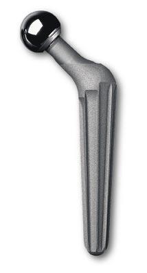
5 Biomechanical Concept The Cone Prosthesis has a conical angle of 5 degrees and fits the proximal medullary cavity better than a conventional flared stem. The concept of conical fixation is derived from positive experience with the 2 degree Wagner revision stem in which fixation is predominantly diaphyseal. In addition to providing rotational stability, the sharp longitudinal ribs of the stem are also beneficial for bony apposition. Schenk s 2 investigations have shown very clearly that bone forms and attaches preferentially on the sharpedged prominences of the implant and less in the hollows of the surface. The circular conical stem is not subject to any rotation force during insertion, i.e., the angle of anteversion can be determined by the surgeon. The stem has 8 sharp longitudinal ribs. The relatively sharp ridges of the ribs cut into the bone, thus allowing for optimum rotational stability. This also explains why the typical thigh pain associated with some uncemented prosthetic systems is practically unknown with the Cone Prosthesis. 1 The depth of penetration of the longitudinal ribs depends on the hardness of the bone. The depth of penetration of these ribs is slight on the whole and usually ranges from mm. Nevertheless, the penetration of the ribs into the bone is important for the depth of fixation of the stem and thus for the length of the leg and the tension in the soft tissues. With a cone angle of 5 degrees, a 1mm variation in the stem diameter leads to a variation of 12mm in the depth of insertion. This means that if a prosthetic stem that is 1mm too thin is inserted into the conically reamed medullary cavity, the stem will be anchored 12mm further down. The same applies if the longitudinal ribs of the stem cut 0.6mm instead of 0.1mm into the bone in the case of soft bone. This phenomenon therefore demands sensitivity on the part of the surgeon when using the awls: softer bone requires slightly less reaming than hard bone. The round stem facilitates unimpeded rotation during implantation to adjust the anteversion angle. 1 Wagner H, Wagner M. Cone prosthesis for the hip joint. Arch Orthop Trauma Surg. 2000; 120: Schenk RK, Wehrli U. Zur Reaktion des Knochens auf eine zementfreie SL-Femur-Revisionprothese. Orthopade. 1989; 18:

6 The Implant The Cone Prosthesis, designed for uncemented fixation, is manufactured from the tissuecompatible titanium-aluminium-niobium alloy Protasul The surface of the prosthesis is grit-blasted which, together with the characteristic shape, promotes bony apposition over a large area. 2 To enhance fixation, the prosthesis has a conical shape, and the core and cover angles are each 5 degrees. There are eight longitudinal sharp conical ribs arranged on the circumference of the stem, which serve for the actual fixation of the prosthesis. support surface. All the other ribs are inserted somewhat further proximally in order to ensure the greatest possible area of contact in the trochanter to provide rotational stability. The CCD angle is 135 degrees for all sizes and the neck length increases as the stem diameter increases. The stem has a standard 12/14 taper to take ceramic, metal or Metasul femoral heads, which are all available in multiple sizes. The stem is available in 12 diameters from 13 to 24mm to fit the width of the patient's medullary cavity. The height of the ribs varies between 1 and 2.5mm depending on the diameter of the stem. The ribs are the same height along their entire length. They are shaped so that they penetrate 0.1 to 0.5mm into the bone, depending on the hardness of the bone, and ideally make contact with bone over their entire length. To achieve this, there is a sharp conical awl for each diameter that has a shape adapted to the implant, i.e., it has a cover angle of 5 degrees. The eight ribs give the prosthesis a high degree of rotational stability. 3 In order to reduce the bending and torsional stresses in the ribs that can occur under loading, they are not knife-edged but have a ridge surface with a radial width of 0.4 to 0.5mm. This shape increases the strength of the prosthetic stem. In order to achieve broad-based support of the prosthesis in the region of the calcar, the medial rib is inserted distal to this area into the convex 5 3 Buhler D, Berlemann U, Lippuner K, Jaeger P Nolte L. Three-dimensional primary stability of cementless femoral stems. Clinical Biomechanics. 1997; 12: 75-85
 which are used for the careful preparation of the medullary canal, as well as trial prostheses, used for determining the best-suited size of implant.")
7 Instruments Surgeons have a set of user-friendly instruments at their disposal for implantation of the Cone Prosthesis. The core instruments are reamers (awls) which are used for the careful preparation of the medullary canal, as well as trial prostheses, used for determining the best-suited size of implant. Reamers (Awls) The Cone Prosthesis, with its circular crosssection, is indicated particularly for slender configurations of the proximal femur, as well as for cases involving pathologic morphology. When these indications are present, an important focus should be placed on the preparation of the medullary canal in the most bone-preserving manner. The use of reamers allows the surgeon to prepare the medullary canal with care and precision, even in cases of poor bone quality of the proximal femur where the use of rasps is not an option. The range of instruments includes a set of conical reamers, graded in millimeter increments corresponding exactly to the diameter of the implants. The medullary canal can therefore be enlarged in a stepwise fashion. The markings on the reamers determine the position of the center of rotation of the femoral head, which enables the surgeon to have continuous control as to the depth of the reamer. Trial Stems Trial stems are used to determine the exact positioning and correct size of the final implant. Trial stems are designed to duplicate the size and fit of the actual implants, to allow the surgeon to determine optimum implant diameter with a high degree of precision. Trial stems are available in sizes identical to those of the corresponding implants. Please note that the trial stems have only four ribs (instead of eight, as on the actual implants) to facilitate easy extraction of the trial prosthesis, and to avoid unnecessary damage to the bone in the trial procedure. The trial stem may be inserted as far as the intended anchoring depth of the implant, affording the surgeon an exact replication of the implant positioning so he or she can accurately assess tension of the soft tissue, range of motion, and degree of anteversion. As a result, accurate final seating of the implant can be achieved. 6
8 Preoperative Planning Fig. 1 The preoperative planning follows standard procedures. A good-quality x-ray with a magnification of 1.15:1 is essential. The usual planning templates fit this scale. To achieve this degree of magnification, it is assumed that the bone lies 10cm above the tabletop of the x-ray machine. In addition, the distance from the tabletop to the level of the film should be 5cm and the distance from the focus of the x-ray tube to the film should be 115cm (Fig. 1). In very obese patients, the bone can lie higher above the table. In this case, either the distance from the tube to the film must be increased or a marker of precisely defined length must be imaged on the x-ray film at the height of the bone at the same time. a b c a = 115cm b = 10cm c = 5cm Determination of the appropriate stem diameter with the planning template. The contour of the template lies on the contour of the cortical bone. This stem diameter is too small. With the correct diameter, the contour of the template must overlap the contour of the cortical bone by 1mm. Planning templates for cone prosthesis, 13 24mm diameter,
9 The first step in planning consists of selecting suitable implants for the acetabulum and the femur using the planning templates on the original x-ray films. When selecting the Cone Prosthesis, it is important that the configuration of the femur allows close contact between the middle third of the prosthetic stem and the cortex, and not just that the tip of the stem fits tightly in the medullary cavity. Selection of the correct stem diameter is particularly important. It is important not to choose a stem diameter that is too small. Such a decision can result in secondary subsidence of the prosthesis in its conical fixation. The outline on the planning template corresponds exactly to the dimensions of the implant. In choosing the diameter, it must be remembered that reaming with the awl removes a thin layer of bone and that the sharp longitudinal ribs cut slightly into the bone during insertion. The outline of the prosthetic stem on the planning template must therefore overlap the inner outline of the cortex in the region of the middle third of the stem by 1mm on each side. Using the planning template, the outline of the selected Cone Prosthesis is then transferred to the planning sketch and the line of resection and the trochanteric reference line are also drawn from the template. The planning sketch is now laid on the x-ray film and the outline of the femur is transferred carefully. The tip of the greater trochanter on the x-ray is at the level of the previously made marking for the desired trochanter position and usually coincides with the trochanteric reference line transferred from the template. Finally, the distance between the line of resection and the proximal limit of the lesser trochanter is measured. This gives the level of resection of the femoral neck. In reconstructing deformed hip joints, further marking points can be given and their distances measured. All longitudinal measurements must be made according to the scale on the template as this takes into account the degree of magnification of the x-ray. All measurements should be entered on the planning sketch so that they can be referred to during the operation. On the outline drawing of the pelvis, the position of the cup implant with the center of rotation is first sketched and the current and desired position of the tip of the trochanter is marked in order to check the leg length. T R X-ray example of preoperative planning with superimposed template 3 years postoperative 8
10 Surgical Technique The Cone Prosthesis can be implanted using all of the usual operative approaches. However, posterior access with the patient in the lateral position is particularly suitable. The awl for the Cone Prosthesis has a straight stem, which is introduced into the axis of the medullary cavity. With the posterior approach, when the hip and knee are flexed, the way to the medullary cavity is free without the need for temporary removal of the greater trochanter and without the instrument exerting pressure on the muscles. With the lateral, transgluteal and anterior approaches, retraction of the muscles is more difficult. Moreover, with the posterior approach, the incision is smaller and there is less blood loss with the patient in the lateral position. This is particularly apparent in obese patients. The incision or resection of the posterior joint capsule is a critical point in the posterior approach. Posterior dislocation of the prosthesis can occur more readily during the healing phase if the prosthetic cup and/or the stem are placed in insufficient anteversion. This problem can be counteracted by a test reduction before definitive fixation of the Cone Prosthesis, but nonetheless, this phenomenon requires particular care. In borderline cases, it can be useful to position the leg in slight external rotation and to avoid hip flexion greater than 60 degrees during the postoperative period. 9
. 2 The gluteus maximus and the fascia lata are split in the direction of the fibers. By retracting the gluteus maximus, the greater trochanter and short external rotators are exposed (Fig. 3). Fig.")
11 1 Fig. 2 Place the patient in lateral position. The skin incision is 3cm posterior to the intertrochanteric ridge, running in the direction of the fibers of the gluteus maximus and fascia lata (Fig. 2). 2 The gluteus maximus and the fascia lata are split in the direction of the fibers. By retracting the gluteus maximus, the greater trochanter and short external rotators are exposed (Fig. 3). Fig. 3 10


12 3 Fig. 4 The sciatic nerve is identified. Division of the tendon of gluteus maximus is very rarely necessary (Fig. 4). 4 The short external rotators including the piriformis muscle are detached from the greater trochanter. Slight internal rotation of the leg facilitates the dissection. The hip joint is then exposed (Fig. 5). Fig. 5 5 After exposure of the hip joint, Hohmann retractors are inserted at the cranial and caudal margins of the femoral neck and the posterior hip capsule is incised or resected. Another Hohmann retractor with a sharp tip is then inserted under the posterior rim of the acetabulum. The head of the femur can be carefully dislocated by a combined movement of internal rotation, flexion and adduction. The resection line is then marked according to preoperative planning. If dislocation cannot be achieved, even after further soft tissue attachment, an in situ osteotomy of the femoral neck is performed (Fig. 6). Fig. 6 11


13 6 Fig. 7 Perform an osteotomy of the femoral neck at the marked site 45 degrees to the femoral axis. The osteotomy with the oscillating saw should involve only the medial 2/3 of the cross section of the femoral neck so that the saw does not run into the greater trochanter. The remaining third is divided with a chisel along the medial surface of the trochanter in the direction of the femoral shaft (Fig. 7). 7 Remove the femoral head. Insert a retractor at the anterior rim of the acetabulum to expose the entire rim of the acetabulum. Proceed at this point with the preparation of the acetabulum (Fig. 8). Fig. 8 12

.")
.")
14 8 Fig. 9 Open the medullary cavity with a hollow chisel. At the same time, taking into account the planned anteversion of degrees, the greater trochanter is grooved inside so that the awl and prosthesis are not subsequently diverted in a varus direction. The cancellous bone is resected sparingly and only enough to allow sufficient room for the awl (Fig. 9). 9 Explore the medullary cavity with the medullary cavity gauge. This is used mainly to check that there is free access to the medullary cavity and locates any bony barriers (Fig. 10). Fig The femoral medullary cavity is widened conically with the modular awls in the longitudinal direction of the femur until noticeable resistance is felt (Fig. 11). Fig
.")

15 11 Fig. 12 The depth of penetration of the awl is checked with a Kirschner wire which is placed on the tip of the trochanter (Fig. 12). 12 The trial stem, with a diameter in accordance with the last awl to have been used, is connected to the impactor/extractor ( ). While connecting, ensure that the tab at the distal tip of the impactor is placed in the designated slot and then firmly connected. The trial stem is inserted in the femur until it is properly seated. The positioning guide on the handle of the impactor/extractor helps in determining the optimal anteversion. When there is severe pre-existing anteversion, make sure that the prosthesis is placed in the corrected position so that the neck of the prosthesis is not sitting on the rim of the cortex of the femoral neck. If necessary, some bone must be removed with a fine chisel or with the calcar curved rasp until there is a sufficient gap between the neck of the prosthesis and the bone. Fig. 13 Select the head trial size as templated and seat it onto the trial taper. Next, the hip is reduced. Leg length, offset and range of motion are checked. This procedure is repeated as necessary using different length trial heads until trial placement is satisfactory. The trial stem is removed with hammer taps on the impactor/extractor (Fig 13). If the trial reduction does not yield the desired result, proceed with the next diameter reamer and repeat the trial step with the appropriately sized trial. 14

16 13 Fig. 14 The prosthesis is inserted by hand until resistance can be felt. The impactor ( ) is used to ensure final seating of the Cone Prosthesis by hammer taps (Fig. 14). The tip of the impactor is inserted into the impacting hole in the shoulder of the prosthesis so that the fork-shaped flange surrounds the neck of the prosthesis. With this instrument, the prosthesis is rotated into the desired anteversion and impacted into its definitive position. The stability of the fixation can be assessed as follows: at first the prosthesis penetrates somewhat deeper into the medullary cavity with each hammer tap until the required stability is reached and the prosthesis does not move any further with continuing hammer taps. At this time there will be a higher pitch in the tone of the hammer taps. Finally, the depth of the penetration according to the preoperative planning is checked with the use of a tape measure. Fig For the final trial reduction, a plastic trial head is seated onto the neck taper (Fig. 15). 15
17 15 Cautious trial reduction is carried out with the assistance of the nylon-headed impactor. The joint is examined by moving the leg in all directions, especially in flexion/internal rotation. If necessary, the prosthesis is re-implanted with adjusted angle of anteversion and the trial reduction is repeated. Finally, soft tissue tension is checked with longitudinal traction on the extended leg. With this traction, the prosthetic joint may open by a few mm, and if necessary a longer prosthetic head must be selected (Fig. 16). If the leg has been lengthened considerably, the tension of the sciatic nerve should be assessed. Fig. 16 Fig The intermediate spaces remaining between the prosthesis and the bone are tightly filled with the chips of cancellous bone which have been obtained during the dissection (Fig. 17). 17 After careful cleaning of the taper, the selected femoral head is mounted with a light rotational movement and rotated further with axial force until it is firmly seated. The ball head is seated with several taps with the nylon-headed impactor (Fig. 18). Fig
18 18 Fig. 19 A trial reduction is performed in order to assess the function of the hip. This is then followed with re-fixation of the short external rotators. A redon drain is then inserted and the appropriate closure technique is performed (Fig. 19). 17


19 Postoperative Treatment and Case Examples Postoperative treatment is carried out in the same way as has proven successful with other hip prostheses. Case 1* Advanced and very painful dysplastic arthritis of the left hip in a 39 year-old woman. Begin the patient's exercise program a few days before the operation during the preoperative investigations and haemodilution. This makes it much easier for the patient because everything is then familiar after the operation. Also begin compression stockings on both legs and breathing exercises a few days prior to the operation. After the operation, the affected leg is laid in a foam splint. Beginning on the first day after the operation, have the patient stand beside his or her bed three times a day. Walking exercises begin on the third postoperative day in the patient s room, and walking to the toilet and in the corridor commence on the fifth day. Partial weight-bearing with 25 30kg with two elbow crutches is checked on the scales. With the leg in elastic suspension, active abduction and extension exercises of the hip are carried out with the patient supine. 3 weeks after implantation of an uncemented Cone Prosthesis and a conical screwed-in cup. 7 years after implantation of the prosthesis there is normal pain-free function, and the bone structure is homogeneous with structural adaptation to the mechanical loading. *Results shown here not indicative of all patients' experience with the Cone Prosthesis. Individual results may vary. 18


20 Climbing stairs and isometric training of the hip muscles in the lateral and prone positions start on the fifteenth postoperative day. Getting into a car is practiced. The patient is discharged home after 3 weeks with instructions to continue partial weight-bearing and the isometric muscle exercises and to omit passive movement exercises. Case 2* Dysplastic arthritis of the right hip in a 57- year-old woman 15 years after intertrochanteric osteotomy. The first follow-up examination takes place three months after the operation. Depending on the X- ray findings, there is usually a gradual transition to full weight-bearing within four weeks. Patients are advised against sport activities for the next few months. 3 weeks after implantation of an uncemented Cone Prosthesis and a monobloc primary cup with metal articulation. 5 years after the hip replacement. There is normal, pain-free function and the bone structure in the prosthesis bed is uniform. 19 *Results shown here not indicative of all patients' experience with the Cone Prosthesis. Individual results may vary.

21 Ordering Information CONE PROSTHESIS 12/14 UNCEMENTED Catalog No. Description Cone Prosthesis ø 13mm Cone Prosthesis ø 14mm Cone Prosthesis ø 15mm Cone Prosthesis ø 16mm Cone Prosthesis ø 17mm Cone Prosthesis ø 18mm Cone Prosthesis ø 19mm Cone Prosthesis ø 20mm Cone Prosthesis ø 21mm Cone Prosthesis ø 22mm Cone Prosthesis ø 23mm Cone Prosthesis ø 24mm TRIAL CASE Catalog No. Description Tray Trial Stems Cone Prosthesis (empty) Standard Container Cover, blue Trial Stem Cone Prosthesis ø 13mm Trial Stem Cone Prosthesis ø 14mm Trial Stem Cone Prosthesis ø 15mm Trial Stem Cone Prosthesis ø 16mm Trial Stem Cone Prosthesis ø 17mm Trial Stem Cone Prosthesis ø 18mm Trial Stem Cone Prosthesis ø 19mm Trial Stem Cone Prosthesis ø 20mm Trial Stem Cone Prosthesis ø 21mm Trial Stem Cone Prosthesis ø 22mm Trial Stem Cone Prosthesis ø 23mm Trial Stem Cone Prosthesis ø 24mm Impactor Positioning Bar INSTRUMENTS Catalog No. Description Tray (empty) Insert (empty) Standard container cover, blue Awl Cone Prosthesis ø 13 mm Awl Cone Prosthesis ø 14 mm Awl Cone Prosthesis ø 15 mm Awl Cone Prosthesis ø 16 mm Awl Cone Prosthesis ø 17 mm Awl Cone Prosthesis ø 18 mm Awl Cone Prosthesis ø 19 mm Awl Cone Prosthesis ø 20 mm Awl Cone Prosthesis ø 21 mm Awl Cone Prosthesis ø 22 mm Awl Cone Prosthesis ø 23 mm Awl Cone Prosthesis ø 24 mm Impactor Cone Prosthesis Extractor Cone Prosthesis Handle with quick coupling Ruler 20cm Calcar Rasp Gauge for the medullary cavity Repositioning Lever Repositioning Top Repositioning Top Repositioning Top Head Trial - Size 22mm/Neutral Head Trial - Size 22mm/+3.5mm Head Trial - Size 22mm/+8mm Head Trial - Size 28mm/Neutral Head Trial - Size 28mm/-4mm Head Trial - Size 28mm/+4mm Head Trial - Size 28mm/+8mm Head Trial - Size 32mm/Neutral Head Trial - Size 32mm/-4mm Head Trial - Size 32mm/+4mm Head Trial - Size 32mm/+8mm 20
22 SALES AIDS Catalog No. Description Demo Sample, Size X-ray template, 15% LITERATURE Literature No. Description Wagner H, Wagner M. Cone prosthesis for the hip joint. Reprint from Arch Orthop Trauma Surg. 2000; 120: Castelli CC et al. Radiographic evaluation of the conus uncemented stem. Reprint from Hip International. Vol. 9 no. 3, 1999; Kim YY et al. Total hip reconstruction in the anatomically distorted hip cemented versus hybrid total hip arthroplasty. Reprint from Arch Orthop Trauma Surg. 1998; 117: D Wagner H, Wagner M. Conical Stem Fixation for Cementless Hip Prostheses for Primary Implantation and Revision. Reprint from Endoprosthetics. E. W. Morscher 1995;
23 22


24 Please refer to package inserts for complete product information, including contraindications, warnings, precautions, and adverse effects. Contact your Zimmer Representative or visit us at Nov Printed in USA 2003 Zimmer
Preoperative Planning. The primary objectives of preoperative planning are to:
 Preoperative Planning The primary objectives of preoperative planning are to: - Determine preoperative leg length discrepancy. - Assess acetabular component size and placement. - Determine femoral component
Encina Taper Stem. Stinson Orthopedics Inc. 303 Twin Dolphin Drive, Suite 600 Redwood City, CA
 Stinson Orthopedics Inc. 303 Twin Dolphin Drive, Suite 600 Redwood City, CA 94065 info@stinsonortho.com www.stinsonortho.com Table of Contents Introduction 3 Features 4 Surgical Technique 5 Preoperative
VerSys LD/Fx Cemented and Press-Fit Hip Prostheses. Surgical Technique IMAGE TO COME. Versatile solutions for total and partial hip replacement
 VerSys LD/Fx Cemented and Press-Fit Hip Prostheses Surgical Technique IMAGE TO COME Versatile solutions for total and partial hip replacement VerSys LD/Fx Cemented and Press-Fit Hip Prostheses VerSys
Stinson Orthopedics Inc. 303 Twin Dolphin Drive, Suite 600 Redwood City, CA
 Stinson Orthopedics Inc. 303 Twin Dolphin Drive, Suite 600 Redwood City, CA 94065 info@stinsonortho.com www.stinsonortho.com Encina HA Stem Table of Contents Introduction 3 Encina HA Stem Features 4 Surgical
Cementless Tapered Femoral Stem Surgical technique
 Cementless Tapered Femoral Stem Surgical technique Contents Operative summary 4 Pre-operative planning 5 Femoral neck osteotomy 5 Femoral canal preparation 5 Intra-medullary (IM) reamer 6 Sequential rasping
THE NATURAL FIT. Surgical Technique. Hip Knee Spine Navigation
 THE NATURAL FIT Surgical Technique Hip Knee Spine Navigation MiniMAX Surgical Technique Hip Knee Spine Navigation INTRODUCTION The MiniMAX TM is a cementless anatomic stem available in 9 right sizes and
Cementless Tapered Femoral Stem Surgical technique
 Cementless Tapered Femoral Stem Surgical technique Contents Operative summary 4 Pre-operative planning 5 Femoral neck osteotomy 5 Femoral canal preparation 5 Intra-medullary (IM) reamer 6 Sequential rasping
Alloclassic Zweymüller Stem
 Alloclassic Zweymüller Stem Surgical Technique A proven concept Disclaimer This document is intended exclusively for physicians and is not intended for laypersons. Information on the products and procedures
*smith&nephew SL-PLUS
 Surgical Technique *smith&nephew SL-PLUS Cementless Femoral Hip System SL-PLUS Standard and Lateral Stem Table of Contents Notes from the Author s Clinic... 3 Indications... 4 Contraindications... 5 Preoperative
CAUTION: Ceramic liners are not approved for use in the United States.
 Total Hip Prostheses, Self-Centering Hip Prostheses and Hemi-Hip Prostheses IMPORTANT: This essential product information sheet does not include all of the information necessary for selection and use of
SURGICAL TECHNIQUE CEMENTED & PRESS-FIT UNIFIED INSTRUMENTATION INTRAOPERATIVE FLEXIBILITY PROVEN BIOMECHANICS
 SURGICAL TECHNIQUE CEMENTED & PRESS-FIT UNIFIED INSTRUMENTATION INTRAOPERATIVE FLEXIBILITY PROVEN BIOMECHANICS INTRODUCTION The Summit Tapered Hip System s comprehensive set of implants and instruments
Section of Modular Hip Prostheses cemented. TMC-3 Modular Hip Prosthesis, cemented. TMC-3 Modular Hüftprothese, zementiert
 Section of Modular Hip Prostheses cemented TMC-3 Modular Hip Prosthesis, cemented TMC-3 Modular Hüftprothese, zementiert Prothèse de hanche modulaire TMC-3, cimentée Indication : The TMC-3 Modular hip
operative technique Kent Hip
 operative technique Kent Hip The Kent Hip Operative Technique The Kent Hip was developed by Mr Cliff Stossel, FRCS in Maidstone, Kent, UK and first implanted in 1986. It was designed to deal with problems
CLS Spotorno Hip Stem. Surgical Technique
 CLS Spotorno Hip Stem Surgical Technique Surgical Technique CLS Spotorno Stem Table of Contents CLS Spotorno Stem 4 Indications for the CLS Spotorno Stem 5 Preoperative Planning 10 Surgical Technique
Clinical Evaluation Surgical Technique
 Clinical Evaluation Surgical Technique Table of Contents EMPERION Specifications 3 EMPERION Surgical Technique 9 EMPERION Catalog 18 Nota Bene: This technique description herein is made available to the
HELIOS h i p s y s t e m
 HELIOS h i p s y s t e m Design The Helios stem is a highly polished, High Nitrogen Stainless Steel (ISO5832-9) dual tapered cemented stem. The design of the stem is based on the clinically lly successful
Surgical Technique VPLWK QHSKHZ 1$126 1HFN 3UHVHUYLQJ +LS 6WHP 1716-e_NANOS_OPT.indd :27
 Surgical Technique NANOS Neck Preserving Hip Stem Table of Contents Introduction... 3 Development/Concept... 4 Indications/Contraindications... 5 Preoperative Planning... 5 Surgical Technique... 6 Prosthesis
Following a tradition of success. VerSys Heritage Primary Hip Prosthesis Surgical Technique
 Following a tradition of success VerSys Heritage Primary Hip Prosthesis Surgical Technique VerSys Heritage Primary Hip Prosthesis 1 Surgical Technique For VerSys Heritage Primary Hip Prosthesis Dennis
Surgical Technique. SL-PLUS Cementless Femoral Hip System
 Surgical Technique *smith&nephew SL-PLUS Cementless Femoral Hip System SL-PLUS Standard and Lateral Stem Table of Contents Comment from the Author s Clinic... 3 Indications... 4 Contraindications... 5
28 Surgical Technique
 Surgical Technique 10 12 14 16 18 20 22 24 28 26 Technique described by James L. Guyton, MD Campbell Clinic Memphis, Tennessee James W. Harkess, MD Campbell Clinic Memphis, Tennessee David G. LaVelle,
Optimum implant geometry
 Surgical Technique Optimum implant geometry Extending proven Tri-Lock heritage The original Tri-Lock was introduced in 1981. This implant was the first proximally coated tapered-wedge hip stem available
Zimmer M/L Taper Hip Prosthesis. Surgical Technique
 Zimmer M/L Taper Hip Prosthesis Surgical Technique Zimmer M/L Taper Hip Prosthesis 1 Zimmer M/L Taper Hip Prosthesis Surgical Technique Table of Contents Preoperative Planning 2 Determination of Leg Length
Bone Preservation Stem
 TRI-LOCK Bone Preservation Stem Featuring GRIPTION Coating Surgical Technique Implant Geometry Extending the TRI-LOCK Stem heritage The original TRI-LOCK Stem was introduced in 1981. This implant was
Surgical Technique. *smith&nephew POLARSTEM Cementless Stem System
 Surgical Technique *smith&nephew POLARSTEM Cementless Stem System POLARSTEM Cementless Stem System Contents Introduction... 3 Indications... 4 Contraindications... 4 Case Studies... 5 Preoperative Planning...
Anterior Approach Surgical Technique. Paragon Stem System. enabling people to enjoy life
 Anterior Approach Surgical Technique Paragon Stem System enabling people to enjoy life Contents Pre-Operative Planning... 2 Suggested Templating Method... 2 Surgical Technique... 3 Surgical Approach...
Surgical Technique. Hip System
 Surgical Technique Hip System INDICATIONS FOR USE The TaperSet Hip System is designed for total or partial hip arthroplasty and is intended to be used with compatible components of the Consensus Hip System.
VerSys Fiber Metal Taper Hip Prosthesis. Surgical Technique
 VerSys Fiber Metal Taper Hip Prosthesis Surgical Technique VerSys Fiber Metal Taper Hip Prosthesis Surgical Technique 1 VerSys Fiber Metal Taper Hip Prosthesis Surgical Technique Table of Contents Preoperative
Revision. Hip Stem. Surgical Protocol
 U2 TM Revision Hip Stem Surgical Protocol U2 Revision Hip Stem Table of Contents Introduction... 1 Preoperative Planning... 2 Femoral Preparation... 3 Trial Reduction... 5 Implant Insertion... 6 Ordering
A further enhanced classic. Wagner SL Revision Hip Stem
 A further enhanced classic Wagner SL Revision Hip Stem The original Wagner SL Revision Stem offers a time-proven solution in the treatment of revision hips. While its underlying anchorage philosophy and
*smith&nephew SL-PLUS Cementless Femoral Hip System. Product Information
 Product Information *smith&nephew SL-PLUS Cementless Femoral Hip System First Came the Philosophy to develop a universal hip system that could be used in almost every indication, immaterial to the patient
Aesculap Trilliance Triple Tapered Polished Hip Stem
 Aesculap Trilliance Triple Tapered Polished Hip Stem Aesculap Orthopaedics Trilliance Triple Tapered Polished Hip Stem CONTENTS 2 Contents Page Trilliance Philosophy 4 Trilliance Design 6 Trilliance Implants
PLR. Proximal Loading Revision Hip System
 PLR Proximal Loading Revision Hip System The PLR splined revision stem is designed to recreate the natural stresses in the revised femur, where proximal bone may be compromised. PLR Hip System Design Considerations
Integral 180 Surgical Technique
 Integral 180 Surgical Technique The Integral 180 and 225 are part of the Alliance Family Total Hip System. The Integral 225 femoral component is marketed for use with bone cement in the United States.
SURGICAL TECHNIQUE. Alpine Cementless Hip Stem
 SURGICAL TECHNIQUE Alpine Cementless Hip Stem The following technique is a general guide for the instrumentation of the Alpine Cementless Hip Stem. It is expected that the surgeon is already familiar with
CLS Spotorno Hip Stem
 CLS Spotorno Hip Stem Surgical Technique Nature as Model Disclaimer This document is intended exclusively for experts in the field, i.e. physicians in particular, and is expressly not for the information
Cemented femoral stem - type CSC
 Cemented femoral stem - type CSC Cemented Femoral Hip Joint Components ARTHROPLASTY Implant Description Preface The cemented femoral stem type CSC with centralizer was designed using the latest knowledge
Taperloc Complete Hip System. Surgical Technique
 Taperloc Complete Hip System Surgical Technique One Surgeon. One Patient. Over 1 million times per year, Biomet helps one surgeon provide personalized care to one patient. The science and art of medical
EXTENDED TROCHANTERIC OSTEOTOMY SURGICAL TECHNIQUE FPO EXTENSIVELY COATED FIXATION
 EXTENDED TROCHANTERIC OSTEOTOMY SURGICAL TECHNIQUE FPO EXTENSIVELY COATED FIXATION SINCE 1983 PREOPERATIVE PLANNING EXPLANTATION OPTIONS the cement from inside the cement canal until the bone/ cement bond
ZMR Over-the-Junction Instruments for Revision Hip Arthroplasty. Surgical Technique IMAGE TO COME
 ZMR Over-the-Junction Instruments for Revision Hip Arthroplasty Surgical Technique IMAGE TO COME ZMR Over-the-Junction Instruments for Revision Hip Arthroplasty Introduction The ZMR Over-the-Junction (OTJ)
Progeny Hip Stem. Surgical Protocol and Product Specifications
 Progeny Hip Stem Surgical Protocol and Product Specifications Progeny Hip Stem Introduction With emphasis on maximum stability and ease of use, the StelKast ProgenyTM Hip System provides the surgeon with
Anatomical Shoulder Glenoid. Surgical Technique
 Anatomical Shoulder Glenoid Surgical Technique Anatomical Shoulder Glenoid Surgical Technique 3 Table of Contents Glenoid Preparation Surgical Steps 4 Anatomical Shoulder Glenoid 4 Glenoid Components
TaperFill. Surgical Technique
 TaperFill Surgical Technique Table of Contents Indications and Contraindications 3 TaperFill Hip Size Charts 4-5 DJO Surgical 9800 Metric Boulevard Austin, TX (800) 456-8696 www.djosurgical.com Preoperative
MetaFix. Cementless Total Hip Replacement Surgical technique
 Cementless Total Hip Replacement Surgical technique Contents Operative summary Acetabular preparation Pre-operative templating Femoral neck osteotomy Femoral canal preparation Femoral punch Tapered IM
U2 PSA. Revision Knee. Surgical Protocol
 U2 PSA TM Revision Knee Surgical Protocol Table of Contents 1 Component Removal... 1 2 Tibial Preparation... 1 2.1 Tibial Canal Preparation... 1 2.2 Proximal Tibial Resection... 2 2.3 Non Offset Tibial
AVANTEON. Operative Technique & Catalogue Information AVANTEON
 AVANTEON Operative Technique & Catalogue Information AVANTEON H I P S Y S T E M Pre-operative Planning The overall aim of pre-operative planning is to establish anatomical data from the patient to guide
CC TRIO VERSAFITCUP. Surgical Technique. each to their own. Hip Knee Spine Navigation
 VERSAFITCUP CC TRIO each to their own Surgical Technique Hip Knee Spine Navigation Versafitcup CC TRIO Surgical Technique Hip Knee Spine Navigation EACH TO THEIR OWN The Versafitcup CC Trio is a range
21st Century Fracture Management ETS. Surgical Protocol
 21st Century Fracture Management ETS Surgical Protocol ETS Operative Technique Step 1 Confirm that a cemented hemiarthroplasty is indicated. An X-ray template of the ETS is provided. This should be used
ACETABULAR CUP SURGICAL TECHNIQUE
 ACETABULAR CUP SURGICAL TECHNIQUE ACETABULAR CUP DEVICE INDICATIONS FOR USE The ICONACY I-Hip total hip replacement is indicated for the following conditions: 1. A severely painful and/or disabled hip
Surgical Technique.
 Surgical Technique www.biomet.co.uk INTRODUCTION design principals Recent advances in imaging technology have enabled orthopaedic surgeons to extend closed treatment of femoral fractures to include more
HIP SYSTEM SURGICAL TECHNIQUE
 HIP SYSTEM SURGICAL TECHNIQUE Introduction...2 Preoperative Planning...3 Preoperative Planning...3 Templating and Radiographs...4 Determination of Leg Length Discrepancy...5 Determining Acetabular Cup
Tradition Hip Primary Surgical Technique
 Design Rationale Many total hip designs in today s marketplace do not take advantage of the known forces present in the femur. Long term stability of a total hip prosthesis requires an implant design and
S U R G I C A L T E C H N I Q U E
 SURGICAL TECHNIQUE RECOVERY FUNCTION SURVIVORSHIP DePuy believes in an approach to total hip replacement that places equal importance on recovery, function and survivorship. The DePuy PROXIMA Hip System
Efficacy Innovation ABG II. Brochure. Surgical Protocol. Cemented Stem
 Efficacy Innovation ABG II Cemented Stem Brochure Surgical Protocol 2 The ABG Hip System The ABG hip implant range has been progressively extended and enhanced over the past years, harnessing extensive
Approach Patients with Confidence
 Surgical Technique Approach Patients with Confidence The ACTIS Total Hip System is the first DePuy Synthes stem specifically designed to be utilized with tissue sparing approaches, such as the anterior
Optimizing function Maximizing survivorship Accelerating recovery
 Surgical Technique Optimizing Function Maximizing Survivorship Accelerating Recovery The company believes in an approach to patient treatment that places equal importance on: Optimizing function Maximizing
Absolut TM Cemented Stem. Surgical Technique
 Absolut TM Cemented Stem Surgical Technique Contents ABSOLUT Cemented Stem 2 Absolut Confidence 2 Absolut Reproducibility 2 Absolut Choice 2 Pre-Operative Planning 3 Suggested Templating Method 3 Surgical
GENERIC, LOGIC, INTEGRALE TO.H.GB.011/1.0
 Surgical technique mechanical instrumentation GENERIC, LOGIC, INTEGRALE TO.H.GB.011/1.0 2 Pre-surgical planning By means of radiological assessment and templates, it is possible to: - determine the position
ADDRESSING CLINICAL ISSUES OF CEMENTLESS HIP ARTHROPLASTY
 E C H E L O N P R I M A R Y H I P S Y S T E M P R O D U C T R A T I O N A L E ADDRESSING CLINICAL ISSUES OF CEMENTLESS HIP ARTHROPLASTY Echelon Primary Total Hip System HIGH OFFSET STANDARD OFFSET Cementless
APS Natural-Hip System
 APS Natural-Hip System Surgical Technique Bone conserving, anatomic fit APS Natural-Hip System Surgical Technique APS Natural-Hip System Surgical Technique Developed in conjunction with Jay Butler, MD
Metha Short Hip Stem System
 Metha Short Hip Stem System Accuracy That Stands Alone Aesculap Orthopaedics Metha Short Hip Stem System Designed For Anatomic Accuracy The Metha Short Hip Stem is designed for anatomic accuracy to restore
NeoGen Femoral Nail System
 NeoGen Femoral Nail System LESS IS MORE TE-2070-04 Surgical Technique BLE OF CONTENT Preface Standard Femoral Mode Recon Mode Post-Operative Management Appendix Products Information Indication Patient
AML Hip System. Design Rationale/ Surgical Technique
 AML Hip System Design Rationale/ Surgical Technique Design Rationale Evolution In 1977, DePuy Synthes Companies introduced the original cementless total hip. The AML Hip launched in order to solve one
FIRST STEM SPECIFICALLY DESIGNED FOR AMIS. Surgical Technique
 FIRST STEM SPECIFICALLY DESIGNED FOR AMIS Surgical Technique Joint Spine Sports Med AMIStem Surgical Technique Joint Spine Sports Med INTRODUCTION This document describes the Surgical Technique for the
Cementless femoral stem type SF
 Cementless femoral stem type SF Cementless Femoral Hip Joint Components ARTHROPLASTY Implant Description Surgical Technique Instrumentation Set Catalogue Preface The cementless stem of a total hip joint
VerSys 6 Beaded FullCoat Plus Hip Prosthesis
 VerSys 6 Beaded FullCoat Plus Hip Prosthesis Surgical Technique Stability without compromise VerSys 6 Beaded FullCoat Plus Hip Prosthesis 1 Surgical Technique For VerSys 6 Beaded FullCoat Plus Hip Prosthesis
SURGICAL TECHNIQUE GUIDE
 DANGER indicates an imminently hazardous situation which, if not avoided, will result in death or serious injury. WARNING indicates a potentially hazardous situation which, if not avoided, could result
ZMR CROSSOVER INSTRUMENTS AND SURGICAL TECHNIQUE. Surgical Technique for Revision Hip Arthroplasty
 ZMR CROSSOVER INSTRUMENTS AND SURGICAL TECHNIQUE Surgical Technique for Revision Hip Arthroplasty A MULTITUDE OF OPTIONS Several fixation options are offered within the ZMR Hip System. Spout, Cone, and
Minimally Invasive System for Total Hip Arthroplasty. Surgical Technique
 Minimally Invasive System for Total Hip Arthroplasty Surgical Technique INTRODUCTION The DePuy MI System was created by an International team of surgeons whose first priority was to achieve patient gain
3. PATIENT POSITIONING & FRACTURE REDUCTION 3 8. DISTAL GUIDED LOCKING FOR PROXIMAL NAIL PROXIMAL LOCKING FOR LONG NAIL 13
 Contents IMPLANT FEATURES 2 1. INDICATIONS 3 2. PRE-OPERATIVE PLANNING 3 3. PATIENT POSITIONING & FRACTURE REDUCTION 3 4. INCISION 4 5. ENTRY POINT 4-6 6. PROXIMAL NAIL INSERTION 6-7 7. PROXIMAL LOCKING
Operating Instruction. Müller Hip Stem
 Müller Hip Stem Ortho Select GmbH Eltastrasse 2 D 78573 Wurmlingen Germany Tel: +49-(0)7461-96632-30 Fax: +49-(0)7461-96632-35 info@ortho-select.de www.ortho-select.de 12009 Introduction and product description
UNDERSTANDING TRADITION, MASTERING INNOVATION. Surgical Technique
 UNDERSTANDING TRADITION, MASTERING INNOVATION Surgical Technique Joint Spine Sports Med MasterLoc Surgical Technique Joint Spine Sports Med INTRODUCTION This document describes the Surgical Technique for
Versys Advocate V-Lign and Non V-Lign Cemented Hip Prosthesis
 Versys Advocate V-Lign and Non V-Lign Cemented Hip Prosthesis Surgical Technique Traditional Design. Innovative Features. Versys Advocate V-Lign and Non V-Lign Cemented Hip Prosthesis 1 Versys Advocate
Allofit /Allofit -S Alloclassic Acetabular Cup System
 Allofit /Allofit -S Alloclassic Acetabular Cup System Product Information Convincing Primary Stability The Allofit acetabular cup was developed in 1993. The goal was to create a reliable implant that was
CPT 6 Stainless Steel Primary Hip System
 CPT 6 Stainless Steel Primary Hip System Surgical Technique The proven, simple solution 1Osteotomy of the Femoral Neck Superimpose the Osteotomy Guide on the proximal femur. Position the guide over the
FLH /11
 FLH 225 04/11 This publication has been issued by: European Central Marketing Waterton Industrial Estate Bridgend, South Wales CF31 3XA, United Kingdom Tel: +44 (0)1656 655221 Fax: +44 (0)1656 645454 www.biomet.com
ZMR Crossover Instruments. Abbreviated Surgical Technique
 ZMR Crossover Instruments Abbreviated Surgical Technique ZMR Crossover Instruments Surgical Technique Introduction ZMR Crossover Instruments facilitate the combination of any Porous Proximal Body with
Surgical Technique. Cup System
 Surgical Technique Cup System INDICATIONS AND USAGE Indications for the use of the CS2 ACETABULAR CUP SYSTEM must be carefully considered with respect to the patient s entire evaluation and alternative
Metasul LDH Large Diameter Head
 Metasul LDH Large Diameter Head Surgical Technique Metasul LDH Large Diameter Head Surgical Technique Enhancing Stability and Increasing Range of Motion Metasul LDH Large Diameter Head Surgical Technique
CLS Spotorno Hip Cup. Surgical Technique. Nature as Model
 CLS Spotorno Hip Cup Surgical Technique Nature as Model Disclaimer This document is intended exclusively for experts in the field, physicians in particular, and it is not intended for laypersons. Information
Wagner SL Revision Hip Stem
 Wagner SL Revision Hip Stem Surgical Technique Femoral Revision Prosthesis for Extensive Bone Loss Disclaimer This document is intended exclusively for experts in the field, i.e. physicians in particular,
EMPERION Modular Hip System Surgical Technique
 Surgical Technique Introduction The EMPERION Modular Hip System is a versatile system that can be used for primary and revision hip surgeries. Using modular proximal bodies, this system addresses the proximal
asterloc Surgical Technique HIP SYSTEM UNDERSTANDING TRADITION, MASTERING INNOVATION Hip Knee Spine Navigation
 asterloc HIP SYSTEM UNDERSTANDING TRADITION, MASTERING INNOVATION Surgical Technique Hip Knee Spine Navigation Masterloc Surgical Technique Hip Knee Spine Navigation INTRODUCTION This document describes
Trabecular Metal Tibial Cone Surgical Technique
 Trabecular Metal Tibial Cone Surgical Technique Provides structural support in areas of bone loss Trabecular Metal Cone 1 Zimmer Trabecular Metal Tibial Cone Surgical Technique Table of Contents Overview
TaperFit. Cemented Total Hip Replacement Surgical technique
 TaperFit Cemented Total Hip Replacement Surgical technique TaperFit Contents Operative summary 4 Pre-operative templating 5 Surgical exposure 5 Femoral neck resection 5 Acetabular preparation 5 Cenator
Sirus Antegrade Femoral Nail System Surgical Technique
 Sirus Antegrade Femoral Nail System Surgical Technique The Cannulated Titanium Nail with Anatomical Shape and Lateral Entry Point Disclaimer This document is intended exclusively for experts in the field,
Surgical Technique. Unisyn Hip System
 Surgical Technique Unisyn Hip System INDICATIONS AND USAGE Significantly impaired joints resulting from rheumatoid, osteo, and post-traumatic arthritis. Revision of failed femoral head replacement, hip
Approach Patients with Confidence
 Approach Patients with Confidence The is the first stem specifically designed to be utilized with tissue sparing approaches, such as the anterior approach, as well as traditional approaches. The implant
MetaFix. Cementless Total Hip Replacement Surgical technique
 MetaFix Cementless Total Hip Replacement Surgical technique Contents Operative summary 4 Acetabular preparation 5 Pre-operative templating 5 Operative technique 6 Femoral neck osteotomy 6 Femoral canal
Manza Cup HA SURGICAL TECHNIQUE.
 1 PRE-OPERATIVE PLANNING. Preoperative assessment of the appropriate size and position of the acetabular component will provide intraoperative guidance for acetabular reaming. To determine the acetabluar
Design Rationale. ECHELON Primary Hip System
 Design Rationale ECHELON Primary Hip System ECHELON Primary Total Hip System Addressing clinical issues of cementless hip arthroplasty Cementless total hip arthroplasty has provided a proven method of
Zimmer Natural Nail System
 Zimmer Natural Nail System Antegrade Femoral Nail Surgical Technique (Piriformis Fossa & Greater Trochanteric Approaches) Zimmer Natural Nail System Antegrade Femoral Surgical Technique 1 Zimmer Natural
Surgical Technique r5.indd 1 12/8/10 10:36 AM
 Surgical Technique The science of simplicity With more than 700,000 implantations and two and a half decades of clinical success, the Corail Total Hip System now has the most extensive experience with
Surgical Technique. Intramedullary locked Nailing With Screws for Humerus Fractures Solid/Cannulated. Humeral Interlocking Nail.
 Screws for Humerus Fractures Surgical Technique Humeral Interlocking Nail Approved by Humerus Nail Kit Code 08050001 Contents Introduction Implant design Indications Pre-operative planning Patient positioning
Optimum implant geometry
 Design Rationale Optimum implant geometry Extending proven Tri-Lock heritage The original Tri-Lock was introduced in 1981. This implant was the first proximally coated tapered-wedge hip stem available
Natural-Hip System. Surgical Technique. Addressing surgical concerns comprehensively
 Natural-Hip System Surgical Technique Addressing surgical concerns comprehensively Natural-Hip System Surgical Technique Natural-Hip System Surgical Technique Developed in conjunction with Aaron A. Hofmann,
Inspiring people, Enriching lives
 TM Inspiring people, Enriching lives Encore Medical, L.P. 9800 Metric Blvd. Austin, Texas 78758 512-832-9500 www.encoremed.com Encore Medical, L.P. Cat. # 0177-170 1000 12/04 rev. A Encore and Revelation
SURGICAL TECHNIQUE. Entrada Hip Stem
 SURGICAL TECHNIQUE Entrada Hip Stem The following is a general technique guide for the Entrada Hip Stem. It is expected that the surgeon is already familiar with the fundamentals of Total Hip Arthroplasty
Hip Resurfacing System
 Hip Resurfacing System The Arthrosurface HemiCAP Hip Hemiarthroplasty System restores the articular surface geometry of the femoral head and preserves functional structures using an innovative 3 dimensional
Zimmer MIS Mini-Incision THA Anterolateral Approach
 Zimmer MIS Mini-Incision THA Anterolateral Approach Retractor Placement Guide Optimizing exposure and preserving soft tissue during MIS THA Minimally invasive surgery allows you to follow the basic principles
TORNIER BIO-RSA. Bony Increased Offset - Reversed Shoulder Arthroplasty SURGICAL TECHNIQUE
 TORNIER BIO-RSA Bony Increased Offset - Reversed Shoulder Arthroplasty SURGICAL TECHNIQUE 2 Table of Contents: Concept...4 Bony Increased Offset Reversed Shoulder Arthroplasty (BIO-RSA ) Concept...4 Surgical