CERVICAL SPINE INJURIES ASSOCIATED WITH LATERAL MASS AND FACET JOINT FRACTURES: NEW CLASSIFICATION AND SURGICAL TREATMENT WITH PEDICLE SCREW FIXATION
|
|
|
- Clifton Parsons
- 6 years ago
- Views:
Transcription
1 Title Cervical spine injuries associated with lateral mass with pedicle screw fixation Author(s)Kotani, Yoshihisa; Abumi, Kuniyoshi; Ito, Manabu; Mi CitationEuropean Spine Journal, 14(1): Issue Date Doc URL Rights The original publication is available at Type article (author version) File Information ESJ14-1.pdf Instructions for use Hokkaido University Collection of Scholarly and Aca
2 Yoshihisa Kotani, M.D. CERVICAL SPINE INJURIES ASSOCIATED WITH LATERAL MASS AND FACET JOINT FRACTURES: NEW CLASSIFICATION AND SURGICAL TREATMENT WITH PEDICLE SCREW FIXATION Yoshihisa Kotani, M.D.*, Kuniyoshi Abumi, M.D.*, Manabu Ito, M.D.*, and Akio Minami, M.D.* *: Dept. of Orthopaedic Surgery, Hokkaido University Graduate School of Medicine, Kita-15, Nishi-7, Kitaku, Sapporo JAPAN,
3 Author Contact Information Corresponding author: Yoshihisa Kotani,M.D. Dept. of Ortho. Surg., Hokkaido University Graduate School of Medicine Kita-15, Nishi-7, Kitaku, Sapporo , Japan Tel: Fax:
4 Abstract OBJECTIVE: To clarify the initial spinal instability and the degree of discoligamentous injuries in cervical lateral mass and facet joint fractures, we retrospectively analyzed the initial fracture patterns, spinal instability and discoligamentous abnormalities on MR imaging. The surgical results using cervical pedicle screw fixation (CPS) for these injuries were retrospectively evaluated in terms of neurological outcome Summary of Background data. There have been no report precisely analyzing fracture patterns and discoligamentous involvements in cervical lateral mass and facet joint fractures. Methods. Thirty-one patients received surgical treatments using a cervical pedicle screw. The lateral mass fractures were divided into following four subtypes of separation, comminution, split, and traumatic spondylolysis. The sagittal and frontal alignments were evaluated at both mainly injured and adjacent spinal segments on radiographs. The initial discoligamentous injuries were investigated in terms of their frequencies, subtype of injuries, and spinal levels. The surgical outcome and complications were also reviewed. Results. The anterior translation of fractured vertebra was demonstrated in 77% of lateral mass fractures, while 24% of anterior translation was observed even in cephalad-adjacent vertebrae. On magnetic resonance imaging, the signal changes in anterior longitudinal ligament (ALL) and intervertebral disc were demonstrated in 76% and 25% of caudal and cephalad segments adjacent to fractured vertebra, respectively. In the facet joint fractures, 3
5 the frequency of translation deformity and discoligamentous abnormality was lower than those in lateral mass fractures. The CPS provided the fusion rate of 100% and excellent capability of deformity correction. The average numbers of stabilized segments were minimized to 1.6 and 1.1 in lateral mass and facet joint fractures, respectively. Conclusions. The evaluation of initial spinal instability and the assessment of soft tissue injury on MRI provide detailed information for these injuries, serving for accurate diagnosis and determination of treatment strategy. In separation fractures or fractures with mild comminution cases, we successfully saved the stabilized segments using CPS. The exclusive posterior stabilization with CPS provides a short fusion as well as a normal spinal alignment even in the lateral mass fractures with severe spinal instability. Running Head: Cervical lateral mass fracture Key words: Cervical lateral mass fracture, Facet joint fracture, Discoligamentous injury, Magnetic resonance imaging, Cervical pedicle screw Mini Abstract The evaluation of initial spinal instability and the assessment of soft tissue injury serve for accurate diagnosis and determination of treatment strategy for cervical lateral mass and facet 4
6 joint fractures. The exclusive posterior stabilization with cervical pedicle screw provides shorter fusion as well as a normal spinal alignment even in the lateral mass fractures with spinal instability. Key Points The anterior translation of fractured vertebra was demonstrated in 77% of lateral mass fractures, while 24% of anterior translation was observed even in cephalad-adjacent vertebrae. On magnetic resonance imaging, the signal changes in anterior longitudinal ligament (ALL) and intervertebral disc were demonstrated in 76% and 25% of caudal and cephalad segments adjacent to fractured vertebra, respectively. In separation fractures or fractures with mild comminution cases, CPS successfully saved the stabilized segments. The exclusive posterior stabilization with CPS provides shorter fusion as well as a normal spinal alignment even in the lateral mass fractures with severe spinal instability. 5
7 Introduction With the recent progression of medical imaging technologies, the increasing number of lateral mass and facet joint fractures were clinically detected in the cervical spine. These often require the conservative treatment, however, the surgical treatment is indicated when there is a neurologic disturbance or definite segmental spinal instability at the injured segment. 1,2,3,4,5 Due to a limited number of clinical reports, these fractures remain unclear in terms of fracture patterns, injury mechanism, frequency of initial sagittal and coronal deformity, and the degree of associated soft tissue injuries. 3,5 These data are required for the accurate diagnosis and the determination of initial treatment strategies. In turn, there were several surgical procedures for lateral mass and facet joint fractures. Jeanneret et al. reported the osteosynthesis of the fractured lateral mass for the fracture-separation of lateral mass without sacrificing the motion segment. 4 Other procedures include posterior spinous process wiring, 6 lateral mass screw-plate fixation, 1,2,5,7,8,9 and the combined anterior and posterior stabilization. 10 We have utilized a cervical pedicle screw fixation (CPS) over 300 cases of several cervical spine disorders since 1990, demonstrating the excellent clinical outcome. 11,12,13 The objectives of this study were twofold: first to retrospectively analyze the initial injury patterns, injury mechanism, spinal instability and discoligamentous abnormalities of 6
8 lateral mass and facet joint fractures and clarify the surgical indication and treatment strategies for these injuries; and secondary, to assure the efficacy of cervical pedicle screw fixation in the surgical treatment of these injuries. 7
9 Materials and Methods Patient Demographics From January 1991 to December 1999, thirty-one patients including twenty-three lateral mass fractures and eight facets joint fractures received surgical treatments in our institution. Twenty-six patients were male and five were female. The average age at surgery was 46 years old (18 63 years old). The injuries were caused by a traffic accident in 20, fall in six, a heavy weight object in two, and others in two. The days from injury to surgery were thirty-two days in average (0 to 257 days). Most patients were treated in other hospitals initially, and subsequently transferred to our institution due to residual deformity or neurological disturbance. The spinal fracture levels were C6 in fourteen, C5 in eight, C7 in six, C4 in two, and C3 in one. One patient suffered from a superior articular process fracture of C4 in combination with inferior articular process fracture of C3. The injury types were classified by evaluating injury x-rays according to Allen s classification. 14 There were 27 compressive-extension injuries, three lateral flexion injuries, and one distractive-flexion injury. The neurological disturbance at surgery was a persistent radiculopathy in 21 patients, while five patients had a myelopathy with various degree of Frankel grade from B to D. 15 The associated injury was recorded in one patient with a cerebral contusion treated conservatively. 8
10 Radiographic analyses of fracture patterns and associated soft tissue injury From x-rays and MRI analyses, the fractures were classified into subtypes. The translation of fractured and adjacent vertebrae in the sagittal and coronal planes, uncovertebral joint subluxation, and the degree of vertebral body destruction were evaluated preoperatively and at follow-up. The signal changes and the rupture of the intervertebral disc and spinal ligaments (Anterior longitudinal ligament: ALL; Posterior longitudinal ligament: PLL; Supraspinous and interspinous ligaments; SSL&ISL) were examined at both a mainly injured spinal segment and adjacent segments on initial MRI films. These changes were classified into partial or complete change, subsequently. The associated bony bruise was also evaluated on MRI films. The occurrence of these parameters was compared between the lateral mass fracture group and the articular process fracture group as well as between each fracture subtype to examine whether the severity of injury corresponded to these changes. Surgical Procedures Thirty patients underwent posterior reduction and stabilization with cervical pedicle screw system. The simultaneous posterior neural decompression was conducted in eight patients: foraminotomy in six and multiple level laminectomies for developmental narrow canal in two. Four patients received additional anterior decompression and fusion for the release of 9
11 rigid deformity or requirement of additional anterior column support. The osteosynthesis for the separation fracture of lateral mass was performed in one patient with a titanium cannulated screw. In early series of transpedicular screw fixation, scale-downed VSP screw (Depuy AcroMed, Raynham, MA) was used in eight patients. In subsequent cases, the cervical transpedicular screw system was used with different screw diameters of 3.5, 4.0, and 4.5 mm. The bone graft was conducted with the locally harvested bone from spinous processes and was properly placed under cervical pedicle screw plate bilaterally. Assessment of surgical outcome and functional recovery At a final follow-up, the fusion status and spinal alignment were evaluated using functional x-rays and CT scans. The number of stabilized segments was compared among fracture subtypes to assess whether the short fusion was achieved in specific fracture subtypes. The neurologic recovery was evaluated using a Frankel grade for myelopathy, and the pain, motor, and sensory changes were descriptively recorded for radiculopathy cases. The early and late complications were recorded in terms of infections, pseudoarthrosis, implant failures, and neurologic deterioration 10
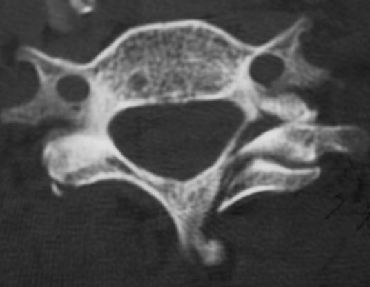
12 Results All cases were followed until a final follow-up (average 5.2 years, 2 to 10 years and 1 month). Radiographic analyses of fracture patterns and associated soft tissue injury The lateral mass fractures were divided into following four subtypes: separation fracture in eleven, comminution type in four, split type in five, and traumatic spondylolysis in two (Figure 1). The separation fracture was defined as two fracture lines of unilateral lamina and pedicle, thereby isolating and separating the unilateral entire articular mass. 4,5,16 The comminution type showed multiple fracture lines in the lateral mass with significant fragmentations, frequently accompanying with the lateral wedging deformity in a coronal plane. The split type fracture had a vertical fracture line on a coronal plane in the unilateral lateral mass, creating an anterior-posterior separation with the invagination of superior articular process of caudal adjacent vertebra. The traumatic spondylolysis showed bilateral horizontal fracture lines at pars interarticularis, leading to a separation between anterior and posterior spinal elements. The superior and inferior articular process fractures, and combination of both fractures at consecutive vertebrae were seen in six, one, and one patient, respectively. 11
13 In terms of spinal alignment on initial radiographs, the anterior translation of fractured vertebra was demonstrated in 77% of whole lateral mass fractures, while 24% and 10% of anterior translation were observed even in cephalad and caudal adjacent vertebrae, respectively (Table 1). The alignment change in coronal plane was detected in 33% of lateral mass fractures. In turn, articular process fractures demonstrated the anterior translation of upper adjacent vertebra in 50%, while 33% and 0% of anterior translation were observed in fractured and caudal adjacent vertebrae, respectively. The alignment change in coronal plane was not detected in any articular process fractures. The subtype analysis in lateral mass fractures demonstrated that the separation fracture, split type and traumatic spondylolysis showed high rates of anterior translation in 91%, 80%, and 100%, respectively. The anterior translation of the adjacent vertebra was 20%, 50%, 0%, and 50% in separation, comminution type, split type, and traumatic spondylolysis, respectively. However, there was no statistical difference between groups due to a small number of patients in each group. The comminution and split type demonstrated higher rates of coronal malalignment in 25% and 40%, respectively, when compared to that of separation fracture (3%) (P<0.05). In terms of vertebral body destruction, both groups of lateral mass fractures and articular process fractures showed 33% of vertebral body destruction rate. However, in the 12
14 subgroups of lateral mass fracture, the separation fracture demonstrated significantly lower rate of 1% than that of other subtypes (50-60%) (P<0.05). On MRI, the main injured segment was the caudal segment adjacent to the fractured vertebra in the lateral mass fractures. The signal changes of ALL and disc in lateral mass fractures were demonstrated in 76% and 24-29% of caudal and cephalad segments adjacent to the fractured vertebra, respectively (Table 2). The PLL and SISL changes at the main injured segment were 35% and 12%, respectively in the lateral mass fractures. The adjacent PLL or SISL change was minimum (0-6%) in lateral mass fractures. Meanwhile, the signal changes of ALL and disc in articular process fractures were demonstrated in 40% and 80% at the cephalad segment adjacent to the fractured vertebra, respectively. The signal changes of PLL and SISL were not detected in both cephalad and caudal segments. Additionally, the caudal adjacent segment did not show any signal change of ALL and disc in articular process fractures. The subtype analyses in lateral mass fractures demonstrated that each subtype showed the same trend as that in whole lateral mass fracture group. There were no significant differences among fracture subtypes in terms of frequency of signal changes. Surgical outcome and functional recovery 13
15 The arthrodesis was successfully achieved in all cases. Although the average number of injured segments was 1.7, 1.5 spinal segments were surgically stabilized in average, demonstrating the tendency of short segment fusion in a whole group. The average number of stabilized segments in either lateral mass fracture or articular process fracture group was 1.6 and 1.1 segments, respectively. In separation type, we successfully saved fixed segments in about a half of cases by fusing the segment just below the fractured vertebra, exclusively (average fixed segment: 1.4). The average number of fixed segments in comminution, split, and traumatic spondylolysis types were 1.6, 2, and 2 segments, respectively. The more comminuted type of injury often requires two-level fixation. The postoperative radiographic analysis demonstrated that there were six cases of mild anterior translation deformity at follow-up. Among them, three cases showed the anterior translation of fractured vertebra due to incomplete reduction during operation. However, no correction loss after postoperative periods was seen in all cases. Other three cases of cephalad vertebra translation occurred due to short-segment fusion. A case of C6 separation fracture was treated with osteosynthesis of unilateral fracture site using a cannulated screw. The reduction of fractured lateral mass was successfully achieved, however, the translation of vertebra remained to some degree. Other two cases of C5 separation and C6 comminution type fractures showed the anterior translation of fractured and cephalad vertebrae, however, because of a lack in soft tissue injury at the adjacent segment on MRI, the main injured 14
16 segment was stabilized exclusively. Even though the mild deformity seen in six cases above, no problem relating neurologic symptom or pain was demonstrated at follow-up. In terms of neurologic symptoms, there were five cases of cervical myelopathy, and 21 cases of radiculopathy, preoperatively. Based on Frankel grading system, there were B in one, C in three, and D in two cases, preoperatively, however, there were no B, C in one case, D in two, and E in three cases. The neurologic improvement more than one grade was seen in all myelopathy cases. All radiculopathy cases recovered well in terms of radicular pain, numbness, and weakness in upper extremities, except three cases of persisting motor weakness between good and fair level. Complications There was one deep infection in C5-7 lateral flexion injury case, which was treated with C5-7 pedicle screw fixation. The continuous irrigation without implant removal successfully resulted in the settlement of infection. There were four cases of screw thread exposure from the pedicle wall detected in postoperative x-ray and CTs. No neurologic injury was observed in those cases, however, one case of vertebral artery injury was demonstrated. In this case, the screw was inserted into a fractured pedicle in separation fracture of lateral mass. The bleeding stopped with bone wax and the patient had no circulatory and neurologic symptoms, consequently. 11 The patient who complained of a pain due to skin irritation of screw head required a hardware removal after completion of arthrodesis. 15
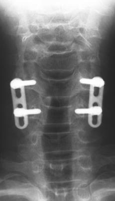
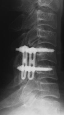
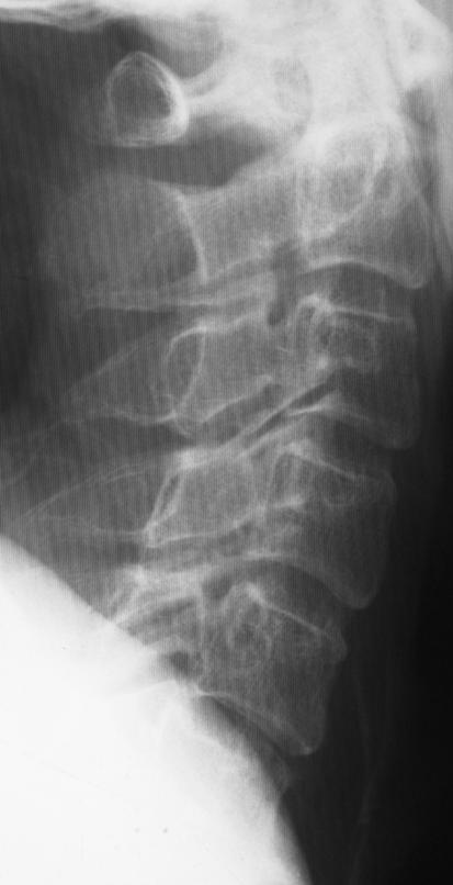
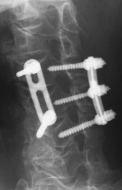
17 Case Presentations Case1 (Figure 2) A 43-year-old male suffered from a separation fracture of left C5 lateral mass due to a traffic accident. The initial neurologic presentation was incomplete Brown-Sequard syndrome with motor and sensory deficits. The imaging studies showed a typical horizontalization and fracture-separation of left C5 lateral mass as well as ruptured ALL and disc injury at C5-6 on sagittal MRI imaging. The posterior fixation and fusion was conducted using cervical pedicle screw system. After four years postoperatively, the arthrodesis was complete and no adjacent segment change was demonstrated. Case 2 (Figure 3) A 33-year-old male sustained a C6 separation fracture of left lateral mass with C6 radiculopathy. The CT scan demonstrated a typical floating lateral mass of C6. The osteosynthesis of fractured pedicle was carried out using a cannulated titanium screw. At five years postoperatively, the successful bony fusion was obtained at the fracture site, however, a slight anterior slippage of C6 vertebra remained without any symptoms. 16
18 Case 3 (Figure 4) A 60-year-old male injured a comminution type of C5 lateral mass fracture with spinal cord injury of Frankel C grade. The supposed injury mechanism was C5-6 compressive-extension injury according to Allen s classification. The comminuted right lateral mass displaced posteriorly, demonstrating two-level instability. Two-level posterior fixation and fusion was carried out with cervical pedicle screw system followed by C4-7 laminoplasty. The successful arthrodesis was achieved at one year postoperatively. 17
19 Discussion Several authors have pointed out a low accuracy in the initial diagnosis of lateral mass and articular process fractures. 5,17,18,19,20 These fractures often involved in the lower cervical spine and initial plain radiographs did not demonstrate the abnormality in many cases. The oblique x-rays are recommended for better visualization of fracture sites, however, in patients who suffered from multiple trauma or neurological injury, it is sometimes difficult to take those views initially. Halliday et al. reported that only 6 of 24 cervical plain radiographs (25%) detected an initial abnormality in the emergency department. 3 Levine et al. reported that standard roentgenograms were effective in making the definitive diagnosis in only nine of twenty-four cases (38%). 5 The use of CT or multidirectional tomography is recommended for better visualization of fracture and neural involvement. 20,21 Fractures of the lateral mass and articular process were generally accepted as being produced by hyperextension or hyperextension combined with rotational injury mechanism. 18,21,22 In our series, most injury types were classified into compressive-extension injuries followed by lateral flexion injuries according to Allen s classification. 14 In lateral flexion injuries, the lateral mass fractures were more comminuted and associated with the asymmetrical vertebral body fracture. 18
20 In this series of lateral mass fracture, we newly divided the fractures into following four subtypes: separation, comminution, split, and traumatic spondylolysis. In these subtypes, comminution and split type fractures were new entities. The comminution type fracture consisted of multiple fracture lines in the lateral mass with significant fragmentations, frequently accompanying with the lateral wedging deformity in a coronal plane. The split type fracture was previously reported by Sim and Yetkin et al. 21,23 Sim reported five cases of these injuries and described that the segmental stability was likely to be adequate and surgical indication was limited. 23 We experienced five cases of this injury type, all presenting an anterior translation, axial rotational deformity, and local lateral wedging deformity in a coronal plane. Although the invagination of superior articular process into the fractured rostal lateral mass possibly prevents the gross initial instability, the significant three-dimensional deformity should be corrected and stabilized to prevent the subsequent persistent severe neck pain. The analysis of spinal alignment abnormality at initial injury demonstrated the higher incidence of anterior translation or frontal plane deformity in lateral mass fractures than that in articular process fractures. In lateral mass fractures, the anterior translation of vertebra was detected in 77% of fractured vertebra, while in 24% of upper adjacent vertebra as well as the frontal plane deformity in 33%. Levine et al. reported an incidence of anterior vertebral translation in twenty-four cases of lateral mass fracture separation. 5 The anterior 19
21 translation of fractured vertebra was observed in 79% of fractured vertebra, as well as in 21% of cephalad-adjacent vertebra. These data were almost equivalent to that in the present study. In turn, fracture subtype analysis demonstrated that the split and comminution type showed significantly higher rates of coronal malalignment and vertebral body destruction when compared to the separation fracture. This indicated that higher injury energy seemed to be provided to those two types of injuries. This serves as the first study to precisely evaluate the initial intervertebral disc or ligamentous injuries at both fractured and adjacent spinal segments in cervical lateral mass and facet joint fractures. Overall, 76% of ALL and disc, 35% of PLL, and 12% of SSL and ISL were injured on MRI at the main injured segment of lateral mass fractures. Halliday et al. reported 50% of ALL, 29% of PLL, and 75% of ISL injuries in twenty-four cases of lateral mass and facet fractures. 3 Considering the main injury mechanism of compressive-extension in Allen s classification, it is reasonable to observe the higher frequency of ALL and disc injuries than that of SSL or ISL injury. The associated lamina fractures often demonstrate the T2 high signal intensity change around ISL resembling SSL and ISL injuries, however, this signal change has to be carefully examined with T1-weighted image in terms of the continuity of SSL black line. Another possible explanation to this contrast is that our series may include more severely injured cases when compared to their series requiring surgeries in only 50% of whole cases. In turn, 24 to 29% of ALL and disc signal changes at the 20
22 cephalad-adjacent segment were demonstrated in this study, serving as the important data for diagnosis of injury type and subsequent treatment strategy. The initial treatment strategy for lateral mass or articular process fracture is determined mainly based on the neurologic disturbance and segmental spinal instability. The suggested simple algorithm for the initial treatment strategy is shown in Figure 5. When the patients have the apparent neurologic disturbance with anterior or lateral vertebral subluxation, the surgical treatment is recommended. When there is neurologic disturbance without anterior or lateral vertebral subluxation, the further investigation has to be performed using CT and MRI in terms of fracture type and soft tissue damage analysis. The multiple discoligamentous damage or comminuted lateral mass fractures with supposed spinal instability require the surgical treatment. In case of adequate spinal stability preserved, the conservative treatment with collar can be selected. In turn, even when the patients are neurologically normal, the spinal instability based on multiple discoligamentous damage or comminuted lateral mass fractures require the surgical intervention. Although the practical decision-making in clinic is not sometimes simple like this algorithm due to severity of neural damage or socioeconomic status, this algorithm is useful in simplifying the principal initial management strategy. In terms of surgical management, we utilized a cervical pedicle screw fixation with a high fusion rate and patient satisfaction. In separation fracture cases, the osteosynthesis 21
23 using a single screw is another choice for surgical treatment (Figure 3). 4 However, our case demonstrated a residual anterior translation of repaired vertebra due to the injured intervertebral disc below. As the moderate or severe disc injury is mostly associated with the separation fracture, the osteosynthesis is only indicated for separation fracture with a minimum disc damage shown on MRI. In turn, the cervical pedicle screw fixation was reported to offer the superior three-dimensional biomechanical stability and pullout strength 24,25. Kotani et al. demonstrated the clear advantage of cervical pedicle screw fixation over that of combined anterior and posterior fixation using Bohlman s triple wiring or posterior lateral mass screw fixation even in the severe discoligamentous injury model 25. This stabilizing capacity clinically provides the excellent correction of fractured vertebra as well as a high fusion rate. Consequently, we successfully saved the stabilized segments in separation fractures or fractures with mild comminution based on the adjacent disc and ligaments evaluation. However, severely comminuted lateral mass fractures with coronal plane malalignment required two-level posterior fixation. Regarding the clinical risk of CPS, the previous studies showed no increased neurovascular complications using CPS over that of other posterior fixation techniques 11. Finally, the exclusive posterior stabilization with cervical pedicle screw system provides a short fusion as well as a normal spinal alignment even in the lateral mass fractures with severe spinal instability. Acknowledgement 22
24 The authors acknowledge Masanori Fujiya, M.D., Michinori Saita, M.D., Yasuhiro Shono, M.D., and Takahiro Iida, M.D. for a cooperative work in the patient data correction. References 1. Cooper PR, Cohen A, Rosiello A, et al: Posterior stabilization of cervical spine fractures and subluxations using plates and screws. Neurosurgery 23: , Fehlings GM, Cooper PR, Errico TJ: posterior plates in the management of cervical instability: long-term results in 44 patients. J Neurosurg 81: , Halliday AL, Henderson BR, Hart BL, and Benzel EC: The management of unilateral lateral mass/facet fractures of the subaxial cervical spine. The use of magnetic resonance imaging to predict instability. Spine 22: , Jeanneret B, Gebhard JS, Magerl F: Transpediclar screw fixation of articular mass fracture-separation: results of an anatomical study and operative technique. J Spinal Disord 7: , Levine AM, Mazel C, Roy-Camille R: Management of fracture separations of the articular mass using posterior cervical plating. Spine 17:S , Bohlman HH: Acute fractures and dislocations of the cervical spine: an analysis of three hundred hospitalized patients and review of the literature. J Bone Joint Surg (Am) 61: , An HS, Coppes MA: Posterior cervical fixation for fracture and degenerative disc disease. Clin Orthop 335: , Heller JG, Silcox DH, Sutterlin CE: Complications of posterior cervical plating. Spine 20, , Shapiro S, Snyder W, Kaufman K, et al: Outcome of 51 cases of unilateral locked cervical facets: interspinous braided cable for lateral mass plate fusion compared with interspinous wire and facet wiring with iliac crest. J Neurosurg (Spine 1) 91:19-24,
25 10. Schultz KD, McLaughlin MR, Haid RW, et al: Single-stage anterior-posterior decompression and stabilization for complex cervical spine disorders. J Neurosurg (Spine 2) 93: , Abumi K Shono Y, Kotani Y, et al: Complications of pedicle screw fixation in reconstructive surgery of the cervical spine. Spine 25: , Abumi K, Ito H, Taneichi H, et al: Transpediclar screw fixations for traumatic lesions of the middle and lower cervical spine: Description of the techniques and preliminary report. J spinal Disord 7:19-28, Abumi K, Kaneda K, Shono Y: One-stage posterior decompression and reconstruction of the cervical spine by using pedicle screw fixation systems. J Neurosurg 90:19-26, Allen B Jr, Ferguson RL, Lehmann TR, et al: A mechanistic cllasification of closed, indirect fractures and dislocations of the lower cervical spine. Spine 7:1-27, Frankel HL, hancock DO, Hyslop G et al: The value of postural reduction in the initial management of closed injuries of the spine with paraplegia and tetraplesia. Paraplesia 7: , Judet R, Roy-Cammile R, Zerah JC: Fracture du rachis cervical, fracture separation du massif articulaire. Rev Chir Orthop 56: , Bucholz Rd, Cheung KC: Halo vest versus spinal fusion for cervical injury: Evidence from an outcome study. J neurosurg 70: , Harris JH, Mirvis SE: Radiology of acute cervical spine trauma. 3 rd ed. Baltimore: William & Wilkins, , Smith GR, Beckly DE, Abel MS: Articular mass fracture: A neglected cause of post-traumatic neck pain? Clin Radiol 27:335-40, Woodring JH, Lee C: Limitations of cervical radiography in the evaluation of acute cervical trauma. J Trauma 34:32-39, Yetkin Z, Osborn AG, Giles D, et al: Uncovertebral and facet joint dislocations in cervical articular pillar fractures: CT evaluation. AJNR 6: ,
26 22. Whitley JE, Forsyth HF: The classification of cervical spine injuries. AJR 83: , Sim E: Vertical facet splitting: a special variant of rotatory dislocations of the cervical spine. J Neurosurg 82: , Jones EL, Heller JG, Silcox DH, Hutton WC: Cervical pedicle screws versus lateral mass screws: Anatomic feasibility and biomechanical comparison. Spine 22: , Kotani Y, Cunnigham BW, Abumi K: Biomechanical analysis of cervical stabilization systems: An assessment of transpediclar screw fixation in the cervical spine. Spine 19: ,
27 Table 1. Translation of fractured and adjacent vertebrae on sagittal and frontal plane (Frequency shown as percentage in each fracture type) Fracture Type Anterior Translation Frontal Translation Upp. Adj. Fractured Low. Adj. Fractured Lateral Mass 24% 77% 10% 33% Articular Proc. 50% 33% 0% 0% Lateral Mass: Lateral Mass Fracture, Articular Proc.: Articular Process Fracture, Upp. Adj.: Upper Adjacent Vertebrae, Fractured: Fractured Vertebra, Low. Adj.: Lower Adjacent Vertebra 26
28 Table 2. Invertebral Disc and Ligamentous Injuries on Initial Magnetic Resonance Imaging (Frequency shown as percentage in each fracture type) Fracture Type Main Injured Segment Adj. Segment ALL Disc PLL SISL ALL Disc Lateral Mass 76% 76% 35% 12% 24% 29% Articular Proc. 40% 80% 0% 0% 0% 0% All signal changes of lateral mass fracture at adjacent segment were observed in cephalad-adjacent segments to fractured vertebrae. Lateral Mass: Lateral Mass Fracture, Articular Proc.: Articular Process Fracture, Adj. Segment: Adjacent segment to main injured segment, ALL: Anterior Longitudinal Ligament, Disc: Intervertebral Disc, PLL: Posterior Longitudinal ligament, SISL: Supraspinous and Interspinous Ligaments 27
29 Figure Legend Figure 1: Fracture subtypes of lateral mass fracture. A: Separation fracture. The separation fracture was defined as two fracture lines of unilateral lamina and pedicle, thereby isolating and separating the unilateral entire articular mass B: Comminution type. There are multiple fracture lines in the lateral mass with significant fragmentations, frequently accompanying with the lateral wedging deformity in a coronal plane. C: Split type. There is vertical fracture line on a coronal plane in the unilateral lateral mass, creating an anterior-posterior separation with the invagination of superior articular process of caudal adjacent vertebra. D: Traumatic Spondylolysis. There are bilateral horizontal fracture lines at pars interarticularis, leading to a separation between anterior and posterior spinal elements Figure 2 A: A 43-year-old male injured a C5 separation fracture of lateral mass with incomplete Brown-Sequard 28
30 syndrome. A-P radiograph showed a typical horizontalization of left C5 lateral mass. B: MRI showed the ALL and disc injury at C5/6 level. C: CT scan demonstrated a typical floating lateral mass on the left side. D.E: Single segment posterior fixation and fusion was conducted with cervical pedicle screw system. After four years postoperatively, the arthrodesis was complete and no adjacent segment change was demonstrated. Figure 3 A, B: A 33-year-old male sustained C6 separation fracture of left lateral mass with C6 radiculopathy. C, D: The osteosynthesis of fractured pedicle was carried out using a cannulated titanium screw. At five years postoperatively, the successful bony fusion was obtained at the fracture site, however, a slight anterior slippage of C6 vertebra remained without any symptom. Figure 4 A,B: A 60-year-old male injured a comminution type of C5 lateral mass fracture with spinal cord injury of Frankel C grade. The supposed injury mechanism was C5-6 compressive-extension injury according to Allen s classification. C,D: The comminuted right lateral mass displaced posteriorly, demonstrating two-level instability. 29
31 E,F,G: Two-level posterior fixation and fusion was carried out with cervical pedicle screw system followed by C4-7 laminoplasty. The successful arthrodesis was shown at one year postoperatively. The oblique x-ray demonstrated the accurate screw purchases. Figure 5: Suggested algorism of treatment strategy for cervical lateral mass or articular process fracture. The algorism was simplified mainly based on the presence of neurologic abnormality and anterior or lateral vertebral subluxation for the emergency use. 30
32 Figure Legend Figure 1: Fracture subtypes of lateral mass fracture. A: Separation fracture. The separation fracture was defined as two fracture lines of unilateral lamina and pedicle, thereby isolating and separating the unilateral entire articular mass B: Comminution type. There are multiple fracture lines in the lateral mass with significant fragmentations, frequently accompanying with the lateral wedging deformity in a coronal plane. C: Split type. There is vertical fracture line on a coronal plane in the unilateral lateral mass, creating an anterior-posterior separation with the invagination of superior articular process of caudal adjacent vertebra. D: Traumatic Spondylolysis. There are bilateral horizontal fracture lines at pars interarticularis, leading to a separation between anterior and posterior spinal elements Figure 2 A: A 43-year-old male injured a C5 separation fracture of lateral mass with incomplete Brown-Sequard syndrome. A-P radiograph showed a typical horizontalization of left C5 lateral mass. B: MRI showed the ALL and disc injury at C5/6 level. C: CT scan demonstrated a typical floating lateral mass on the left side. D.E: Single segment posterior fixation and fusion was conducted with cervical pedicle screw system. After four years postoperatively, the arthrodesis was complete and no adjacent segment change was demonstrated.
33 Figure 3 A, B: A 33-year-old male sustained C6 separation fracture of left lateral mass with C6 radiculopathy. C, D: The osteosynthesis of fractured pedicle was carried out using a cannulated titanium screw. At five years postoperatively, the successful bony fusion was obtained at the fracture site, however, a slight anterior slippage of C6 vertebra remained without any symptom. Figure 4 A,B: A 60-year-old male injured a comminution type of C5 lateral mass fracture with spinal cord injury of Frankel C grade. The supposed injury mechanism was C5-6 compressive-extension injury according to Allen s classification. C,D: The comminuted right lateral mass displaced posteriorly, demonstrating two-level instability. E,F,G: Two-level posterior fixation and fusion was carried out with cervical pedicle screw system followed by C4-7 laminoplasty. The successful arthrodesis was shown at one year postoperatively. The oblique x-ray demonstrated the accurate screw purchases. Figure 5: Suggested algorism of treatment strategy for cervical lateral mass or articular process fracture. The algorism was simplified mainly based on the presence of neurologic abnormality and anterior or lateral vertebral subluxation for the emergency use.
34


35 A B C D


36 5 B A C D E


37 6 A B C D


38 5 C 5 A B D


39 6 E F G
40 D Neurologic Disturbance Neurologically Normal Sublux (+) Sublux (-) Sublux (+) Sublux (-) Neuro. Severity MRI Evaluation Spinal Instability MRI Evaluation Surgery Conservative Surgery Conservative
SUBAXIAL CERVICAL SPINE TRAUMA- DIAGNOSIS AND MANAGEMENT
 SUBAXIAL CERVICAL SPINE TRAUMA- DIAGNOSIS AND MANAGEMENT 1 Anatomy 3 columns- Anterior, middle and Posterior Anterior- ALL, Anterior 2/3 rd body & disc. Middle- Posterior 1/3 rd of body & disc, PLL Posterior-
Fractures of the thoracic and lumbar spine and thoracolumbar transition
 Most spinal column injuries occur in the thoracolumbar transition, the area between the lower thoracic spine and the upper lumbar spine; over half of all vertebral fractures involve the 12 th thoracic
Subaxial Cervical Spine Trauma
 Subaxial Cervical Spine Trauma Pooria Salari, MD Assistant Professor Of Orthopaedics Department of Orthopaedic Surgery St. Louis University School of Medicine St. Louis, Missouri, USA Initial Evaluation
Common fracture & dislocation of the cervical spine. Theerachai Apivatthakakul Department of Orthopaedic Chiangmai University
 Common fracture & dislocation of the cervical spine Theerachai Apivatthakakul Department of Orthopaedic Chiangmai University Objective Anatomy Mechanism and type of injury PE.and radiographic evaluation
Subaxial Cervical Spine Trauma Dr Hesarikia BUMS
 Subaxial Cervical Spine Trauma Dr. Hesarikia BUMS Subaxial Cervical Spine From C3-C7 ROM Majority of cervical flexion Lateral bending Approximately 50% rotation Ligamentous Anatomy Anterior ALL, PLL, intervertebral
Subaxial Cervical Spine Trauma. Introduction. Anatomic Considerations 7/23/2018
 Subaxial Cervical Spine Trauma Sheyan J. Armaghani, MD Florida Orthopedic Institute Assistant Professor USF Dept of Orthopedics Introduction Trauma to the cervical spine accounts for 5 of all spine injuries
AO CLASSIFICATIONS THORACO-LUMBAR SPINAL INJURIES
 AO CLASSIFICATIONS THORACO-LUMBAR SPINAL INJURIES T H E A O / A S I F ( A R B E I T S G E M E I N S C H A F T F Ü R O S T E O S Y N T H E S E F R A G E N / A S S O C I A T I O N F O R T H E S T U D Y O
Comparative Study of Surgical Approaches for Distractive Flexion Injuries of Sub-Axial Cervical Spine
 Open Journal of Modern Neurosurgery, 2018, 8, 342-351 http://www.scirp.org/journal/ojmn ISSN Online: 2163-0585 ISSN Print: 2163-0569 Comparative Study of Surgical Approaches for Distractive Flexion Injuries
Imaging of Cervical Spine Trauma Tudor H Hughes, M.D.
 Imaging of Cervical Spine Trauma Tudor H Hughes, M.D. General Considerations Most spinal fractures are due to a single episode of major trauma. Fatigue fractures of the spine are unusual except in the
Classification of Thoracolumbar Spine Injuries
 Classification of Thoracolumbar Spine Injuries Guillem Saló Bru 1 IMAS. Hospitals del Mar i de l Esperança. ICATME. Institut Universitari Dexeus USP. UNIVERSITAT AUTÒNOMA DE BARCELONA Objectives of classification
Traumatic C6 7 subluxation with anomalous course of vertebral arteries treated with pedicle screw/rod fixation
 J Neurosurg Spine 7:65 70, 2007 Traumatic C6 7 subluxation with anomalous course of vertebral arteries treated with pedicle screw/rod fixation Case report MASASHI YAMAZAKI, M.D., PH.D., 1 TAKANA KOSHI,
Posterior Cervical Arthrodesis by Lateral Mass Screws Fixation A Long term Follow-up Study
 Original Article Posterior Cervical Arthrodesis by Lateral Mass Screws Fixation A Long term Follow-up Study Bhaskar G 1, Sharath Kumar Maila 2, Lakshman Rao A 3, Mastan Reddy A 4 1 Professor I/C 2, 3 Assistant
Spinal Cord Injuries: The Basics. Kadre Sneddon POS Rounds October 1, 2003
 Spinal Cord Injuries: The Basics Kadre Sneddon POS Rounds October 1, 2003 Anatomy Dorsal columntouch, vibration Corticospinal tract- UMN Anterior horn-lmn Spinothalamic tractpain, temperature (contralateral)
ISPUB.COM. Fracture Through the Body of the Axis. B Johnson, N Jayasekera CASE REPORT
 ISPUB.COM The Internet Journal of Orthopedic Surgery Volume 8 Number 1 B Johnson, N Jayasekera Citation B Johnson, N Jayasekera.. The Internet Journal of Orthopedic Surgery. 2007 Volume 8 Number 1. Abstract
Bilateral spondylolysis of inferior articular processes of the fourth lumbar vertebra: a case report
 Upsala Journal of Medical Sciences. 2012; 117: 72 77 CASE REPORT Bilateral spondylolysis of inferior articular processes of the fourth lumbar vertebra: a case report TOMOAKI KOAKUTSU 1,2, NAOKI MOROZUMI
C2 Body Fracture: Report of Cases Managed Conservatively by Philadelphia Collar
 C2 Body Fracture: Report of Cases Managed Conservatively by Philadelphia Collar The Harvard community has made this article openly available. Please share how this access benefits you. Your story matters.
A rare case of spinal injury: bilateral facet dislocation without fracture at the lumbosacral joint
 J Orthop Sci (2012) 17:189 193 DOI 10.1007/s00776-011-0082-y CASE REPORT A rare case of spinal injury: bilateral facet dislocation without fracture at the lumbosacral joint Kei Shinohara Shigeru Soshi
factor for identifying unstable thoracolumbar fractures. There are clinical and radiological criteria
 NMJ-Vol :2/ Issue:1/ Jan June 2013 Case Report Medical Sciences Progressive subluxation of thoracic wedge compression fracture with unidentified PLC injury Dr.Thalluri.Gopala krishnaiah* Dr.Voleti.Surya
Lateral Mass and Pedicle Screws Fixation of Cervical Spine
 Lateral Mass and Pedicle Screws Fixation of Cervical Spine Anderson Spine 1991 Paul A Anderson University of Wisconsin AAOS Biomedical Engineering Committee Co-chair ASTM F05-24 Purpose Anatomy Surgical
Dorsal Cervical Surgeries and Techniques
 Dorsal Cervical Approaches Dorsal Cervical Surgeries and Techniques Gregory R. Trost, MD Professor and Vice Chair of Neurological Surgery University of Wisconsin-Madison Advantages Straightforward Easily
Title. Author(s)Kotani, Yoshihisa; Abumi, Kuniyoshi; Ito, Manabu; Mi. CitationJournal of Neurosurgery : Spine, 99(3): Issue Date
Kotani, Yoshihisa; Abumi, Kuniyoshi; Ito, Manabu; Mi. CitationJournal of Neurosurgery : Spine, 99(3): Issue Date") Title Improved accuracy of computer-assisted cervical pedi Author(s)Kotani, Yoshihisa; Abumi, Kuniyoshi; Ito, Manabu; Mi CitationJournal of Neurosurgery : Spine, 99(3): 257-263 Issue Date 2003-10 Doc URL
Title Improved accuracy of computer-assisted cervical pedi Author(s)Kotani, Yoshihisa; Abumi, Kuniyoshi; Ito, Manabu; Mi CitationJournal of Neurosurgery : Spine, 99(3): 257-263 Issue Date 2003-10 Doc URL
ASJ. Cervical Pedicle Screw Fixation: Anatomic Feasibility of Pedicle Morphology and Radiologic Evaluation of the Anatomical Measurements
 Asian Spine Journal Asian Spine Clinical Journal Study Asian Spine J 2014;8(3):273-280 Cervical http://dx.doi.org/10.4184/asj.2014.8.3.273 pedicle screw fixation 273 Cervical Pedicle Screw Fixation: Anatomic
University of Groningen. Thoracolumbar spinal fractures Leferink, Vincentius Johannes Maria
 University of Groningen Thoracolumbar spinal fractures Leferink, Vincentius Johannes Maria IMPORTANT NOTE: You are advised to consult the publisher's version (publisher's PDF) if you wish to cite from
Comprehension of the common spine disorder.
 Objectives Comprehension of the common spine disorder. Disc degeneration/hernia. Spinal stenosis. Common spinal deformity (Spondylolisthesis, Scoliosis). Osteoporotic fracture. Anatomy Anatomy Anatomy
Treatment of thoracolumbar burst fractures by vertebral shortening
 Eur Spine J (2002) 11 :8 12 DOI 10.1007/s005860000214 TECHNICAL INNOVATION Alejandro Reyes-Sanchez Luis M. Rosales Victor P. Miramontes Dario E. Garin Treatment of thoracolumbar burst fractures by vertebral
Key Primary CPT Codes: Refer to pages: 7-9 Last Review Date: October 2016 Medical Coverage Guideline Number:
 National Imaging Associates, Inc. Clinical guidelines CERVICAL SPINE SURGERY: ANTERI CERVICAL DECOMPRESSION WITH FUSION CERVICAL POSTERI DECOMPRESSION WITH FUSION CERVICAL ARTIFICIAL DISC CERVICAL POSTERI
Complications of Pedicle Screw Fixation in Reconstructive Surgery of the Cervical Spine
 SPINE Volume 25, Number 8, pp 962 969 2000, Lippincott Williams & Wilkins, Inc. Complications of Pedicle Screw Fixation in Reconstructive Surgery of the Cervical Spine Kuniyoshi Abumi, MD, Yasuhiro Shono,
Objectives. Comprehension of the common spine disorder
 Objectives Comprehension of the common spine disorder Disc degeneration/hernia Spinal stenosis Common spinal deformity (Spondylolisthesis, Scoliosis) Osteoporotic fracture Destructive spinal lesions Anatomy
Title. Author(s)Sudo, Hideki; Taneichi, H.; Kaneda, K. CitationSpinal Cord, 44(2): Issue Date Doc URL
Sudo, Hideki; Taneichi, H.; Kaneda, K. CitationSpinal Cord, 44(2): Issue Date Doc URL") Title Secondary medulla oblongata involvement following mi instability in a patient with ossification of the po Author(s)Sudo, Hideki; Taneichi, H.; Kaneda, K. CitationSpinal Cord, 44(2): 126-129 Issue
Title Secondary medulla oblongata involvement following mi instability in a patient with ossification of the po Author(s)Sudo, Hideki; Taneichi, H.; Kaneda, K. CitationSpinal Cord, 44(2): 126-129 Issue
Stage Operation for Unstable Lumbar Spine Fracture- Dislocation with Incomplete Paraplegia: A Case Series
 C a s e R e p o r t J. of Advanced Spine Surgery Volume 2, Number 2, pp 60~65 Journal of Advanced Spine Surgery JASS Stage Operation for Unstable Lumbar Spine Fracture- Dislocation with Incomplete Paraplegia:
Thoracolumbar spine trauma classifications: evolution or more confusion
 Thoracolumbar spine trauma classifications: evolution or more confusion Poster No.: C-1713 Congress: ECR 2012 Type: Educational Exhibit Authors: J. P. Salazar, J. Halaburda Berni, C. Torrents, L. Casas;
CERVICAL SPINE INJURIES IN THE ELDERLY
 CERVICAL SPINE INJURIES IN THE ELDERLY ISADOR H. LIEBERMAN, JOHN K. WEBB From University Hospital, Queen s Medical Centre, Nottingham, England We reviewed 41 patients over the age of 65 years (mean 76.5)
ELY ASHKENAZI Israel Spine Center at Assuta Hospital Tel Aviv, Israel
 nterior cervical decompression using the Hybrid Decompression Fixation technique, a combination of corpectomies and or discectomies, in the management of multilevel cervical myelopathy J ORTHOP TRUM SURG
APPLYING BOTH ANTERIOR AND POSTERIOR APPROACH FOR MID CERVICAL TRAUMA: CASE REPORT
 APPLYING BOTH ANTERIOR AND POSTERIOR APPROACH FOR MID CERVICAL TRAUMA: CASE REPORT Dr. Sunil Pahari *1, Prof. Dr. Lu Hou Gen 2, Prof. Dr. Liu Jun 3 and Prof. Dr. Zhi Yu Sun 4 1,2,3,4Department of Orthopaedic
Lower cervical trauma an orthopaedician domain
 International Journal of Research in Orthopaedics http://www.ijoro.org Original Research rticle DOI: http://dx.doi.org/10.18203/issn.2455-4510.intjresorthop20173175 Lower cervical trauma an orthopaedician
102 Results RESULTS. Age Mean=S.D Range 42= years -84 years Number % <30 years years >50 years
 102 Results RESULTS A total of 50 cases were studied 39 males and 11females.Their age ranged between 16 years and 84 years (mean 42years). T1 and T2WI were acquired for all cases in sagittal and axial
Incidence and Risk Factors for Late Neurologic Deterioration after C3-6 Laminoplasty in Patients with Cervical Spondylotic Myelopathy
 Incidence and Risk Factors for Late Neurologic Deterioration after C3-6 Laminoplasty in Patients with Cervical Spondylotic Myelopathy Sakaura H, Miwa T, Kuroda Y, Ohwada T Dept. of Orthop. Surg., Kansai
102 Posterior Cervical Arthrodesis
 102 Posterior Cervical Arthrodesis Gabriel Widi, Mohammed Faraz Khan, and Glen Manzano Indications Posterior and posterolateral access to the cervical spine: Laminectomy for decompression Intradural tumor
Dr Ajit Singh Moderator Dr P S Chandra Dr Rajender Kumar
 BIOMECHANICS OF SPINE Dr Ajit Singh Moderator Dr P S Chandra Dr Rajender Kumar What is biomechanics? Biomechanics is the study of the consequences of application of external force on the spine Primary
Cover Page. The handle holds various files of this Leiden University dissertation.
 Cover Page The handle http://hdl.handle.net/1887/29800 holds various files of this Leiden University dissertation. Author: Moojen, Wouter Anton Title: Introducing new implants and imaging techniques for
5/19/2017. Interspinous Process Fixation with the Minuteman G3. What is the Minuteman G3. How Does it Work?
 Interspinous Process Fixation with the Minuteman G3 LLOYDINE J. JACOBS, MD CASTELLVI SPINE MEETING MAY 13, 2017 What is the Minuteman G3 The world s first spinous process plating system that is: Minimally
Systematic review Cervical artificial disc replacement versus fusion in the cervical spine: a systematic review (...)
") Systematic review Cervical artificial disc replacement versus fusion in the cervical spine: a systematic review (...) 59 59 66 Cervical artificial disc replacement versus fusion in the cervical spine:
Systematic review Cervical artificial disc replacement versus fusion in the cervical spine: a systematic review (...) 59 59 66 Cervical artificial disc replacement versus fusion in the cervical spine:
Delayed surgical treatment for a traumatic bilateral cervical facet joint dislocation using a posterior-anterior approach: a case report
 Shimada et al. Journal of Medical Case Reports 2013, 7:9 JOURNAL OF MEDICAL CASE REPORTS CASE REPORT Open Access Delayed surgical treatment for a traumatic bilateral cervical facet joint dislocation using
Imaging of Orthopedic Spinal Devices: What Every Radiologist Should Know.
 Imaging of Orthopedic Spinal Devices: What Every Radiologist Should Know. Poster No.: C-1656 Congress: ECR 2016 Type: Educational Exhibit Authors: E. Federici, C. Dell'atti, M. Bartocci, D. Beomonte Zobel,
Original article: Multidetector computed tomographic evaluation of cervical spine trauma
 Original article: Multidetector computed tomographic evaluation of cervical spine trauma 1Sajid Ansari *, 2 R.K. Rauniyar, 3 Kaleem Ahmad, 4 Mukesh Kumar Gupta 1Assistant Professor, Department of Radiodiagnosis,
Int J Clin Exp Med 2018;11(2): /ISSN: /IJCEM Yi Yang, Hao Liu, Yueming Song, Tao Li
: /ISSN: /IJCEM Yi Yang, Hao Liu, Yueming Song, Tao Li") Int J Clin Exp Med 2018;11(2):1278-1284 www.ijcem.com /ISSN:1940-5901/IJCEM0063093 Case Report Dislocation and screws pull-out after application of an Isobar TTL dynamic stabilisation system at L2/3 in
Int J Clin Exp Med 2018;11(2):1278-1284 www.ijcem.com /ISSN:1940-5901/IJCEM0063093 Case Report Dislocation and screws pull-out after application of an Isobar TTL dynamic stabilisation system at L2/3 in
Anterior Cervical Subluxation: An Unstable Position
 275 Anterior Cervical Subluxation: An Unstable Position, 1 A. T. Scher1 The radioiogic signs of cervical anterior subluxation are subtle. Even when recognized, the injury may not be considered significant.
Interbody fusion cage for the transforaminal approach. Travios. Surgical Technique
 Interbody fusion cage for the transforaminal approach Travios Surgical Technique Image intensifier control This description alone does not provide sufficient background for direct use of DePuy Synthes
The craniocervical junction
 Anver Jameel, MD The craniocervical junction A biomechanical and anatomical unit that extends from the skull base to C2 Includes the clivus, foramen magnum and contiguous occipital bone, the occipital
Therapeutic Strategy for Traumatic Instability of Subaxial Cervical Spine
 Chinese Medicine, 2009, 1, 23-29 Published Online September 2009 in SciRes (www.scirp.org/journal/cm) Therapeutic Strategy for Traumatic Instability of Subaxial Cervical Spine - ABSTRACT A simple, safe
SpineFAQs. Lumbar Spondylolisthesis
 SpineFAQs Lumbar Spondylolisthesis Normally, the bones of the spine (the vertebrae) stand neatly stacked on top of one another. The ligaments and joints support the spine. Spondylolisthesis alters the
Traumatic thoracic spinal fracture dislocation with minimal or no cord injury
 J Neurosurg (Spine 3) 96:333 337, 2002 Traumatic thoracic spinal fracture dislocation with minimal or no cord injury Report of four cases and review of the literature SCOTT SHAPIRO M.D., TODD ABEL, M.D.,
ASJ. A Rare Hyperextension Injury in Thoracic Spine Presenting with Delayed Paraplegia. Asian Spine Journal. Introduction
 sian Spine Journal 126 Dong-Eun Case Shin Report et al. http://dx.doi.org/10.4184/asj.2013.7.2.126 Rare Hyperextension Injury in Thoracic Spine Presenting with Delayed Paraplegia Dong-Eun Shin, Ki-Sik
Facet orientation in patients with lumbar degenerative spondylolisthesis
 35 J. Tokyo Med. Univ., 71 1 35 0 Facet orientation in patients with lumbar degenerative spondylolisthesis Wuqikun ALIMASI, Kenji ENDO, Hidekazu SUZUKI, Yasunobu SAWAJI, Hirosuke NISHIMURA, Hidetoshi TANAKA,
Asymmetric T5 Pedicle Subtraction Osteotomy (PSO) for complex posttraumatic deformity
 for complex posttraumatic deformity") Eur Spine J (2013) 22:2130 2135 DOI 10.1007/s00586-013-2942-y OPEN OPERATING THEATRE (OOT) Asymmetric T5 Pedicle Subtraction Osteotomy (PSO) for complex posttraumatic deformity Ibrahim Obeid Fethi Laouissat
Eur Spine J (2013) 22:2130 2135 DOI 10.1007/s00586-013-2942-y OPEN OPERATING THEATRE (OOT) Asymmetric T5 Pedicle Subtraction Osteotomy (PSO) for complex posttraumatic deformity Ibrahim Obeid Fethi Laouissat
Outline. Epidemiology Indications for C-spine imaging Modalities Interpretation Types of fractures
 C-Spine Plain Films Outline Epidemiology Indications for C-spine imaging Modalities Interpretation Types of fractures Epidemiology 7000-10000 c-spine injuries treated each year Additional 5000 die at the
Two Consecutive Levels of Unilateral Cervical Spondylolysis on Opposite Sides 두개의연속된척추에서반대쪽편측에발생한경추척추분리증
 Case Report pissn 1738-2637 / eissn 2288-2928 http://dx.doi.org/10.3348/jksr.2015.73.3.181 Two Consecutive Levels of Unilateral Cervical Spondylolysis on Opposite Sides 두개의연속된척추에서반대쪽편측에발생한경추척추분리증 Kyeong
Disclosures. Cervical Spine Stabilization. Adequate Fixation?
 Disclosures Safety of reconstruction of complex cervical spine pathology using pedicle screws inserted with navigation Alexander A. Theologis, MD; Shane Burch, MD Theologis OREF Burch Medtronic May 9,
2. The vertebral arch is composed of pedicles (projecting from the body) and laminae (uniting arch posteriorly).
 and laminae (uniting arch posteriorly).") VERTEBRAL COLUMN 2018zillmusom I. VERTEBRAL COLUMN - functions to support weight of body and protect spinal cord while permitting movements of trunk and providing for muscle attachments. A. Typical vertebra
VERTEBRAL COLUMN 2018zillmusom I. VERTEBRAL COLUMN - functions to support weight of body and protect spinal cord while permitting movements of trunk and providing for muscle attachments. A. Typical vertebra
SpineFAQs. Neck Pain Diagnosis and Treatment
 SpineFAQs Neck Pain Diagnosis and Treatment Neck pain is a common reason people visit their doctor. Neck pain typically doesn't start from a single injury. Instead, the problem usually develops over time
Accelerated spondylotic changes adjacent to the fused segment following central cervical corpectomy: magnetic resonance imaging study evidence
 See the Editorial and the Response in this issue, p 1. J Neurosurg (Spine 1) 100:2 6, 2004 Accelerated spondylotic changes adjacent to the fused segment following central cervical corpectomy: magnetic
Pediatric cervical spine injuries with neurological deficits, treatment options, and potential for recovery
 SICOT J 2017, 3, 53 Ó The Authors, published by EDP Sciences, 2017 DOI: 10.1051/sicotj/2017035 Available online at: www.sicot-j.org CASE REPORT OPEN ACCESS Pediatric cervical spine injuries with neurological
Cervical corpectomy for sub-axial retro-vertebral body lesions
 Eshra Egyptian Journal of Neurosurgery (2019) 33:2 https://doi.org/10.1186/s41984-018-0004-9 Egyptian Journal of Neurosurgery RESEARCH Open Access Cervical corpectomy for sub-axial retro-vertebral body
Induction and Maintenance of Lordosis in MultiLevel ACDF Using Allograft. Saad Khairi, MD Jennifer Murphy Robert S. Pashman, MD
 Induction and Maintenance of Lordosis in MultiLevel ACDF Using Allograft Saad Khairi, MD Jennifer Murphy Robert S. Pashman, MD Purpose Is lordosis induced by multilevel cortical allograft ACDF placed on
Technique Guide. C1/C2 Access System. Percutaneous transarticular screw fixation.
 Technique Guide C1/C2 Access System. Percutaneous transarticular screw fixation. C1/C2 Access System Table of Contents General introduction 2 Indications/Contraindications 2 Image intensifier-assisted
It consist of two components: the outer, laminar fibrous container (or annulus), and the inner, semifluid mass (the nucleus pulposus).
, and the inner, semifluid mass (the nucleus pulposus).") Lumbar Spine The lumbar vertebrae are the last five vertebrae of the vertebral column. They are particularly large and heavy when compared with the vertebrae of the cervical or thoracicc spine. Their bodies
Lumbar Spine The lumbar vertebrae are the last five vertebrae of the vertebral column. They are particularly large and heavy when compared with the vertebrae of the cervical or thoracicc spine. Their bodies
Departement of Neurosurgery A.O.R.N A. Cardarelli- Naples.
 Percutaneous posterior pedicle screw fixation in the treatment of thoracic, lumbar and thoraco-lumbar junction (T12-L1) traumatic and pathological spine fractures. Report of 45 cases. G. Vitale, A. Punzo,
Traumatic spondylolisthesis of the axis has been
 Bulletin Hospital for Joint Diseases Volume 60, Number 2 2001-2002 61 Traumatic Spondylolisthesis of the Axis 42 Cases Myung-Sang Moon MD Jeong-Lim Moon MD Young-Wan Moon MD Doo-Hoon Sun MD PhD and Won-Tai
Fractures of the C-2 vertebral body
 J Neurosurg 81:206-212, 1994 Fractures of the C-2 vertebral body EDWARD C. BENZEL, M.D., BLAINE L. HART, M.D., PERRY A. BALL, M.D., NEVAN G. BALDWIN, M.D., WILLIAM W. ORRISON, M.D., AND MARY ESPINOSA,
Although unstable thoracolumbar injuries are a common
 ORIGINAL ARTICLE Anterior-Only Stabilization of Three-Column Thoracolumbar Injuries Rick C. Sasso, MD, Natalie M. Best, BS, Thomas M. Reilly, MD, and Robert A. McGuire, Jr., MD Objective: The optimal treatment
We are IntechOpen, the world s leading publisher of Open Access books Built by scientists, for scientists. International authors and editors
 We are IntechOpen, the world s leading publisher of Open Access books Built by scientists, for scientists 4,000 116,000 120M Open access books available International authors and editors Downloads Our
Degenerative spondylolisthesis at the L4 L5 in a 32-year-old female with previous fusion for idiopathic scoliosis: A case report
 Journal of Orthopaedic Surgery 2003: 11(2): 202 206 Degenerative spondylolisthesis at the L4 L5 in a 32-year-old female with previous fusion for idiopathic scoliosis: A case report RB Winter Clinical Professor,
Posterior Instrumentation of Thoracolumbar Fracture
 Posterior Instrumentation of Thoracolumbar Fracture Jin-Young Lee, MD, and Gab-Lae Kim, MD Department of Orthopedic Surgery, Hallym University College of Medicine, Seoul, Korea Abstract The thoracolumbar
Treatment of chronic traumatic C7 T1 grade III spondylolisthesis with mild neurological deficit: case report
 Case Report Treatment of chronic traumatic C7 T1 grade III spondylolisthesis with mild neurological deficit: case report Jacinto Mata-Gómez 1, Marta Ortega-Martínez 1, Julio Valencia-Anguita 2, Ignacio
Interspinous Fusion Devices. Midterm results. ROME SPINE 2012, 7th International Meeting Rome, 6-7 December 2012
 Interspinous Fusion Devices. Midterm results. ROME SPINE 2012, 7th International Meeting Rome, 6-7 December 2012 Posterior distraction and decompression Secure Fixation and Stabilization Integrated Bone
Case SCIWORA in patient with congenital block vertebra
 Case 15428 SCIWORA in patient with congenital block vertebra Lucas Walgrave 1, Charlotte Vanhoenacker 1-2, Thomas Golinvaux 3, Filip Vanhoenacker3-5 1: Leuven University Hospital, Department of Radiology,
Posterior surgical procedures are those procedures
 9 Cervical Posterior surgical procedures are those procedures that have been in use for a long time with established efficacy in the treatment of radiculopathy and myelopathy caused by pathologies including
Increased Fusion Rates With Cervical Plating for Two- Level Anterior Cervical Discectomy and Fusion
 Increased Fusion Rates With Cervical Plating for Two- Level Anterior Cervical Discectomy and Fusion SPINE Volume 25, Number 1, pp 41 45 2000, Lippincott Williams & Wilkins, Inc. Jeffrey C. Wang, MD, Paul
Anterior cervical diskectomy icd 10 procedure code
 Home Anterior cervical diskectomy icd 10 procedure code Access to discounts at hundreds of restaurants, travel destinations, retail stores, and service providers. AAPC members also have opportunities to
ProDisc-C versus fusion with Cervios chronos prosthesis in cervical degenerative disc disease: Is there a difference at 12 months?
 Original research ProDisc-C versus fusion with Cervios chronos prosthesis in cervical degenerative disc ( ) 51 51 56 ProDisc-C versus fusion with Cervios chronos prosthesis in cervical degenerative disc
3D titanium interbody fusion cages sharx. White Paper
 3D titanium interbody fusion cages sharx (SLM selective laser melted) Goal of the study: Does the sharx intervertebral cage due to innovative material, new design, and lordotic shape solve some problems
Biomechanical Behavior of L3-L5 Vertebrae in Six Cadaveric Spines of Fusion Cage with or without Posterolateral and Bilateral Instrumentation
 Cronicon OPEN ACCESS EC ORTHOPAEDICS Research Article Biomechanical Behavior of L3-L5 Vertebrae in Six Cadaveric Spines of Fusion Cage with or without Posterolateral and Bilateral Takahiro Funato 1 *,
Involvement of the spine is common in rheumatoid. Incidence been reported to be 85% radiologically but only 30% have neurological signs and symptoms.
 RHEUMATOID SPINE Involvement of the spine is common in rheumatoid. Incidence been reported to be 85% radiologically but only 30% have neurological signs and symptoms. When neurology is present it may manifest
The imaging features of spondylolisthesis : what the clinician needs to know
 The imaging features of spondylolisthesis : what the clinician needs to know Poster No.: C-1018 Congress: ECR 2011 Type: Authors: Educational Exhibit D. Shah 1, C. J. Burke 1, A. C. andi 2, R. Houghton
Surgical Management of Cervical Spondyloarthropathy in Hemodialysis Patients
 The Open Orthopaedics Journal, 2010, 4, 39-43 39 Open Access Surgical Management of Cervical Spondyloarthropathy in Hemodialysis Patients Panayiotis Spinos 1, Charalambos Matzaroglou *,2, Meni Partheni
Radiculopathy Caused by Osteoporotic Vertebral Fractures in the Lumbar Spine
 Neurol Med Chir (Tokyo) 51, 484 489, 2011 Radiculopathy Caused by Osteoporotic Vertebral Fractures in the Lumbar Spine Manabu SASAKI, 1 Masanori AOKI, 2 Kazuya NISHIOKA, 3 and Toshiki YOSHIMINE 4 1 Department
Spinal Deformity Pathologies and Treatments
 Spinal Deformity Pathologies and Treatments Scoliosis Spinal Deformity 3-dimensional deformity affecting all 3 planes Can be difficult to visualize with 2-dimensional radiographs Kyphosis Deformity affecting
Neck Pain: Help! Eric M. Massicotte, MD, MSc, MBA, FRCSC Associate Professor University of Toronto
 Neck Pain: Help! Eric M. Massicotte, MD, MSc, MBA, FRCSC Associate Professor University of Toronto Copyright 2017 by Sea Courses Inc. All rights reserved. No part of this document may be reproduced, copied,
CERVICAL SPONDYLOSIS & CERVICAL DISC DISEASE
 CERVICAL SPONDYLOSIS & CERVICAL DISC DISEASE Cervical spondylosis l Cervical osteophytosis l Most common progressive disease in the aging cervical spine l Seen in 95% of the people by 65 years Pathophysiology
How to Determine the Severity of a Spinal Sprain Outline
 Spinal Trauma How to Determine the Severity of a Spinal Sprain Outline Instructor: Dr. Jeffrey A. Cronk, DC, CICE Director of Education, Spinal Kinetics. CICE, American Board of Independent Medical Examiners.
PREPARED FOR. Marsha Eichhorn DATE OF INJURY : N/A DATE OF ANALYSIS : 12/14/2016 DATE OF IMAGES : 12/8/2016. REFERRING DOCTOR : Dr.
 Accent on Health Chiropractic 405 Firemans Ave PREPARED FOR Marsha Eichhorn DATE OF INJURY : N/A DATE OF ANALYSIS : 12/14/2016 DATE OF IMAGES : 12/8/2016 REFERRING DOCTOR : Dr. David Bohn This report contains
Case Report Adjacent Lumbar Disc Herniation after Lumbar Short Spinal Fusion
 Case Reports in Orthopedics, Article ID 456940, 4 pages http://dx.doi.org/10.1155/2014/456940 Case Report Adjacent Lumbar Disc Herniation after Lumbar Short Spinal Fusion Koshi Ninomiya, Koichi Iwatsuki,
Misdiagnosis in cervical spondylosis myelopathy.
 Journal of the International Society of Head and Neck Trauma (ISHANT) Case report Misdiagnosis in cervical spondylosis myelopathy. Dr. Reinel A. Junco Martin. Neurosurgeon. Assistant professor Miguel Enriquez
The vault bones Frontal Parietals Occiput Temporals Sphenoid Ethmoid
 The Vertebral Column Head, Neck and Spine Bones of the head Some consider the bones of the head in terms of the vault bones and the facial bones hanging off the front of them The vault bones Frontal Parietals
Management Of Posttraumatic Spinal Instability (Neurosurgical Topics, No 3) READ ONLINE
 READ ONLINE") Management Of Posttraumatic Spinal Instability (Neurosurgical Topics, No 3) READ ONLINE If you are searching for a ebook Management of Posttraumatic Spinal Instability (Neurosurgical Topics, No 3) in pdf
Management Of Posttraumatic Spinal Instability (Neurosurgical Topics, No 3) READ ONLINE If you are searching for a ebook Management of Posttraumatic Spinal Instability (Neurosurgical Topics, No 3) in pdf
Thoracic Fracture-Dislocations Without Spinal Cord Injury - Two Cases Reports -
 Thoracic Fracture-Dislocations Without Spinal Cord Injury - Two Cases Reports - Dong Eun Shin, MD, Seung Yong Rhee, MD, Hak Sun Kim, MD # Department of Orthopaedic Surgery, Bundang CHA Hospital, College
Original Date: October 2015 LUMBAR SPINAL FUSION FOR
 National Imaging Associates, Inc. Clinical guidelines Original Date: October 2015 LUMBAR SPINAL FUSION FOR Page 1 of 9 INSTABILITY AND DEGENERATIVE DISC CONDITIONS FOR CMS (MEDICARE) MEMBERS ONLY CPT4
MRI of chronic spinal cord injury
 The British Journal of Radiology, 76 (2003), 347 352 DOI: 10.1259/bjr/11881183 E 2003 The British Institute of Radiology Pictorial review MRI of chronic spinal cord injury 1 K POTTER, FRCR and 1 A SAIFUDDIN,
LUMBAR SPINAL STENOSIS
 LUMBAR SPINAL STENOSIS Always occurs in the mobile segment. Factors play role in Stenosis Pre existing congenital or developmental narrowing of the lumbar spinal canal Translation of one anatomic segment
Management of Skip-Lesions in Dialysis-Related Cervical Spondyloarthropathy
 ISPUB.COM The Internet Journal of Neurosurgery Volume 3 Number 1 Management of Skip-Lesions in Dialysis-Related Cervical Spondyloarthropathy S Chandela, J Llena, J Houten Citation S Chandela, J Llena,