Developmental Dysplasia of the Hip
|
|
|
- Kevin Sims
- 5 years ago
- Views:
Transcription


1

2 Developmental Dysplasia of the Hip Abnormal relationship of femoral head to the acetabulum Formerly known as congenital hip dislocation Believed to be developmental Most dislocations are evident at births Some develop later in infancy For this reason, it is now called developmental dysplasia of the hip
3 Developmental Dysplasia of the Hip (DDH) General term encompassing a wide range of disorders of the hip Dislocation Subluxation Instability/inadequate acetabular development Reducible subluxed or dislocated hips Irreducible hips and dislocations resulting from teratologic etiologies


4
5 Terminology Encountered Hip dysplasia Developmental dysplasia of hip (DDH) Developmental dislocation of hip (DDH) Hip dislocation Congenital dislocation of hip (CDH) Acetabular dysplasia The severity and time of occurrence determines the name
6 DDH Incidence Incidence 5-7 per 1000 cases Can be higher if taken into account the minority of adults who undergo hip replacement for osteoarthritis have a background of previously undetected and asymptomatic hip dysplasia per 1000 Usually unilateral (80% of cases) and on the left Different than immature hips which resolve within 2-8 weeks Higher in Caucasian/Native-American populations Females (8x higher)
7 Multifactorial Causes/Risk Factors Previous family history Firstborn Children Oligohydramnios Breech position Abnormal laxity of ligaments and hip capsule Teratologic
8 Causes/Risk Factors If a child has DDH, the risk of another child having it is 6% ( 1 in 17 ) If a parent has DDH, the risk of a child having it is 12% ( 1 in 8 ) If a parent and a child have DDH, the risk of a subsequent child having DDH is 36% ( 1 in 3 ) Lack of space & restriction of movement in utero Extreme hip flexion with knee extension Due to hormones secreted by mothers to lax the ligaments (stretch easier) to allow easier vaginal delivery Girls have more laxity than boys Occur during fetal development and associated with other abnormalities Arthrogryposis, spina bifida, foot deformities, torticollis


9 Fetal Positions
10 Signs & Symptoms Asymmetrical gluteal creases Asymmetrical thigh creases Asymmetrical legs Hip clicks/pops (different than snapping) Not all babies


11 Asymmetric Gluteal, Thigh and Labial Folds Galeazzi Test/knee height difference
12


13 The Ortolani and Barlow maneuvers have been the standard techniques for detecting hip instability in newborns


14
15 Sonographic Methods Static Proposed by Graf Coronal images: to assess anatomy and morphology Dynamic - Real-time assessment of hip in a transverse plane - Proposed by Harcke

16 Graf Sonographic Anatomic Classification
17 Static Evaluation of Hip Measurements Alpha angle: formed by the acetabular roof to the vertical cortex of the ilium > 60 is considered normal Between mild dysplasia < 43 severe dysplasia Beta angle: formed by the vertical cortex of the ilium and the triangular labral fibrocartilage (echogenic triangle) Normally < 77 degrees Bony coverage The percentage of the femoral epiphysis covered by the acetabular roof. A value of >50% is considered normal
18 Sonographic Techniques High-frequency Linear-array transducer (dependent on baby s body habitus) 5-9 MHz Place baby in a supine position Others recommend RPO or LPO Place a folded towel or wedge to support baby An oblique position enables the examiner to maintain the planes of interest through movements of adduction and abduction. Research also suggests examining the infant with its feet toward the examiner. (if possible, I know it is hard) When examining the right hip, hold the transducer in the left hand while the right hand guides the positions and movements. When examining the left hip, the right hand holds the transducer while the left hand guides the positions. Place transducer on lateral or posterolateral aspect of hip joint


19

20 Coronal Transverse
; 3, greater trochanter; 4, iliac bone; 5, lower limb of the ilium and bony acetabular roof; 6, cartilaginous acetabular roof; 7, acetabular")
21 Superior Lateral Sonographic Appearance Egg-in-a-spoon in a coronal plane Inferior Medial 1, chondro-osseous junction between the bony part and the cartilaginous part of the femoral neck; 2, cartilaginous part of the femoral head (hyaline cartilage); 3, greater trochanter; 4, iliac bone; 5, lower limb of the ilium and bony acetabular roof; 6, cartilaginous acetabular roof; 7, acetabular labrum; 8, synovial fold.


22 Angles


23 Bony Coverage


24 Ultrasonography of a 2-month-old girl shows that the α angle is abnormal, measuring 56


25

26 Normal Transverse showing cup-like appearance formed by metaphysis & ischium F= femoral head M=femoral metaphysis I= ischium T= triradiate cartilage Arrow= cartilaginous labrum

27 Abnormal hip: Transverse view of hip with stress showing subluxation of femoral head from its normal position and disruption of cup-like configuration. This hip was reducible.


28 Treatment Pavlik Harness usually for younger patients (less than six months of age)

29 I can t Look Anymore..

30 Thank YOU so much for your kind attention Rochester Institute of Technology
Ultrasound Scanning of Neonatal Hips
 Ultrasound Scanning of Neonatal Hips Dr. Dickson S F Tsang Associate Consultant Queen Mary Hospital Why? How? What? Outline IAAHS 2nd April, 2011 Outline Why? Why performing hip ultrasound (USG)? Why USG?
Society for Pediatric Radiology 2015 Hands on Session. DDH: Pitfalls and Practical Tips
 Society for Pediatric Radiology 2015 Hands on Session DDH: Pitfalls and Practical Tips Michael A. DiPietro, M.D. John F. Holt Collegiate Professor of Radiology Professor of Pediatrics and Communicable
DDH: Pathology Diagnosis, and Treatment before Walking Age
 DDH: Pathology Diagnosis, and Treatment before Walking Age 영남의대 김세동 Ⅰ. Terminology of hip dysplasia a. Congenital dysplasia or dislocation of the hip(cdh): Hippocrates Congenital -Existing at Birth but
Evaluation of three ultrasound techniques used for the diagnosis of developmental dysplasia of the hip (DDH)
") Evaluation of three ultrasound techniques used for the diagnosis of developmental dysplasia of the hip (DDH) Poster No.: C-2049 Congress: ECR 2012 Type: Scientific Exhibit Authors: E. M. D. B. Pacheco,
Evaluation of three ultrasound techniques used for the diagnosis of developmental dysplasia of the hip (DDH) Poster No.: C-2049 Congress: ECR 2012 Type: Scientific Exhibit Authors: E. M. D. B. Pacheco,
What is a Hip Dysplasia?
 What is a Hip Dysplasia? Hip dysplasia, developmental dysplasia of the hip (DDH)[1] or congenital dysplasia of the hip (CDH)[2] is a congenital or acquired deformation or misalignment of the hip joint.
Clinical Practice & Referral Guideline - Developmental Dysplasia of the Hip
 Clinical Practice & Referral Guideline - Developmental Dysplasia of the Hip *This guideline was developed from the American Academy of Pediatrics Clinical Practice Guideline: Early Detection of Developmental
Radiological Sequelae of developmental dysplasia of the hip: a Review
 Radiological Sequelae of developmental dysplasia of the hip: a Review Poster No.: P-0037 Congress: ESSR 2012 Type: Scientific Exhibit Authors: S. G. Flanagan, J. Sarkodieh, K. Mcdonald, M. Ramachandran,
Friday Teaching. Bones
 Friday Teaching Bones Regarding slipped femoral capital epiphysis It represents Salter Harris type V injury 20% are bilateral There is slight widening of the joint space Slip is typically posteromedial
Guidelines, Policies and Statements. Statement on the Use of Ultrasound in the Diagnosis of Developmental Hip Dysplasia and Dislocation
 Guidelines, Policies and Statements Statement on the Use of Ultrasound in the Diagnosis of Developmental Hip Dysplasia and Dislocation Approved by Council June 2018 Disclaimer and Copyright The ASUM Standards
)371( COPYRIGHT 2016 BY THE ARCHIVES OF BONE AND JOINT SURGERY RESEARCH ARTICLE. Research performed at Dr. Sheikh Children Hospital, Mashhad, Iran
371( COPYRIGHT 2016 BY THE ARCHIVES OF BONE AND JOINT SURGERY RESEARCH ARTICLE. Research performed at Dr. Sheikh Children Hospital, Mashhad, Iran") )371( COPYRIGHT 2016 BY THE ARCHIVES OF BONE AND JOINT SURGERY RESEARCH ARTICLE Assessment of Diagnostic Value of Single View Static & Dynamic Technique in Diagnosis of Developmental Dysplasia of Hip:
)371( COPYRIGHT 2016 BY THE ARCHIVES OF BONE AND JOINT SURGERY RESEARCH ARTICLE Assessment of Diagnostic Value of Single View Static & Dynamic Technique in Diagnosis of Developmental Dysplasia of Hip:
DEVELOPMENTAL DYSPLASIA OF THE HIP CURRENT TRENDS APLLIED IN ARAD
 DEVELOPMENTAL DYSPLASIA OF THE HIP CURRENT TRENDS APLLIED IN ARAD PAVEL Adrian Ionel 1, BOIA Eugen Sorin 2, 1 PhD, Victor Babes University of Medicine and Pharmacy, Timisoara, Romania 2 Prof., PhD, MD,
DDH. Abnormal hip development Traditionally CDH (congenital dysplasia of the hip) Today DDH(developmental dysplasia of the hip)
 Today DDH(developmental dysplasia of the hip)") DDH Update on Screening Kathryn A Keeler, MD Assistant Professor University of Missouri-Kansas City School of Medicine, Department of Orthopaedic Surgery and Department of Pediatrics Children s Mercy Kansas
DDH Update on Screening Kathryn A Keeler, MD Assistant Professor University of Missouri-Kansas City School of Medicine, Department of Orthopaedic Surgery and Department of Pediatrics Children s Mercy Kansas
Developmental Dysplasia of the Hip From Birth to Six Months
 From Birth to Six Months James T. Guille, MD, Peter D. Pizzutillo, MD, and G. Dean MacEwen, MD Abstract The term developmental dysplasia or dislocation of the hip (DDH) refers to the complete spectrum
Hip Dysplasia for the Primary Care Physician George Gantsoudes, MD. November 4, 2017
 Hip Dysplasia for the Primary Care Physician George Gantsoudes, MD November 4, 2017 Introduction Developmental Dysplasia of the Hip DDH - preferred term Teratologic hips Subluxation Dislocation-usually
Hip ultrasound for developmental dysplasia: the 50% rule
 Pediatr Radiol (2017) 47:817 821 DOI 10.1007/s00247-017-3802-4 COMMENTARY Hip ultrasound for developmental dysplasia: the 50% rule H. Theodore Harcke 1 & B. Pruszczynski 2 Received: 27 October 2016 /Revised:
The Pavlik harness is a positioning device commonly
 RESEARCH PAPERS Ultrasound Evaluation of Hip Position in the Pavlik Harness Leslie E. Grissom, MD*, H. Theodore Harcke, MD*, S. Jay Kumar, MOt, George S. Bassett, MOt, G. Dean MacEwen, MOt Fifty infants
DDH: Pathology, Diagnosis & Treatment before Walking Age 고려대학안암병원
 DDH: Pathology, Diagnosis & Treatment before Walking Age 이 순혁 고려대학안암병원 Developmental Hip Dysplasia (DDH) Klisic 1988 AAOS 1991 Congenital Hip Dislocation Not always congenital or dislocated Causes, Risk
Developmental Dysplasia of the Hip
 1 Developmental Dysplasia of the Hip Developmental dysplasia of the hip (DDH) or otherwise known as congenital dislocation of the hip (CDH) is a developmental (ongoing) process, which can often go undetected
Treatment of DDH before Walking Age 고려대학안암병원
 Treatment of DDH before Walking Age 이 순혁 고려대학안암병원 Subluxated Hip Always to deg. hip The more, the earlier Even in 2nd Decade Dysplastic Hip Eventually to osteoarthritis but later Etiology of end-stage
Case Developmental dysplasia of hip
 Case 13303 Developmental dysplasia of hip Hidayatullah Hamidi, Sahar Maroof French medical institute for children, Kabul, Afghanistan Email: Hedayatullah.hamidi@gmail.com Maroofsahar1@gmail.com French
Childhood hip conditions. Belen Carsi Paediatric Orthopaedic Consultant
 Childhood hip conditions Belen Carsi Paediatric Orthopaedic Consultant Developmental Dysplasia of the Hip Legg-Calve-Perthes disease Slipped Capital femoral epiphysis Limp Arthritis Developmental Dysplasia
The Hip Baby?? Baby Hippie??
 In Need of a Title? The Hip Baby?? Baby Hippie?? Review of Developmental Dysplasia of the Hip in the Newborn OCR Symposium 2018 Ryan L. Hartman, MD Specialty: Pediatric and Sports Orthopaedics 23 month
Hip Dysplasia David S. Feldman, MD
 Hip Dysplasia David S. Feldman, MD Chief of Pediatric Orthopedic Surgery Professor of Orthopedic Surgery & Pediatrics NYU Langone Medical Center & NYU Hospital for Joint Diseases Overview Hip dysplasia
L side 65% Torticollis, Plagiocephaly, Metatarsus varus Flat foot.
 DEVELOPMENTAL DISLOCATION OF THE HIP [DDH] Older terminology was Congenital dislocation of the hip. DDH means developmental dysplasia of the hip. DDH is better than CDH as dislocation is not always congenital.
Professionally Approved By: Dr P. Wou, Consultant Radiologist October 2017
 GUIDELINES FOR SONOGRAPHERS PERFORMING ULTRASOUND EXAMINATION OF PAEDIATRIC PATIENTS FOR DEVELOPMENTAL DYSPLASIA OF THE HIP Clinical Guideline Register No: 14014 Status: Public Developed in response to:
Peggers Super Summaries: Paediatric Hip
 EMBRYOLOGY Development o Mesenchymal stem cells cartilage blood supply bone Dates o 6/40 Limb development o 8-11/40 hip development (acetabulum and hip formed from one bone splitting by apoptosis) o 16/40
Four weeks of Intrauterine life
 Objective Congenital & Developmental Malformation Overview of Musculoskeletal dev. Abnormal pattern of dev. Common upper & lower ext. abnormalities READ : SPINE and more information in text book Definition
AIUM Practice Guideline for the Performance of an Ultrasound Examination for Detection and Assessment of Developmental Dysplasia of the Hip
 AIUM Practice Guideline for the Performance of an Ultrasound Examination for Detection and Assessment of Developmental Dysplasia of the Hip 2008 by the American Institute of Ultrasound in Medicine The
Ultrasonographic Findings in Developmental Dysplasia of the Hip in Infants
 DEVELOPMENTAL THE IRAQI POSTGRADUATE DYSPLASIA MEDICAL OF JOURNAL THE HIPIN INFANTS Ultrasonographic Findings in Developmental Dysplasia of the Hip in Infants Haider Qasim Hamood ABSTRACT: BACKGROUND:
Hip Joint DX 612 Orthopedics and Neurology
 Hip Joint DX 612 Orthopedics and Neurology James J. Lehman, DC, MBA, DABCO University of Bridgeport College of Chiropractic Hip Anatomy Palpation Point tenderness Edema Symmetry Hip ROM Hip Contracture
Hip Anatomy. Hip Joint DX 612 Orthopedics and Neurology. Hip ROM. Palpation
 Hip Joint DX 612 Orthopedics and Neurology Hip Anatomy James J. Lehman, DC, MBA, DABCO University of Bridgeport College of Chiropractic Palpation Hip ROM Point tenderness Edema Symmetry Hip Contracture
THE IMPORTANCE OF ULTRASONOGRAPHY IN EARLY DIAGNOSIS AND TREATMENT OF DDH
 THE IMPORTANCE OF ULTRASONOGRAPHY IN EARLY DIAGNOSIS AND TREATMENT OF DDH Pavel Adrian Ionel 1, Boia Eugen Sorin 2 1 PhD student, Victor Babes University of Medicine and Pharmacy, Timisoara, Romania 2
Ultrasound and radiography findings in developmental dysplasia of the hip: a pictorial review
 Ultrasound and radiography findings in developmental dysplasia of the hip: a pictorial review Poster No.: C-2542 Congress: ECR 2012 Type: Educational Exhibit Authors: S. P. Ivanoski; Ohrid/MK Keywords:
Infant Hip Sonography Training Phantom
 US-13 Infant Hip Sonography Training Phantom Product Supervision: Univ. Prof., Prof. hc. Reinhard Graf, M.D. Instruction Manual Contents Please read General information Training procedure Before training/training
Ultrasound Evaluation of Pavlik Harness in Treatment of Infants with Developmental Dysplasia of the Hip: Prone Axial Approach to Harness in Situ
 Ultrasound Evaluation of Pavlik Harness in Treatment of Infants with Developmental Dysplasia of the Hip: Prone Axial Approach to Harness in Situ C Fernández, MD; M Guasp, MD; J Gómez Fernández-Montes,
NOT FOR PUBLICATION, QUOTATION, OR CITATION RESOLUTION NO. 15
 BE IT RESOLVED, Sponsored by: RESOLUTION NO. 15 that the American College of Radiology adopt the ACR AIUM SPR SRU Practice Guideline for the Performance of the Ultrasound Examination for Detection and
Characterization of Developmental Hip Dysplasia in Saudi infants using Ultrasonography
 IOSR Journal of Dental and Medical Sciences (IOSR-JDMS) e-issn: 2279-0853, p-issn: 2279-0861.Volume 17, Issue 7 Ver. 3 (July. 2018), PP 60-64 www.iosrjournals.org Characterization of Developmental Hip
Combination of some findings of two different screening methods in DDH: Presentation of our findings in a large population
 Applied Science Reports www.pscipub.com/asr App. Sci. Rep. 1 (1), 2013: 14-18 PSCI Publications Combination of some findings of two different screening methods in DDH: Presentation of our findings in a
SCREENING THE NEWBORN FOR DEVELOPMENTAL DYSPLASIA OF THE HIP: REVIEW
 SCREENING THE NEWBORN FOR DEVELOPMENTAL DYSPLASIA OF THE HIP: REVIEW Dr. Upendra Yadav *1, 3, Dr. Zhu Xiao Fang 3, Dr. Ajit Kumar Yadav 1, 2, Dr. Sudhir Kumar Yadav 4 and Dr. Jeetendra Yadav 4 1 Yangtze
Successful Pavlik treatment in late-diagnosed developmental dysplasia of the hip
 International Orthopaedics (SICOT) (2012) 36:1661 1668 DOI 10.1007/s00264-012-1587-5 ORIGINAL PAPER Successful Pavlik treatment in late-diagnosed developmental dysplasia of the hip Michiel A. J. van de
CLINICAL GUIDELINES ID TAG Developmental Dysplasia of hips Regional Guideline Mr Aidan Cosgrove. Title:
 Title: Author: Designation: Speciality / Division: Directorate: CLINICAL GUIDELINES ID TAG Developmental Dysplasia of hips Regional Guideline Mr Aidan Cosgrove Paediatric Orthopaedics Orthopaedic Orthopaedics
To classify the joints relative to structure & shape
 To classify the joints relative to structure & shape To describe the anatomy of the hip joint To describe the ankle joint To memorize their blood & nerve supply JOINTS: Joints are sites where skeletal
Case 27 Clinical Presentation
 53 Case 27 Clinical Presentation 40-year-old man presents with acute shoulder pain and normal findings on radiographs. 54 RadCases Musculoskeletal Radiology Imaging Findings (,) Coronal images of the shoulder
Residual Dysplasia After Successful Pavlik Harness Treatment. Early Ultrasound Predictors
 ORIGINAL ARTICLE Residual Dysplasia After Successful Pavlik Harness Treatment Early Ultrasound Predictors Venelin Alexandrov Alexiev, MD,* H. Theodore Harcke, MD, and S. Jay Kumar, MD Abstract: The purpose
Epidemiology, Clinical Screening and early Management of Developmental Dysplasia of the Hip in Sulaimani City Center
 Epidemiology, Clinical Screening and early Management of Developmental Dysplasia of the Hip in Sulaimani City Center Dr. Rebwar Abdullah Hasan* Dr. Omar Ali Rafiq** ABSTRACT Background and Objectives:
Developmental Hip Dysplasia and Dislocation: Part I
 This is an enhanced PDF from The Journal of Bone and Joint Surgery The PDF of the article you requested follows this cover page. Developmental Hip Dysplasia and Dislocation: Part I Stuart L. Weinstein,
First practical session. Bones of the gluteal region
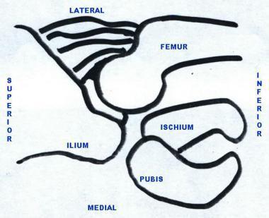
 First practical session 2017 Bones of the gluteal region The Hip bone The hip bone is made of: 1 The ilium: superior in position 2 The ischium:postero-inferior in position 3 The pubis: antero-inferior
University of Central Florida. Victor Huayamave University of Central Florida. Doctoral Dissertation (Open Access) Electronic Theses and Dissertations
 Electronic Theses and Dissertations") University of Central Florida Electronic Theses and Dissertations Doctoral Dissertation (Open Access) Biomechanics of Developmental Dysplasia of the Hip - An engineering study of closed reduction utilizing
University of Central Florida Electronic Theses and Dissertations Doctoral Dissertation (Open Access) Biomechanics of Developmental Dysplasia of the Hip - An engineering study of closed reduction utilizing
The Lower Limb. Anatomy RHS 241 Lecture 2 Dr. Einas Al-Eisa
 The Lower Limb Anatomy RHS 241 Lecture 2 Dr. Einas Al-Eisa The bony pelvis Protective osseofibrous ring for the pelvic viscera Transfer of forces to: acetabulum & head of femur (when standing) ischial
Stephanie W. Mayer, MD. Director of Child and Young Adult Hip Preservation Sports Medicine Center Children s Hospital Colorado
 Stephanie W. Mayer, MD Director of Child and Young Adult Hip Preservation Sports Medicine Center Children s Hospital Colorado University of Colorado Sports Medicine Assistant Team Physician, Colorado Avalanche
Treatment of congenital subluxation and dislocation of the hip by knee splint harness
 Prosthetics and Orthotics International, 1994,18, 34-39 Treatment of congenital subluxation and dislocation of the hip by knee splint harness M. FUKUSHIMA Fukushima Orthopaedic Clinic, Hiroshima City,
Adolescent Hip Dysplasia
 Adolescent Hip Dysplasia The hip is a "ball-and-socket" joint. In a normal hip, the ball at the upper end of the femur (thighbone) fits firmly into the socket, which is a curved portion of the pelvis called
FAI syndrome with or without labral tear.
 Case This 16-year-old female, soccer athlete was treated for pain in the right groin previously. Now has acute onset of pain in the left hip. The pain was in the groin that was worse with activities. Diagnosis
The early diagnosis of developmental dysplasia of the hip
 Current Orthopaedics (2002) 16, 57^ 64 c 2002 Published by Elsevier Science Ltd. doi:10.1054/ycuor.235, available online at http://www.idealibrary.com on CHILDREN The early diagnosis of developmental dysplasia
The Hip from Cradle to Grave. Haemish Crawford Ascot Hospital Starship Children s Hospital
 The Hip from Cradle to Grave Haemish Crawford Ascot Hospital Starship Children s Hospital Developmental dysplasia hip DDH Irritable vs. septic hip Perthes disease Slipped Upper Femoral Epiphysis (SUFE)
SURGICAL AND APPLIED ANATOMY
 Página 1 de 6 Copyright 2001 Lippincott Williams & Wilkins Bucholz, Robert W., Heckman, James D. Rockwood & Green's Fractures in Adults, 5th Edition SURGICAL AND APPLIED ANATOMY Part of "37 - HIP DISLOCATIONS
Developmental dysplasia of the hip: What has changed in the last 20 years?
 Submit a Manuscript: http://www.wjgnet.com/esps/ Help Desk: http://www.wjgnet.com/esps/helpdesk.aspx DOI: 10.5312/wjo.v6.i11.886 World J Orthop 2015 December 18; 6(11): 886-901 ISSN 2218-5836 (online)
A comparison of ultrasonography and radiography in the management of infants with suspected developmental dysplasia of the hip
 Acta Orthop. Belg., 2013, 79, 524-529 ORIGINAL STUDY A comparison of ultrasonography and radiography in the management of infants with suspected developmental dysplasia of the hip Hakan Atalar, Halil Dogruel,
Congenital Dislocation Of The Hip In Newborns Of Mashhad City
 ISPUB.COM The Internet Journal of Pediatrics and Neonatology Volume 4 Number 1 Congenital Dislocation Of The Hip In Newborns Of Mashhad City G Mamouri, F Khatami, A Hamedi Citation G Mamouri, F Khatami,
BIOMECHANICAL EXAMINATION OF THE PEDIATRIC LOWER EXTREMITY
 BIOMECHANICAL EXAMINATION OF THE PEDIATRIC LOWER EXTREMITY B.Resseque, D.P.M. ARCH HEIGHT OFF WEIGHTBEARING Evaluate arch height by placing a ruler from the heel to the first metatarsal head Compare arch
Ultrasound in the selective screening of developmental dysplasia of the hip
 European Review for Medical and Pharmacological Sciences 2011; 15: 394-398 Ultrasound in the selective screening of developmental dysplasia of the hip A.A. AFAQ, S. STOKES, H. FAREED*, H.G. ZADEH*, M.
CLINICS IN SPORTS MEDICINE
 Clin Sports Med 25 (2006) 365 369 CLINICS IN SPORTS MEDICINE A Acetabular labrum, tears of, hip arthroscopy in, 264 Acetabular rim, trimming of, and labral repair, new method for, 293 297 Acetabulum, femoral
Non-arthritic anterior hip pain in the younger patient: examination and intervention strategies
 Non-arthritic anterior hip pain in the younger patient: examination and intervention strategies Melodie Kondratek, PT, DScPT, OMPT Bryan Kuhlman, PT, DPT, OMPT Oakland University Orthopedic Spine and Sports
Scholars Journal of Applied Medical Sciences (SJAMS)
") Scholars Journal of Applied Medical Sciences (SJAMS) Abbreviated Key Title: Sch. J. App. Med. Sci. Scholars Academic and Scientific Publisher A Unit of Scholars Academic and Scientific Society, India www.saspublisher.com
Scholars Journal of Applied Medical Sciences (SJAMS) Abbreviated Key Title: Sch. J. App. Med. Sci. Scholars Academic and Scientific Publisher A Unit of Scholars Academic and Scientific Society, India www.saspublisher.com
PAEDIATRIC ORTHOPAEDICS BRENT WEATHERHEAD, MD, FRCSC PAEDIATRIC ORTHOPAEDIC SURGEON MEDICAL DIRECTOR, REBALANCE
 PAEDIATRIC ORTHOPAEDICS BRENT WEATHERHEAD, MD, FRCSC PAEDIATRIC ORTHOPAEDIC SURGEON MEDICAL DIRECTOR, REBALANCE DISCLOSURES I HAVE NO INDUSTRY CONFLICTS TO DECLARE I AM AN ORTHOPAEDIC SURGEON TRAINED IN
Disparity between Clinical and Ultrasound Examinations in Neonatal Hip Screening
 Original Article Clinics in Orthopedic Surgery 2016;8:203-209 http://dx.doi.org/10.4055/cios.2016.8.2.203 Disparity between Clinical and Ultrasound Examinations in Neonatal Hip Screening Bong Soo Kyung,
Hip Biomechanics and Osteotomies
 Hip Biomechanics and Osteotomies Organization Introduction Hip Biomechanics Principles of Osteotomy Femoral Osteotomies Pelvic Osteotomies Summary Inroduction Osteoarthritis is very prevalent Primary OA
Revised 2018 (Resolution 24)* PREAMBLE
* PREAMBLE") The American College of Radiology, with more than 30,000 members, is the principal organization of radiologists, radiation oncologists, and clinical medical physicists in the United States. The College
The American College of Radiology, with more than 30,000 members, is the principal organization of radiologists, radiation oncologists, and clinical medical physicists in the United States. The College
Developmental Dysplasia of the Hip, (DDH) including Femoral and Pelvic Osteotomy
 including Femoral and Pelvic Osteotomy") Developmental Dysplasia of the Hip, (DDH) including Femoral and Pelvic Osteotomy Leicester Children s Hospital Information for Patients, Parents and Carers DRAFT What is developmental dysplasia of the
Developmental Dysplasia of the Hip, (DDH) including Femoral and Pelvic Osteotomy Leicester Children s Hospital Information for Patients, Parents and Carers DRAFT What is developmental dysplasia of the
The Hip (Iliofemoral) Joint. Presented by: Rob, Rachel, Alina and Lisa
 Joint. Presented by: Rob, Rachel, Alina and Lisa") The Hip (Iliofemoral) Joint Presented by: Rob, Rachel, Alina and Lisa Surface Anatomy: Posterior Surface Anatomy: Anterior Bones: Os Coxae Consists of 3 Portions: Ilium Ischium Pubis Bones: Pubis Portion
The Hip (Iliofemoral) Joint Presented by: Rob, Rachel, Alina and Lisa Surface Anatomy: Posterior Surface Anatomy: Anterior Bones: Os Coxae Consists of 3 Portions: Ilium Ischium Pubis Bones: Pubis Portion
THE HIP. Cooler than cool, the pinnacle of what is "it". Beyond all trends and conventional coolness.
 THE HIP Cooler than cool, the pinnacle of what is "it". Beyond all trends and conventional coolness. Objectives Hip anatomy Causes of hip pain Hip exam Anatomy Bones Ilium Anterior Superior Iliac Spine
Sonographic Evaluation of Developmental Hip Dysplasia using Graf s Method
 IOSR Journal of Dental and Medical Sciences (IOSR-JDMS) e-issn: 2279-0853, p-issn: 2279-0861.Volume 17, Issue 7 Ver. 3 (July. 2018), PP 63-67 www.iosrjournals.org Sonographic Evaluation of Developmental
The hip: Built for endurance and mobility
 The hip: Built for endurance and mobility The hip joint Some anatomical landmarks Innominate Ilium, pubis, ischium Sacrum Iliac crests Asis Psis Pubic tubercle Acetabulum Femur Head of femur Neck of femur
Natural history of hip instability in infants (without subluxation or dislocation): a three year follow-up
: a three year follow-up") Pruszczynski et al. BMC Musculoskeletal Disorders 2014, 15:355 RESEARCH ARTICLE Open Access Natural history of hip instability in infants (without subluxation or dislocation): a three year follow-up Blazej
Pruszczynski et al. BMC Musculoskeletal Disorders 2014, 15:355 RESEARCH ARTICLE Open Access Natural history of hip instability in infants (without subluxation or dislocation): a three year follow-up Blazej
The most relevant diagnostic criteria for developmental dysplasia of the hip: a study of British specialists
 Williams et al. BMC Musculoskeletal Disorders (2016) 17:38 DOI 10.1186/s12891-016-0867-4 RESEARCH ARTICLE Open Access The most relevant diagnostic criteria for developmental dysplasia of the hip: a study
Bones of Lower Limb. Dr. Heba Kalbouneh Associate Professor of Anatomy and Histology
 Bones of Lower Limb Dr. Heba Kalbouneh Associate Professor of Anatomy and Histology Bones of the lower limb Hip Bone Made up of 3 bones: 1) Ilium (flat), superior in position 2) Ischium (L), postero-inferior
Ultrasound in the Diagnosis of Developmental Hip Dysplasia and Dislocation A Protocol
 Reference Number: UHB 353 Version Number: 1 Date of Next Review: 5 th Apr 2020 Previous Trust/LHB Reference Number: N/A Ultrasound in the Dysplasia and Dislocation A Protocol Introduction and Aim The aim
Joints of the lower limb
 Joints of the lower limb 1-Type: Hip joint Synovial ball-and-socket joint 2-Articular surfaces: a- head of femur b- lunate surface of acetabulum Which is deepened by the fibrocartilaginous labrum acetabulare
RESULTS OF THE EARLY TREATMENT OF DEVELOPMENTAL DYSPLASIA OF THE HIP
 RESULTS OF THE EARLY TREATMENT OF DEVELOPMENTAL DYSPLASIA OF THE HIP Dana Vasilescu, Dan Cosma University of Medicine and Pharmacy Iuliu Haţieganu Cluj-Napoca 13, E. Isac st., 400023 Cluj-Napoca, România
Is ultrasound screening for DDH in babies born breech sufficient?
 J Child Orthop (2010) 4:3 8 DOI 10.1007/s11832-009-0217-2 ORIGINAL CLINICAL ARTICLE Is ultrasound screening for DDH in babies born breech sufficient? Meghan Imrie Vanessa Scott Philip Stearns Tracey Bastrom
A novel method for assessing postoperative femoral head reduction in developmental dysplasia of the hip
 J Child Orthop (2014) 8:319 324 DOI 10.1007/s11832-014-0600-5 ORIGINAL CLINICAL ARTICLE A novel method for assessing postoperative femoral head reduction in developmental dysplasia of the hip Anthony Cooper
Hip pain rating after preforming MRI with gadolinium arthrography and intra-articular lidocaine
 Hip pain rating after preforming MRI with gadolinium arthrography and intra-articular lidocaine Poster No.: C-1352 Congress: ECR 2014 Type: Scientific Exhibit Authors: J. García Yavar, J. Cabezudo, S.
The Hip Joint. Shenequia Howard David Rivera
 The Hip Joint Shenequia Howard David Rivera Topics Of Discussion Movement Bony Anatomy Ligamentous Anatomy Muscular Anatomy Origin/Insertion/Action/Innervation Common Injuries MOVEMENT Flexion Extension
ESSENTIALS OF INFANT HIP SONOGRAPHY According to GRAF
 ESSENTIALS OF INFANT HIP SONOGRAPHY According to GRAF R. Graf K. Lercher S. Scott T. Spieß Copyright Sonocenter Stolzalpe Authors: R. Graf A-8850 Murau, Hagersiedlung 7 K. Lercher A-8852 Stolzalpe S. Scott
Joints. Judi Laprade. Illustrations from: Essential Clinical Anatomy 3 rd ed. (ECA3) Moore, K. and Agur, A. Lippincott Williams and Wilkins, 2007
 Moore, K. and Agur, A. Lippincott Williams and Wilkins, 2007") Slide 1 Joints Judi Laprade Illustrations from: Essential Clinical Anatomy 3 rd ed. (ECA3) Moore, K. and Agur, A. Lippincott Williams and Wilkins, 2007 Grant s Atlas of Anatomy 12 th ed. (GA12) Agur, A.
Slide 1 Joints Judi Laprade Illustrations from: Essential Clinical Anatomy 3 rd ed. (ECA3) Moore, K. and Agur, A. Lippincott Williams and Wilkins, 2007 Grant s Atlas of Anatomy 12 th ed. (GA12) Agur, A.
CONGENITAL HIP DISEASE IN YOUNG ADULTS CLASSIFICATION AND TREATMENT WITH THA. Th. KARACHALIOS, MD, DSc PROF IN ORTHOPAEDICS
 CONGENITAL HIP DISEASE IN YOUNG ADULTS CLASSIFICATION AND TREATMENT WITH THA Th. KARACHALIOS, MD, DSc PROF IN ORTHOPAEDICS EDITOR IN CHIEF HIP INTERNATIONAL UNIVERSITY OF THESSALIA, LARISA HELLENIC REPUBLIC
1. Discuss some common pediatric problems seen in the clinic. Diagnosis Clinical examination (at birth and subsequent well-baby examinations)
") 1 Pediatric Orthopaedics for Primary Care Providers 2 Disclosure Statement No conflicts related to this presentation 3 4 Goals 1. Discuss some common pediatric problems seen in the clinic 2. Examination
1 Pediatric Orthopaedics for Primary Care Providers 2 Disclosure Statement No conflicts related to this presentation 3 4 Goals 1. Discuss some common pediatric problems seen in the clinic 2. Examination
It is formed by fusion of 3 bones: I. Ilium (superior bone). II. Pubis (antero-inferior bone). III. Ischium (postero-inferior bone).
. II. Pubis (antero-inferior bone). III. Ischium (postero-inferior bone).") It is formed by fusion of 3 bones: I. Ilium (superior bone). II. Pubis (antero-inferior bone). III. Ischium (postero-inferior bone). Pubis Acetabulum Ana (242 ) The three constituent of bones of the hip
It is formed by fusion of 3 bones: I. Ilium (superior bone). II. Pubis (antero-inferior bone). III. Ischium (postero-inferior bone). Pubis Acetabulum Ana (242 ) The three constituent of bones of the hip
LAB Notes#1. Ahmad Ar'ar. Eslam
 LAB Notes#1 Ahmad Ar'ar Eslam 1 P a g e Anatomy lab Notes Lower limb bones :- Pelvic girdle: It's the connection between the axial skeleton and the lower limb; it's made up of one bone called the HIP BONE
ORDER OF VERBAL EXAMS
 ORDER OF VERBAL EXAMS The students are able to register for the exam on the NEPTUN system. The students pick two titles, from the title list available at the beginning of the Semester. This list can be
Labral Tears/FAI. Andrew Parker, MD
 Labral Tears/FAI Andrew Parker, MD Athletic Hip Injuries Incidence of hip injuries has increased dramatically over the last decade In part due to better recognition with improved imaging and arthroscopy,
Musculoskeletal Examination
 Musculoskeletal Examination Statement of Goals Know how to perform a complete musculoskeletal examination. Learning Objectives A. Describe the anatomy of the musculoskeletal system including the bony structures,
Pediatric Orthopedic Pathology Pathology 2 Dr. Gary Mumaugh
 Pediatric Orthopedic Pathology Pathology 2 Dr. Gary Mumaugh Congenital Defects - Clubfoot (congenital equinovarus) Forefoot is adducted and supinated o Positional equinovarus o Idiopathic congenital equinovarus
The Efficacy of Pavlik Harness as a Treatment of Developmental Dislocation of the Hip
 The Efficacy of Pavlik Harness as a Treatment of Developmental Dislocation of the Hip Firas A. Suleiman, MD*, Fadi Al Rousan, MD*, Ahmad Almarzoq, MD *, Razi Altarawneh, MD*, Hidar Soudi, MD* ABSTRACT
COMMON MUSCULOSKELETAL PROBLEMS GROWTH AND DEVELOPMENT PATHOLOGIC VS. NORMAL
 COMMON MUSCULOSKELETAL PROBLEMS GROWTH AND DEVELOPMENT PATHOLOGIC VS. NORMAL Clifford L. Craig, M.D. M2 Musculoskeletal Fall 2008 I. ANGULAR AND TORSIONAL DEFORMITIES OF THE LOWER LIMBS Examination Relaxed,
Hip Injuries & Arthroscopy in Athletes
 Hip Injuries & Arthroscopy in Athletes John P Salvo, MD Sports Medicine Rothman Institute Philadelphia, PA EATA Annual Meeting January, 2011 Hip Injuries & Arthroscopy in Anatomy History Physical Exam
Pelvic Girdle
 ARTICULATIONS OF LOWER EXTREMITY Pages 429-437 Pelvic Girdle formed by connection of the hip bones and the sacrum Sacroiliac Joints compound joints synovial joint - anterior, between the auricular surfaces
PEDIATRIC HIP DISORDERS VINAY NAROTAM, MD ASSISTANT PROFESSOR UNIVERSITY OF NORTH CAROLINA SCHOOL OF MEDICINE DEPARTMENT OF ORTHOPAEDICS
 1 PEDIATRIC HIP DISORDERS VINAY NAROTAM, MD ASSISTANT PROFESSOR UNIVERSITY OF NORTH CAROLINA SCHOOL OF MEDICINE DEPARTMENT OF ORTHOPAEDICS Disclosures 2 I have no relevant financial relationships with
CONGENITAL DISLOCATION OF THE HIP AND COMPUTERISED
 CONGENITAL DISLOCATION OF THE HIP AND COMPUTERISED AXIAL TOMOGRAPHY J. G. EDELSON, M. HIRSCH, H. WEINBERG, D. ATTAR, E. BARMEIR From the Soroka Medical Center, Beer-Sheba CT scans of 18 hips with typical