First practical session. Bones of the gluteal region
|
|
|
- Bernard Alexander
- 6 years ago
- Views:
Transcription
1 First practical session 2017 Bones of the gluteal region

2 The Hip bone The hip bone is made of: 1 The ilium: superior in position 2 The ischium:postero-inferior in position 3 The pubis: antero-inferior in position Anatomical position of the hip bone It is very important to understand the anatomical position of the hip bone, in anatomical position: 1-The Anterior superior iliac spine and the pubictubercle lie in the same vertical plane. 2-The ischial spine and the upper border of the symphysis pubis lie in the same horizontal plane. It means that the pelvis is looking forward in the anatomical position
3 The 2 hip bones with the sacrum form the pelvis Now look! where does the pelvis look? It is looking right at you! Never upwards hip bone sacroiliac joints sacrum symphysis pubis femur During your first practical session, make sure to have a look at the anatomical position of the pelvis

4 The fact that the pelvis is facing (looking) forward is important to understand how structures passing from the pelvis smoothly join the thigh Notice, the external iliac artery as it passes from the pelvis into thigh to become the femoral artery What do you think about The femoral nerve and vein?


5 The ilium, ischium and pubis meet one another by means of triradiate (Y-shaped) cartilage at the Acetabulum. While the inferior ramus of the pubis meets with the ramus of the ischium by cartilaginous union Ossifies near the age of 7 years X-ray? at puberty the triradiate cartilage starts to ossify and near the age of 17 the triradiate cartilage will be replaced by bony union

6 Anteroposterior radiograph of the pelvis of a boy aged Ilium. 2. Part of triradiate growth cartilage. 3. Superior femoral epiphysis. 4. Cartilaginous growth plates. 5. Ossifying greater trochanter.6. Ischium. 7. Pubis. 8. Cartilage between pubic and ischial rami.
7 The hip bones articulate with the sacrum at the sacroiliac joints posteriorly while anteriorly they articulate with one Thus the two hip bones form the pelvic girdle where the ilium corresponds to the scapula in the upper limb, the pubis corresponds to the clavicle while the ischium corresponds to the coracoid process another at the symphysis pubis. hip bone sacroiliac joints sacrum symphysis pubis femur
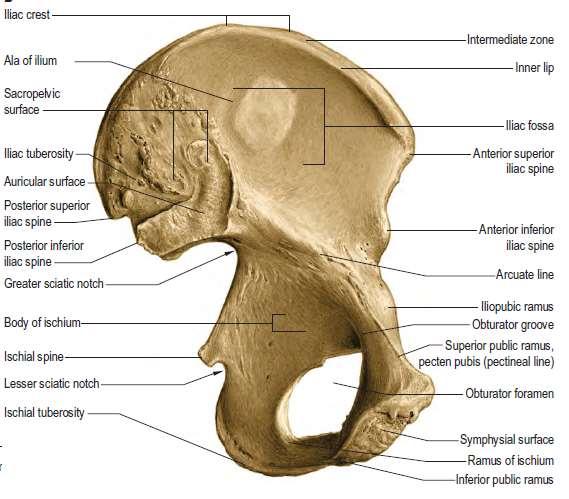
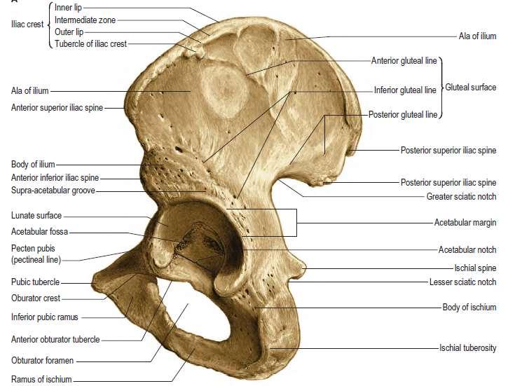
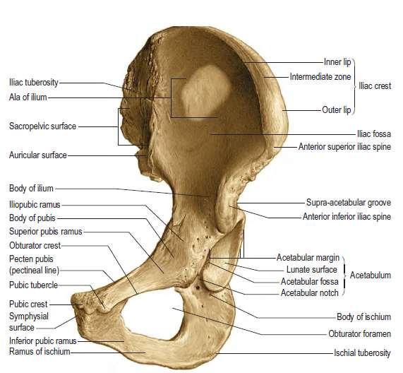
8 Two parts: 1-Ala 2- Body 1-The Ilium Superior border Is made by the iliac crest Three surfaces 1 gluteal surface 2 iliac fossa 3 sacropelvic surface Posterior border Begins at the posterior superior iliac spine (P.S.I.S) And ends at the posterior inferior iliac spine (P.I.I.S) Anterior border Begins at the anterior superior iliac spine (A.S.I.S) and ends at the anterior Inferior iliac spine (A.I.I.S) the anterior superior spine of the ilium is easily felt and may be visible in the thin subject
9
10 Iliac fossa Medial border Forms the arcuate line Which extends to the ilio-pubic eminence The sacropelvic surface presents: 1- Iliac tuberosity: rough area that gives attachment to the interosseous and dorsal sacroiliac ligaments 2- auricular surface: Smooth area articulates with the sacrum to form the sacroiliac joint 3- pelvic surface Smooth area
11

12 The gluteal surface is divided into 4 parts by three lines: 1- Posterior gluteal line MAKE SURE you know the names of the muscles that are attached to the areas between these lines 2- Middle gluteal line Or anterior 3- Inferior gluteal line Which muscle is attached to the area between The inferior and middle gluteal lines?
13


14 Pubic bone Formed of a body and two rami: superior and inferior The body is flattened and has: 1- an upper border called pubic crest that ends laterally by the pubic tubercle 2- symphyseal surface which articulates with the opposite pubis to form the pubic symphysis The inferior ramus of the pubic bone joins the ischial ramus to form the conjoined tendon. The superior pubic ramus has a pectineal line on its medial surface Pectineal line
15
16 The ischium Body Greater sciatic notch Ischial ramus Ischial spine lesser sciatic notch Ischial tuberosity


17 The ischial tuberosity is covered by gluteus maximus when one stands. In the sitting position the muscle slips away laterally.to palpate this bony point, therefore, feel for it uncovered by gluteus maximus in the flexed position of the hip. Ischial tuberosity Divided by a transverse ridge into: An upper quadrangular and a lower triangular parts The upper quadrangular part is divided by an obliqueridge into: 1 Upper lateral part for the attachment of semimembranousus 2 lower Medial for the attachment of semitendinosus and long head of biceps The lower triangular part is divided by a longitudinal ridge into: 1-lateral part that gives attachment to the adductor part of the adductor magnus muscle 2-medial part ( subcutaneous part)


18 The Acetabulum It is a C-shaped cavity located on the lateral aspect of the hip bone directed laterally, downwards and forwards It is notched inferiorly by the acetabular notch which is bridged by the transverse acetabular ligament ( part of the acetabular labrum) The acetabular ligament converts the acetabular notch into foramen Its cavity presents a horse-shoe shaped articular surface called Lunate surface Ilium forms the superior 2/5 of the lunate surface The ischium forms the posterior 2/5 Of the lunate surface anterior The pubis forms the anterior 1/5 of the lunate surface The Lunate surface surrounds a non articular depression called acetabular fossa which is occupied by fat tissue in living
19 OBTURATOR foramen Covered by a membrane in living subjects


20 Muscle attachment to the hip bone Muscles and ligaments attached to the external surface of the right hip bone

21 Muscle attachment to the hip and upper end of the femur bones IMPORTANT IMPORTANT IMPORTANT IMPORTANT IMPORTANT

22 ligaments in the gluteal region 1- SACROTUBEROUS LIGAMENT 2- SACROSPINOUS LIGAMENT
23 Attachment of the inguinal ligament
24 Bones the thigh
25
 head of femur greater")

26 Long bone? fovea capitis neck Left femur (anterior view) head of femur greater trochanter intertrochanteric line Upper end Lower end And a shaft Lesser trochanter Shaft or body Adductor tubercle Medial epicondyle Medial condyle lateral epicondyle lateral condyle Pattelar surface


27 Distal end of femur: articular surface.
28 The greater trochanter of the femur lies a hand s breadth below the iliac crest; it is best palpated with the hip abducted so that the overlying hip abductors (tensor fasciae latae and gluteus medius and minimus) are relaxed. important

29 L ef t f e m u r ( p o s t e r i o r v i e w ) greater trochanter Gluteal tuberosity intertrochanteric creast Lesser trochanter Spiral line lateral lip of linea aspera Medial lip of linea aspera linea aspera lateral surface lateral supracondylar line medial surface medial supracondylar line Adductor tubercle lateral condyle Intercondylar notch medial condyle
Bones of Lower Limb. Dr. Heba Kalbouneh Associate Professor of Anatomy and Histology
 Bones of Lower Limb Dr. Heba Kalbouneh Associate Professor of Anatomy and Histology Bones of the lower limb Hip Bone Made up of 3 bones: 1) Ilium (flat), superior in position 2) Ischium (L), postero-inferior
Copyright 2003 Pearson Education, Inc. publishing as Benjamin Cummings. Dr. Nabil Khouri MD, MSc, Ph.D
 Dr. Nabil Khouri MD, MSc, Ph.D Pelvic Girdle (Hip) Organization of the Lower Limb It is divided into: The Gluteal region The thigh The knee The leg The ankle The foot The thigh and the leg have compartments
It is formed by fusion of 3 bones: I. Ilium (superior bone). II. Pubis (antero-inferior bone). III. Ischium (postero-inferior bone).
. II. Pubis (antero-inferior bone). III. Ischium (postero-inferior bone).") It is formed by fusion of 3 bones: I. Ilium (superior bone). II. Pubis (antero-inferior bone). III. Ischium (postero-inferior bone). Pubis Acetabulum Ana (242 ) The three constituent of bones of the hip
It is formed by fusion of 3 bones: I. Ilium (superior bone). II. Pubis (antero-inferior bone). III. Ischium (postero-inferior bone). Pubis Acetabulum Ana (242 ) The three constituent of bones of the hip
LAB Notes#1. Ahmad Ar'ar. Eslam
 LAB Notes#1 Ahmad Ar'ar Eslam 1 P a g e Anatomy lab Notes Lower limb bones :- Pelvic girdle: It's the connection between the axial skeleton and the lower limb; it's made up of one bone called the HIP BONE
The Hip (Iliofemoral) Joint. Presented by: Rob, Rachel, Alina and Lisa
 Joint. Presented by: Rob, Rachel, Alina and Lisa") The Hip (Iliofemoral) Joint Presented by: Rob, Rachel, Alina and Lisa Surface Anatomy: Posterior Surface Anatomy: Anterior Bones: Os Coxae Consists of 3 Portions: Ilium Ischium Pubis Bones: Pubis Portion
The Hip (Iliofemoral) Joint Presented by: Rob, Rachel, Alina and Lisa Surface Anatomy: Posterior Surface Anatomy: Anterior Bones: Os Coxae Consists of 3 Portions: Ilium Ischium Pubis Bones: Pubis Portion
Bony Anatomy. Femur. Femoral Head Femoral Neck Greater Trochanter Lesser Trochanter Intertrochanteric Crest Intertrochanteric Line Gluteal Tuberosity
 Hip Anatomy Bony Anatomy Femur Femoral Head Femoral Neck Greater Trochanter Lesser Trochanter Intertrochanteric Crest Intertrochanteric Line Gluteal Tuberosity Bony Anatomy Pelvic Girdle Acetabulum 3 bones
The Lower Limb. Anatomy RHS 241 Lecture 2 Dr. Einas Al-Eisa
 The Lower Limb Anatomy RHS 241 Lecture 2 Dr. Einas Al-Eisa The bony pelvis Protective osseofibrous ring for the pelvic viscera Transfer of forces to: acetabulum & head of femur (when standing) ischial
Lectures of Human Anatomy
 Lectures of Human Anatomy Lower Limb Gluteal Region and Hip Joint By DR. ABDEL-MONEM AWAD HEGAZY M.B. with honor 1983, Dipl."Gynecology and Obstetrics "1989, Master "Anatomy and Embryology" 1994, M.D.
The Appendicular Skeleton
 8 The Appendicular Skeleton PowerPoint Lecture Presentations prepared by Jason LaPres Lone Star College North Harris 8-1 The Pectoral Girdle The Pectoral Girdle Also called shoulder girdle Connects the
Gluteal region DR. GITANJALI KHORWAL
 Gluteal region DR. GITANJALI KHORWAL Gluteal region The transitional area between the trunk and the lower extremity. The gluteal region includes the rounded, posterior buttocks and the laterally placed
Hip joint and pelvic girdle. Lower Extremity. Pelvic Girdle 6/5/2017
 Hip joint and pelvic girdle Lower Extremity The relationship between the pelvic girdle and hip is similar to that between the shoulder girdle and shoulder joint. The lower limbs are attached to the axial
Copyright 2003 Pearson Education, Inc. publishing as Benjamin Cummings. Dr. Nabil khouri
 Dr. Nabil khouri Appendicular Skeleton The appendicular skeleton is made up of the bones of the upper and lower limbs and their girdles Two girdles: Pectoral girdles attach the upper limbs to the body
The os coxae or hip bone consists of three flat bones, ilium, ischium and pubis, which fuse together to form the acetabulum.
 The os coxae The os coxae or hip bone consists of three flat bones, ilium, ischium and pubis, which fuse together to form the acetabulum. The ilium extends from the acetabulum upwards forming the lateral
Chapter 8. The Appendicular Skeleton. Lecture Presentation by Lee Ann Frederick University of Texas at Arlington Pearson Education, Inc.
 Chapter 8 The Appendicular Skeleton Lecture Presentation by Lee Ann Frederick University of Texas at Arlington An Introduction to the Appendicular Skeleton The Appendicular Skeleton 126 bones Allows us
Lab Activity 9. Appendicular Skeleton Martini Chapter 8. Portland Community College BI 231
 Lab Activity 9 Appendicular Skeleton Martini Chapter 8 Portland Community College BI 231 Appendicular Skeleton Upper & Lower extremities Shoulder Girdle Pelvic Girdle 2 Humerus 3 Humerus: Proximal End
Main Menu. Joint and Pelvic Girdle click here. The Power is in Your Hands
 1 Hip Joint and Pelvic Girdle click here Main Menu K.6 http://www.handsonlineeducation.com/classes//k6entry.htm[3/23/18, 2:01:12 PM] Hip Joint (acetabular femoral) Relatively stable due to : Bony architecture
Lesson 24. A & P Hip
 Lesson 24 A & P Hip 1 Aims of the Session This session will allow candidates to have an understanding of the bony prominences and soft tissues of the hip 2 Learning Outcomes By the end of the lesson the
Posterior compartment of the thigh. Dr. Heba Kalbouneh Associate Professor of Anatomy and Histology
 Posterior compartment of the thigh Dr. Heba Kalbouneh Associate Professor of Anatomy and Histology Posterior compartment of the thigh 1-Muscles: Biceps femoris Semitendinosus Semimembranosus Adductor magnus
The University Of Jordan Faculty Of Medicine THE LOWER LIMB. Dr.Ahmed Salman Assistant Prof. of Anatomy. The University Of Jordan
 The University Of Jordan Faculty Of Medicine THE LOWER LIMB Dr.Ahmed Salman Assistant Prof. of Anatomy. The University Of Jordan Gluteal Region Cutaneous nerve supply of (Gluteal region) 1. Lateral cutaneous
The thigh. Prof. Oluwadiya KS
 The thigh Prof. Oluwadiya KS www.oluwadiya.com The Thigh: Boundaries The thigh is the region of the lower limb that is approximately between the hip and knee joints Anteriorly, it is separated from the
PELVIS & SACRUM Dr. Jamila El-Medany Dr. Essam Eldin Salama
 PELVIS & SACRUM Dr. Jamila El-Medany Dr. Essam Eldin Salama Learning Objectives At the end of the lecture, the students should be able to : Describe the bony structures of the pelvis. Describe in detail
Biology 218 Human Anatomy. Adapted from Martini Human Anatomy 7th ed. Chapter 7 The Skeletal System Appendicular Division
 Adapted from Martini Human Anatomy 7th ed. Chapter 7 The Skeletal System Appendicular Division Introduction The appendicular skeleton includes: Pectoral girdle Shoulder bones Upper limbs Pelvic girdle
The Hip Joint. Shenequia Howard David Rivera
 The Hip Joint Shenequia Howard David Rivera Topics Of Discussion Movement Bony Anatomy Ligamentous Anatomy Muscular Anatomy Origin/Insertion/Action/Innervation Common Injuries MOVEMENT Flexion Extension
The Lower Limb. Sevda LAFCI FAHRİOĞLU, MD.PhD.
 The Lower Limb Sevda LAFCI FAHRİOĞLU, MD.PhD. The Lower Limb The bones of the lower limb form the inferior part of the appendicular skeleton the organ of locomotion for bearing the weight of body stronger
Located more distal and anterior together with Trapezoid, anterior to scaphoid Trapezium rarely to be fractured.
 The hand The hand consists of the 3 groups: Proximal part: carpals bones (8) Middle part: metacarpal bones (5) Distal part: fingers or phalanges bones (3 for each finger except for the thumb just 2 bones).
Bones of the Lower Limb Bone Structure Description Notes. border of the superior ramus. inferolaterally from the pubic symphysis
 Bones of the Lower Limb Bone Structure Description Notes pubis an angulated bone the forms the anterior part of the pelvis one of three bones that form the os coxae: ilium, ischium, pubis; its forms 1/5
Lower limb summary. Anterior compartment of the thigh. Done By: Laith Qashou. Doctor_2016
 Lower limb summary Done By: Laith Qashou Doctor_2016 Anterior compartment of the thigh Sartorius Anterior superior iliac spine Upper medial surface of shaft of tibia 1. Flexes, abducts, laterally rotates
Anatomage Table Instructors Guide- Lower Limb
 The Lower Limb Anatomage Table Instructors Guide- Lower Limb Table of Contents Lower Limb 1- The Skeletal System...3 1: Hip Bone...3 2: Hip Joint and Femur...4 3: Patella and Knee Joint...7 4: Tibia, Fibula,
Muscles of the Thigh. 6.1 Identify, describe the attachments of and deduce the actions of the muscles of the thigh: Anterior group
 Muscles of the Thigh 6.1 Identify, describe the attachments of and deduce the actions of the muscles of the thigh: Anterior group Sartorius: This is a long strap like muscle with flattened tendons at each
Skeletal System Module 13: The Pelvic Girdle and Pelvis
 OpenStax-CNX module: m47993 1 Skeletal System Module 13: The Pelvic Girdle and Pelvis Donna Browne Based on The Pelvic Girdle and Pelvis by OpenStax College This work is produced by OpenStax-CNX and licensed
Identify the muscles associated with the medial compartment of the thigh. Identify the attachment points of the medial thigh muscles.
 L 8 A B O R A T O R Y Thigh MEDIAL THIGH Identify the muscles associated with the medial compartment of the thigh. Identify the attachment points of the medial thigh muscles. Identify the actions of these
SKELETAL SYSTEM 206. AXIAL SKELETON 80 APPENDICULAR SKELETON 126 (see Figure 6.1) Clavicle. Clavicle. Pectoral girdles. Scapula. Scapula.
 Clavicle. Clavicle. Pectoral girdles. Scapula. Scapula.") SKELETAL SYSTEM 206 AXIAL SKELETON 80 APPENDICULAR SKELETON 126 (see Figure 6.1) Pectoral girdles 4 Clavicle Scapula 2 2 Clavicle Scapula Humerus 2 Humerus Upper limbs 60 Radius 2 Ulna Carpal bones Metacarpal
SKELETAL SYSTEM 206 AXIAL SKELETON 80 APPENDICULAR SKELETON 126 (see Figure 6.1) Pectoral girdles 4 Clavicle Scapula 2 2 Clavicle Scapula Humerus 2 Humerus Upper limbs 60 Radius 2 Ulna Carpal bones Metacarpal
Biology 218 Human Anatomy
 Chapter 8 Adapted from Tortora 10 th ed. LECTURE OUTLINE A. Introduction (p. 203) 1. The appendicular skeleton contains 126 bones that form: i. two pectoral (shoulder) girdles two upper limbs i one pelvic
Anatomy of the lower limb
 Anatomy of the lower limb 1. Bones of the lower limb Pelvis Hip bone/coxal bone Acetabulum o Acetabular margin o Acetabular fossa o Acetabular notch o Lunate surface Ischiopubic ramus Obturator foramen
Chapter 8 Outline. Pectoral Girdle Upper Limb Pelvic Girdle Lower Limb Aging of the Appendicular Skeleton Development of the Appendicular Skeleton
 Chapter 8 Outline Pectoral Girdle Upper Limb Pelvic Girdle Lower Limb Aging of the Appendicular Skeleton Development of the Appendicular Skeleton Figure 8.1 Appendicular Skeleton Pectoral Girdle Clavicle
Amy Warenda Czura, Ph.D. 1 SCCC BIO130 Lab 7 Appendicular Skeleton & Articulations
 The Skeletal System II: Appendicular Skeleton and Articulations Exercises 11, 13 (begins: page 145 in 9 th and 10 th editions) Exercises 10, 11 (begins: page 147 in 11 th edition, page 149 in 12 th edition)
The Appendicular Skeleton
 8 The Appendicular Skeleton PowerPoint Lecture Presentations prepared by Jason LaPres Lone Star College North Harris An Introduction to the Appendicular Skeleton Learning Outcomes 8-1 Identify the bones
Human Anatomy Biology 351
 Human Anatomy Biology 351 Lower Limb Please place your name on the back of the last page of this exam. You must answer all questions on this exam. Because statistics demonstrate that, on average, between
Figure 7: Bones of the lower limb
 BONES OF THE APPENDICULAR SKELETON The appendicular skeleton is composed of the 126 bones of the appendages and the pectoral and pelvic girdles, which attach the limbs to the axial skeleton. Although the
ANATYOMY OF The thigh
 ANATYOMY OF The thigh 1- Lateral cutaneous nerve of the thigh Ι) Skin of the thigh Anterior view 2- Femoral branch of the genitofemoral nerve 5- Intermediate cutaneous nerve of the thigh 1, 2 and 3 are
The psoas minor is medial to the psoas major. The iliacus is a fan-shaped muscle that when contracted helps bring the swinging leg forward in walking
 1 p.177 2 3 The psoas minor is medial to the psoas major. The iliacus is a fan-shaped muscle that when contracted helps bring the swinging leg forward in walking and running. The iliopsoas and adductor
Anterior and Medial compartments of the thigh. Dr. Heba Kalbouneh Associate Professor of Anatomy and Histology
 Anterior and Medial compartments of the thigh Dr. Heba Kalbouneh Associate Professor of Anatomy and Histology Terms Related to Movements Movement Flexion Extension Abduction Adduction Medial (internal)
ANATOMY TEAM GLUTEAL REGION & BACK OF THIGH
 ANATOMY TEAM GLUTEAL REGION & BACK OF THIGH OBJECTIVES By the end of this lecture, the student should be able to identify and discuss: Contents of gluteal region: Groups of Glutei muscles and small muscles
Figure 1 - Hip and Pelvis
 Hip Figure 1 - Hip and Pelvis The terms hip and pelvis are frequently used interchangeably, but strictly speaking, the pelvis is a girdle of bones and the hip is a joint. The pelvis consists of The sacrum
LOWER LIMB. As we know the bony part of the body is divided into Axial and Appendicular (upper and lower Limbs)
") LOWER LIMB As we know the bony part of the body is divided into Axial and Appendicular (upper and lower Limbs) Bones of the Lower limb: 1-Pelvic Girdle: composed of: 1. Right hip bone : is formed by 3
LOWER LIMB As we know the bony part of the body is divided into Axial and Appendicular (upper and lower Limbs) Bones of the Lower limb: 1-Pelvic Girdle: composed of: 1. Right hip bone : is formed by 3
Applied anatomy of the hip and buttock
 CHAPTER CONTENTS The hip joint e9 Capsule and ligaments e9 s e0 Flexor muscles................... e0 Extensor muscles.................. e Abductor muscles.................. e Adductor muscles..................
Pectoral (Shoulder) Girdle
 Girdle") Chapter 8 Skeletal System: Appendicular Skeleton Pectoral girdle Pelvic girdle Upper limbs Lower limbs 8-1 Pectoral (Shoulder) Girdle Consists of scapula and clavicle Clavicle articulates with sternum
Chapter 8 Skeletal System: Appendicular Skeleton Pectoral girdle Pelvic girdle Upper limbs Lower limbs 8-1 Pectoral (Shoulder) Girdle Consists of scapula and clavicle Clavicle articulates with sternum
Muscles of Lesson Five. Muscular Nomenclature and Kinesiology - Two. Muscles of Lesson Five, cont. Chapter 16
 Chapter 16 Muscular Nomenclature and Kinesiology - Two Lessons 5-6 Muscles of Lesson Five Iliopsoas (psoas major, iliacus) Hip outward rotators (piriformis, gemellus superior, gemellus inferior, obturator
Acland's DVD Atlas of Human Anatomy. Transcript for Volume Robert D Acland
 Acland's DVD Atlas of Human Anatomy Transcript for Volume 2 2007 Robert D Acland This free downloadable pdf file is to be used for individual study only. It is not to be reproduced in any form without
Mohammad Ashraf. Abdulrahman Al-Hanbali. Ahmad Salman. 1 P a g e
 - 7 Mohammad Ashraf Abdulrahman Al-Hanbali Ahmad Salman 1 P a g e Structures under the cover of Gluteus Maximus: 1-Bones: Ileum, Femur (Head, greater trochanter and gluteal tuberosity), Ischium (ischial
Muscles of the lower extremities. Dr. Nabil khouri MD, MSc, Ph.D
 Muscles of the lower extremities Dr. Nabil khouri MD, MSc, Ph.D Posterior leg Popliteal fossa Boundaries Biceps femoris (superior-lateral) Semitendinosis and semimembranosis (superior-medial) Gastrocnemius
Human Anatomy Biology 351
 Human Anatomy Biology 351 Lower Limb Please place your name on the back of the last page of this exam. You must answer all questions on this exam. Because statistics demonstrate that, on average, between
rotation of the hip Flexion of the knee Iliac fossa of iliac Lesser trochanter Femoral nerve Flexion of the thigh at the hip shaft of tibia
 Anatomy of the lower limb Anterior & medial compartments of the thigh Dr. Hayder The fascia lata encloses the entire thigh like a sleeve/stocking. Three intramuscular fascial septa (lateral, medial, and
PRE-LAB EXERCISES. Before we get started, look up the definitions of these common bone marking terms: Canal: Condyle: Facet: Fissure:
 1 PRE-LAB EXERCISES When studying the skeletal system, the bones are often sorted into two broad categories: the axial skeleton and the appendicular skeleton. This lab focuses on the appendicular skeleton,
Appendicular Skeleton. Prof. Abdulameer Al-Nuaimi
 Appendicular Skeleton Prof. Abdulameer Al-Nuaimi a.alnuaimi@sheffield.ac.uk abdulameerh@yahoo.com Hi Prof, It is great to hear from you, I really enjoyed your teaching last year. You taught me the hardest
Lecture 10 Arteries and veins of the upper limb
 Lecture 10 Arteries and veins of the upper limb 1. Identify the Subclavian, axillary, brachial (deep and superficial), radial and ulnar arteries and superficial/deep palmar arches 2. Describe the major
Muscles of Gluteal Region
 1 The Gluteal Region In the gluteal region the skin is tough with many layers underneath. Directly under it is the superficial fascia followed by the deep fascia then the muscles and the bones of the thigh.
Topic 7: Hip and pelvis. Parts of the hip. Parts of the femur
 Topic 7: Hip and pelvis Parts of the hip Parts of the femur Classifying the hip joint Ball and socket Synovial Multiaxial Movements of the hip: Abduction/adduction Flexion/extension Medial/lateral rotation
BONES JOINTS MUSCLES OF THE LOWER LIMB
 BONES JOINTS MUSCLES OF THE LOWER LIMB LOWER LIMB: BONES LOWER LIMB GLUTEAL REGION consists of 6 major segments: FEMORAL REGION (THIGH) KNEE REGION LEG REGION TALOCRURAL REGION (ANKLE) FOOT REGION LOWER
Biology 152 Appendicular Skeleton Anatomy Objectives
 Biology 152 Appendicular Skeleton Anatomy Objectives We will learn proper bone names, left/right/medial, and the parts of bones in this exercise. Start by learning the names of the bones. As you gain comfort
bio4165 lab quiz 1 Posterior View Anterior View Lateral View Anterior View bio fall.quarter lab.quiz.1...page.1 of 6
 B A Posterior View D C E Lateral View bio.4165...fall.quarter.2005...lab.quiz.1...page.1 of 6 F I G 35 Posterior View H bio.4165...fall.quarter.2005...lab.quiz.1...page.2 of 6 J Posterior View L K Inferior
Joints of the lower limb
 Joints of the lower limb 1-Type: Hip joint Synovial ball-and-socket joint 2-Articular surfaces: a- head of femur b- lunate surface of acetabulum Which is deepened by the fibrocartilaginous labrum acetabulare
C. Bones of the Pelvic Girdle
 C. Bones of the Pelvic Girdle 1. 2 coxal bones (a.k.a hip bones): -bony pelvis is made up of hip bones, sacrum, & coccyx -pelvic bones are large & heavy & attach to the axial skeleton via sacrum/coccyx
Pelvic Girdle
 ARTICULATIONS OF LOWER EXTREMITY Pages 429-437 Pelvic Girdle formed by connection of the hip bones and the sacrum Sacroiliac Joints compound joints synovial joint - anterior, between the auricular surfaces
Chapter 8B. The Skeletal System: Appendicular Skeleton. The Appendicular Skeleton. Clavicle. Pectoral (Shoulder) Girdle
 Girdle") The Appendicular Skeleton Chapter 8B The Skeletal System: Appendicular Skeleton 126 bones Pectoral (shoulder) girdle Pelvic (hip) girdle Upper limbs Lower limbs Functions primarily to facilitate movement
The Appendicular Skeleton Chapter 8B The Skeletal System: Appendicular Skeleton 126 bones Pectoral (shoulder) girdle Pelvic (hip) girdle Upper limbs Lower limbs Functions primarily to facilitate movement
BIOLOGY 113 LABORATORY Skeletal System
 BIOLOGY 113 LABORATORY Skeletal System Objectives Distinguish between the axial and appendicular skeleton. Distinguish between the cranium and facial skeleton. Locate and name the bones of the skull and
Contents of the Posterior Fascial Compartment of the Thigh
 Contents of the Posterior Fascial Compartment of the Thigh 1-Muscles: B i c e p s f e m o r i s S e m i t e n d i n o s u s S e m i m e m b r a n o s u s a small part of the adductor magnus (h a m s t
lesser trochanter of femur lesser trochanter of femur iliotibial tract (connective tissue) medial surface of proximal tibia
 medial surface of proximal tibia") LOWER LIMB MUSCLES OF THE APPENDICULAR SKELETON The muscles that act on the lower limb fall into three groups: those that move the thigh, those that move the lower leg, and those that move the ankle, foot,
LOWER LIMB MUSCLES OF THE APPENDICULAR SKELETON The muscles that act on the lower limb fall into three groups: those that move the thigh, those that move the lower leg, and those that move the ankle, foot,
Baraa Ayed حسام أبو عوض. Ahmad Salman. 1 P a g e
 4 Baraa Ayed حسام أبو عوض Ahmad Salman 1 P a g e Today we are going to cover these concepts: Iliotibial tract Anterior compartment of the thigh and the hip Medial compartment of the thigh Femoral triangle
To classify the joints relative to structure & shape
 To classify the joints relative to structure & shape To describe the anatomy of the hip joint To describe the ankle joint To memorize their blood & nerve supply JOINTS: Joints are sites where skeletal
Slide Read the tables it is about the difference between male & female pelvis.
 I didn t include the slides, this is only what the doctor read or said because he skipped a lot of things because we took it previously, very important to go back to the slides (*there is an edited version)
In-Depth Foundations: Anatomy Terms to Know
 Be familiar with / able to identify and define all the following parts. The Spine Cranium Vertebrae Cervical, Thoracic, Lumbar Sacrum Coccyx Bones of Upper Body Cranium Mastoid process; Occipital condyle,
Chapter 7 Part C The Skeleton
 Chapter 7 Part C The Skeleton Part 2 The Appendicular Skeleton Consists of bones of the limbs and their girdles Pectoral girdle Attaches upper limbs to body trunk Pelvic girdle Attaches lower limbs to
Anatomy images for MSS practical exam- 2019
 Anatomy images for MSS practical exam- 2019 Ilium Ischium Pubis Acetabulaum Iliac crest Iliac tubercle ASIS (muscle and ligament attached) AIIS (muscle attached) PSIS PIIS Ischial spine Ischial tuberosity
ANATYOMY OF The thigh
 ANATYOMY OF The thigh 1- Lateral cutaneous nerve of the thigh Ι) Skin of the thigh Anterior view 2- Femoral branch of the genitofemoral nerve 5- Intermediate cutaneous nerve of the thigh 1, 2 and 3 are
Bio 113 Anatomy and Physiology The Muscles. Muscles of the Head and Neck. Masseter. Orbicularis occuli. Orbicularis oris. Sternocleidomastoid
 Bio 113 Anatomy and Physiology The Muscles Muscles of the Head and Neck Masseter Orbicularis occuli Orbicularis oris Sternocleidomastoid Temporalis BIO 113 Fall 2011 Muscles Page 1 of 5 Muscles of the
Riverside Community College Anatomy & Physiology 2B SPRING 2012 EXAM #1-ABC (Nervous System)
") Riverside Community College Anatomy & Physiology 2B SPRING 2012 EXAM #1-ABC (Nervous System) Name: 1) This vertebra is an example of a(n). 1) A) thoracic B) axis C) atlas D) lumbar E) sacral 1 2) W hich
Riverside Community College Anatomy & Physiology 2B SPRING 2012 EXAM #1-ABC (Nervous System) Name: 1) This vertebra is an example of a(n). 1) A) thoracic B) axis C) atlas D) lumbar E) sacral 1 2) W hich
Introduction to Anatomy. Dr. Maher Hadidi. Tala Ar ar. Mar/10th/2013
 Sheet Introduction to Anatomy Dr. Maher Hadidi Tala Ar ar 15 Mar/10th/2013 Lower limb The skeleton of the lower limb is the lower appendicular skeleton which consists of 2 parts: 1- Pelvic girdle. 2- Bones
10/12/2010. Upper Extremity. Pectoral (Shoulder) Girdle. Clavicle (collarbone) Skeletal System: Appendicular Skeleton
 Girdle. Clavicle (collarbone) Skeletal System: Appendicular Skeleton") Skeletal System: Appendicular Skeleton Pectoral girdle Pelvic girdle Upper limbs Lower limbs 8-1 Pectoral (Shoulder) Girdle Consists of scapula and clavicle Clavicle articulates with sternum (Sternoclavicular
Skeletal System: Appendicular Skeleton Pectoral girdle Pelvic girdle Upper limbs Lower limbs 8-1 Pectoral (Shoulder) Girdle Consists of scapula and clavicle Clavicle articulates with sternum (Sternoclavicular
5 Testing the Muscles of the Lower Extremity
 C H A P T E R 5 Testing the Muscles of the Lower Extremity Hip Flexion Hip Flexion, Abduction, and External Rotation with Knee Flexion Hip Extension Hip Abduction Hip Abduction from Flexed Position Hip
this makes sense, however this is lower order thinking and does not solve the lower leg
 Functional Knee Valgus in a Barbell Squat 1 One of the most common lower leg dysfunction we see in athletes, particularly general population is functional knee valgus, or better referred to as the knees
Chapter 7: Skeletal System: Gross Anatomy
 Chapter 7: Skeletal System: Gross Anatomy I. General Considerations A. How many bones in an average adult skeleton? B. Anatomic features of bones are based on II. Axial Skeleton A. Skull 1. Functionally
ANATOMY. Lecturer : Maher Hadidi Done by: ,,Subject : lecture#: ~ Date:
 ANATOMY,,Subject : Lecturer : Maher Hadidi Done by: ~~ lecture#: ~ Date: GftJteal Reaion ' ' v Highest point of iliac cres Safe site for (/M) injection Horizantal line Sciatic nerve Ischial tubrosity
Gluteal Region and Back of Thigh
 Gluteal Region and Back of Thigh Musculoskeletal block- Anatomy-lecture 14 Editing file Objectives Know contents of gluteal region: 1. Groups of Glutei muscles and small muscles (Lateral Rotators). 2.
Chapter 8 The Skeletal System: The Appendicular Skeleton. Copyright 2009 John Wiley & Sons, Inc.
 Chapter 8 The Skeletal System: The Appendicular Skeleton Appendicular Skeleton The primary function is movement It includes bones of the upper and lower limbs Girdles attach the limbs to the axial skeleton
Axial skeleton bones and markings
 Axial skeleton bones and markings Skull Cranial bones Frontal x 1 Supraorbital foramen Occipital x 1 Foramen magnum Occipital condyles Superior nuchal line Inferior nuchal line Anterior cranial fossa External
Copyright 2010 Pearson Education, Inc.
 E. VERTEBRAL COLUMN 1. The vertebral column extends from the skull to the pelvis and forms the vertical axis of the skeleton. 2. The vertebral column is composed of vertebrae that are separated by intervertebral
Bone List Anatomy
 1 Frontal Bone Skull 2 Parietal Bone Skull 3 Occipital Bone Skull 4 Temporal Bone Skull 5 Coronal Suture Skull 6 Sagittal Suture Skull 7 Squamous suture Skull 8 Lambdoid Suture Skull 9 Surpaorbital Ridge
The Dance Hall by Vincent van Gogh,1888
 The Dance Hall by Vincent van Gogh,1888 Articulations of the pelvic girdle Lumbosacral joints, sacroiliac joints & pubic symphysis The remaining joints of the lower limb Hip joint Knee joint Tibiofibular
5.1 Identify, describe the attachments of and deduce the actions of the muscles of the thigh:
 5.1 Identify, describe the attachments of and deduce the actions of the muscles of the thigh: Anterior group Proximal attachment Distal attachment Sartorius ASIS» Upper part of shaft tibia (middle surface)»
Anatomy images for MSS practical exam- 2019
 Anatomy images for MSS practical exam- 2019 Ilium Ischium Pubis Acetabulaum Iliac crest Iliac tubercle ASIS (muscle and ligament attached) AIIS (muscle attached) PSIS PIIS Ischial spine Ischial tuberosity
1-Muscles: 2-Blood supply: Branches of the profunda femoris artery. 3-Nerve supply: Sciatic nerve
 1-Muscles: B i c e p s f e m o r i s S e m i t e n d i n o s u s S e m i m e m b r a n o s u s a small part of the adductor magnus (h a m s t r i n g p a r t o r i s c h i a l p a r t ) 2-Blood supply:
Exercise Science Section 2: The Skeletal System
 Exercise Science Section 2: The Skeletal System An Introduction to Health and Physical Education Ted Temertzoglou Paul Challen ISBN 1-55077-132-9 Role of the Skeleton Protection Framework Attachments for
External Acoustic Meatus. Mastoid Process. Zygomatic Process. Temporal Bone
 Bone lab review 1. Frontal Bone 2. Supra-Orbital Foramen 3. Orbit (Orbital Cavity) 4. Superior Orbital Fissure 5. Inferior Orbital Fissure 6. Zygomatic Bone 7. Infra-Orbital Foramen 8. Maxilla 9. Mandible
Muscle Testing of Knee Extensors. Yasser Moh. Aneis, PhD, MSc., PT. Lecturer of Physical Therapy Basic Sciences Department
 Muscle Testing of Knee Extensors Yasser Moh. Aneis, PhD, MSc., PT. Lecturer of Physical Therapy Basic Sciences Department Muscle Testing of Knee Extensors othe Primary muscle Quadriceps Femoris -Rectus
Perpendicular Plate Zygomatic Bone. Mental Foramen Mandible
 Glabella Frontal Middle Nasal Concha Nasal Lacrimal Perpendicular Plate Zygomatic Inferior Nasal Concha Maxilla Mental Mandible Skull (anterior view) Squamosal Suture Coronal Suture Frontal Parietal Nasal
The National Ribat University
 بسم هللا الرحمن الرحيم The National Ribat University Faculty of Graduate Studies and Scientific Research Measurements of Acetabular Angle among Sudanese Population in Omdurman Teaching Hospital (march
Human Anatomy Biology 255
 Human Anatomy Biology 255 Exam #4 Please place your name and I.D. number on the back of the last page of this exam. You must answer all questions on this exam. Because statistics demonstrate that, on average,
Anatomy & Physiology. Muscles of the Lower Limbs.
 Anatomy & Physiology Muscles of the Lower Limbs http://www.ishapeup.com/musclecharts.html Muscles of the Lower Limbs Among the strongest muscles in the body. Because pelvic girdle is composed of heavy,
Anatomy & Physiology Pelvic Girdles 10.1 General Information
 Anatomy & Physiology Pelvic Girdles 10.1 General Information ICan2Ed, Inc. In human anatomy, the pelvis (plural pelves or pelvises) is the lower part of. The area of the body that is between the abdomen