The Hip Joint. Shenequia Howard David Rivera
|
|
|
- Homer Pierce
- 5 years ago
- Views:
Transcription

1 The Hip Joint Shenequia Howard David Rivera
2 Topics Of Discussion Movement Bony Anatomy Ligamentous Anatomy Muscular Anatomy Origin/Insertion/Action/Innervation Common Injuries
3 MOVEMENT Flexion Extension Abduction Adduction Internal Rotation External Rotation
4 Bony Anatomy The hip jt is the hip bone fused with the ilium, ischium, and pubis The hip bone forms the bony connection between the sacrum and femur
5 Ilium Composes the largest part of the hip bone and contributes the superior part of the acetabulum Anteriorly, the ilium has an anterior superior iliac spine and inferior to it an anterior inferior iliac spine Iliac crest Gluteal Line Posterior superior iliac spine
6 Ischium Composes the posteroinferior part of the hip bone The superior part of the body of the ischium fuses with the pubis and ilium, forming the posterioinferior aspect of the acetabulum Ramus of the ischium Ischial spine Ischial tuberosity the body weight rest on it in the sitting position
7 PUBIS Composes the anteromedial part of the hip bone Contributes the anterior part of acetabulum Is divided into a flattened body and two rami, superior and inferior Body of pubis Pubic crest
8 ACETABULUM Is the large cup-shaped cavity or socket on the lateral aspect of the hip bone Articulates with the head of the femur to form the hip joint The Ilium, Ishium, and Pubis join to form the acetabulum
9 FEMUR The largest and heaviest bone in the body The head of the femur projects superomedially and slightly anterior The head is attached to the femoral body by the neck of the femur Lesser trochanter Greater trochanter Intertrochanteric line
10 BONY ANATOMY OF THE FEMUR
11 BONY ANATOMY OF THE FEMUR

12 BONY ANATOMY OF THE HIP

13 BONY ANATOMY OF THE HIP

14 BONY ANATOMY OF THE HIP
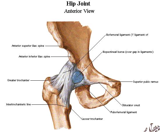
15 LIGAMENTS Illiofemoral ligament -also known as the Y ligament -runs from the base of the AIIS to the intertrochantic line -reinforces the fibrous capsule anteriorly -strongest ligament in the hip -prevents hyperextension of the hip during standing by screwing the femoral head into the acetabulum
16 LIGAMENTS Pubicfemoral ligament -runs from the anterior pubis ramus to the anterior surface of the intertrochantic fossa -reinforces the fibrous capsule inferiorly and anteriorly -tighten during abduction and extension -prevents overabduction of the hip joint
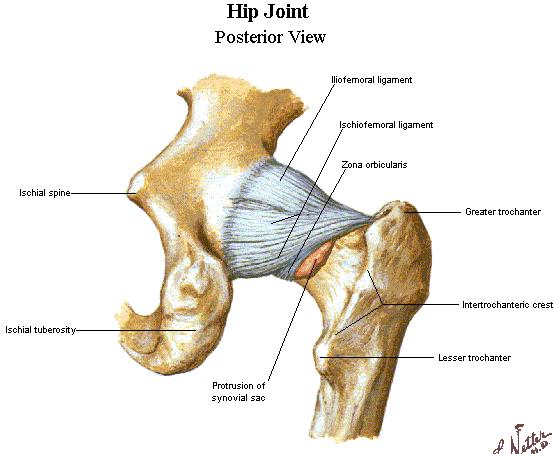
17 LIGAMENTS Ischiofemoral ligament -the ischial portion of the acetabulum and spirls to the neck of the femur and base of the greater trochanter -prevents hyperextension of the hip -fibers relaxed during flexion
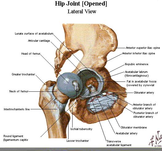
18 LIGAMENTS Ligamentum teres -known also as the ligament of the head of the femur -attaches to the acetabular notch and the transverse acetabular ligament to the pit in the head of the femur -is weak -supplies the blood for the femur head
19 Ligamentous Anatomy
20 Ligamentous Anatomy
21 Ligamentous Anatomy

22 Muscular Anatomy
23 Muscular Anatomy

24 Muscular Anatomy
25 Muscular Anatomy
26 Muscular Anatomy

27 Muscular anatomy
28 Movements of the Hip and its main Muscles Flexion Illiopsoas, sartorius, tensor fascia lata, rectus femoris, pectineus, adductor longus, adductor brevis, adductor magnus, gracilis Extension Hamstrings, adductor magnus, gluteus maximus Abduction Gluteus medius, gluteus minimus, tensor fascia lata
29 Adduction Adductor longus, adductor brevis, adductor magnus, gracilis, pectineus, oburator externus Rotation Medial Gluteus medius, gluteus minimus, tensor fascia lata Lateral Obturator externus, obturator internus, gemelli, piriformis, quadratus femoris, gluteus maximus
30 Origin/Insertion/Action Adductor Brevis O - Inferior Pubic Ramus I - Pectineal Line and Linea Aspera A - adducts, flexes, and medially rotates femur Gracillis O - pubic Symphysis and inferior pubic ramus I - medial surface of the tibia A - adducts thigh, flexes medially and medially rotates thigh, flexes leg
31 Origin/Insertion/Action Pectineus -O - Superior ramus of pubis -I - Pectineal line of femur -A adducts and flexes thigh

32 Origin/Insertion/Action Adductor Longus O - med portion of the superior pubic ramus I - linea aspera of femur A - adducts, flexes, and medially rotates the femur

33 Origin/Insertion/Action Adductor Magnus O - ischiopubic ramus and ischial tuberosity I - linea aspera of the femur; the ischiocondylar part inserts on the adductor tubercle of the femur A - adducts, flexes, and medially rotates the femur; extends the femur Inn - post div of oburator nerve; tibial nerve
34 Origin/Insertion/Action Biceps Femoris Longus -O - long head: ischial tuberosity, short head: linea aspera of femur -I - lateral side of head of fibula -A - extends the thigh

35 Origin/Insertion/Action Gluteus Maximus -O - ilium posterior to posterior gluteal line -I - end in the iliotibial tract that inserts into the lateral condyle of the tibia -A - extends the thigh

36 Origin/Insertion/Action Gluteus Medius -O external surface of ilium -I lateral surface of greater trochanter of femur -A abducts and internally rotates the thigh
37 Origin/Insertion/Action Gluteus Minimus -O external surface of ilium -I anterior surface of greater trochanter of femur -A abducts and internal rotates
38 Origin/Insertion/Action Piriformis -O lateral border of ischial tuberosity -I superior border of greater trochanter of femur -A external rotation extended thigh and abducts flexed thigh
39 Origin/Insertion/Action Quadratus femoris -O lateral border of ischial tuberosity -I quadrate tubercle on intertrochanteric crest of femur -A - external rotation extended thigh and abducts flexed thigh

40 Origin/Insertion/Action Obturator Externus -O pelvis surface of obturator membrane -I medial surface of greater trochanter -A - external rotation extended thigh and abducts flexed thigh

41 Origin/Insertion/Action Iliopsoas -O sides of T12-L5 vertebrae, iliac crest -I lesser trochanter of femur, pectineal line, lesser trochanter -A flexing the thigh

42 Origin/Insertion/Action Rectus Femoris -O AIIS and ilium superior to acetabulum -I base of patella -A flex thigh

43 Origin/Insertion/Action Sartorius -O ASIS -I superior part of medial surface of tibia -A flexes, abducts, and external rotates thigh Tensor Fascia Lata -O ASIS -I iliotibial tract -A- abducts, medial rotates, and flexes thigh
44 Origin/Insertion/Action Semimembranosus -O ischial tuberosity -I posterior part of medial condyle of tibia -A extend thigh

45 Origin/Insertion/Action Semitendinosus -O ischial tuberosity -I medial surface of superior part of tibia -A extend thigh

46 Origin/Insertion/Action Vastus lateralis -O greater trochanter and lateral lip of linea aspera of femur Vastus medialis -O intertrochanteric line and medial lip of linea aspera of femur Vastus intermedius -O anterior and lateral surfaces of body of femur *Same for all 3 -I base of patella and A helps flex thigh

47 Common Injuries Dislocation -femoral head moves out of the acetabulum -usually it goes posterior into notch -position typically flexion, adduction, and internal rotation -common mechanism: knee to dashboard during traffic collision -signs and symptoms: extreme pain, obvious deformity, unwilling to move the extremity
48 COMMON INJURIES Hip Pointer -contusion to the iliac crest -signs and symptoms: pain, swelling, and ecchymosis -severe limit to motion -palpable hematoma
49 COMMON INJURIES Piriformis Syndrome -sciatic nerve through piriformis -pressure on the sciatic nerve due to muscle spasm, trigger points, or tightness causing posterior thigh pain -other signs and symptoms: pain, limited ROM, pt tenderness deep to the gluteals
50 COMMON INJURIES Hip Fracture -most frequently occurs through the femoral neck -a direct blow to the lateral hip -signs and symptoms: pain, swelling, and loss of function -the involved leg will appear shortened and will be externally rotated
51 COMMON INJURIES Trochanteric Bursitis -cause is abnormal friction or irritation of the bursa between the IT band and greater trochanter, direct blow, or improper biomechanics -usually a sport such as running -signs and symptoms: local pain, swelling, pt tenderness, and crepitus over the greater trochanter -patient may complain of hip snapping
52 COMMON INJURIES Ischial Bursitis -lies over the ischial tuberosity -may become painful and inflamed with excessive friction -signs and symptoms: pain with sitting, pt tenderness over ischial tuberosity, pain w/ passive hip flexion and active/resistive hip extension -often difficult to differentiate from proximal hamstring tendinitis
53 COMMON INJURIES Hip Joint Sprain -less common -excessive forcible exertion of the extremity that stretch or tear the surrounding ligaments -signs and symptoms: pain and decrease ROM
54 COMMON INJURIES Hip Joint Strains -resulting from overstretching or from a rapid, forceful contraction of the muscle -explosive starts and slipping of the foot during cutting are common mechanisms for hip flexor and adductor strains -these injuries frequently occur during the beginning of practice and preseason training -signs and symptoms: pain, pt tenderness, muscle spasm, swelling, ecchymosis, and decreased ROM
55 COMMON INJURIES Avulsion Fracture -results from a violent contraction or tractioning of the attaching muscle -common sites: ASIS, AIIS, lesser trochanter, and ischial tuberosity -signs and symptoms: complain of a sudden sharp pain at time of injury, unwilling to move the extremity, pt tenderness along the bone, also may have a muscle bulging away from the attachment, and swelling
56 COMMON INJURIES Legg-Calve-Perthes Disease -characterized by avascular necrosis of the proximal femoral epiphysis -a chronic condition that develops slowly in children -often in males than in females -signs and symptoms: pain in the hip or groin that radiate to the knee, limping, decreased ROM, and hip flexor tightness may be noted -physician should be consulted to rule out serious pathologies such as this
57 COMMON INJURIES Avascular Necrosis of the Femoral Head -blood supply to the femur head is severed of is occluded for a prolonged period of time. -this is a common complication following hip dislocations, fractures, and chronic synovitis and often necessitates a hip replacement
58 COMMON INJURIES Chronic Synovitis -inflammatory process at the hip that is characterized by chronic irritation and excess secretion of synovial fluid within the capsule -this condition is very difficult to detect -may lead to avascular necrosis of the femoral head
The Hip (Iliofemoral) Joint. Presented by: Rob, Rachel, Alina and Lisa
 Joint. Presented by: Rob, Rachel, Alina and Lisa") The Hip (Iliofemoral) Joint Presented by: Rob, Rachel, Alina and Lisa Surface Anatomy: Posterior Surface Anatomy: Anterior Bones: Os Coxae Consists of 3 Portions: Ilium Ischium Pubis Bones: Pubis Portion
The Hip (Iliofemoral) Joint Presented by: Rob, Rachel, Alina and Lisa Surface Anatomy: Posterior Surface Anatomy: Anterior Bones: Os Coxae Consists of 3 Portions: Ilium Ischium Pubis Bones: Pubis Portion
Main Menu. Joint and Pelvic Girdle click here. The Power is in Your Hands
 1 Hip Joint and Pelvic Girdle click here Main Menu K.6 http://www.handsonlineeducation.com/classes//k6entry.htm[3/23/18, 2:01:12 PM] Hip Joint (acetabular femoral) Relatively stable due to : Bony architecture
Lesson 24. A & P Hip
 Lesson 24 A & P Hip 1 Aims of the Session This session will allow candidates to have an understanding of the bony prominences and soft tissues of the hip 2 Learning Outcomes By the end of the lesson the
The thigh. Prof. Oluwadiya KS
 The thigh Prof. Oluwadiya KS www.oluwadiya.com The Thigh: Boundaries The thigh is the region of the lower limb that is approximately between the hip and knee joints Anteriorly, it is separated from the
Hip joint and pelvic girdle. Lower Extremity. Pelvic Girdle 6/5/2017
 Hip joint and pelvic girdle Lower Extremity The relationship between the pelvic girdle and hip is similar to that between the shoulder girdle and shoulder joint. The lower limbs are attached to the axial
Human Anatomy Biology 351
 Human Anatomy Biology 351 Lower Limb Please place your name on the back of the last page of this exam. You must answer all questions on this exam. Because statistics demonstrate that, on average, between
Lectures of Human Anatomy
 Lectures of Human Anatomy Lower Limb Gluteal Region and Hip Joint By DR. ABDEL-MONEM AWAD HEGAZY M.B. with honor 1983, Dipl."Gynecology and Obstetrics "1989, Master "Anatomy and Embryology" 1994, M.D.
Muscles of the Thigh. 6.1 Identify, describe the attachments of and deduce the actions of the muscles of the thigh: Anterior group
 Muscles of the Thigh 6.1 Identify, describe the attachments of and deduce the actions of the muscles of the thigh: Anterior group Sartorius: This is a long strap like muscle with flattened tendons at each
The psoas minor is medial to the psoas major. The iliacus is a fan-shaped muscle that when contracted helps bring the swinging leg forward in walking
 1 p.177 2 3 The psoas minor is medial to the psoas major. The iliacus is a fan-shaped muscle that when contracted helps bring the swinging leg forward in walking and running. The iliopsoas and adductor
Bony Anatomy. Femur. Femoral Head Femoral Neck Greater Trochanter Lesser Trochanter Intertrochanteric Crest Intertrochanteric Line Gluteal Tuberosity
 Hip Anatomy Bony Anatomy Femur Femoral Head Femoral Neck Greater Trochanter Lesser Trochanter Intertrochanteric Crest Intertrochanteric Line Gluteal Tuberosity Bony Anatomy Pelvic Girdle Acetabulum 3 bones
Muscles of Lesson Five. Muscular Nomenclature and Kinesiology - Two. Muscles of Lesson Five, cont. Chapter 16
 Chapter 16 Muscular Nomenclature and Kinesiology - Two Lessons 5-6 Muscles of Lesson Five Iliopsoas (psoas major, iliacus) Hip outward rotators (piriformis, gemellus superior, gemellus inferior, obturator
Gluteal region DR. GITANJALI KHORWAL
 Gluteal region DR. GITANJALI KHORWAL Gluteal region The transitional area between the trunk and the lower extremity. The gluteal region includes the rounded, posterior buttocks and the laterally placed
Lower limb summary. Anterior compartment of the thigh. Done By: Laith Qashou. Doctor_2016
 Lower limb summary Done By: Laith Qashou Doctor_2016 Anterior compartment of the thigh Sartorius Anterior superior iliac spine Upper medial surface of shaft of tibia 1. Flexes, abducts, laterally rotates
Identify the muscles associated with the medial compartment of the thigh. Identify the attachment points of the medial thigh muscles.
 L 8 A B O R A T O R Y Thigh MEDIAL THIGH Identify the muscles associated with the medial compartment of the thigh. Identify the attachment points of the medial thigh muscles. Identify the actions of these
lesser trochanter of femur lesser trochanter of femur iliotibial tract (connective tissue) medial surface of proximal tibia
 medial surface of proximal tibia") LOWER LIMB MUSCLES OF THE APPENDICULAR SKELETON The muscles that act on the lower limb fall into three groups: those that move the thigh, those that move the lower leg, and those that move the ankle, foot,
LOWER LIMB MUSCLES OF THE APPENDICULAR SKELETON The muscles that act on the lower limb fall into three groups: those that move the thigh, those that move the lower leg, and those that move the ankle, foot,
rotation of the hip Flexion of the knee Iliac fossa of iliac Lesser trochanter Femoral nerve Flexion of the thigh at the hip shaft of tibia
 Anatomy of the lower limb Anterior & medial compartments of the thigh Dr. Hayder The fascia lata encloses the entire thigh like a sleeve/stocking. Three intramuscular fascial septa (lateral, medial, and
Figure 1 - Hip and Pelvis
 Hip Figure 1 - Hip and Pelvis The terms hip and pelvis are frequently used interchangeably, but strictly speaking, the pelvis is a girdle of bones and the hip is a joint. The pelvis consists of The sacrum
Human Anatomy Biology 351
 Human Anatomy Biology 351 Lower Limb Please place your name on the back of the last page of this exam. You must answer all questions on this exam. Because statistics demonstrate that, on average, between
Muscles of the lower extremities. Dr. Nabil khouri MD, MSc, Ph.D
 Muscles of the lower extremities Dr. Nabil khouri MD, MSc, Ph.D Posterior leg Popliteal fossa Boundaries Biceps femoris (superior-lateral) Semitendinosis and semimembranosis (superior-medial) Gastrocnemius
Sports Medicine 15. Unit I: Anatomy. The knee, Thigh, Hip and Groin. Part 4 Anatomies of the Lower Limbs
 Sports Medicine 15 Unit I: Anatomy Part 4 Anatomies of the Lower Limbs The knee, Thigh, Hip and Groin Anatomy of the lower limbs In Part 3 of this section we focused upon 11 of the 12 extrinsic muscles
Muscles of the Hip 1. Tensor Fasciae Latae O: iliac crest I: lateral femoral condyle Action: abducts the thigh Nerve: gluteal nerve
 Muscles of the Hip 1. Tensor Fasciae Latae O: iliac crest I: lateral femoral condyle Action: abducts the thigh Nerve: gluteal nerve 2. Gluteus Maximus O: ilium I: femur Action: abduct the thigh Nerve:
Human Anatomy Biology 255
 Human Anatomy Biology 255 Exam #4 Please place your name and I.D. number on the back of the last page of this exam. You must answer all questions on this exam. Because statistics demonstrate that, on average,
The University Of Jordan Faculty Of Medicine THE LOWER LIMB. Dr.Ahmed Salman Assistant Prof. of Anatomy. The University Of Jordan
 The University Of Jordan Faculty Of Medicine THE LOWER LIMB Dr.Ahmed Salman Assistant Prof. of Anatomy. The University Of Jordan Gluteal Region Cutaneous nerve supply of (Gluteal region) 1. Lateral cutaneous
First practical session. Bones of the gluteal region
 First practical session 2017 Bones of the gluteal region The Hip bone The hip bone is made of: 1 The ilium: superior in position 2 The ischium:postero-inferior in position 3 The pubis: antero-inferior
Anatomy & Physiology. Muscles of the Lower Limbs.
 Anatomy & Physiology Muscles of the Lower Limbs http://www.ishapeup.com/musclecharts.html Muscles of the Lower Limbs Among the strongest muscles in the body. Because pelvic girdle is composed of heavy,
The Lower Limb. Anatomy RHS 241 Lecture 2 Dr. Einas Al-Eisa
 The Lower Limb Anatomy RHS 241 Lecture 2 Dr. Einas Al-Eisa The bony pelvis Protective osseofibrous ring for the pelvic viscera Transfer of forces to: acetabulum & head of femur (when standing) ischial
Baraa Ayed حسام أبو عوض. Ahmad Salman. 1 P a g e
 4 Baraa Ayed حسام أبو عوض Ahmad Salman 1 P a g e Today we are going to cover these concepts: Iliotibial tract Anterior compartment of the thigh and the hip Medial compartment of the thigh Femoral triangle
Anatomage Table Instructors Guide- Lower Limb
 The Lower Limb Anatomage Table Instructors Guide- Lower Limb Table of Contents Lower Limb 1- The Skeletal System...3 1: Hip Bone...3 2: Hip Joint and Femur...4 3: Patella and Knee Joint...7 4: Tibia, Fibula,
The Muscular System. Chapter 10 Part D. PowerPoint Lecture Slides prepared by Karen Dunbar Kareiva Ivy Tech Community College
 Chapter 10 Part D The Muscular System Annie Leibovitz/Contact Press Images PowerPoint Lecture Slides prepared by Karen Dunbar Kareiva Ivy Tech Community College Table 10.14: Muscles Crossing the Hip and
Mohammad Ashraf. Abdulrahman Al-Hanbali. Ahmad Salman. 1 P a g e
 - 7 Mohammad Ashraf Abdulrahman Al-Hanbali Ahmad Salman 1 P a g e Structures under the cover of Gluteus Maximus: 1-Bones: Ileum, Femur (Head, greater trochanter and gluteal tuberosity), Ischium (ischial
Anterior and Medial compartments of the thigh. Dr. Heba Kalbouneh Associate Professor of Anatomy and Histology
 Anterior and Medial compartments of the thigh Dr. Heba Kalbouneh Associate Professor of Anatomy and Histology Terms Related to Movements Movement Flexion Extension Abduction Adduction Medial (internal)
Applied anatomy of the hip and buttock
 CHAPTER CONTENTS The hip joint e9 Capsule and ligaments e9 s e0 Flexor muscles................... e0 Extensor muscles.................. e Abductor muscles.................. e Adductor muscles..................
CHAPTER 8: THE BIOMECHANICS OF THE HUMAN LOWER EXTREMITY
 CHAPTER 8: THE BIOMECHANICS OF THE HUMAN LOWER EXTREMITY _ 1. The hip joint is the articulation between the and the. A. femur, acetabulum B. femur, spine C. femur, tibia _ 2. Which of the following is
ANATOMY TEAM GLUTEAL REGION & BACK OF THIGH
 ANATOMY TEAM GLUTEAL REGION & BACK OF THIGH OBJECTIVES By the end of this lecture, the student should be able to identify and discuss: Contents of gluteal region: Groups of Glutei muscles and small muscles
Acland's DVD Atlas of Human Anatomy. Transcript for Volume Robert D Acland
 Acland's DVD Atlas of Human Anatomy Transcript for Volume 2 2007 Robert D Acland This free downloadable pdf file is to be used for individual study only. It is not to be reproduced in any form without
Muscles of Gluteal Region
 1 The Gluteal Region In the gluteal region the skin is tough with many layers underneath. Directly under it is the superficial fascia followed by the deep fascia then the muscles and the bones of the thigh.
Bones of the Lower Limb Bone Structure Description Notes. border of the superior ramus. inferolaterally from the pubic symphysis
 Bones of the Lower Limb Bone Structure Description Notes pubis an angulated bone the forms the anterior part of the pelvis one of three bones that form the os coxae: ilium, ischium, pubis; its forms 1/5
this makes sense, however this is lower order thinking and does not solve the lower leg
 Functional Knee Valgus in a Barbell Squat 1 One of the most common lower leg dysfunction we see in athletes, particularly general population is functional knee valgus, or better referred to as the knees
The Lower Limb II. Anatomy RHS 241 Lecture 3 Dr. Einas Al-Eisa
 The Lower Limb II Anatomy RHS 241 Lecture 3 Dr. Einas Al-Eisa Tibia The larger & medial bone of the leg Functions: Attachment of muscles Transfer of weight from femur to skeleton of the foot Articulations
Joints of the lower limb
 Joints of the lower limb 1-Type: Hip joint Synovial ball-and-socket joint 2-Articular surfaces: a- head of femur b- lunate surface of acetabulum Which is deepened by the fibrocartilaginous labrum acetabulare
Copyright 2003 Pearson Education, Inc. publishing as Benjamin Cummings. Dr. Nabil Khouri MD, MSc, Ph.D
 Dr. Nabil Khouri MD, MSc, Ph.D Pelvic Girdle (Hip) Organization of the Lower Limb It is divided into: The Gluteal region The thigh The knee The leg The ankle The foot The thigh and the leg have compartments
ANATYOMY OF The thigh
 ANATYOMY OF The thigh 1- Lateral cutaneous nerve of the thigh Ι) Skin of the thigh Anterior view 2- Femoral branch of the genitofemoral nerve 5- Intermediate cutaneous nerve of the thigh 1, 2 and 3 are
Myology of the Knee. PTA 105 Kinesiology
 Myology of the Knee PTA 105 Kinesiology Objectives Describe the planes of motion and axes of rotation of the knee joint Visualize the origins and insertions of the muscles about the knee List the innervations
BONES JOINTS MUSCLES OF THE LOWER LIMB
 BONES JOINTS MUSCLES OF THE LOWER LIMB LOWER LIMB: BONES LOWER LIMB GLUTEAL REGION consists of 6 major segments: FEMORAL REGION (THIGH) KNEE REGION LEG REGION TALOCRURAL REGION (ANKLE) FOOT REGION LOWER
Topic 7: Hip and pelvis. Parts of the hip. Parts of the femur
 Topic 7: Hip and pelvis Parts of the hip Parts of the femur Classifying the hip joint Ball and socket Synovial Multiaxial Movements of the hip: Abduction/adduction Flexion/extension Medial/lateral rotation
Thigh, Hip, & Low Back Evaluation.
 Thigh, Hip, & Low Back Evaluation www.fisiokinesiterapia.biz Thigh Injuries Quad contusions - Myositis Ossificans Trochanteric bursitis - snapping hip Ischial bursitis - bench-warmer s bursitis Strains
5 Testing the Muscles of the Lower Extremity
 C H A P T E R 5 Testing the Muscles of the Lower Extremity Hip Flexion Hip Flexion, Abduction, and External Rotation with Knee Flexion Hip Extension Hip Abduction Hip Abduction from Flexed Position Hip
CHAPTER 19. The Hip and Pelvis KEY TERMS OBJECTIVES
 CHAPTER 19 The Hip and Pelvis KEY TERMS adductor muscles coccyx greater sciatic notch hamstring muscles hip flexors iliac crest iliac crest contusion iliac fossa ilium ischium obturator foramina pubis
Muscle Testing of Knee Extensors. Yasser Moh. Aneis, PhD, MSc., PT. Lecturer of Physical Therapy Basic Sciences Department
 Muscle Testing of Knee Extensors Yasser Moh. Aneis, PhD, MSc., PT. Lecturer of Physical Therapy Basic Sciences Department Muscle Testing of Knee Extensors othe Primary muscle Quadriceps Femoris -Rectus
In-Depth Foundations: Anatomy Terms to Know
 Be familiar with / able to identify and define all the following parts. The Spine Cranium Vertebrae Cervical, Thoracic, Lumbar Sacrum Coccyx Bones of Upper Body Cranium Mastoid process; Occipital condyle,
Muscles of the Gluteal Region
 Muscles of the Gluteal Region 1 Some of the most powerful in the body Extend the thigh during forceful extension Stabilize the iliotibial band and thoracolumbar fascia Related to shoulders and arms because
Overview. Overview. Introduction. Introduction Anatomy History Examination Common Disorders. Introduction Anatomy History Examination Common Disorders
 Common Hip Disorders in Figure Skaters 14 th Annual Meeting of Sports Medicine and Science in Figure Skating January 25, 2009 8:15-8:45am Robert J. Dimeff, MD Medical Director of Sports Medicine Overview
DISSECTION SCHEDULE. Session I - Hip (Front) & Thigh (Superficial)
 & Thigh (Superficial)") DISSECTION SCHEDULE Session I - Hip (Front) & Thigh (Superficial) Surface anatomy Inguinal region Gluteal region Thigh Leg Foot bones Hip bone Femur Superficial fascia Great saphenous vein Superficial
DISSECTION SCHEDULE Session I - Hip (Front) & Thigh (Superficial) Surface anatomy Inguinal region Gluteal region Thigh Leg Foot bones Hip bone Femur Superficial fascia Great saphenous vein Superficial
LOWER LIMB. As we know the bony part of the body is divided into Axial and Appendicular (upper and lower Limbs)
") LOWER LIMB As we know the bony part of the body is divided into Axial and Appendicular (upper and lower Limbs) Bones of the Lower limb: 1-Pelvic Girdle: composed of: 1. Right hip bone : is formed by 3
LOWER LIMB As we know the bony part of the body is divided into Axial and Appendicular (upper and lower Limbs) Bones of the Lower limb: 1-Pelvic Girdle: composed of: 1. Right hip bone : is formed by 3
Evaluation of the Hip
 Evaluation of the Hip Adam Lewno, DO PCSM Fellow, University of Michigan Primary Care Sports Update 2017 Disclosures Financial: None Images: I would like to acknowledge the work of the original owners
Scapula Spine Lateral edge of clavicle. Medial border Scapula. Medial border of Scapula, between superior angle and root of spine. Scapula.
 Muscle attachments and actions answer sheet Muscle Origins insertions Movements Joints crossed Trapezius Base of skull Spinous process of C7 Thoracic Spine Lateral edge of clavicle Elevation Retraction
Bones of Lower Limb. Dr. Heba Kalbouneh Associate Professor of Anatomy and Histology
 Bones of Lower Limb Dr. Heba Kalbouneh Associate Professor of Anatomy and Histology Bones of the lower limb Hip Bone Made up of 3 bones: 1) Ilium (flat), superior in position 2) Ischium (L), postero-inferior
ANATYOMY OF The thigh
 ANATYOMY OF The thigh 1- Lateral cutaneous nerve of the thigh Ι) Skin of the thigh Anterior view 2- Femoral branch of the genitofemoral nerve 5- Intermediate cutaneous nerve of the thigh 1, 2 and 3 are
Posterior compartment of the thigh. Dr. Heba Kalbouneh Associate Professor of Anatomy and Histology
 Posterior compartment of the thigh Dr. Heba Kalbouneh Associate Professor of Anatomy and Histology Posterior compartment of the thigh 1-Muscles: Biceps femoris Semitendinosus Semimembranosus Adductor magnus
THE HIP. Cooler than cool, the pinnacle of what is "it". Beyond all trends and conventional coolness.
 THE HIP Cooler than cool, the pinnacle of what is "it". Beyond all trends and conventional coolness. Objectives Hip anatomy Causes of hip pain Hip exam Anatomy Bones Ilium Anterior Superior Iliac Spine
Lecture 08 THIGH MUSCLES ANTERIOR COMPARTMENT. Dr Farooq Khan Aurakzai. Dated:
 Lecture 08 THIGH MUSCLES ANTERIOR COMPARTMENT BY Dr Farooq Khan Aurakzai Dated: 11.02.2017 INTRODUCTION to the thigh Muscles. The musculature of the thigh can be split into three sections by intermuscular
MUSCLES OF THE LOWER LIMBS
 MUSCLES OF THE LOWER LIMBS Naming, location and general function Dr. Nabil khouri ROLES THAT SHOULD NOT BE FORGOTTEN Most anterior compartment muscles of the hip and thigh Flexor of the femur at the hip
ANATYOMY OF The thigh
 ANATYOMY OF The thigh 1- Lateral cutaneous nerve of the thigh Ι) Skin of the thigh Anterior view 2- Femoral branch of the genitofemoral nerve 1, 2 and 3 are From the lumber plexus 5- Intermediate cutaneous
Prevention and Treatment of Injuries. The Femur. Quadriceps 12/11/2017
 Prevention and Treatment of Injuries The Thigh, Hip, Groin, and Pelvis Oak Ridge High School Conroe, Texas The Femur Is the longest and the second strongest bone in the body and is designed to permit maximum
lower limb Anterior Compartment: lecture 3 The deep fascia ( fascia lata) divides the thigh into 3 compartments:
 divides the thigh into 3 compartments:") lower limb lecture 3 The deep fascia ( fascia lata) divides the thigh into 3 compartments: 1. Anterior Extensor compartment 2. Medial Adductor compartment 3. Posterior Flexor compartment Anterior Compartment:
lower limb lecture 3 The deep fascia ( fascia lata) divides the thigh into 3 compartments: 1. Anterior Extensor compartment 2. Medial Adductor compartment 3. Posterior Flexor compartment Anterior Compartment:
RN(EC) ENC(C) GNC(C) MN ACNP *** MECHANISM OF INJURY.. MOST IMPORTANT ***
 ENC(C) GNC(C) MN ACNP *** MECHANISM OF INJURY.. MOST IMPORTANT ***") HISTORY *** MECHANISM OF INJURY.. MOST IMPORTANT *** Age of patient - Certain conditions are more prevalent in particular age groups (Hip pain in children may refer to the knee from Legg-Calve-Perthes
HISTORY *** MECHANISM OF INJURY.. MOST IMPORTANT *** Age of patient - Certain conditions are more prevalent in particular age groups (Hip pain in children may refer to the knee from Legg-Calve-Perthes
Anatomy of the lower limb
 Anatomy of the lower limb 1. Bones of the lower limb Pelvis Hip bone/coxal bone Acetabulum o Acetabular margin o Acetabular fossa o Acetabular notch o Lunate surface Ischiopubic ramus Obturator foramen
Bio 113 Anatomy and Physiology The Muscles. Muscles of the Head and Neck. Masseter. Orbicularis occuli. Orbicularis oris. Sternocleidomastoid
 Bio 113 Anatomy and Physiology The Muscles Muscles of the Head and Neck Masseter Orbicularis occuli Orbicularis oris Sternocleidomastoid Temporalis BIO 113 Fall 2011 Muscles Page 1 of 5 Muscles of the
LAB Notes#1. Ahmad Ar'ar. Eslam
 LAB Notes#1 Ahmad Ar'ar Eslam 1 P a g e Anatomy lab Notes Lower limb bones :- Pelvic girdle: It's the connection between the axial skeleton and the lower limb; it's made up of one bone called the HIP BONE
Hip Anatomy. Bony. The Athletic Hip: Anatomy and Common Injuries. Kyle Wilkens MSPAS, PA-C, ATC/L October 8, 2013
 The Athletic Hip: Anatomy and Common Injuries Kyle Wilkens MSPAS, PA-C, ATC/L October 8, 2013 Hip Anatomy Bony Femur Ischium Ilium Pubis Sacrum Coccyx Soft Tissue Joints Muscles -Pubic Symphisis Labrum
It is formed by fusion of 3 bones: I. Ilium (superior bone). II. Pubis (antero-inferior bone). III. Ischium (postero-inferior bone).
. II. Pubis (antero-inferior bone). III. Ischium (postero-inferior bone).") It is formed by fusion of 3 bones: I. Ilium (superior bone). II. Pubis (antero-inferior bone). III. Ischium (postero-inferior bone). Pubis Acetabulum Ana (242 ) The three constituent of bones of the hip
It is formed by fusion of 3 bones: I. Ilium (superior bone). II. Pubis (antero-inferior bone). III. Ischium (postero-inferior bone). Pubis Acetabulum Ana (242 ) The three constituent of bones of the hip
The Knee. Clarification of Terms. Osteology of the Knee 7/28/2013. The knee consists of: The tibiofemoral joint Patellofemoral joint
 The Knee Clarification of Terms The knee consists of: The tibiofemoral joint Patellofemoral joint Mansfield, p273 Osteology of the Knee Distal Femur Proximal tibia and fibula Patella 1 Osteology of the
ANATOMY. Lecturer : Maher Hadidi Done by: ,,Subject : lecture#: ~ Date:
 ANATOMY,,Subject : Lecturer : Maher Hadidi Done by: ~~ lecture#: ~ Date: GftJteal Reaion ' ' v Highest point of iliac cres Safe site for (/M) injection Horizantal line Sciatic nerve Ischial tubrosity
Diagnostic Approach to Hip Pain. Zoë J. Foster, MD October 3, 2018
 Diagnostic Approach to Hip Pain Zoë J. Foster, MD October 3, 2018 Disclosures I have nothing to disclose. Objectives Review examination of the hip, including special tests Discuss differential diagnosis
Surgical Anatomy of the Hip. Joseph H. Dimon
 Surgical Anatomy of the Hip Joseph H. Dimon The hip joint is a deep joint surrounded by large and powerful muscles necessary for its proper function. Essential neurovascular structures lie in front and
Practical 1 Worksheet
 Practical 1 Worksheet ANATOMICAL TERMS 1. Use the word bank to fill in the missing words. reference side stand body arms palms anatomical forward All anatomical terms have a(n) point which is called the
Gluteal Region and Back of Thigh
 Gluteal Region and Back of Thigh Musculoskeletal block- Anatomy-lecture 14 Editing file Objectives Know contents of gluteal region: 1. Groups of Glutei muscles and small muscles (Lateral Rotators). 2.
MUSCULOSKELETAL LOWER LIMB
 MUSCULOSKELETAL LOWER LIMB Spinal Cord Lumbar and Sacral Regions Spinal cord Dorsal root ganglion Conus medullaris Cauda equina Dorsal root ganglion of the fifth lumbar nerve End of subarachnoid space
SURGICAL AND APPLIED ANATOMY
 Página 1 de 6 Copyright 2001 Lippincott Williams & Wilkins Bucholz, Robert W., Heckman, James D. Rockwood & Green's Fractures in Adults, 5th Edition SURGICAL AND APPLIED ANATOMY Part of "37 - HIP DISLOCATIONS
SURGICAL EXPOSURES SURGERY OF THE HIP
 1 of 24 11/19/03 1:11 PM SURGICAL EXPOSURES SURGERY OF THE HIP by R. CALANDRUCCIO In: Atlas of Orthopaedic Surgery Volume 3 Lower Extremity; Editors: Laurin, CA, Riley Jr. LH, Roy-Camille R Reprinted with
Where should you palpate the pulse of different arteries in the lower limb?
 Where should you palpate the pulse of different arteries in the lower limb? The femoral artery In the femoral triangle, its pulse is easily felt just inferior to the inguinal ligament midway between the
Lower Limb Nerves. Clinical Anatomy
 Lower Limb Nerves Clinical Anatomy Lumbar Plexus Ventral rami L1 L4 Supplies: Abdominal wall External genitalia Anteromedial thigh Major nerves.. Lumbar Plexus Nerves relation to psoas m. : Obturator n.
5.1 Identify, describe the attachments of and deduce the actions of the muscles of the thigh:
 5.1 Identify, describe the attachments of and deduce the actions of the muscles of the thigh: Anterior group Proximal attachment Distal attachment Sartorius ASIS» Upper part of shaft tibia (middle surface)»
Anatomy images for MSS practical exam- 2019
 Anatomy images for MSS practical exam- 2019 Ilium Ischium Pubis Acetabulaum Iliac crest Iliac tubercle ASIS (muscle and ligament attached) AIIS (muscle attached) PSIS PIIS Ischial spine Ischial tuberosity
Lumbar Plexus. Ventral rami L1 L4 Supplies: Major nerves.. Abdominal wall External genitalia Anteromedial thigh
 Lower Limb Nerves Lectures Objectives Describe the structure and relationships of the plexuses of the lower limb. Describe the course, relationships and structures supplied for the major nerves of the
GET HIP! CAPA 2015 Annual Conference WHAT IS HIP? HIP JOINT. Bradford H. Stiles, M.D., FAAFP
 GET HIP! Bradford H. Stiles, M.D., FAAFP WHAT IS HIP? HIP JOINT Synovial ball-and-socket joint Articulation between femoral head and acetabulum Acetabulum formed by the confluence of pelvis bones (ilium,
SKELETAL MUSCLE ANATOMY
 SKELETAL MUSCLE ANATOMY OUTLINE I. Anatomical Terms of Motion II. Head, Face & Neck Muscles III. Anterior Torso Muscles IV. Posterior Torso Muscles V. Arm & Shoulder Muscles VI. Leg & Hip Muscles 2 ANATOMICAL
Apophysis. Apophyseal Avulsion. Apophyseal avulsion injuries 3/2/2017
 Apophysis 0 Differentiate from Epiphysis: The end of long bones which undergo endochondral ossification to produce longitudinal growth of the bones. i.e. growth plates 0 Apophysis refers to any eminence,
Lowe w r e r B ody Resistance Training
 Lower Body Resistance Training Tibialis Anterior Extensor Hallucis Longus Extensor Digitorum Longus Proneus Tertius AROM 25-40 degrees Extensor active-insufficiency Flexion & Eversion (Pronation) Flexion
The Lower Limb. Sevda LAFCI FAHRİOĞLU, MD.PhD.
 The Lower Limb Sevda LAFCI FAHRİOĞLU, MD.PhD. The Lower Limb The bones of the lower limb form the inferior part of the appendicular skeleton the organ of locomotion for bearing the weight of body stronger
TABLE OF MUSCLES OF LOWER EXTREMITY 2018zillmusom ANTERIOR THIGH
 TABLE OF MUSCLES OF LOWER EXTREMITY 2018zillmusom ANTERIOR THIGH MUSCLE ORIGIN INSERTION ACTION NERVE Iliopsoas Ilium, vertebra Femur Flex hip joint Femoral nerve (T12-L5) Pectineus Pubis Femur Flex hip
Muscles to know. Lab 21. Muscles of the Pelvis and Lower Limbs. Muscles that Position the Lower Limbs. Generally. Muscles that Move the Thigh
 Muscles to know Lab 21 Muscles of the Pelvis, Leg and Foot psoas major iliacus gluteus maximus gluteus medius sartorius quadriceps femoris (4) gracilus adductor longus biceps femoris semitendinosis semimembranosus
To classify the joints relative to structure & shape
 To classify the joints relative to structure & shape To describe the anatomy of the hip joint To describe the ankle joint To memorize their blood & nerve supply JOINTS: Joints are sites where skeletal
The os coxae or hip bone consists of three flat bones, ilium, ischium and pubis, which fuse together to form the acetabulum.
 The os coxae The os coxae or hip bone consists of three flat bones, ilium, ischium and pubis, which fuse together to form the acetabulum. The ilium extends from the acetabulum upwards forming the lateral
musculoskeletal system anatomy nerves of the lower limb 2 done by: Dina sawadha & mohammad abukabeer
 musculoskeletal system anatomy nerves of the lower limb 2 done by: Dina sawadha & mohammad abukabeer #Sacral plexus : emerges from the ventral rami of the spinal segments L4 - S4 and provides motor and
Figure 7: Bones of the lower limb
 BONES OF THE APPENDICULAR SKELETON The appendicular skeleton is composed of the 126 bones of the appendages and the pectoral and pelvic girdles, which attach the limbs to the axial skeleton. Although the
Hip Injuries & Arthroscopy in Athletes
 Hip Injuries & Arthroscopy in Athletes John P Salvo, MD Sports Medicine Rothman Institute Philadelphia, PA EATA Annual Meeting January, 2011 Hip Injuries & Arthroscopy in Anatomy History Physical Exam
HIP ANATOMY. The hip or coxafemoral joint includes the concave acetabulum of the innominate of the
 HIP ANATOMY The Joint The hip or coxafemoral joint includes the concave acetabulum of the innominate of the pelvis and the convex head of the femur (1) (Table 12.1). Structurally, the hip is a very stable
Chapter Four. The Lower Extremities
 Chapter Four The Lower Extremities 40 Ligamentous Articular Strain The lower extremities are among the most important structures of the body and yet are often overlooked. Each lower extremity contains
Lower Limb Dr. Robin Paudel
 Lower Limb n What is a limb? n Skeleton n Joints n Pelvis or limb girdle n Hip/Hip Muscles n Lumber and sacral plexus getting spinal nerves out onto limb n Muscles anterior and posterior compartments n