Contents. Design Rationale Surgical Goals System Basics
|
|
|
- Natalie Gilmore
- 5 years ago
- Views:
Transcription
1
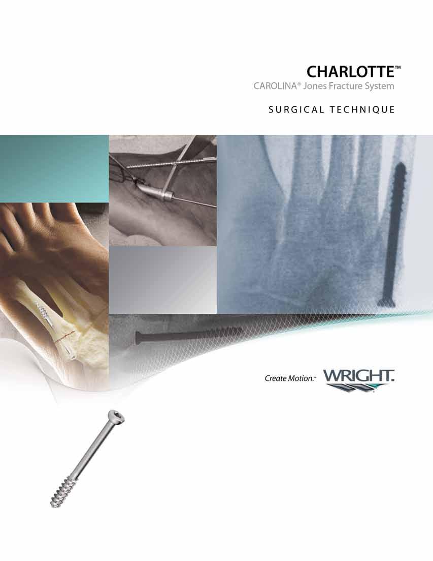
2 Contents Preface 3 Chapter Chapter Chapter 3 11 Appendix A 12 Appendix B 13 Appendix C Introduction Design Rationale Surgical Goals System Basics CHARLOTTE CAROLINA Jones Surgical Technique Identification and Incision Identify Crucial Anatomy and Place K-wire Tap and Measure Install Screw Mini IGNITE Graft Surgical Technique Iliac Crest Bone Marrow Aspirate Surgical Technique Bone Marrow Aspirate Surgical Technique Ordering Information CHARLOTTE CAROLINA Jones Fracture System Surgical Technique Proper surgical procedures and techniques are the responsibility of the medical professional. The following guidelines are furnished for information purposes only. Each surgeon must evaluate the appropriateness of the procedures based on his or her personal medical training and experience. Prior to use of the system, the surgeon should refer to the product package insert for complete warnings, precautions, indications, contraindications and adverse effects. Package inserts are also available by contacting Wright Medical Technology, Inc.

3 Introduction Preface Designed in conjunction with: Robert Anderson, MD OrthoCarolina, Charlotte, NC Treatment of Jones Fractures has been a notoriously difficult challenge for the Foot and Ankle specialist. The CHARLOTTE CAROLINA Jones Fracture System has been designed in accord with state-of-the-art research and modern clinical methods. James Nunley, MD Duke University Durham, NC W. Hodges Davis, MD Bruce Cohen, MD Carroll Jones, MD OrthoCarolina, Charlotte, NC CHARLOTTE CAROLINA Jones Fracture System 3
4 Design Rationale chapter 1 Surgical Goals To create the screw entry point high and inside on the proximal fragment, allowing proper intramedullary screw placement. To implant a screw with excellent strength, distal bite and compression in order to ensure stability across the fracture site. To minimize screw prominence issues by utilizing a low-profile head. System Basics All screws are manufactured from solid-core surgical grade stainless steel for maximum strength, stiffness and fatigue life. Screws are available in 4.5, 5.5 and 6.5mm diameters from 40 to 70mm in length. The instrument set includes: 3.2mm Cannulated Drill (P/N ) and 3.2mm Solid-core Drill (P/N ). 4.5, 5.5 and 6.5mm Cannulated Taps (P/N , , ). Each Tap (and corresponding screw) has the same thread pitch, only the diameter differs so that the Taps may be used in series without damaging the bone thread path. 2.0mm Tissue Protector (P/N ) for use with the K-wire. 4.5mm Tissue Protector (P/N ) for use with Drill and 4.5mm Tap. 5.5mm Tissue Protector (P/N ) for use with 5.5mm Tap. Universal Tissue Protector (P/N ) for use with all Taps, Drills and Drivers. Percutaneous Bone Grafting Use of mini IGNITE Power Mix should be considered with the CHARLOTTE CAROLINA Jones Fixation System. IGNITE Injectable Stimulus for Small Bone Fracture Callus Formation is designed to percutaneously graft a fracture site through an 11 gauge needle, preserving the periosteal blood supply. The combination of osteoinductive demineralized bone matrix, osteoconductive calcium sulfate and osteogenic progenitor cells from the patient allows a healing response at the fracture site. 4 Chapter 1 Design Rationale
 may be used in a standard Jacobs chuck. Figure 1 Perform surgery under regional block anesthesia.")

5 Surgical Technique chapter 2 Presented by Robert Anderson, MD and James Nunley, MD Patient Setup and Surgical Planning It is helpful to have a drill handpiece with a Hudson-style adapter. If one cannot be obtained, the adapter (P/N ) may be used in a standard Jacobs chuck. Figure 1 Perform surgery under regional block anesthesia. A tourniquet is usually used to provide a bloodless field, especially when an open technique is used. Position patient supine with a bump under the ipsilateral hip so that the body is rotated towards the side of the operating room table that corresponding to the non-affected foot. Make sure that the ipsilateral knee can be flexed so that it may be placed plantigrade on the edge of the table or sterile operating room fluoroscopy unit. Figure 1 Figure 2 Place a K-wire (P/N ) on the lateral aspect of the foot and use fluoroscopy to position the pin overlapping and parallel to metatarsal shaft. This position should correspond to the target screw placement on both AP and lateral images. Trace two lines on the skin that corresponding to the pin alignment in both views. Figure 2 Chapter 2 CHARLOTTE CAROLINA Jones Fracture System 5


. Advance the pin to 1/2 the length of the shaft, or at a minimum just past the fracture line.")
6 Approach and K-wire Placement Figure 3 Make a 1-2cm extensile incision approximately 2cm proximal to the base of the 5th metatarsal. Identify and protect the sural nerve and peroneus brevis tendon. Using the K-wire tissue protector (P/N ) insert the K-wire high and inside on the base of the 5th metatarsal. Figure 3 The K-wire will be opposed to the lateral aspect of the cuboid. Use fluoroscopy to direct the tip of the pin into the center of the intramedullary canal (continuously checking AP, lateral and oblique views). Advance the pin to 1/2 the length of the shaft, or at a minimum just past the fracture line. To prevent deviating medially, the proximal end of the pin should lie against the lateral calcaneal skin while being advanced. Figures 4 and 5 Figure 4 Figure 5 6 Chapter 2 CHARLOTTE CAROLINA Jones Fracture System

 advancing continuously under fluoroscan until proper depth is obtained.")
7 Figure 6 Using the 4.5mm drill tissue protector (P/N ) and continuous fluoroscopic guidance, advance the cannulated drill (P/N ) avoiding penetration of the 5th metatarsal cortex. Figures 6 and 7 Because the 5th metatarsal is not a straight bone, the guide wire may tend to curve; this will limit advancement of the drill. In this case, redirect the wire prior to further drilling. Figure 7 ALTERNATIVE DRILLING TECHNIQUE Use the cannulated drill to enter the proximal cortex only. Remove the K-wire and drill with the solid 1/8 drill (P/N ) advancing continuously under fluoroscan until proper depth is obtained. Use of this technique decreases the risk of perforation or damage to the K-wire or cannulated drill. Chapter 2 CHARLOTTE CAROLINA Jones Fracture System 7
 and 4.5mm tissue protector (P/N 56010045), tap to the intended screw length.")
 and 5.")
 if necessary. Figure 8 Alternatively, the universal tissue protector may be used for drilling, tapping and screw insertion steps.")

8 Tap and Measure The tap should be advanced through the straight portion of the distal fragment in A/P and lateral views; care should be taken not to advance into the curved portion, which could malreduce the fracture. Figure 8 Using the 4.5mm cannulated tap (P/N ) and 4.5mm tissue protector (P/N ), tap to the intended screw length. (Figure 8) The tap can be considered a trial since the profile of the tap is the same as the screw. The tap should feel snug within the intramedullary canal; if diameter is undersized, tap to the next size screw using the 5.5mm cannulated tap (P/N ) and 5.5 tissue protector (P/N ). Check under fluoroscan. Continue sequentially tapping with the 6.5mm cannulated tap (P/N ) and 6.5mm/universal tissue protector (P/N ) if necessary. Figure 8 Alternatively, the universal tissue protector may be used for drilling, tapping and screw insertion steps. Figure 9 Correct tissue protector placement. Figure 9A Incorrect tissue protector placement can lead to inaccurate screw measurement. Figure 10 When the correct tap is in the desired position, measure screw length from the grooves in the tap against the tissue protector. Check with fluoroscopic imaging to be sure the angled tip of the tissue protector is seated securely against the proximal 5th metatarsal bone. Figures 9, 9A and 10 8 Chapter 2 CHARLOTTE CAROLINA Jones Fracture System


9 Figure 11 ALTERNATIVE MEASURING TECHNIQUE The guide pin may be used to measure screw length. If using the guide pin to measure screw length, guide pin must be advanced until its tip approximates the location of the tip of the screw. Use the depth gauge (P/N ) to determine screw length. Figure 11 2ND ALTERNATIVE MEASURING TECHNIQUE Figure 12 Place a screw of the estimated length on the lateral side of the foot and check with fluoroscopic imaging that the proper length was chosen. This is especially helpful when the fracture is quite distal to confirm that all the threads are distal to the fracture line. Figure 12 Chapter 2 CHARLOTTE CAROLINA Jones Fracture System 9
, insert screw until fully engaged.")

10 Install Screw Obtain the correct screw diameter and length. Using the hex driver (P/N ), insert screw until fully engaged. Use fluoroscan to check final screw placement. Figures 13 and 14 Figure 13 Figure CHARLOTTE Chapter 2 CHARLOTTE Carolina Jones CAROLINA Fracture Jones SystemFracture System

, localize the defect site distal to the screw insertion site.")

, mix thoroughly according to instructions provided with the kit.")
11 chapter Surgical Chapter Technique 13 Jones Title Fracture IGNITE Graft Presented by Robert Anderson, MD Ortho Carolina, Charlotte, NC Step One Stabilize Fracture With Hardware Fracture/nonunion must have stable fixation prior to mini IGNITE Graft injection. Placement of the CHARLOTTE CAROLINA Jones Screw is shown. Refer to the CHARLOTTE CAROLINA Jones Fracture System surgical technique for a recommended step-by-step technique on placement. Step Two Localize Defect Under Fluoro Using the blue delivery needle with trocar (supplied in the mini IGNITE Power Mix kit), localize the defect site distal to the screw insertion site. Confirm placement under fluoroscopic guidance. Step Three Create Extra-Cortical Envelope Maintaining needle placement, replace the trocar with the supplied mini IGNITE periosteal elevator. With a gentle sweeping motion, bluntly dissect between bone and soft tissue to create a envelope to receive the IGNITE Graft and bridge the defect. NOTE: INJECTION TIME after mixing marrow and IGNITE powder is approximately 5 MINUTES. Step Four Mix and Inject Maintain needle placement. After aspirating 4cc of red marrow (see Appendix A/B), mix thoroughly according to instructions provided with the kit. Dock syringe onto the pre-placed needle and slowly inject the mini IGNITE Power Mix Graft. Step Five Allow to Coagulate for 3 5 Minutes and Remove Needle Slowly Following injection, maintain needle placement for 3-5 minutes to allow material to coagulate and minimize backflow. Chapter 31 IGNITE Description Graft of Surgical SectionTechnique 11


12 Iliac Ordering Crest Bone Information Marrow Aspirate Appendix A Anterior Approach Medical Health Center, Denver, CO Step One Prepare Site Prep and drape site as for iliac crest graft harvest. Palpate the anterior iliac crest. Placement 1cm posterior to the anterior superior iliac spine will avoid damage to the lateral femoral cutaneous nerve of the thigh. Step Two Insert Needle Introduce the aspiration needle into the central portion of the crest taking care to avoid penetrating the lateral overhanging lip. Step Three Prepare for Aspiration Remove the trocar and place the aspiration syringe on to needle. NOTE: Bone marrow aspiration should be completed after site preparation. See mixing instructions for more details. Step Four Aspirate Bone Marrow Begin aspiration of red marrow. If marrow does not aspirate easily, reposition needle slightly. If marrow still does not aspirate, redirect needle by removing syringe, replacing the trocar and following steps 1-3. Step Five Redirect, Continue Aspiration Redirect needle every 2cc during aspiration to avoid aspirating peripheral blood. The mini IGNITE kit includes diluent for reconstituting the IGNITE powder. The operating medical professional has the option of reconstituting the powder using the included diluent or patient s own bone marrow aspirate (BMA). The selection of patient s own BMA or the included diluent is the responsibility of the operating medical professional. 12 CHARLOTTE Iliac Appendix Crest Bone A Iliac Marrow CAROLINA Crest Aspirate Bone Jones Marrow Fracture Anterior Aspirate System Approach
 to ensure a quality aspirate harvest.")


13 Bone Marrow Aspirate Alternative Bone Marrow Harvest Sites Presented by Steve Brigido, DPM Coordinated Health, East Stroudsburg, PA Appendix B NOTE: Mesenchymal stem cell concentrations are not well understood in these sites. Care should be taken to redirect the needle frequently (every cc) to ensure a quality aspirate harvest. Proximal Tibia Using the ported 11G aspiration needle from the mini IGNITE Power Mix kit, insert the needle into the anterior medial proximal metaphysis at a 30 angle taking care to avoid the distal aspect of the patellar tendon laterally and the distal insertion of the medial collateral ligament medially. Begin aspiration redirecting the needle every ccs. Distal Tibia Using the ported 11G aspiration needle from the mini IGNITE Power Mix kit, insert the needle into the medial face of the tibia at the distal metaphysis. The needle is inserted at a 30 angle, and when inserted properly there are no neurovascular structures to avoid. Begin aspiration redirecting the needle every ccs. Calcaneous Using the ported 11G aspiration needle from the mini IGNITE Power Mix kit, insert the needle into the wall of the lateral calcaneal body at a 30 angle taking care to avoid the sural nerve and the peroneal tendon complex. Begin aspiration redirecting the needle every ccs. The mini IGNITE kit includes diluent for reconstituting the IGNITE powder. The operating medical professional has the option of reconstituting the powder using the included diluent or patient s own bone marrow aspirate (BMA). The selection of patient s own BMA or the included diluent is the responsibility of the operating medical professional. The alternative bone marrow harvest sites shown are optional at the discretion of the medical professional based on individual patient needs and circumstances. These alternative harvest sites are not intended to replace or change standard surgical procedures and techniques. CHARLOTTE Appendix B Bone CAROLINA Marrow Aspirate Jones Fracture System 13
14 Ordering Information Appendix C CHARLOTTE CAROLINA Jones Fixation System KIT1/A Part Number Description Qty Screw 4.5 x 40mm Screw 4.5 x 45mm Screw 4.5 x 50mm Screw 4.5 x 55mm Screw 4.5 x 60mm Screw 4.5 x 65mm Screw 4.5 x 70mm Screw 5.5 x 40mm Screw 5.5 x 45mm Screw 5.5 x 50mm Screw 5.5 x 55mm Screw 5.5 x 60mm Screw 5.5 x 65mm Screw 5.5 x 70mm Screw 6.5 x 40mm Screw 6.5 x 45mm Screw 6.5 x 50mm Screw 6.5 x 55mm Screw 6.5 x 60mm Screw 6.5 x 65mm Screw 6.5 x 70mm Drill 3.2mm Cannulated Drill 3.2mm Solid K-Wire 2.0 x 228mm 4 14 CHARLOTTE Appendix C Ordering CAROLINA Information Jones Fracture System
15 Appendix C CHARLOTTE CAROLINA Jones Distal Thread Lengths Overall Screw Length (mm) Distal Thread Length (mm) CHARLOTTE CAROLINA Jones Fixation Instruments Part No. Description Qty Quick Connect Handle Quick Connect Adapter Hex Driver 3.5mm Tissue Protector 4.5 Drill/Tap Tissue Protector 5.5 Tap Tissue Protector 6.5 Tap/Universal Tissue Protector K-Wire Screw Tap 4.5mm Cannulated Screw Tap 5.5mm Cannulated Screw Tap 6.5mm Cannulated Depth Gauge Surgical Tray (thermoform) 1 Devices with lumens require moistening with distilled or demineralized water before sterilization. ANSI/AAMI ST79:2006: Comprehensive guide to steam sterilization and sterility assurance in health care facilities. Appendix CHARLOTTE C Ordering CAROLINA Information Jones Fracture System 15

16 ordering information 876P-0400 mini IGNITE Power Mix 4cc - Injectable Stimulus for Small Bone Fracture Callus Formation mini IGNITE Power Mix - Injectable Stimulus for Small Bone Fracture Callus Formation Surgical Technique - SK Wright Medical Technology, Inc Airline Road Arlington, TN USA phone toll-free Wright Medical EMEA Krijgsman DM Amstelveen The Netherlands Trademarks and Registered marks of Wright Medical Technology, Inc Wright Medical Technology, Inc. All Rights Reserved. SO Rev.05.10
CHARLOTTE. 7.0 Multi-Use Compression Screw System SURGICAL TECHNIQUE
 CHARLOTTE 7.0 Multi-Use Compression Screw System SURGICAL TECHNIQUE Contents Chapter 1 1 Chapter 2 2 Chapter 3 3-9 Chapter 4 10-12 Appendix A 13-14 Design Rationale Screw Behavior Surgical Technique Procedure
CHARLOTTE. 7.0 Multi-Use Compression Screw System SURGICAL TECHNIQUE
 CHARLOTTE 7.0 Multi-Use Compression Screw System SURGICAL TECHNIQUE Contents Chapter 1 1 Chapter 2 2 2 3 4 Appendix A 5 6 Appendix B 7 8 Preoperative Planning Surgical Technique Subtalar Arthrodesis Guide
CHARLOTTE. 3.0 and 4.3 Multi-Use Compression Screws SURGIC A L T ECHNIQUE
 CHARLOTTE 3.0 and 4.3 Multi-Use Compression Screws SURGIC A L T ECHNIQUE CHARLOTTE 3.0 and 4.3 Multi-Use Compression Screws SURGICAL TECHNIQUE Surgical Technique as described by: Robert Anderson, MD Bruce
CHARLOTTE. Snap-Off Screws SURGICAL TECHNIQUE
 CHARLOTTE Snap-Off Screws SURGICAL TECHNIQUE CHARLOTTE snap-off screws surgical technique SURGICAL ADVISORS ROBERT ANDERSON, MD BRUCE COHEN, MD W. HODGES DAVIS, MD Proper surgical procedures and techniques
CHARLOTTE Lisfranc. Recontruction System SURGICAL TECHNIQUE
 CHARLOTTE Lisfranc Recontruction System SURGICAL TECHNIQUE Contents Preface 3 Chapter 1 4 Chapter 2 4 4 4 4 4 Chapter 3 5 5 5 6 6 6 7 7 8 Chapter 4 8 8 9 10 Appendix 10 Introduction Indications and Contraindications
DARCO. LPS Plate SURGICAL TECHNIQUE
 DARCO LPS Plate SURGICAL TECHNIQUE Contents Preface 3 Chapter 1 4 Chapter 2 5 5 6 7 9 Appendix 10 10 11 Design Rationale Preoperative Planning Surgical Technique Surgical Approach Joint Preparation Surgical
PediLoc 3.5mm and 4.5mm Contour Femur Plate Surgical Technique
 PediLoc 3.5mm and 4.5mm Contour Femur Plate Surgical Technique Surgical Technique Contour Femur Plate The technique description herein is made available to the healthcare professional to illustrate the
CHARLOTTE. Compression Staple
 CHARLOTTE Compression Staple CHARLOTTE compression staple surgical technique SURGICAL ADVISORS ROBERT ANDERSON, MD BRUCE COHEN, MD W. HODGES DAVIS, MD Proper surgical procedures and techniques are the
DARCO. Bow 2 Plate SURGIC AL TECHNIQUE
 DARCO Bow 2 Plate SURGIC AL TECHNIQUE Contents 2 Preface 3 Chapter 1 4 Chapter 2 5 6 7 8 9 Appendix 10 10 11 Intended Use Indications/Contraindications Design Rationale Preoperative Planning Surgical Technique
SURGICAL TECHNIQUE GUIDE: JONES FRACTURE USING THE PRECISION JONES FRACTURE SCREW SYSTEM
 PRODUCT DESCRIPTION The PRECISION Jones Fracture Screw System offers extensive options of Type II Anodized Titanium screws. System-specific instrumentation is designed to address procedural challenges
CHARLOTTE. Compression Staple SURGIC A L T ECHNIQUE
 CHARLOTTE Compression Staple SURGIC A L T ECHNIQUE CHARLOTTE Compression Staple SURGICAL TECHNIQUE Surgical Technique as described by: Robert Anderson, MD Bruce Cohen, MD W. Hodges Davis, MD Contents Chapter
CANNULINK. Intraossous Fixation System SURGICAL TECHNIQUE
 CANNULINK Intraossous Fixation System SURGICAL TECHNIQUE Contents Chapter 1 4 Introduction The CANNULINK Advantage Indications for Use Preoperative Planning Chapter 2 5 Surgical Technique CANNULINK Standard
System. Humeral Nail. Surgical Technique
 System Humeral Nail Surgical Technique Contents IMPLANT FEATURES 2 1. INDICATIONS 3 2. PRE-OPERATIVE PLANNING 3 3. PATIENT POSITIONING & FRACTURE REDUCTION 3 4. INCISION 4 5. ENTRY POINT 4-6 6. PROXIMAL
Contents. Chapter 1 4 Chapter Chapter Chapter Chapter 5 15
 Contents Chapter 1 4 Chapter 2 5 5 Chapter 3 6 6 7 Chapter 4 8 8 8 8 9 10 10 11 12 13 14 14 Chapter 5 15 Introduction Intended Use Indications Device Description Implant Options and Sizing Instrumentation
ORTHOLOC 3Di. Foot Reconstruction System Midfoot Fusion Plate SURGICAL TECHNIQUE
 ORTHOLOC 3Di Foot Reconstruction System Midfoot Fusion Plate SURGICAL TECHNIQUE ORTHOLOC Foot Reconstruction System Midfoot Fusion Plate SURGICAL TECHNIQUE Surgeon Design Team The ORTHOLOC 3Di Foot Reconstruction
Surgical Technique. CONQUEST FN Femoral Neck Fracture System
 Surgical Technique CONQUEST FN Femoral Neck Fracture System Table of Contents Introduction... 3 Indications... 3 Product Overview... 4 Surgical Technique... 5 Patient Positioning... 5 Reduce the Fracture...
3. PATIENT POSITIONING & FRACTURE REDUCTION 3 8. DISTAL GUIDED LOCKING FOR PROXIMAL NAIL PROXIMAL LOCKING FOR LONG NAIL 13
 Contents IMPLANT FEATURES 2 1. INDICATIONS 3 2. PRE-OPERATIVE PLANNING 3 3. PATIENT POSITIONING & FRACTURE REDUCTION 3 4. INCISION 4 5. ENTRY POINT 4-6 6. PROXIMAL NAIL INSERTION 6-7 7. PROXIMAL LOCKING
PediLoc 3.5mm and 4.5mm Bowed Femur Plate Surgical Technique
 PediLoc 3.5mm and 4.5mm Bowed Femur Plate Surgical Technique 2957 Bow Broch_REV_B.indd 1 2/10/11 12:47 PM Surgical Technique Bowed Femur Plate The technique description herein is made available to the
ORTHOLOC 3Di. Foot Reconstruction System SURGIC AL TECHNIQUE
 ORTHOLOC 3Di Foot Reconstruction System S C R E W TA R G E T I N G G U I D E SURGIC AL TECHNIQUE SURGEON DESIGN TEAM The ORTHOLOC 3Di Foot Reconstruction System was developed in conjuction with: ORTHOLOC
wave Calcaneal Fracture Plate
 wave Calcaneal Fracture Plate s u r g i c a l t e c h n i q u e Tornier WAVE Calcaneal fracture plate system surgical procedure Indications for Use: The Tornier Calcaneal Fracture Plate System is indicated
STS. Subtalar Spacer System SURGICAL TECHNIQUE
 STS Subtalar Spacer System SURGICAL TECHNIQUE Contents Chapter 1 4 Introduction 4 Device Description Chapter 2 5 Intended Use 5 Indications Chapter 3 6 Surgical Technique 9 Postoperative Protocol 9 Explant
MICRONAIL. Intramedullary Distal Radius System SURGICAL TECHNIQUE
 MICRONAIL II Intramedullary Distal Radius System SURGICAL TECHNIQUE Contents Introduction 3 4 Chapter 1 5 Chapter 2 6 Appendix A 18 Appendix B 19 Surgeon Design Team Introduction Product Information Surgical
PHALINX. Hammertoe Fixation SURGICAL TECHNIQUE
 PHALINX Hammertoe Fixation SURGICAL TECHNIQUE Contents Chapter 1 4 Product Information 4 Device Description Chapter 2 5 Intended Use 5 Indications 5 Contraindications Chapter 3 6 Surgical Technique 6
Biomet Large Cannulated Screw System
 Biomet Large Cannulated Screw System s u r g i c a l t e c h n i q u e A Complete System for Simplified Fracture Fixation 6.5mm & 7.3mm The Titanium, Self-drilling, Self-tapping Large Cannulated Screw
Technique Guide. LCP Proximal Femoral Hook Plate 4.5/5.0. Part of the LCP Periarticular Plating System.
 Technique Guide LCP Proximal Femoral Hook Plate 4.5/5.0. Part of the LCP Periarticular Plating System. Table of Contents Introduction Features and Benefits 2 AO ASIF Principles 4 Indications 5 Surgical
AcUMEDr. FoREARM ROD SYSTEM
 AcUMEDr FoREARM ROD SYSTEM FoREARM ROD SYSTEM Since 1988 Acumed has been designing solutions to the demanding situations facing orthopedic surgeons, hospitals and their patients. Our strategy has been
Imbibe Bone marrow aspiration needle. Operative technique
 Imbibe Bone marrow aspiration needle Operative technique Imbibe Bone Marrow Aspiration Needle Imbibe Bone marrow aspiration needle Contents 1. Smart design... 3 2. Operative technique... 4 Posterior iliac
Zimmer MIS Periarticular 3.5mm Proximal Tibial Locking Plate
 Zimmer MIS Periarticular 3.5mm Proximal Tibial Locking Plate Surgical Technique The Science of the Landscape Zimmer MIS Periarticular 3.5mm Proximal Tibial Locking Plate Surgical Technique 1 Zimmer MIS
Orthopedic Bone Nail System - Distal Femoral Nail Surgical Technique Manual
 Orthopedic Bone Nail System - Distal Femoral Nail Surgical Technique Manual Note: The surgical procedures should be performed under the guidance of qualified skilled orthopedic surgeons, and this surgical
US Patent No. 6,309,396B1. G. David Ritland, M.D.
 US Patent No. 6,309,396B1 G. David Ritland, M.D. The Femur Finder is designed to direct the guide wire into the medullary canal. Do not use it to manipulate the fracture fragments, reduce the fracture,
Conventus CAGE PH Surgical Techniques
 Conventus CAGE PH Surgical Techniques Conventus Orthopaedics The Conventus CAGE PH (PH Cage) is a permanent implant comprised of an expandable scaffold, made from nitinol and titanium, which is deployed
CSS. Cannulated Screw System SURGICAL TECHNIQUE
 CSS Cannulated Screw System SURGICAL TECHNIQUE Contents Chapter 1 4 Product Information 4 Device Description Chapter 2 5 Intended Use 5 Indications 5 Contraindications Chapter 3 6 Surgical Technique Appendix
LCP Medial Distal Tibia Plate, without Tab. The Low Profile Anatomic Fixation System with Angular Stability and Optimal Screw Orientation.
 LCP Medial Distal Tibia Plate, without Tab. The Low Profile Anatomic Fixation System with Angular Stability and Optimal Screw Orientation. Technique Guide LCP Small Fragment System Table of Contents Introduction
Surgical Technique. Customer Service:
 Patent and Patent Pending CAUTION: Federal Law (USA) restricts this device to sale by or on the order of a physician. INDICATIONS FOR USE The Axis Charcot Fixation System in diameters of 5.5, 6.5 and 7.5mm
ORTHOLOC Calcaneal Fracture System SURGICAL TECHNIQUE
 ORTHOLOC Calcaneal Fracture System SURGICAL TECHNIQUE ORTHOLOC Calcaneal Fracture System SURGICAL TECHNIQUE Contents Chapter 1 4 Chapter 2 5 Chapter 3 6 7 8 9 Appendix 1 10-12 ORTHOLOC Calcaneal Fracture
Zimmer Trabecular Metal Ankle Interpositional Spacer and Trabecular Metal Ankle Fusion Spacer
 Zimmer Trabecular Metal Ankle Interpositional Spacer and Trabecular Metal Ankle Fusion Spacer Surgical Technique 2 Zimmer Trabecular Metal Ankle Interpositional Spacer and Trabecular Metal Ankle Fusion
NeoGen Tibia Nail System
 NeoGen Tibia Nail System LESS IS MORE TE-2070-03 Surgical Technique BLE OF CONTENT Preface Surgical Technique Appendix Products Information Patient Preparation Entry Portal Fracture Reduction Canal Preparation
Surgical Technique. Fibula Rod System
 Surgical Technique Fibula Rod System Acumed is a global leader of innovative orthopaedic and medical solutions. We are dedicated to developing products, service methods, and approaches that improve patient
Locking Ankle Plating System. Surgical Technique
 Locking Ankle Plating System Surgical Technique Acumed is a global leader of innovative orthopaedic and medical solutions. We are dedicated to developing products, service methods, and approaches that
Technique Guide. 2.7 mm/3.5 mm LCP Distal Fibula Plates. Part of the Synthes locking compression plate (LCP) system.
 system.") Technique Guide 2.7 mm/3.5 mm LCP Distal Fibula Plates. Part of the Synthes locking compression plate (LCP) system. Table of Contents Introduction 2.7 mm/3.5 mm LCP Distal Fibula Plates 2 AO Principles
Technique Guide 2.7 mm/3.5 mm LCP Distal Fibula Plates. Part of the Synthes locking compression plate (LCP) system. Table of Contents Introduction 2.7 mm/3.5 mm LCP Distal Fibula Plates 2 AO Principles
Surgical Technique. Targeter Systems Overview
 Surgical Technique Targeter Systems Overview PERI-LOC Locked Plating System Targeter Systems Overview Table of contents Product overview... 2 Introduction... 2 Indications... 2 Design features and benefits...
Surgical Technique. Calcaneal Locking Plate
 Surgical Technique Calcaneal Locking Plate PERI-LOC Locked Plating System Calcaneal Locking Plate Surgical TechniqueCatalog Infor Table of Contents Introduction...2 Indications...3 Plate Features...3 Patient
Knee Surgical Technique
 Knee Surgical Technique COMPASS Universal Hinge by Jimmy Tucker, M.D. Orthopaedic Surgeon Director, Arkansas Sports Medicine, P.A. Little Rock, Arkansas Table of contents Design features 3 Indications
Zimmer NexGen MIS Tibial Component. Cemented Surgical Technique IMAGE TO COME
 Zimmer NexGen MIS Tibial Component Cemented Surgical Technique IMAGE TO COME Zimmer NexGen MIS Tibial Component Cemented Surgical Technique 1 Zimmer NexGen MIS Tibial Component Cemented Surgical Technique
Technique Guide. 3.5 mm LCP Low Bend Medial Distal Tibia Plates. Part of the Synthes locking compression plate (LCP) system.
 system.") Technique Guide 3.5 mm LCP Low Bend Medial Distal Tibia Plates. Part of the Synthes locking compression plate (LCP) system. Table of Contents Introduction 3.5 mm LCP Low Bend Medial Distal Tibia Plates
Technique Guide 3.5 mm LCP Low Bend Medial Distal Tibia Plates. Part of the Synthes locking compression plate (LCP) system. Table of Contents Introduction 3.5 mm LCP Low Bend Medial Distal Tibia Plates
NeoGen Femoral Nail System
 NeoGen Femoral Nail System LESS IS MORE TE-2070-04 Surgical Technique BLE OF CONTENT Preface Standard Femoral Mode Recon Mode Post-Operative Management Appendix Products Information Indication Patient
Ankle Fracture System SURGICAL TECHNIQUE
 ORTHOLOC 3Di Ankle Fracture System SURGICAL TECHNIQUE SURGEON DESIGN TEAM The ORTHOLOC 3Di Ankle Fracture System was developed in conjuction with: Robert B. Anderson, MD OrthoCarolina Charlotte, NC ORTHOLOC
SALVATION 3Di. Plating System SURGICAL TECHNIQUE
 SALVATION 3Di Plating System SURGICAL TECHNIQUE Contents Chapter 1 4 Introduction Chapter 2 5 Intended use 5 Indications 5 Contraindications Chapter 3 6 Device Description Chapter 4 7 Preoperative Planning
Surgical Technique 4.5/8.5MM BEAMING SYSTEM. Customer Service:
 Patent and Patent Pending CAUTION: Federal Law (USA) restricts this device to sale by or on the order of a physician. INDICATIONS FOR USE The 4.5/8.5 screw system is intended for fixation arthrodesis of
The Flower Medial Column Fusion Plate
 The Flower Medial Column Fusion Plate PROCEDURE GUIDE www.flowerortho.com The Flower Foot & Ankle Application NC FUSION PLATE 2-HOLE COMPRESSION PLATE TMT FUSION PLATE LAPIDUS FUSION PLATE COMPRESSION
ORTHOLOC Calcaneal Fracture System SURGIC AL TECHNIQUE
 ORTHOLOC Calcaneal Fracture System SURGIC AL TECHNIQUE Contents Chapter 1 4 Chapter 2 5 Chapter 3 6 7 8 9 Appendix 1 10-12 ORTHOLOC Calcaneal Fracture System - Introduction - ORTHOLOC Polyaxial Locking
Surgical Technique. Forearm Fracture Solutions
 Surgical Technique Forearm Fracture Solutions Acumed is a global leader of innovative orthopaedic and medical solutions. We are dedicated to developing products, service methods, and approaches that improve
Surgical Technique. Cannulated Angled Blade Plate 3.5 and 4.5, 90
 Surgical Technique Cannulated Angled Blade Plate 3.5 and 4.5, 90 Cannulated Angled Blade Plate 3.5 and 4.5, 90 Table of contents Indications/Contraindications 2 Implants 3 Surgical technique 5 Implant
MICA. Minimally Invasive Foot Surgery CHEVRON OSTEOTOMY SURGIC AL TECHNIQUE
 MICA Minimally Invasive Foot Surgery CHEVRON OSTEOTOMY SURGIC AL TECHNIQUE Contents Chapter 1 4 Introduction Chapter 2 5 Indications and Warnings Chapter 3 6 Patient Positioning and Set Up Chapter 4 7
Surgical Technique. Distal Radius and Foot
 Surgical Technique Distal Radius and Foot JET-X BAR Unilateral Fixator Distal Radius and Foot Surgical Technique Contents Design Features...2 Distal Radius Surgical Technique Indications...10 Surgical
Biologically-Assisted ACL Reconstruction. Surgical Technique
 Biologically-Assisted ACL Reconstruction Surgical Technique One Surgeon. One Patient. Over 1 million times per year, Biomet helps one surgeon provide personalized care to one patient. The science and art
Femur. Monoaxial Locking Plate System. Operative Technique. Distal Lateral Femur Universal Holes Targeting Instrumentation.
 Femur AxSOS 3 Titanium Monoaxial Locking Plate System Femur Fractures Operative Technique Distal Lateral Femur Universal Holes Targeting Instrumentation This publication sets forth detailed recommended
AcUMEDr. Olecranon Threaded Compression Rod
 AcUMEDr Olecranon Threaded Compression Rod Olecranon Threaded Compression Rod Since 1988, Acumed has been designing solutions to the demanding situations facing orthopaedic surgeons, hospitals and their
Surgical Technique Guide
 Surgical Technique Guide Minimally Invasive, Intramedullary Device For Distal Radius Fragility Fractures The Sonoma WRx Wrist Fracture Repair Device is flexible, inserting easily through a small incision
Olecranon Locking Plate II
 INDEX Indications Patient Position Fracture Reduction and Fixation Surgical Technique Step 1 Surgical Approach Step 2 Implantation Step 3 Proximal Locking Screw Insertion Step 4 Distal Screw Insertion
Technique Guide. 3.5 mm LCP Low Bend Medial Distal Tibia Plate Aiming Instruments. Part of the 3.5 mm LCP Percutaneous Instrument System.
 Technique Guide 3.5 mm LCP Low Bend Medial Distal Tibia Plate Aiming Instruments. Part of the 3.5 mm LCP Percutaneous Instrument System. Table of Contents Introduction 3.5 mm LCP Low Bend Medial Distal
FLT105 12/02.
 FLT105 12/02 www.biometmerck.co.uk Disclaimer Biomet Merck Ltd, as the manufacturer of this device, does not practice medicine and does not recommend this or any other surgical technique for use on a
A sequenced approach to flush graft placement. GLENOID BONE LOSS SYSTEM Procedural Solution
 A sequenced approach to flush graft placement GLENOID BONE LOSS SYSTEM Procedural Solution One comprehensive system, two ways of treating glenoid bone loss The GLENOID BONE LOSS SYSTEM provides you with
LCP Anterolateral Distal Tibia Plate 3.5. The low profile anatomic fixation system with optimal plate placement and angular stability.
 LCP Anterolateral Distal Tibia Plate 3.5. The low profile anatomic fixation system with optimal plate placement and angular stability. Technique Guide LCP Small Fragment System Table of Contents Introduction
Supplemental Use Guide Standard Triple Arthrodesis
 Acutrak 2 Headless Compression Screw System 4.7 mm and 7.5 mm Screws Supplemental Use Guide Standard Triple Arthrodesis Acumed is a global leader of innovative orthopaedic and medical solutions. We are
LCP Anterolateral Distal Tibia Plate 3.5. The low profile anatomic fixation system with optimal plate placement and angular stability.
 LCP Anterolateral Distal Tibia Plate 3.5. The low profile anatomic fixation system with optimal plate placement and angular stability. Technique Guide LCP Small Fragment System Table of Contents Introduction
Fibula Rod System. Lateral Malleolus Fracture Indications:
 Fibula Rod System Fibula Rod System Since 1988, Acumed has been designing solutions for the demanding situations facing orthopaedic surgeons, hospitals and their patients. Our strategy has been to know
OptiLock Periarticular Plating System For Distal Tibial Fractures. Surgical Technique
 OptiLock Periarticular Plating System For Distal Tibial Fractures Surgical Technique Contents Introduction... Page 1 Indications & Contraindications... Page 6 System Features... Page 7 Surgical Technique...
Cannulated Angled Blade Plate 3.5 and 4.5, 90.
 Cannulated Angled Blade Plate 3.5 and 4.5, 90. Technique Guide This publication is not intended for distribution in the USA. Instruments and implants approved by the AO Foundation. Table of Contents Introduction
Technique Guide. Locking Attachment Plate. For treatment of periprosthetic fractures.
 Technique Guide Locking Attachment Plate. For treatment of periprosthetic fractures. Table of Contents Introduction Locking Attachment Plate 2 Indications 4 Surgical Technique Patient Positioning 5 Preparation
Implantable K-wire SURGIC A L T ECHNIQUE
 Implantable K-wire SURGIC A L T ECHNIQUE Contents Chapter 1 4 Product Information 4 Device Description 4 Indications 4 Contraindications Chapter 2 5 Surgical Technique 5 Hammertoe Correction 6 MTP Joint
DARCO. CPS Plate SURGICAL TECHNIQUE
 DARCO CPS Plate SURGICAL TECHNIQUE Contents Preface 3 Chapter 1 4 Chapter 2 5 Chapter 3 6 Appendix 9 Introduction - CPS Features Preoperative Planning Surgical Technique - Surgical Approach and Retraction
Technique Guide. 6.5 mm Midfoot Fusion Bolt. For intramedullary fixation of the medial column of the foot.
 Technique Guide 6.5 mm Midfoot Fusion Bolt. For intramedullary fixation of the medial column of the foot. Table of Contents Introduction 6.5 mm Midfoot Fusion Bolt 2 AO Principles 4 Indications 5 Surgical
Lag Screw Device Intended for symphyseal fracture fixation of the mandible
 Lag Screw Device Intended for symphyseal fracture fixation of the mandible SUrgicaL TecHNiqUe Lag Screw Device Intended for symphyseal fracture fixation of the mandible Simplifies the lag screw fixation
Zimmer Small Fragment Universal Locking System. Surgical Technique
 Zimmer Small Fragment Universal Locking System Surgical Technique Zimmer Small Fragment Universal Locking System 1 Zimmer Small Fragment Universal Locking System Surgical Technique Table of Contents Introduction
Surgical Technique. Anterolateral and Medial Distal Tibia Locking Plates
 Surgical Technique Anterolateral and Medial Distal Tibia Locking Plates PERI-LOC Periarticular Locked Plating System Anterolateral and Medial Distal Tibia Locking Plates Surgical Technique Contents Product
SMV Scientific Bone Plate and Screw System Surgical Technique
 SMV Scientific Bone Plate and Screw System Surgical Technique Description: The SMV Scientific Bone Plate and Screw System consists of non-locking plates and bone screw fasteners in a variety of lengths,
Lateral TTC Plate SURGICAL TECHNIQUE
 MAXLOCK EXTREME Lateral TTC Plate SURGICAL TECHNIQUE Contents Overview 2 Exposure 3 Surgical Technique 4 Implants and Instruments 10 11 Proper surgical procedures and techniques are the responsibility
Surgical Technique. Clavicle Locking Plate
 Surgical Technique Clavicle Locking Plate PERI-LOC Locked Plating System Clavicle Locking Plate Surgical Technique Table of Contents Introduction...2 Indications...3 Plate Features...3 Patient Positioning...4
MIS Cemented Tibial Component
 MIS Cemented Tibial Component NexGen Complete Knee Solution Surgical Technique Table of Contents Surgical Exposure... 2 Finish the Tibia... 2 Position Based on Anatomic Landmarks... 3 Lateral Posterior
Integra. ADVANSYS Plating System SURGICAL TECHNIQUE
 Integra ADVANSYS Plating System SURGICAL TECHNIQUE Table of Contents Dorsal Lisfranc Plate Indications...2 Contraindications...2 Description...2 Surgical Technique...3 Surgical Site Preparation...3 Step
STRENGTH FROM WITHIN. Surgical Technique for Representatives
 STRENGTH FROM WITHIN Surgical Technique for Representatives Table of Contents PREOPERATIVE TEMPLATING... p.3 OUTRIGGER DRILL GUIDE OVERVIEW... p.4 PATIENT POSITIONING...p.5 STEP 1: REDUCE THE FRACTURE...p.5-6
ACL Reconstruction for BTB Grafts
 Transtibial ACL Reconstruction System for BTB Grafts Surgical Technique Designed in conjunction with John C. Garrett, M.D., Atlanta, GA ACL Reconstruction for BTB Grafts Reference Anatomical Constants
Zimmer ITST Intertrochanteric/ Subtrochanteric Fixation System. Abbreviated Surgical Technique
 Zimmer ITST Intertrochanteric/ Subtrochanteric Fixation System Abbreviated Surgical Technique ITST System Abbreviated Surgical Technique Indications The ITST Intramedullary Nail is indicated for use in
AFX. Femoral Implant. System. The AperFix. AM Portal Surgical Technique Guide. with the. The AperFix System with the AFX Femoral Implant
 The AperFix System AFX with the Femoral Implant AM Portal Surgical Technique Guide The Cayenne Medical AperFix system with the AFX Femoral Implant is the only anatomic system for soft tissue ACL reconstruction
N-Force Fixation System. Surgical Technique
 N-Force Fixation System Surgical Technique Table of Contents Features... 2 Indications/Contraindications... 3 Surgical Technique 4.0 mm...6 11 Step 1: Preoperative Radiographic Fracture Assessment and
TABLE OF CONTENTS. 2 (8144 Rev 2)
") 1 (8144 Rev 2) TABLE OF CONTENTS Introduction Conventus CAGE TM - Proximal Humerus...3 Indications and Contraindications...4 Surgical Summary...5 Patient Positioning & Approach...6 Surgical Technique Plate
1 (8144 Rev 2) TABLE OF CONTENTS Introduction Conventus CAGE TM - Proximal Humerus...3 Indications and Contraindications...4 Surgical Summary...5 Patient Positioning & Approach...6 Surgical Technique Plate
Locking Radial Head Plates
 Locking Radial Head Plates Locking Radial Head Plates Since 1988, Acumed has been designing solutions to the demanding situations facing orthopaedic surgeons, hospitals and their patients. Our strategy
Correction System. Surgical Technique
 Nextra Hammertoe Correction System Surgical Technique Maximized Bone Purchase* Stable and Secure Phalanx Optimized Screw Design Adjustable Bone-to-Bone Apposition Progressive Ratchet Tightening Mechanism
2.7 mm/3.5 mm LCP Distal Fibula Plate
 Part of the DePuy Synthes Locking Compression Plate (LCP ) System 2.7 mm/3.5 mm LCP Distal Fibula Plate Surgical Technique Table of Contents Introduction 2.7 mm/3.5 mm LCP Distal Fibula Plates 2 AO Principles
PediLoc Extension Osteotomy Plate (PLEO)
") PediLoc Extension Osteotomy Plate (PLEO) Left PLEO Plates Sizes: 6, 8 and 10 hole plates Right PLEO Plates Sizes: 6, 8 and 10 hole plates PediLoc Extension Osteotomy Plate The technique description herein
PediLoc Extension Osteotomy Plate (PLEO) Left PLEO Plates Sizes: 6, 8 and 10 hole plates Right PLEO Plates Sizes: 6, 8 and 10 hole plates PediLoc Extension Osteotomy Plate The technique description herein
GREENS SURGICALS. Redefining Excellence INSTRUMENT SYSTEM PREPARED BY: DR. VINAY KUMAR
 GREENS SURGICALS Redefining Excellence TIBIA AND FEMUR INSTRUMENT SYSTEM PREPARED BY: DR. VINAY KUMAR OPERATIVE TECHNIQUES INDEX SR.NO CONTENTS 1 LIST OF INSTRUMENT FOR TIBIA AND FEMUR. 2 RADIO GRAPH OF
humerus InSafeLOCK Nail
 humerus InSafeLOCK Nail Introduction Content Humerus InSafeLOCK Nail is an innovative intramedullary nailing system, developed for humerus problems. Humerus fractures have 5-6 % incidence of all bone fractures.
DARCO MFS. Locked plating system for reconstructive forefoot surgery.
 DARCO MFS Locked plating system for reconstructive forefoot surgery. DARCO MFS Locked plating system for reconstructive forefoot surgery System Basics The DARCO MFS plating system for the forefoot has
The Vilex FUZETM. Dual Thread Screw & Intramedullary Nail in One Implant. The Ultimate TTC Arthrodesis Internal Fixator
 The Vilex FUZETM Dual Thread Screw & Intramedullary Nail in One Implant The Ultimate TTC Arthrodesis Internal Fixator Introduction The Vilex FUZE TM TTC Arthrodesis Compression Nail combines the attributes
Surgical Technique. Proximal Humerus Locking Plate
 Surgical Technique Proximal Humerus Locking Plate PERI-LOC Upper Extremity Locked Plating System 3.5mm & 4.5mm Proximal Humerus Locking PlatesCatalog Infor Table of Contents Introduction.........................................................2
PROXIMAL TIBIAL PLATE
 SURGICAL NÁSTROJE TECHNIQUE PRO ARTROSKOPII PROXIMAL INSTRUMENTS TIBIAL FOR PLATE ARTHROSCOPY Proximal Tibial Plate Description of medical device The Proximal Tibial Plate is used in epyphyseal and metaphyseal
3.5 mm Locking Attachment Plate
 For Treatment of Periprosthetic Fractures 3.5 mm Locking Attachment Plate Surgical Technique Table of Contents Introduction 3.5 mm Locking Attachment Plate 2 Indications 4 Surgical Technique Preparation
HammerFUZE HAMMERTOE COMPRESSION SYSTEM SURGICAL TECHNIQUE
 HammerFUZE HAMMERTOE COMPRESSION SYSTEM SURGICAL TECHNIQUE HammerFUZE HAMMERTOE COMPRESSION SYSTEM SURGICAL TECHNIQUE Design Rationale The HammerFUZE Hammertoe Compression System was designed to simplify
Distal Ulnar Locking Plate
 INDEX Indications Patient Position Surgical Technique - Step 1 Approach - Step 2 Plate Contouring - Step 3 Fracture Reduction - Step 4 Distal Plate Fixation - Step 5 Confirm Proper Reconstruction - Step