humerus InSafeLOCK Nail
|
|
|
- Aubrey Wright
- 5 years ago
- Views:
Transcription


1 humerus InSafeLOCK Nail

 have the risk of insufficient stability and dislocation.")
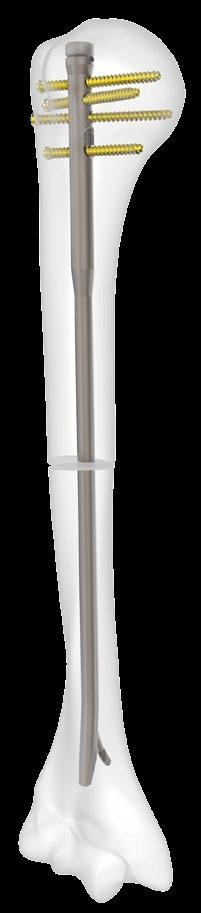
2 Introduction Content Humerus InSafeLOCK Nail is an innovative intramedullary nailing system, developed for humerus problems. Humerus fractures have 5-6 % incidence of all bone fractures. Nowadays, conservative and surgical methods are used for the treatment of humerus fractures. Other surgical options have negative effects as; plate-screw fixation, an oversized opening of soft tissue and radial nerves damage risk. Multiple Elastic Pins and unlocked nails (like Küntscher) have the risk of insufficient stability and dislocation Introduction Features Features of InSafeLOCK Nail Indications Surgical Technique Set Detail Implants Tray Tray Tray 3 Sample Cases Features Humerus InSafeLOCK Nail; Perforated and rounded, Titanium flexible and biocompatible, Usable for both right and left humerus fractures, No need to fluoroscopy for distal and proximal locking, New, ergonomic, easy instruments for insertionextraction and locking, Advantage of application as carved and non-carved, Humerus InSafeLOCK nail shaft diameter; 7 mm, 8 mm, 9 mm, Length options; 80, 00, 0, 40, 60, 80, 300 mm, Proximal diameter is 9 mm of all nails and 8 cm of proximal part length. 3 cm of the distal tip was angled 5 anteriorly for nail can be easily distracted and contributes to rotational stability. Warning: This description is not suf icient by itself for direct and proper use of the instrument set intraoperatively. Instruction by a surgeon who is thoroughly trained and experienced in handling these instruments and in doing the procedure are highly recommended. The distal part of the nail with oval shaped designed hole in the posterior direction, the hole is designed for endopin. No soft tissue damage at the distal part and no need to use fluoroscopy for the distal locking.
3 usable for both right and left Endopin (internally safe locking pin) Proximal diameter 9 mm Proximal length 8 cm threaded part 80, 00, 0, 40, 60, 80, 300 mm length choices elastic material Ti6Al7Nb 7,0 mm, 8,0 mm and 9,0 mm three different diameter choices special designed locking hole in the distalposterior of the nail no need to fluoroscopy for distal locking 5 angled distal part to anterior threaded endopin squeeze the posterior cortex and stuck the nail to anterior cortex posteriorly distal locking from inside to outside threaded part cannulated distally angled end, opposite locking hole on the posterior


4 4 locking holes in the proximal, three holes are static and the other one is dynamic. 4 mm diameter locking screws allow multiple locking in different planes at the humerus head. multiple locking choices at the different angles on the proximal 0 static transverse screw 8 angled static screw angled static screw dynamic compression screw calcar screw mm compression angled static screw Lateral targeting arm mm set screw compression screw end cap 5 5 static transverse, dynamic compression, calcar screw angled static screw Medial 3
, No distal locking screw loosening,")
5 Features of InSafeLOCK Nail Usable for both right and left, Can be placed to the most distal part (until the proximal edge of the olecranon fossa), No need to fluoroscopy or extra guide usage for distal locking, No distal locking complications as indicated below; No tissue incision, No radial nerves damaged (radial, median and musculocutaneous nerves), No distal locking screw loosening, migration and irritation problems, Shorter application period and operation time. Indications Proximal humerus fractures (, 3, 4 parts), Metaphyseal, Metaphyseal diafizer, and diaphyseal all fractures (3 cm from end of the olecranon fossa to proximal), Lengthening operations, over the nailing, In tumor resections, Shortening osteotomies, In deformity correction surgeries. Contraindications; Active systemic infection, Local infection at the entrance of the nail, Sensitivity and allergy of implant material, The bone is excessively contaminated of open fractures. 4
 After measuring the humerus canal, humerus length is determined through the Surgical Ruler before the pre-operative time.")
6 Surgical Technique Humerus InSafeLOCK Nail applied with the technique of antegrade operation DETERMINATION OF NAIL S DIAMETER AND LENGHT ARE: a) Sturdy humerus is measured digitally. length b) After measuring the humerus canal, humerus length is determined through the Surgical Ruler before the pre-operative time. diameter PATIENT POSITION: The patient is positioned supine or sunbed on the table. The fracture is reducted. 3 INCISION: It is reached to humerus top in 3 cm incision from acromion to distal with the anterolateral approach. 5


7 4 NAIL ENTRY POINT: Entering the medulla directly from the top of the head. Showed in the figure; Entry point of the nail is positioned posterolateral of biceps tendon between big tubercule and humerus head in the medial of the groove. AP LATERAL posterior lateral medial anterior Sulcus Biceps Tendon The K-wire 3X400 mm Ø should be placed metaphyseal in the medulla canal with fluoroscopic control. 6

.")

8 5 OPENING NAIL ENTRY POINT: Entry Reamer opened under the guidance of K-wire in the Entry Reamer Sleeve. All nail diameters are 9 mm. Entry Reamer is designed for this diameter. The nail holder diameter is mm and enlargement process is performed from top of the bone to 0 mm depth with the reamer ( mm). 6 SENDING THE GUIDE WIRE: The Long Guide Wire (Ø3X650 mm) is inserted into the intramedullary canal after opening the entry point if carving of diaphease will be done. Reduction Device can be used by fixated to T-handle in order to pass easily the fracture line and assist in reduction. guide wire pusher 7

9 7 DETERMINING NAIL LENGHT: The Guide Wire is transmitted to the distal humerus, the tip of the wire is positioned at the top of the olecranon fossa, with a reamer. The length of the nail is determined by Length Measuring Device of the wire outside the bone with the aid of length measurement device. Nail size should be 6-0 mm shorter than the measured size if compression is applied in the fracture line. Otherwise, there is a risk of overflowing the bone surface of the nail after compression. 8 UNREAMED TECHNIQUE: If the unreamed technical to be preferred; first of all Guide Wire (Ø X650 mm) should be placed in the canal and The nail should be attached to the Targeting Arm must be inserted into the canal via this wire. 8

 which is compatible with the inner diameter of the nail is placed through the Reamer while")
10 9 CARVED TECHNIQUE: The canal carving process is started through Guide Wire (Ø 3X650 mm) with the smallest Flexible Reamer (Ø 7 mm) and the size is continued by 0.5 mm increment. guide wire pusher While the Reamer is removing, the Guide Wire Pusher is used behind the reamer to prevent the Thick Guide Wire going back. The carving process continues until the cortex contact is felt. The nail diameter should be -.5 mm smaller than the last used reamer. 0 GUIDE-WIRE CHANGING: At the end of the canal carving process, Guide-Wire (Ø X650 mm) which is compatible with the inner diameter of the nail is placed through the Reamer while the last Reamer is in the humerus, the Guide Wire (Ø 3X650 mm) is removed. Ø 3X650 Ø X650 9
 or R (Right)). Nail Holder and nail are connected by Connection Screw.")
11 TARGETING DEVICE NAIL ASSEMBLY The determined nail is attached to the Targeting Arm as shown in the picture and the other instrument is checked. The laser marks on the top of the nail help to matchup the direction of the nail (L (Left) or R (Right)). Nail Holder and nail are connected by Connection Screw. The mm Wrench is used to tighten the bolt. The Targeting Arm is attached tightly to the Nail Holder with the Wrench. NAIL PLACEMENT: The nail that is set up to the Targeting Arm, is taken forward through as the distal tip is sloped to anteriorly straight (distal on the humerus), the channel from insertion site (through the guide wire in the channel). The nail is improved by hand and by rotational movements with passing the broken line. Posterior Anterior AP LATERAL 0
 is tightly connected to the Connection Screw by mm Wrench.")


12 3 NAILING: In the case of nailing needed, Connection Screw is attached to the threaded slot on the Nail Holder. Nail Hammer (inside threaded end) is tightly connected to the Connection Screw by mm Wrench. Nail clamping is applied with the Sliding Hammer which is located on the Sliding Hammer Impactor. This process should be done with soft impacts which will not damage the cortex. For slight forward movements; the process should be applied by hitting the nail and screw with U Hammer through K-wire. 4 NAIL PROXIMAL POSITION: Nail placement could be tracked with 0, 5, 0, 5 and 0 mm nail marks on the Nail Holder. With this aim, K-wire (Ø 3X50 mm) to be sent from 0 to 5 levels on the Targeting Arm, indicates the nail connection point (0 mm) and nail placement depth (5 mm). Regarding the levels of the proximal tip of the nail in the bone; If the compression needed the nail placement is applied at a depth of 0 mm from the bone surface. When the nail passes to the distal fragment, the Guide Wire is removed.


13 5 PREPARATION FOR DISTAL LOCKING: For distal locking, there is no need to use fluoroscopy or guide. Internal locking is performed without the incision. In this way, tissue nerve injury and aesthetic disadvantages that may occur are prevented in the patient, the duration of surgery will be significantly reduced. The elbow is placed in a supine position with a 90 flexion before the distal pin, which provides distal fixation, is delivered. Nail Holder should be target tub. Major in proximal and epithelium condyle in distal. For endopin holding point, posterior cortex is drilled through the top of the nail (through nail channel) with Ø 3X400 mm K-wire (with motor aid).


14 6 DISTAL LOCKING: Endopin,5x00 mm is sent through with the Screwdriver from the Connection Screw for Nail Holder insertion site. The grooves in the tip of the Endopin are matched with the grooves in the nail channel. Endopin is improved to distal tip by clockwise rotation of the inserter Final placement is determined by considering the laser mark on the inserter. Endopin; Distal nail tip makes compress to the anterior cortex and performs distal locking by providing attachment holding point of posterior cortex with grooved tip. laser mark 3


15 7 PROXIMAL LOCKING DEFINITION: The humeral nail could be used for non-stabilized or fragmented humerus fractures. Proximal locking, depending on the choice of the surgeon, performed with the aid of a guide through 4 screw holes designed at different angles. The targeting arm is designed to provide four options for locking over the nail in the proximal direction. These are; Static Transverse, Static Angle, Dynamic Compression and Calcar Screw. When necessary, the dynamic hole in the distal could be compressed to 6 mm by applying a transverse screw for compression purposes. Angled Static Screw Static Transverse Screw Dynamic Compression Screw Angled Static Screw Calcar Screw 4
16 PREPARATION: Static locking will help to protect the length of the bone and the rotational stability of the fracture. For this purpose, the Proximal Screw Guide with a Trocar inserted into the hole defined in the Targeting Arm is marked to skin. Following the incision, the soft tissue is passed and the contact with the bone is provided. The Trocar is removed, replaced with the Proximal Drill Guide and the hole is opened for the nail lock screw by drilling the lateral cortex to the appropriate depth with 3.x300 mm Measured Drill. On counter-cortex connection, the screw size is determined by the measure of the Drill Guide tip. If the direction of the drilling is towards the joint surface, the bone density is taken into consideration and care should be taken to avoid subchondral penetration. The use of scoping is recommended in this step. Depth Gauge (through the drill guide) could be used to determine the length of the proximal screws as an alternative. 8 PROXIMAL LOCKING: The Drill Guide is removed and the Ø 4 mm Proximal Locking Screw, which is determined via the Screw Guide, is inserted into the bone with the Screwdriver (Ø4X9x50 mm). For the exact placement, the laser mark on the screwdriver must be at the guide alignment. Other Proximal Locking Screws are applied following the same steps. At the humerus, the suitability and length of the screw sites are checked on both plans. 5


. Locking screws could be applied to the other proximal holes after this application.")
17 COMPRESSION TECHNIQUE: The distal fragment is approximated to the main fragment with 4 mm compression screws (or direct compression device) which is based on 4 mm screw inserted transversally through the oblong hole and the fracture gap is closed. 6 mm compression could be provided with the system. Compression could be done in 3 different ways; WITH COMPRESSION HEAD SCREW; Before implementation, the Targeting Arm should be separated. Compressed Head Screw is applied with 4x9x50 mm Hexagon Screwdriver. Locking screws could not be applied to the other proximal holes after this application. Compression Screw WITH SET SCREW; When the Set Screw is a small piece, it is placed to grooved canal in the nail with a.5x00 mm Screwdriver over the Targeting Screw. Then the compression is performed with a.5 x 00 mm Normal Screwdriver (due to risk of tip fracture). Locking screws could be applied to the other proximal holes after this application. The process is completed by inserting the appropriate head bolt after the nail has been detected. Set Screw WITH THREADED COMPRESSION SHAFTS Threaded Compression Shaft is applied over the Targeting Arm by attaching to the Screwdriver. Compression is performed paying attention to the laser marks on Compression Shaft. During this application, temporary detection of the proximal fragment is provided with the drill bit sent to the static hole without the Compression Shaft removed. After Compression Shaft is removed, the lock screws are applied from the other proximal holes Trying to improve more than 6 mm while applying compression, or applying too much force could cause bending of the transverse going screw in the oblong hole. It is recommended that this procedure be performed at the time of scopy control. The process is completed by attaching the appropriate top screw after the nail detection is done. Threaded Compression Shaft 6
18 9 CLOSING THE WOUND: Incision wound is closed by using the standard technique. 0 REMOVING NAIL PROCESS: The removal operation should be performed by applying the reverse of the nail placement procedure. The End Cap is removed. The connection is made between Targeting Arm and the nail. The screws located in the proximal part of the nail are removed. If the set screw is available, must be removed before the compression screw. Endopin is reached from the top of the Targeting Arm using a Screwdriver Ø.5X00 mm. The Endopin should provide a complete fitting with the Screwdriver recess by turning the screwdriver clockwise and Endopin in the nail channel is removed. Ø.5X00 mm Screwdriver with holder tip could be used in order to catch and remove the Endopin which is released in the nail inner channel during removal. The Nail Extractor is connected to the nail by Connection Screw for Nail Extractor. The Nail is removed from the humerus channel by Hammer which is located on Sliding Hammer Rod. When extra power required during removal, Sliding U Hammer could be used over the Nail Extractor. 7
19 Set Detail Implants Code Barcode Description Qty HIN - HUMERUS INSAFELOCK NAIL Ø 7.0 X 80 MM HIN - HUMERUS INSAFELOCK NAIL Ø 7.0 X 00 MM HIN - HUMERUS INSAFELOCK NAIL Ø 7.0 X 0 MM HIN - HUMERUS INSAFELOCK NAIL Ø 7.0 X 40 MM HIN - HUMERUS INSAFELOCK NAIL Ø 7.0 X 60 MM HIN - HUMERUS INSAFELOCK NAIL Ø 7.0 X 80 MM HIN - HUMERUS INSAFELOCK NAIL Ø 7.0 X 300 MM HIN - HUMERUS INSAFELOCK NAIL Ø 8.0 X 80 MM HIN - HUMERUS INSAFELOCK NAIL Ø 8.0 X 00 MM HIN - HUMERUS INSAFELOCK NAIL Ø 8.0 X 0 MM HIN - HUMERUS INSAFELOCK NAIL Ø 8.0 X 40 MM HIN - HUMERUS INSAFELOCK NAIL Ø 8.0 X 60 MM HIN - HUMERUS INSAFELOCK NAIL Ø 8.0 X 80 MM HIN - HUMERUS INSAFELOCK NAIL Ø 8.0 X 300 MM HIN - HUMERUS INSAFELOCK NAIL Ø 9.0 X 80 MM HIN - HUMERUS INSAFELOCK NAIL Ø 9.0 X 00 MM HIN - HUMERUS INSAFELOCK NAIL Ø 9.0 X 0 MM HIN - HUMERUS INSAFELOCK NAIL Ø 9.0 X 40 MM HIN - HUMERUS INSAFELOCK NAIL Ø 9.0 X 60 MM HIN - HUMERUS INSAFELOCK NAIL Ø 9.0 X 80 MM HIN - HUMERUS INSAFELOCK NAIL Ø 9.0 X 300 MM HIN INSAFELOCK ENDOPIN SCREW 80 MM HIN INSAFELOCK ENDOPIN SCREW 00 MM HIN INSAFELOCK ENDOPIN SCREW 0 MM HIN INSAFELOCK ENDOPIN SCREW 40 MM HIN INSAFELOCK ENDOPIN SCREW 60 MM HIN INSAFELOCK ENDOPIN SCREW 80 MM HIN INSAFELOCK ENDOPIN SCREW 300 MM 8
20 Tray No Code Barcode Description Qty HIN.DESIGN TRAY THREADED COMPRESSION SHAFT GRADUATED DRILL BIT Ø3.X300 MM SCREW DRIVER QUICK TIP.5X00 MM SCREW DRIVER QUICK TIP HOLDER.5X00 MM SCREW DRIVER QUICK HEX. TIP 4X9x50 MM SOFT SCREW DRIVER QUICK LARGE HUM. NAIL PROXIMAL SCREW SLEEVE HUM. NAIL PROXIMAL DRILL GUIDE HUM. NAIL TROCAR DEPTH GAUGE - HIN PROX. SCREW LENGTH HIN LOCK. SCREW BOX CORTEX SCREW FOR NAILS TI Ø4.0X8 MM CORTEX SCREW FOR NAILS TI Ø4.0X0 MM CORTEX SCREW FOR NAILS TI Ø4.0X MM CORTEX SCREW FOR NAILS TI Ø4.0X4 MM CORTEX SCREW FOR NAILS TI Ø4.0X6 MM CORTEX SCREW FOR NAILS TI Ø4.0X8 MM CORTEX SCREW FOR NAILS TI Ø4.0X30 MM CORTEX SCREW FOR NAILS TI Ø4.0X3 MM CORTEX SCREW FOR NAILS TI Ø4.0X34 MM CORTEX SCREW FOR NAILS TI Ø4.0X36 MM CORTEX SCREW FOR NAILS TI Ø4.0X38 MM CORTEX SCREW FOR NAILS TI Ø4.0X40 MM CORTEX SCREW FOR NAILS TI Ø4.0X4 MM CORTEX SCREW FOR NAILS TI Ø4.0X44 MM CORTEX SCREW FOR NAILS TI Ø4.0X46 MM CORTEX SCREW FOR NAILS TI Ø4.0X48 MM CORTEX SCREW FOR NAILS TI Ø4.0X50 MM CORTEX SCREW FOR NAILS TI Ø4.0X5 MM CORTEX SCREW FOR NAILS TI Ø4.0X54 MM CORTEX SCREW FOR NAILS TI Ø4.0X56 MM CORTEX SCREW FOR NAILS TI Ø4.0X58 MM CORTEX SCREW FOR NAILS TI Ø4.0X60 MM CORTEX SCREW FOR NAILS TI Ø4.0X6 MM CORTEX SCREW FOR NAILS TI Ø4.0X64 MM CORTEX SCREW FOR NAILS TI Ø4.0X66 MM CORTEX SCREW FOR NAILS TI Ø4.0X68 MM CORTEX SCREW FOR NAILS TI Ø4.0X70 MM HIN END CAP SCREW BOX HUMERUS INSAFELOCK NAIL SET SCREW HUMERUS INSAFELOCK LOCK. COMPRESSION SCREW (TI) HUMERUS INSAFELOCK END CAP (TI) STANDARD HUMERUS INSAFELOCK END CAP (TI) + 5 MM HUMERUS INSAFELOCK END CAP (TI) + 0 MM HUMERUS INSAFELOCK END CAP (TI) + 5 MM HUMERUS INSAFELOCK END CAP (TI) + 0 MM 9


21


22 Tray No Code Barcode Description Qty HIN.DESIGN TRAY FLEXIBLE REAMER Ø 7 FLEXIBLE REAMER Ø 7.5 FLEXIBLE REAMER Ø 8 FLEXIBLE REAMER Ø 8.5 FLEXIBLE REAMER Ø 9 FLEXIBLE REAMER Ø 9.5 FLEXIBLE REAMER Ø 0 FLEXIBLE REAMER Ø 0.5 HUM. NAIL ENTRY REAMER INTRAMED. NAIL ENTRY REAMER SLEEVE Ø0X80MM INTRAMED. NAIL CANNULE AWL PUSH ROD HUM. NAIL REDUCTION DEVICE INTRAMED. NAIL K-WIRE GUIDE Ø3X05MM T QUICK HANDLE


23 Tray 3 No Code Barcode Description Qty HIN 3.DESIGN TRAY CONNECTION SCREW FOR HUM. NAIL HOLDER CONNECTION SCREW FOR HUM. NAIL EXTRACTOR SURGICAL RULER S.S 300 MM GUIDE WIRE PUSHER K-WIRE TUBE ØXØ8X60 MM KIRSCHNER WIRE TROCAR POINT 3X50 MM K-WIRE TUBE ØXØ8X400 MM KIRSCHNER WIRE TROCAR POINT Ø3X400 MM HUM. NAIL HOLDER HUM. NAIL TARGETING ARM HUM. NAIL LENGTH MEASURING DEVICE SLIDING NAIL IMPACTOR&EXTRACTOR HIN SLIDING HAMMER SLIDING HAMMER STOPPER HIN SLIDING U-HAMMER WRENCH MM T SCREW DRIVER Ø 5 MM CONTAINER 560x70x70 GUIDE WIRE X650 MM GUIDE WIRE 3X650 MM


24 Sample Cases Case follow-up post-op pre-op


25 Case follow-up post-op pre-op


26 Case 3 follow-up post-op pre-op day 45 day 85


27 Case 4 follow-up pre-op post-op day 7 day 54
28 REV00/ TST Medical Devices reserves the right to update, change or modify contents and products at any time without any notice or liability.
LOCKING TEP LOCKING TITANIUM ELASTIC PIN INTRAMEDULLARY NAIL
 LOCKING TEP LOCKING TITANIUM ELASTIC PIN INTRAMEDULLARY NAIL ... Index -3 3-8 8 9 9 0 7 Introduction Features Indicatiıons Surgical Technique Femoral Surgical Technique Tibial Surgical Technique Ulna Radius
PATENTED A-PFN. Antirotator Proximal Femoral Nail. Medical Devices
 PATENTED A-PFN Antirotator Proximal Femoral Nail Medical Devices Introductions Intertrochanteric femoral fractures constitute 0% of all the bone fractures. They are frequently seen in elderly patients
INTRAMEDULLARY. Medical Devices. Femur Intramedullary Nail
 FEMUR INTRAMEDULLARY Femur Intramedullary Nail Multi-functional Standard and Recon Antegrade and Retrograde Application Different Multi-Planner Locking Choices Medical Devices Introductions The new, multifunctional
Orthopedic Bone Nail System - Distal Femoral Nail Surgical Technique Manual
 Orthopedic Bone Nail System - Distal Femoral Nail Surgical Technique Manual Note: The surgical procedures should be performed under the guidance of qualified skilled orthopedic surgeons, and this surgical
Double Engine Orthopedic Bone Nail System Universal Humeral Nail
 Double Engine Orthopedic Bone Nail System ----------- Universal Humeral Nail Surgical Technique Manual Note: The surgical procedures should be performed under the guidance of qualified skilled orthopedic
System. Humeral Nail. Surgical Technique
 System Humeral Nail Surgical Technique Contents IMPLANT FEATURES 2 1. INDICATIONS 3 2. PRE-OPERATIVE PLANNING 3 3. PATIENT POSITIONING & FRACTURE REDUCTION 3 4. INCISION 4 5. ENTRY POINT 4-6 6. PROXIMAL
3. PATIENT POSITIONING & FRACTURE REDUCTION 3 8. DISTAL GUIDED LOCKING FOR PROXIMAL NAIL PROXIMAL LOCKING FOR LONG NAIL 13
 Contents IMPLANT FEATURES 2 1. INDICATIONS 3 2. PRE-OPERATIVE PLANNING 3 3. PATIENT POSITIONING & FRACTURE REDUCTION 3 4. INCISION 4 5. ENTRY POINT 4-6 6. PROXIMAL NAIL INSERTION 6-7 7. PROXIMAL LOCKING
A locking plate system that expands a surgeon s options in trauma surgery. Zimmer NCB Plating System
 A locking plate system that expands a surgeon s options in trauma surgery Zimmer NCB Plating System The Power of Choice The power of having true intraoperative options is at your fingertips. Using standard
LCP Distal Humerus Plates
 The anatomic fixation system for the distal humerus with angular stability Surgical technique LCP Locking Compression Plate Contents Indications and contraindications 2 Implants 3 Instruments 5 Preparation
LCP Medial Distal Tibia Plate, without Tab. The Low Profile Anatomic Fixation System with Angular Stability and Optimal Screw Orientation.
 LCP Medial Distal Tibia Plate, without Tab. The Low Profile Anatomic Fixation System with Angular Stability and Optimal Screw Orientation. Technique Guide LCP Small Fragment System Table of Contents Introduction
NeoGen Tibia Nail System
 NeoGen Tibia Nail System LESS IS MORE TE-2070-03 Surgical Technique BLE OF CONTENT Preface Surgical Technique Appendix Products Information Patient Preparation Entry Portal Fracture Reduction Canal Preparation
Zimmer ITST Intertrochanteric/ Subtrochanteric Fixation System. Abbreviated Surgical Technique
 Zimmer ITST Intertrochanteric/ Subtrochanteric Fixation System Abbreviated Surgical Technique ITST System Abbreviated Surgical Technique Indications The ITST Intramedullary Nail is indicated for use in
NeoGen Femoral Nail System
 NeoGen Femoral Nail System LESS IS MORE TE-2070-04 Surgical Technique BLE OF CONTENT Preface Standard Femoral Mode Recon Mode Post-Operative Management Appendix Products Information Indication Patient
The Titanium Tibial Nail System
 The Titanium Tibial Nail System Solid Tibial Nails (UTN) and Cannulated Tibial Nails (CTN) Surgical Technique This publication is not intended for distribution in the USA. Instruments and implants approved
3. Insert Tocar Sleeves Insert the NCB tissue protection sleeve assembly 1.6 to 10mm through a skin incision (Fig. 38).
.") NCB Proximal Humerus Plating System Surgical Technique 19 2. Temporary Plate Fixation The plate can be temporary fixed to the bone with 1.6mm K-wire through the proximal cannulated fixation screw of the
NCB Proximal Humerus Plating System Surgical Technique 19 2. Temporary Plate Fixation The plate can be temporary fixed to the bone with 1.6mm K-wire through the proximal cannulated fixation screw of the
TRAUMATOLOGY. Trochanter
 TRAUMATOLOGY Trochanter 1 References Prof. Dr. András Sárváry Head of Department Fővárosi Önkormányzat Péterfy Sándor úti Kórház Rendelőintézet és Baleseti Központ Budapest The following surgical description
Surgical Technique. Intramedullary locked Nailing With Screws for Humerus Fractures Solid/Cannulated. Humeral Interlocking Nail.
 Screws for Humerus Fractures Surgical Technique Humeral Interlocking Nail Approved by Humerus Nail Kit Code 08050001 Contents Introduction Implant design Indications Pre-operative planning Patient positioning
Technique Guide. 3.5 mm LCP Olecranon Plates. Part of the Synthes locking compression plate (LCP) system.
 system.") Technique Guide 3.5 mm LCP Olecranon Plates. Part of the Synthes locking compression plate (LCP) system. Table of Contents Introduction 3.5 mm LCP Olecranon Plates 2 AO Principles 3 Indications 3 Clinical
Technique Guide 3.5 mm LCP Olecranon Plates. Part of the Synthes locking compression plate (LCP) system. Table of Contents Introduction 3.5 mm LCP Olecranon Plates 2 AO Principles 3 Indications 3 Clinical
Sirus Antegrade Femoral Nail System Surgical Technique
 Sirus Antegrade Femoral Nail System Surgical Technique The Cannulated Titanium Nail with Anatomical Shape and Lateral Entry Point Disclaimer This document is intended exclusively for experts in the field,
LCP Distal Tibia Plate
 Surgical Technique LCP Locking Compression Plate Original Instruments and Implants of the Association for the Study of Internal Fixation AO/ASIF Table of contents Indications 3 Implants/Instruments 5 Surgical
A locking plate system that expands a surgeon s options in trauma surgery. Zimmer NCB Plating System
 A locking plate system that expands a surgeon s options in trauma surgery Zimmer NCB Plating System The Power of Choice The power of having true intraoperative options is at your fingertips. Using standard
Zimmer Natural Nail System
 Zimmer Natural Nail System Antegrade Femoral Nail Surgical Technique (Piriformis Fossa & Greater Trochanteric Approaches) Zimmer Natural Nail System Antegrade Femoral Surgical Technique 1 Zimmer Natural
TITANIUM TIBIAL NAIL SySTEM
 TITANIUM TIBIAL NAIL SySTEM Solid and Cannulated Nails SURGICAL TEChNIqUE Table of contents Introduction Indications 2 Preoperative Implant Selection 6 Surgical Technique Instruments for Opening the Tibia
Table of contents. Introduction... 3 Indications... 3 Contraindications... 3 TRIGEN Humeral Nail Specifications... 6
 Surgical Technique Table of contents Introduction... 3 Indications... 3 Contraindications... 3 TRIGEN Humeral Nail Specifications... 6 Surgical Technique Patient positioning... 7 Establish the incision
Surgical Technique. CONQUEST FN Femoral Neck Fracture System
 Surgical Technique CONQUEST FN Femoral Neck Fracture System Table of Contents Introduction... 3 Indications... 3 Product Overview... 4 Surgical Technique... 5 Patient Positioning... 5 Reduce the Fracture...
Olecranon Osteotomy Nail. For simple fractures and osteotomies of the olecranon.
 Olecranon Osteotomy Nail. For simple fractures and osteotomies of the olecranon. Technique Guide Discontinued June 2016; AVAILABLE FOR IMPLANT REMOVAL PURPOSES ONLY DSEM/TRM/0517/0843 Table of Contents
Surgical Technique. Forearm Fracture Solutions
 Surgical Technique Forearm Fracture Solutions Acumed is a global leader of innovative orthopaedic and medical solutions. We are dedicated to developing products, service methods, and approaches that improve
GREENS SURGICALS. Redefining Excellence INSTRUMENT SYSTEM PREPARED BY: DR. VINAY KUMAR
 GREENS SURGICALS Redefining Excellence TIBIA AND FEMUR INSTRUMENT SYSTEM PREPARED BY: DR. VINAY KUMAR OPERATIVE TECHNIQUES INDEX SR.NO CONTENTS 1 LIST OF INSTRUMENT FOR TIBIA AND FEMUR. 2 RADIO GRAPH OF
Surgical Technique. Fibula Rod System
 Surgical Technique Fibula Rod System Acumed is a global leader of innovative orthopaedic and medical solutions. We are dedicated to developing products, service methods, and approaches that improve patient
Titanium Solid Humeral Nail System
 For Antegrade or Retrograde Insertion With Spiral Blade, Conventional and Compression Locking Titanium Solid Humeral Nail System Surgical Technique Table of Contents INTRODUCTION Foreword.... 2 Indications....
Olecranon Locking Plate II
 INDEX Indications Patient Position Fracture Reduction and Fixation Surgical Technique Step 1 Surgical Approach Step 2 Implantation Step 3 Proximal Locking Screw Insertion Step 4 Distal Screw Insertion
Technique Guide. 3.5 mm LCP Periarticular Proximal Humerus Plate. Part of the Synthes locking compression plate (LCP) system.
 system.") Technique Guide 3.5 mm LCP Periarticular Proximal Humerus Plate. Part of the Synthes locking compression plate (LCP) system. Table of Contents Introduction 3.5 mm LCP Proximal Humerus Plate 2 AO Principles
Technique Guide 3.5 mm LCP Periarticular Proximal Humerus Plate. Part of the Synthes locking compression plate (LCP) system. Table of Contents Introduction 3.5 mm LCP Proximal Humerus Plate 2 AO Principles
OPERATING MANUAL AND TECHNIQUE GUIDE FOR TITANIUM FEMORAL AND TIBIAL NAILING SYSTEMS
 OPERATING MANUAL AND TECHNIQUE GUIDE FOR TITANIUM FEMORAL AND TIBIAL NAILING SYSTEMS ORTHO-MEDICAL GMBH TITANIUM FEMORAL NAIL OPERATIVE TECHNIQUE Introduction: Why a new type of femoral nail? The latest
3.5 mm LCP Olecranon Plates
 Part of the DePuy Synthes Locking Compression Plate (LCP ) System 3.5 mm LCP Olecranon Plates Surgical Technique Table of Contents Introduction 3.5 mm LCP Olecranon Plates 2 AO Principles 3 Indications
Technique Guide. 3.5 mm LCP Low Bend Medial Distal Tibia Plate Aiming Instruments. Part of the 3.5 mm LCP Percutaneous Instrument System.
 Technique Guide 3.5 mm LCP Low Bend Medial Distal Tibia Plate Aiming Instruments. Part of the 3.5 mm LCP Percutaneous Instrument System. Table of Contents Introduction 3.5 mm LCP Low Bend Medial Distal
LCP Anterolateral Distal Tibia Plate 3.5. The low profile anatomic fixation system with optimal plate placement and angular stability.
 LCP Anterolateral Distal Tibia Plate 3.5. The low profile anatomic fixation system with optimal plate placement and angular stability. Technique Guide LCP Small Fragment System Table of Contents Introduction
U2 PSA. Revision Knee. Surgical Protocol
 U2 PSA TM Revision Knee Surgical Protocol Table of Contents 1 Component Removal... 1 2 Tibial Preparation... 1 2.1 Tibial Canal Preparation... 1 2.2 Proximal Tibial Resection... 2 2.3 Non Offset Tibial
Distal Cut First Femoral Preparation
 Surgical Technique Distal Cut First Femoral Preparation Primary Total Knee Arthroplasty LEGION Total Knee System Femoral preparation Contents Introduction...3 DCF femoral highlights...4 Preoperative planning...6
TIBIAL NAILING SYSTEM OPTIONS MADE EASY
 S U R G I C A L T E C H N I Q U E TIBIAL NAILING SYSTEM OPTIONS MADE EASY TABLE OF CONTENTS DESIGN SUMMARY INSTRUMENT OVERVIEW AND JIG OPTIONS 1 2 ENTRY AND CANAL PREP NAIL INSERTION LOCKING NAIL REMOVAL
Conventus CAGE PH Surgical Techniques
 Conventus CAGE PH Surgical Techniques Conventus Orthopaedics The Conventus CAGE PH (PH Cage) is a permanent implant comprised of an expandable scaffold, made from nitinol and titanium, which is deployed
TABLE OF CONTENTS. 2 (8144 Rev 2)
") 1 (8144 Rev 2) TABLE OF CONTENTS Introduction Conventus CAGE TM - Proximal Humerus...3 Indications and Contraindications...4 Surgical Summary...5 Patient Positioning & Approach...6 Surgical Technique Plate
1 (8144 Rev 2) TABLE OF CONTENTS Introduction Conventus CAGE TM - Proximal Humerus...3 Indications and Contraindications...4 Surgical Summary...5 Patient Positioning & Approach...6 Surgical Technique Plate
3.5 mm LCP Extra-articular Distal Humerus Plate
 Part of the DePuy Synthes Locking Compression Plate (LCP ) System 3.5 mm LCP Extra-articular Distal Humerus Plate Surgical Technique Table of Contents Introduction 3.5 mm LCP Extra-articular Distal Humerus
LCP Anterolateral Distal Tibia Plate 3.5. The low profile anatomic fixation system with optimal plate placement and angular stability.
 LCP Anterolateral Distal Tibia Plate 3.5. The low profile anatomic fixation system with optimal plate placement and angular stability. Technique Guide LCP Small Fragment System Table of Contents Introduction
LCP Low Bend Medial Distal Tibia Plates 3.5 mm. Anatomic plates with low profile head for intra- and extraarticular fractures.
 LCP Low Bend Medial Distal Tibia Plates 3.5 mm. Anatomic plates with low profile head for intra- and extraarticular fractures. Surgical Technique This publication is not intended for distribution in the
Fibula Rod System. Lateral Malleolus Fracture Indications:
 Fibula Rod System Fibula Rod System Since 1988, Acumed has been designing solutions for the demanding situations facing orthopaedic surgeons, hospitals and their patients. Our strategy has been to know
Phoenix Tibial Nail System
 Surgical Technique Phoenix Tibial Nail System Featuring CoreLock Technology Each nail features CoreLock Technology, a preassembled, embedded locking mechanism for locking all proximal oblique screws, which
Surgical Technique 1
 Surgical Technique 1 TRIGEN META-NAIL Semi-extended Instrument Set Surgical Technique Table of Contents Indications... 2... TRIGEN META-NAIL Tibial Nail Specifications... 3 Instruments for opening the
Humeral Nails With Multiple Locking Options for Simple and Complex Fractures. MultiLoc. Humeral Nails. Surgical Technique
 Humeral Nails With Multiple Locking Options for Simple and Complex Fractures MultiLoc Humeral Nails Surgical Technique Table of Contents Introduction MultiLoc Humeral Nails 2 Screw Configurations 4 AO
Surgical Technique.
 Surgical Technique www.biomet.co.uk INTRODUCTION design principals Recent advances in imaging technology have enabled orthopaedic surgeons to extend closed treatment of femoral fractures to include more
AcUMEDr. FoREARM ROD SYSTEM
 AcUMEDr FoREARM ROD SYSTEM FoREARM ROD SYSTEM Since 1988 Acumed has been designing solutions to the demanding situations facing orthopedic surgeons, hospitals and their patients. Our strategy has been
Technique Guide. 3.5 mm LCP Low Bend Medial Distal Tibia Plates. Part of the Synthes locking compression plate (LCP) system.
 system.") Technique Guide 3.5 mm LCP Low Bend Medial Distal Tibia Plates. Part of the Synthes locking compression plate (LCP) system. Table of Contents Introduction 3.5 mm LCP Low Bend Medial Distal Tibia Plates
Technique Guide 3.5 mm LCP Low Bend Medial Distal Tibia Plates. Part of the Synthes locking compression plate (LCP) system. Table of Contents Introduction 3.5 mm LCP Low Bend Medial Distal Tibia Plates
TRK REVISION KNEE Surgical Technique
 1 TRK REVISION KNEE Surgical Technique 1. 2. 3. 4. 5. 6. 7. 8. 9. 10. INTERCONDYLAR RESECTION...... page FEMORAL STEM...... page NON CEMENTED FEMORAL STEM...... page TRIAL FEMORAL COMPONENTS...... page
NCB Distal Femur System. Surgical Technique
 NCB Distal Femur System Surgical Technique NCB Distal Femur System Surgical Technique 3 Surgical Technique NCB Distal Femur System Table of Contents Introduction 4 Indications 8 Preoperative Planning
Zimmer Periarticular Proximal Humeral Locking Plate
 Zimmer Periarticular Proximal Humeral Locking Plate Surgical Technique The Science of the Landscape Zimmer Periarticular Proximal Humeral Locking Plate 1 Surgical Technique Table of Contents Introduction
Suprapatellar Instrumentation for Titanium Cannulated Tibial Nail. Expert nailing system.
 Suprapatellar Instrumentation for Titanium Cannulated Tibial Nail. Expert nailing system. Technique Guide Expert Nailing System Table of Contents Introduction Suprapatellar Instrumentation for Titanium
Zimmer MIS Periarticular 3.5mm Proximal Tibial Locking Plate
 Zimmer MIS Periarticular 3.5mm Proximal Tibial Locking Plate Surgical Technique The Science of the Landscape Zimmer MIS Periarticular 3.5mm Proximal Tibial Locking Plate Surgical Technique 1 Zimmer MIS
PediNail Pediatric Femoral Nail
 PediNail Pediatric Femoral Nail Surgical Technique Table of Contents Indications...3 Patient Positioning...3 Approach...4 Reaming...5 Nail Placement...6 Proximal Interlocking...7 Distal Interlocking...8
pediatric orthopedic implants fixator
 pediatric orthopedic implants DDDAF fixator DAF HUMERUS FIXATOR It is a single plan external fixation tool and used for the treatment of the fractures of humerus, radius and ulna for adults. It is also
Medical Devices HUMERUS. WRIStX
 Medical Devices DDDAF HUMERUS WRIStX DAF HUMERUS FIXATOR It is a single plan external fixation tool and used for the treatment of the fractures of humerus, radius and ulna for adults. It is also used at
Surgical technique. UHN/PHN Humeral Nailing System.
 Surgical technique UHN/PHN Humeral Nailing System. 358.590 Radiographic Ruler for UHN 292.260 Kirschner Wire 2.5 mm with trocar tip, length 280 mm, Stainless Steel 351.120 Awl with T-Handle, cannulated,
Biomet Pediatric Locking Nail System
 Innovation Meets Evolution...EBI Trauma is now: Biomet Pediatric Locking Nail System Surgical Technique Contents Introduction...................... Page 1 Design Rationale................... Page 2 Patient
Indications TRIGEN META-NAIL Tibial Nail Specifications... 3 Instruments for opening the proximal tibia... 4
 Surgical Technique TRIGEN META-NAIL Semi-extended Instrument Set Surgical Technique Table of Contents Indications... 2... TRIGEN META-NAIL Tibial Nail Specifications... 3 Instruments for opening the proximal
Surgical Technique. Cannulated Angled Blade Plate 3.5 and 4.5, 90
 Surgical Technique Cannulated Angled Blade Plate 3.5 and 4.5, 90 Cannulated Angled Blade Plate 3.5 and 4.5, 90 Table of contents Indications/Contraindications 2 Implants 3 Surgical technique 5 Implant
Technique Guide. PHILOS and PHILOS Long. The anatomic fixation system for the proximal humerus.
 Technique Guide PHILOS and PHILOS Long. The anatomic fixation system for the proximal humerus. Table of Contents Introduction PHILOS and PHILOS Long 2 AO Principles 4 Indications 5 Surgical Technique
M/DN Tibial and Humeral Intramedullary Fixation. Surgical Technique
 M/DN Tibial and Humeral Intramedullary Fixation Surgical Technique M/DN Tibial and Humeral Intramedullary Fixation Surgical Technique 1 Surgical Techniques for Fixation of Fractures with M/DN Tibial and
OBSOLETED. LCP Medial Distal Tibia Plate, without Tab. The Low Profile Anatomic Fixation System with Angular Stability and Optimal Screw Orientation.
 LCP Medial Distal Tibia Plate, without Tab. The Low Profile Anatomic Fixation System with Angular Stability and Optimal Screw Orientation. Surgical Technique LCP Small Fragment System This publication
Surgical Technique Guide PANTERA. Proximal Humerus Fracture Fixation Plate System
 Surgical Technique Guide PANTERA Proximal Humerus Fracture Fixation Plate System Installing the PANTERA is a 4-Step Process: The following technique is designed to optimize the surgical exercise. Step
Operasjonsteknikk. Tibia
 Operasjonsteknikk Tibia TRIGEN META-NAIL Tibial Nail System Surgical Technique Table of Contents Indications...2 Implant Specifications...3 Surgical Technique Patient Positioning...4 Incision and Entry
Technique Guide. SureLock Distal Targeting Device. C-arm guided targeting for trochanteric fixation nail.
 Technique Guide SureLock Distal Targeting Device. C-arm guided targeting for trochanteric fixation nail. Table of Contents Introduction SureLock Distal Targeting Device 2 Surgical Technique Preoperative
T2 Tibial. Tibial Fractures. Nailing System. Operative Technique
 T2 Tibial Nailing System Tibial Fractures Operative Technique T2 Tibial Nailing System Contributing Surgeons Prof. Dr. med. Volker Bühren Chief of Surgical Services Medical Director of Murnau Trauma Center
Expert A2FN. Designed for small statured patients.
 Expert A2FN. Designed for small statured patients. Technique Guide Expert Nailing System This publication is not intended for distribution in the USA. Instruments and implants approved by the AO Foundation
Surgical Technique. Anterolateral and Medial Distal Tibia Locking Plates
 Surgical Technique Anterolateral and Medial Distal Tibia Locking Plates PERI-LOC Periarticular Locked Plating System Anterolateral and Medial Distal Tibia Locking Plates Surgical Technique Contents Product
Zimmer NexGen MIS Tibial Component. Cemented Surgical Technique IMAGE TO COME
 Zimmer NexGen MIS Tibial Component Cemented Surgical Technique IMAGE TO COME Zimmer NexGen MIS Tibial Component Cemented Surgical Technique 1 Zimmer NexGen MIS Tibial Component Cemented Surgical Technique
UHN/PHN Humeral Nailing System.
 UHN/PHN Humeral Nailing System. Surgical Technique This publication is not intended for distribution in the USA. Instruments and implants approved by the AO Foundation. 358.590 Radiographic Ruler for
operative technique Kent Hip
 operative technique Kent Hip The Kent Hip Operative Technique The Kent Hip was developed by Mr Cliff Stossel, FRCS in Maidstone, Kent, UK and first implanted in 1986. It was designed to deal with problems
Humeral SuturePlate. Surgical Technique
 Humeral SuturePlate Surgical Technique The humeral SuturePlate is an anatomically designed, low profile, titanium polyaxial locking plate and screw system. Multiple chamfered suture eyelets along the margin
Suprapatellar Instrumentation for Expert Tibial Nail.
 Suprapatellar Instrumentation for Expert Tibial Nail. Surgical Technique EXPERT Nailing System This publication is not intended for distribution in the USA. Instruments and implants approved by the AO
Locking Radial Head Plates
 Locking Radial Head Plates Locking Radial Head Plates Since 1988, Acumed has been designing solutions to the demanding situations facing orthopaedic surgeons, hospitals and their patients. Our strategy
S U R G I C A L T E C H N I Q U E David A. McQueen, MD Return to Menu
 S U R G I C A L T E C H N I Q U E David A. McQueen, MD TOTAL KNEE INSTRUMENTS Wichita Fusion Nail Introduction...1 Preoperative Planning...2 Surgical Technique...3-8 Wichita Fusion Nail Surgical Technique
Expert A2FN. Designed for small statured patients.
 Expert A2FN. Designed for small statured patients. Surgical Technique Expert Nailing System This publication is not intended for distribution in the USA. Instruments and implants approved by the AO Foundation.
Mandible External Fixator II. Provides treatment for fractures of the maxillofacial area.
 Mandible External Fixator II. Provides treatment for fractures of the maxillofacial area. Technique Guide This publication is not intended for distribution in the USA. Instruments and implants approved
Aesculap Targon TX. Intramedullary Nail for Tibial Fractures...going to X-tremes. Aesculap Orthopaedics
 Aesculap Targon TX Intramedullary Nail for Tibial Fractures...going to X-tremes Aesculap Orthopaedics Surgical Technique Preoperative Planning KH483 X-ray template Targon TX (KH483200 : 375 mm-420 mm KH48320
Zimmer MIS Periarticular Distal Femoral Locking Plate
 For Clinical Evaluations Zimmer MIS Periarticular Distal Femoral Locking Plate Surgical Technique The Science of the Landscape Zimmer MIS Periarticular Distal Femoral Locking Plate Surgical Technique
For Distal Femur Fractures. 95º Condylar Plate. Quick Reference Chart
 For Distal Femur Fractures 95º Condylar Plate Quick Reference Chart 95 Condylar Plate. Quick reference chart for distal femur fractures. Insert guide wires Fix condylar fragments with 6.5 mm cancellous
PROXIMAL HUMERAL NAILING SYSTEM OPERATIVE TECHNIQUE
 PROXIMAL HUMERAL NAILING SYSTEM OPERATIVE TECHNIQUE PROXIMAL HUMERAL NAILING SYSTEM Contributing Surgeons: Rupert Beikert, M.D. Senior Trauma Surgeon, Murnau Trauma Center Murnau, Germany Rosemary Buckle,
Surgical Technique. Proximal Humerus Locking Plate
 Surgical Technique Proximal Humerus Locking Plate PERI-LOC Upper Extremity Locked Plating System 3.5mm & 4.5mm Proximal Humerus Locking PlatesCatalog Infor Table of Contents Introduction.........................................................2
Technique Guide. Locking Attachment Plate. For treatment of periprosthetic fractures.
 Technique Guide Locking Attachment Plate. For treatment of periprosthetic fractures. Table of Contents Introduction Locking Attachment Plate 2 Indications 4 Surgical Technique Patient Positioning 5 Preparation
Triathlon TS Knee System. Surgical Protocol
 Triathlon TS Knee System Surgical Protocol Triathlon TS Knee System Surgical Protocol Table of Contents Acknowledgments..........................................................2 Exposure...................................................................4
Arcos Interlocking Distal Stem. Surgical Technique Addendum to the Arcos Modular Femoral Revision System
 Arcos Interlocking Distal Stem Surgical Technique Addendum to the Arcos Modular Femoral Revision System One Surgeon. One Patient. Over 1 million times per year, Biomet helps one surgeon provide personalized
3.5 MM VA-LCP PROXIMAL TIBIA PLATE SYSTEM
 3.5 MM VA-LCP PROXIMAL TIBIA PLATE SYSTEM Part of the DePuy Synthes Variable Angle Periarticular Plating System SURGICAL TECHNIQUE TABLE OF CONTENTS INTRODUCTION 3.5 mm VA-LCP Proximal Tibial Plate 2 AO
Femur Condylar Plate System Procedural Steps.
 Femur Condylar Plate System Procedural Steps www.carbo-fix.com 1 Table of Contents Introduction..3 Instrumentation Set... 8 Procedural Steps:...... 12 Ordering Information 19 2 Introduction The CarboFix
Femoral Recon Nail System FRN
 Greater Trochanter Piriformis Fossa Approaches For Intramedullary Fixation of Femoral Shaft Fractures Femoral Recon Nail System FRN Surgical Technique Table of Contents AO Principles 2 Indications and
Expert TN. Tibial Nail.
 Expert TN. Tibial Nail. Surgical Technique Expert Nailing System This publication is not intended for distribution in the USA. Instruments and implants approved by the AO Foundation. Image intensifier
Intramedullary Nail Systems
 Intramedullary Nail Systems TianJin ZhengTian Medical Instrument Co., td. A Company of NATON Medical Group Facility Add: East of Guihuazhi oad, West of Jingyi oad, Airport Economic Zone, Tianjin, 0008,
Humeral Nailing System
 UHN/PHN Humeral Nailing System Surgical Technique Image intensifier control This description alone does not provide sufficient background for direct use of DePuy Synthes products. Instruction by a surgeon
3.5 mm Locking Attachment Plate
 For Treatment of Periprosthetic Fractures 3.5 mm Locking Attachment Plate Surgical Technique Table of Contents Introduction 3.5 mm Locking Attachment Plate 2 Indications 4 Surgical Technique Preparation
Humerus Block. Discontinued December 2016 DSEM/TRM/0115/0296(1) Surgical Technique. This publication is not intended for distribution in the USA.
 Surgical Technique. This publication is not intended for distribution in the USA.") Humerus Block Surgical Technique Discontinued December 2016 DSEM/TRM/0115/0296(1) This publication is not intended for distribution in the USA. Instruments and implants approved by the AO Foundation. Contents
Humerus Block Surgical Technique Discontinued December 2016 DSEM/TRM/0115/0296(1) This publication is not intended for distribution in the USA. Instruments and implants approved by the AO Foundation. Contents
Cannulated Angled Blade Plate 3.5 and 4.5, 90.
 Cannulated Angled Blade Plate 3.5 and 4.5, 90. Technique Guide This publication is not intended for distribution in the USA. Instruments and implants approved by the AO Foundation. Table of Contents Introduction