Greater Trochanter: Anatomy and Pathology
|
|
|
- Everett Pierce
- 5 years ago
- Views:
Transcription


 minimus (blue) Greater Trochanter Greater")

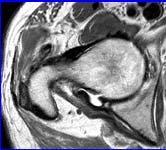
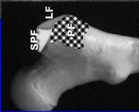
1 Greater Trochanter: Anatomy and Pathology Jon A. Jacobson, M.D. Professor of Radiology Director, Division of Musculoskeletal Radiology University of Michigan Disclosures: Consultant: Bioclinica Book Royalties: Elsevier Advisory Panel: GE, Philips Note: all images from the textbook Fundamentals of Musculoskeletal Ultrasound are copyrighted by Elsevier Inc. Greater Trochanter: gluteal tendons Lateral Posterior medius (red) minimus (blue) Greater Trochanter Greater Trochanter Yellow arrow = gluteus medius White arrow = gluteus minimus Inferior 1 2 FACETS: = anterior; = lateral; S = superoposterior; = posterior Pfirrmann et al. Radiology 2001; 221:469 Axial MRI 3 Superior 4 1


2 Greater Trochanter Subgluteus Medius Bursa Trochanteric Bursa Greater Trochanter TFL Medius Minimus Subgluteus Minimus Bursa Glut Max Yellow arrow = gluteus medius White arrow = gluteus minimus Posterior : anterior facet : lateral facet : posterior facet Minimus and Medius: Long Axis Minimus: Long Axis Medius Minimus Facet Facet From: Philippon et al. Orth J Sports Med 2014 Medius: Long Axis Iliotibial Tract Lateral Facet 2


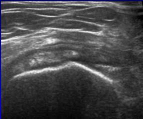
3 Trochanteric Pain Syndrome: Most commonly caused by gluteus minimus and medius tendon abnormalities 1 Trochanteric bursitis: uncommon 20% of symptomatic patients 2 Not actually inflamed 3 Not associated with pain 4 1 Kong A et al. Eur Rad 2007; 17: Long SS et al. AJR 2013; 201: Sylva F et al. Clin Rheumatol 2008; 14:82 4 Blankenbaker DG et al. Skeletal Radiol 2008; 37:903 Trochanteric Bursitis Transverse Trochanteric Bursal Fluid + Glut Min Tear Trochanteric Bursitis Posterior Glut Max Axial Trochanteric Bursitis: Septic Trochanteric Bursa: infection + gas Note posterior location of bursa T1w Greater Trochanter 3


* S")
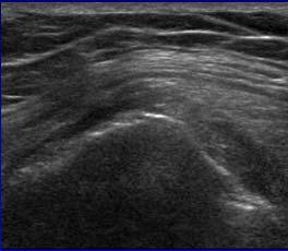
4 Trochanteric Region Bursae Trochanteric Bursitis Trochanteric: deep to gluteus maximus Subgluteus medius Subgluteus minimus Axial or coronal plane Transverse Arthrogram Muscle and Tendon Injury Tear: Anechoic or hypoechoic defect Partial-thickness tear Full-thickness tear: retraction Tendinosis: Hypoechoic, enlarged No inflammation (not tendinitis) Tendinosis: Medius Gluteal Tendon Pathology: Tendinosis: hypoechoic, no defects Partial tear: anechoic clefts Complete tear: discontinuous tendon >2 mm cortical irregularity (depth) Associated with tendon tear Positive predictive value = 90% (xray)* S *Steinert et al. Radiology 2010; 257:754 4


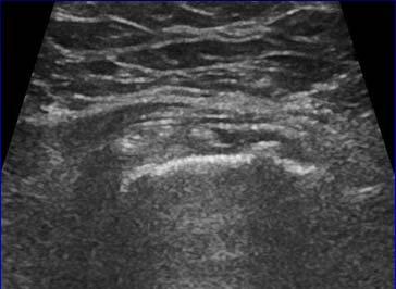
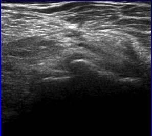
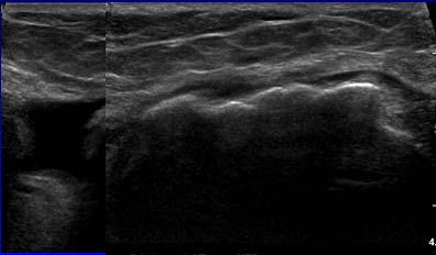
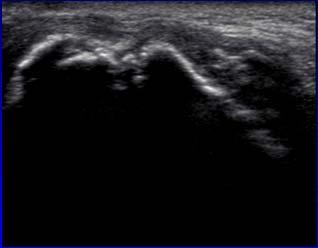
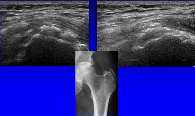
5 Medius: tendinosis Tear: Minimus Short Axis Long Axis Tear: Medius Tear: Medius after THA S >2 mm cortical irregularity depth (x-ray) = 90% positive predictive value for gluteus tendon tear Steinert et al. Radiology 2010; 257:754 Post-operative: Medius Calcific Tendinosis: Medius S Long Axis Short Axis 5


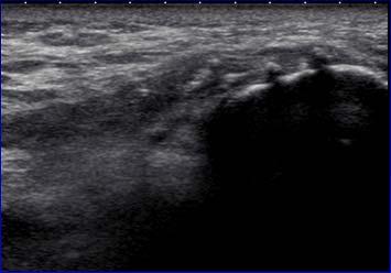
6 Medius Fenestration Greater Trochanter Needle Potential Treatment Algorithm: If bursa: aspirate, inject steroids If tendinosis: Tenotomy or fenestration Inject steroids superficial to tendon 72% of patients significantly improved 1 If tendon tear: platelet-rich plasma injection? Normal 1 Labrosse, et al AJR 2010; 194:202 Snapping Hip Syndrome Painful snap with hip motion Intraarticular Extraarticular: : iliopsoas tendon Lateral: iliotibial tract or gluteus maximus Snapping Hip: lateral Transverse over greater trochanter Hip external rotation / flexion Abrupt motion of iliotibial tract or gluteus maximus over greater trochanter Maximus Snapping Maximus / Iliotibial Band Medius TFL Maximus Iliotibial Band Gmin 6


7 Snapping Hip Syndrome: iliotibial tract Take-home points: Iliotibial Band Gmin Trochanteric anatomy Bursitis: rare Gluteal tendons abnormalities: frequent Snapping hip: dynamic 7
Ultrasound of the Hip: Anatomy, Pathology, and Procedures
 Ultrasound of the Hip: Anatomy, Pathology, and Procedures Jon A. Jacobson, M.D. Professor of Radiology Director, Division of Musculoskeletal Radiology University of Michigan Outline Hip Joint Native hip
Snapping Hip and Impingement
 Snapping Hip and Impingement Jon A. Jacobson, M.D. Professor of Radiology Director, Division of Musculoskeletal Radiology University of Michigan Disclosures: Consultant: Bioclinica Advisory Board: GE,
Tendon Fenestration. Disclosures. Outline: questions. Introduction: Peritendon Steroid Injections. Jon A. Jacobson, MD. Patellar Tendon: tendinosis
 Tendon Fenestration Jon A. Jacobson, MD Professor of Radiology Director, Division of Musculoskeletal Radiology University of Michigan Disclosures Consultant: Bioclinica Advisory Board: GE, Philips Book
Common Applications for Sonography and Guided Intervention: Shoulder
 Common Applications for Sonography and Guided Intervention: Shoulder Jon A. Jacobson, M.D. Professor of Radiology Director, Division of Musculoskeletal Radiology University of Michigan Disclosures: Consultant:
Rotator Cuff and Biceps Pathology
 Rotator Cuff and Biceps Pathology Jon A. Jacobson, M.D. Professor of Radiology Director, Division of Musculoskeletal Radiology University of Michigan Disclosures: Consultant: Bioclinica Advisory Board:
Ultrasound of the Knee
 Ultrasound of the Knee Jon A. Jacobson, M.D. Professor of Radiology Director, Division of Musculoskeletal Radiology University of Michigan Disclosures: Consultant: Bioclinica Book Royalties: Elsevier Advisory
Knee, Ankle, and Foot: Normal and Abnormal Features with MRI and Ultrasound Correlation. Disclosures. Outline. Joint Effusion. Suprapatellar recess
 Knee, Ankle, and Foot: Normal and Abnormal Features with MRI and Ultrasound Correlation Jon A. Jacobson, M.D. Professor of Radiology Director, Division of Musculoskeletal Radiology University of Michigan
Ultrasound Evaluation of Masses
 Ultrasound Evaluation of Masses Jon A. Jacobson, M.D. Professor of Radiology Director, Division of Musculoskeletal Radiology University of Michigan Disclosures: Consultant: Bioclinica Advisory Panel: GE,
Lateral Elbow Pathology
 Lateral Elbow Pathology Jon A. Jacobson, M.D. Professor of adiology Director, Division of Musculoskeletal adiology University of Michigan Disclosures: Consultant: Bioclinica Advisory Board: GE, Philips
Greater Trochanteric Pain Syndrome
 43 Andrea S. Klauser, MD 1 Carlo Martinoli, MD 2 Alberto Tagliafico, MD 3 Rosa Bellmann-Weiler, MD 4 Gudrun M. Feuchtner, MD, PhD 1 Marius Wick, MD 1 Werner R. Jaschke, MD, PhD 1 1 Department of Diagnostic
Greater Trochanteric Pain Syndrome
 ORIGINAL RESEARCH Greater Trochanteric Pain Syndrome Percutaneous Tendon Fenestration Versus Platelet-Rich Plasma Injection for Treatment of Gluteal Tendinosis Jon A. Jacobson, MD, Corrie M. Yablon, MD,
MRI of the Hip. Jon A. Jacobson, M.D. Professor of Radiology Director, Division of Musculoskeletal Radiology University of Michigan
 MRI of the Hip Jon A. Jacobson, M.D. Professor of Radiology Director, Division of Musculoskeletal Radiology University of Michigan Take Home Points Joint effusion: does not collect dependently Imaging
MR Imaging in Athlete s Hip/Pelvis
 MR Imaging in Athlete s Hip/Pelvis Tara Lawrimore, MD FRCPC Department of Radiology Musculoskeletal Division Massachusetts General Hospital Harvard Medical School No disclosures MR and Hip Pain in the
Musculoskeletal Ultrasound Fundamentals
 Fundamentals Benjamin D. Levine, M.D. Associate Professor of Radiology Musculoskeletal Imaging Dept. of Radiological Sciences UCLA Health System I. Image Optimization II. Image Interpretation Artifacts
CAN SOFT TISSUES STRUCTURES DIFFERENTIATE BETWEEN DYSPLASIA AND CAM-FAI OF THE HIP?
 CAN SOFT TISSUES STRUCTURES DIFFERENTIATE BETWEEN DYSPLASIA AND CAM-FAI OF THE HIP? A Le Bouthillier, KS Rakhra 1, PE Beaulé 2, RCB Foster 1 1 Department of Medical Imaging 2 Division of Orthopaedic Surgery
Viviane Khoury, MD. Assistant Professor Department of Radiology University of Pennsylvania
 U Penn Diagnostic Imaging: On the Cape Chatham, MA July 11-15, 2016 Viviane Khoury, MD Assistant Professor Department of Radiology University of Pennsylvania Hip imaging has changed in recent years: new
Complex Fractures and Hip Dislocations
 IMAGING OF HIP PAIN Patients may present with acute (< 2 weeks) or chronic hip pain. Acute pain may be related or not related to an acute traumatic event such as fall or trauma from a motor vehicle accident.
Evaluation of the Hip
 Evaluation of the Hip Adam Lewno, DO PCSM Fellow, University of Michigan Primary Care Sports Update 2017 Disclosures Financial: None Images: I would like to acknowledge the work of the original owners
Ultrasound of the Shoulder
 Ultrasound of the Shoulder Patrick Battaglia, DC, DACBR Logan University, Department of Radiology Outline Review ultrasound appearance of NMSK tissues Present indications for ultrasound of the shoulder.
US finding of the shoulder (with live demonstration) 인제의대상계백병원 안재기
 인제의대상계백병원 안재기") US finding of the shoulder (with live demonstration) 인제의대상계백병원 안재기 Shoulder US Biceps tendon & Rotator Cuff Long Head of Biceps Tendon Subscapularis tendon Supraspinatus tendon Infraspinatus tendon Teres
US finding of the shoulder (with live demonstration) 인제의대상계백병원 안재기 Shoulder US Biceps tendon & Rotator Cuff Long Head of Biceps Tendon Subscapularis tendon Supraspinatus tendon Infraspinatus tendon Teres
The psoas minor is medial to the psoas major. The iliacus is a fan-shaped muscle that when contracted helps bring the swinging leg forward in walking
 1 p.177 2 3 The psoas minor is medial to the psoas major. The iliacus is a fan-shaped muscle that when contracted helps bring the swinging leg forward in walking and running. The iliopsoas and adductor
Technical Note. Arthroscopic Anatomy and Surgical Techniques for Peritrochanteric Space Disorders in the Hip
 Technical Note Arthroscopic Anatomy and Surgical Techniques for Peritrochanteric Space Disorders in the Hip James E. Voos, M.D., Jonas R. Rudzki, M.D., Michael K. Shindle, M.D., Hal Martin, D.O., and Bryan
Greater Trochanteric Pain Syndrome (GTPS): Assessment & Management
: Assessment & Management") Greater Trochanteric Pain Syndrome (GTPS): Assessment & Management Rachael Mary McMillan Physiotherapist, Alphington Sports Medicine Clinic & FFA Australian Women s National Football Teams PhD Candidate
Greater Trochanteric Pain Syndrome (GTPS): Assessment & Management Rachael Mary McMillan Physiotherapist, Alphington Sports Medicine Clinic & FFA Australian Women s National Football Teams PhD Candidate
Ultrasound of Mid and Hindfoot Pathology
 Ultrasound of Mid and Hindfoot Pathology Levon N. Nazarian, M.D. Professor of Radiology Thomas Jefferson University Hospital Disclosures None relevant to this presentation Educational Objective Following
The evaluation and management of patients with
 Greater Trochanteric Hip Pain 1.5 ANCC Contact Hours Diane M. Kimpel Chadwick C. Garner Kevin M. Magone Jedediah H. May Matthew W. Lawless In the patient with lateral hip pain, there is a broad differential
The Hip (Iliofemoral) Joint. Presented by: Rob, Rachel, Alina and Lisa
 Joint. Presented by: Rob, Rachel, Alina and Lisa") The Hip (Iliofemoral) Joint Presented by: Rob, Rachel, Alina and Lisa Surface Anatomy: Posterior Surface Anatomy: Anterior Bones: Os Coxae Consists of 3 Portions: Ilium Ischium Pubis Bones: Pubis Portion
The Hip (Iliofemoral) Joint Presented by: Rob, Rachel, Alina and Lisa Surface Anatomy: Posterior Surface Anatomy: Anterior Bones: Os Coxae Consists of 3 Portions: Ilium Ischium Pubis Bones: Pubis Portion
Hip Tendinopathy. Outline. Tendon Anatomy 6/6/2011. Tendinopathy Hip adductor Iliopsoas Gluteus medius / minimus Hamstring New treatments
 Hip Tendinopathy Kelly C. McInnis, DO Massachusetts General Hospital Sports Medicine Center Physical Medicine and Rehabilitation Outline Tendinopathy Hip adductor Iliopsoas Gluteus medius / minimus Hamstring
MRI Diagnosis of Tears of the Hip Abductor Tendons (Gluteus Medius and Gluteus Minimus)
") Oliver Cvitanic 1 Gregory Henzie 1 Nicholas Skezas 2 Jack Lyons 2 Jon Minter 3 Received October 2, 2002; accepted after revision July 10, 2003. 1 Southwest Oklahoma MRI, 13301 N Meridian Ave., Ste. 600A,
Oliver Cvitanic 1 Gregory Henzie 1 Nicholas Skezas 2 Jack Lyons 2 Jon Minter 3 Received October 2, 2002; accepted after revision July 10, 2003. 1 Southwest Oklahoma MRI, 13301 N Meridian Ave., Ste. 600A,
ANTERIOR TOTAL HIP ARTHOPLASTY
 ANTERIOR TOTAL HIP ARTHOPLASTY And Other Approaches Bill Rhodes PTA 236 Total Hip Arthoplasty (THA) Background THA, also know as Total Hip Replacement Regarded as the most valued development in orthopedics
CLINICS IN SPORTS MEDICINE
 Clin Sports Med 25 (2006) 365 369 CLINICS IN SPORTS MEDICINE A Acetabular labrum, tears of, hip arthroscopy in, 264 Acetabular rim, trimming of, and labral repair, new method for, 293 297 Acetabulum, femoral
STAIRS. What s Hip: Top 5 Hip Problems in Primary Care. I have no relevant disclosures. Top 5 (or 6) Pathologies. Big 3- Questions to Ask
 Pathologies. Big 3- Questions to Ask") I have no relevant disclosures. What s Hip: Top 5 Hip Problems in Primary Care Alan Zhang MD Assistant Professor Sports Medicine and Hip Arthroscopy UCSF Department of Orthopaedic Surgery December, 2015
I have no relevant disclosures. What s Hip: Top 5 Hip Problems in Primary Care Alan Zhang MD Assistant Professor Sports Medicine and Hip Arthroscopy UCSF Department of Orthopaedic Surgery December, 2015
Sonographic evaluation of gluteus medius and minimus tendinopathy
 Eur Radiol (2003) 13:1339 1347 DOI 10.1007/s00330-002-1740-4 MUSCULOSKELETAL David A. Connell Cheryl Bass Christopher J. Sykes David Young Elton Edwards Sonographic evaluation of gluteus medius and minimus
Pragmatic ultrasound in the diagnosis of soft tissue rheumatic pain. Plamen Todorov
 Pragmatic ultrasound in the diagnosis of soft tissue rheumatic pain Plamen Todorov INTRODUCTION Soft tissue rheumatism: nonsystemic, focal pathological syndromes involving the periarticular structures.
Musculoskeletal Imaging Review
 Musculoskeletal Imaging Review Kassarjian et al. MRI of the Quadratus Femoris Musculoskeletal Imaging Review Ara Kassarjian 1 Xavier Tomas 2 Luis Cerezal 3 Ana Canga 4,5 Eva Llopis 6 Kassarjian A, Tomas
HIP_CASE 2_OA. Hip Forces. Function of the Hip. Property of VOMPTI, LLC. For Use of Participants Only. No Use or Reproduction Without Consent 1
 HIP_CASE 2_OA Orthopaedic Manual Physical Therapy Series Charlottesville 2017-2018 Eric Magrum DPT, OCS, FAAOMPT 62 yo female AM stiffness Hip pain diffuse, variable ant>lateral>post Gradual onset Tennis
Hip Cases from Clinic: Refining your history and physical
 Hip Cases from Clinic: Refining your history and physical Alan Zhang MD Assistant Professor Sports Medicine and Hip Arthroscopy UCSF Department of Orthopaedic Surgery 11/20/2017 Case #1 Healthy 21 M College
Incidental Benign Musculoskeletal Findings on PET-CT: an Educational Pictorial Review
 Incidental Benign Musculoskeletal Findings on PET-CT: an Educational Pictorial Review Poster No.: P-0008 Congress: ESSR 2014 Type: Educational Poster Authors: F. Moloney, J. Ryan, M. Twomey, S. McSweeney;
Human Anatomy Biology 351
 Human Anatomy Biology 351 Lower Limb Please place your name on the back of the last page of this exam. You must answer all questions on this exam. Because statistics demonstrate that, on average, between
Pure bone marrow aspirate injection for chronic greater trochanteric pain syndrome: a case report
 Case Report For reprint orders, please contact: reprints@futuremedicine.com Pure bone marrow aspirate injection for chronic greater trochanteric pain syndrome: a case report Rachel G Henderson*,1,2 & Ricardo
MRI of Quadratus Femoris Muscle Tear: Another Cause of Hip Pain
 MRI of Quadratus Femoris Muscle Tear Musculoskeletal Imaging Clinical Observations Seth D. O Brien 1 Liem T. Bui-Mansfield 1,2,3 O Brien SD, Bui-Mansfield LT Keywords: hip pain, MRI, muscle tear, quadratus
Hip Injuries & Arthroscopy in Athletes
 Hip Injuries & Arthroscopy in Athletes John P Salvo, MD Sports Medicine Rothman Institute Philadelphia, PA EATA Annual Meeting January, 2011 Hip Injuries & Arthroscopy in Anatomy History Physical Exam
FUNCTIONAL ANATOMY AND EXAM OF THE HIP, GROIN AND THIGH
 FUNCTIONAL ANATOMY AND EXAM OF THE HIP, GROIN AND THIGH Peter G Gerbino, MD, FACSM Orthopedic Surgeon Monterey Joint Replacement and Sports Medicine Monterey, CA TPC, San Diego, 2017 The lecturer has no
Effectiveness of Ultrasound-Guided Corticosteroid Injection for the Treatment of Gluteus Medius Tendinopathy
 Musculoskeletal Imaging Original Research Labrosse et al. Ultrasound-Guided Corticosteroid Injection for Tendinopathy Musculoskeletal Imaging Original Research Julie M. Labrosse 1 Étienne Cardinal 1 Bernard
External Snapping Hip Syndrome:
 J Korean Soc Radiol 2010;62:185-190 External Snapping Hip Syndrome: Emphasis on the MR Imaging 1 Jung Eun Choi, M.D., Mi Sook Sung, M.D. 2, Ki Haeng Lee, M.D. 3, ae Young Lee, M.D., Jeong Mi Park, M.D.
A Patient s Guide to Trochanteric Bursitis of the Hip
 A Patient s Guide to Trochanteric Bursitis of the Hip Iain is a specialist in musculoskeletal imaging and the diagnosis of musculoskeletal pain. This information is provided with the hope that you can
Lectures of Human Anatomy
 Lectures of Human Anatomy Lower Limb Gluteal Region and Hip Joint By DR. ABDEL-MONEM AWAD HEGAZY M.B. with honor 1983, Dipl."Gynecology and Obstetrics "1989, Master "Anatomy and Embryology" 1994, M.D.
Young Adult Hip problems. Aresh Hashemi-Nejad FRCS(Orth)
") Young Adult Hip problems Aresh Hashemi-Nejad FRCS(Orth) RNOH founded 1837 by William Little 14 year old presenting with limp Knee pain on and off 4 months Limps Aresh Hashemi-Nejad FRCS(Orth) The Royal
Young Adult Hip problems Aresh Hashemi-Nejad FRCS(Orth) RNOH founded 1837 by William Little 14 year old presenting with limp Knee pain on and off 4 months Limps Aresh Hashemi-Nejad FRCS(Orth) The Royal
Human Anatomy Biology 351
 Human Anatomy Biology 351 Lower Limb Please place your name on the back of the last page of this exam. You must answer all questions on this exam. Because statistics demonstrate that, on average, between
Ultrasound-Guided Tendon Fenestration
 85 Mary M. Chiavaras, MD, PhD 1 Jon A. Jacobson, MD 2 1 Department of Radiology, McMaster University, Hamilton General Hospital, Hamilton, Ontario, Canada 2 Department of Radiology, University of Michigan,
Joel S Sellers, DO, FAOASM CAQSM, RMSK
 Joel S Sellers, DO, FAOASM CAQSM, RMSK This is a sports slide of an Olympic wrestler Chris Taylor 1 This is a Sports Illustrated slide of jockey Johnny Sellers This is a slide of Coach Jim Sellers 2 This
MUSCULAR DAMAGE AFTER THA: TRANSGLUTEAL VS ANTERIOR APPROACH
 MUSCULAR DAMAGE AFTER THA: TRANSGLUTEAL VS ANTERIOR APPROACH Fabian Kalberer Department of Orthopedics, Balgrist University of Zurich www.balgrist.ch CHANGING PATIENT S EXPECTATIONS RESIDUAL WEAKNESS,
Ultrasound Guided Therapeutic Injections in the Treatment of Shoulder Pain: A Multimedia Review
 Ultrasound Guided Therapeutic Injections in the Treatment of Shoulder Pain: A Multimedia Review Poster No.: P-0127 Congress: ESSR 2015 Type: Educational Poster Authors: A. Karsandas, J. Tuckett, R. Sinha,
Soft-Tissue Pseudotumors of the Hip
 Soft-Tissue Pseudotumors of the Hip Poster No.: C-1564 Congress: ECR 2016 Type: Educational Exhibit Authors: M. R. Kaleel, J. Czajka, M. O'Loughlin, H. Baweja ; 1 1 2 2 2 2 HARTFORD, Connecticut/US, Hartford/US
Ultrasound-Guided Calcific Tendinitis Lavage: Application, Technique, and Outcome
 Ultrasound-Guided Calcific Tendinitis Lavage: Application, Technique, and Outcome Andrew Schapiro MD, Humberto Rosas MD, Kenneth Lee MD University of Wisconsin Hospital and Clinics aschapiro@uwhealth.org
Tears in the gluteus medius and minimus tendons
 Endoscopic Repair of Full-Thickness Gluteus Medius Tears Benjamin G. Domb, M.D., and Dominic S. Carreira, M.D. Abstract: Tears in the gluteus medius and minimus tendons recently have emerged as an important
The Young Adult Hip: FAI. Jason Snibbe, M.D. Snibbe Orthopedics Team Physician, University of Southern California
 The Young Adult Hip: FAI Jason Snibbe, M.D. Snibbe Orthopedics Team Physician, University of Southern California Introduction Femoroacetabular Impingment(FAI) Presentation and Exam Imaging Surgical Management
Technical application and the level of discomfort associated with an intramuscular
 Original article (short communication) Technical application and the level of discomfort associated with an intramuscular electromyographic investigation into gluteus minimus and gluteus medius. Adam I.
Main Menu. Joint and Pelvic Girdle click here. The Power is in Your Hands
 1 Hip Joint and Pelvic Girdle click here Main Menu K.6 http://www.handsonlineeducation.com/classes//k6entry.htm[3/23/18, 2:01:12 PM] Hip Joint (acetabular femoral) Relatively stable due to : Bony architecture
4/1/2016. Total Hip Arthroplasty. DAHR Procedure. Direct Anterior Hip Replacement. DAHR Procedure. DAHR Procedure
 Mercy Orthopedist Types of Approaches Total Hip Arthroplasty Mercy Has a total of 16 Orthopedist that perform all three different approaches Posterior Anterior Lateral Direct Anterior Direct Anterior Hip
2. Iliotibial Band syndrome
 2. Iliotibial Band syndrome Iliotibial band (ITB) syndrome (so called runners knee although often seen in other sports e.g. cyclists and hill walkers). It is usually an overuse injury with pain felt on
Hip joint and pelvic girdle. Lower Extremity. Pelvic Girdle 6/5/2017
 Hip joint and pelvic girdle Lower Extremity The relationship between the pelvic girdle and hip is similar to that between the shoulder girdle and shoulder joint. The lower limbs are attached to the axial
MRI of the Shoulder What to look for and how to find it? Dr. Eric Handley Musculoskeletal Radiologist Cherry Creek Imaging
 MRI of the Shoulder What to look for and how to find it? Dr. Eric Handley Musculoskeletal Radiologist Cherry Creek Imaging MRI of the Shoulder Benefits of Ultrasound: * Dynamic * Interactive real time
Ultrasound assessment of most frequent shoulder disorders
 Ultrasound assessment of most frequent shoulder disorders Poster No.: C-2026 Congress: ECR 2014 Type: Educational Exhibit Authors: S. P. Ivanoski; Ohrid/MK Keywords: Trauma, Athletic injuries, Arthritides,
Ultrasonographic Examination of the Adult Hip
 Journal of Medical Ultrasound (2012) 20, 201e209 Available online at www.sciencedirect.com journal homepage: www.jmu-online.com REVIEW ARTICLE Ultrasonographic Examination of the Adult Hip Yun-Tai Lin
CT Findings of Traumatic Posterior Hip Dislocation after Reduction 1
 CT Findings of Traumatic Posterior Hip Dislocation after Reduction 1 Sung Kyoung Moon, M.D., Ji Seon Park, M.D., Wook Jin, M.D. 2, Kyung Nam Ryu, M.D. Purpose: To evaluate the CT images of reduced hips
The Hip Joint. Shenequia Howard David Rivera
 The Hip Joint Shenequia Howard David Rivera Topics Of Discussion Movement Bony Anatomy Ligamentous Anatomy Muscular Anatomy Origin/Insertion/Action/Innervation Common Injuries MOVEMENT Flexion Extension
CHAPTER 8: THE BIOMECHANICS OF THE HUMAN LOWER EXTREMITY
 CHAPTER 8: THE BIOMECHANICS OF THE HUMAN LOWER EXTREMITY _ 1. The hip joint is the articulation between the and the. A. femur, acetabulum B. femur, spine C. femur, tibia _ 2. Which of the following is
Lesson 24. A & P Hip
 Lesson 24 A & P Hip 1 Aims of the Session This session will allow candidates to have an understanding of the bony prominences and soft tissues of the hip 2 Learning Outcomes By the end of the lesson the
Human Anatomy Biology 255
 Human Anatomy Biology 255 Exam #4 Please place your name and I.D. number on the back of the last page of this exam. You must answer all questions on this exam. Because statistics demonstrate that, on average,
Gluteus Medius Tears After Hip Arthroplasty. John Urse, DO, FAOAO Jason Spangler, DO Dzi-Viet Nguyen, DO Grandview Medical Center Dayton, OH
 Gluteus Medius Tears After Hip Arthroplasty John Urse, DO, FAOAO Jason Spangler, DO Dzi-Viet Nguyen, DO Grandview Medical Center Dayton, OH Disclosures AANA (Arthroscopy Association of North America) Lodging
This guideline is structured with clinical indications outlined for each of the following applications: Arthroscopic; Open, non-arthroplasty;
 National Imaging Associates, Inc. Clinical guidelines: HIP ARTHROSCOPY & OPEN, NON- ARTHROPLASTY HIP REPAIR CPT CODES: Femoroacetabular Impingement (FAI) Hip Surgery: 29914, 29915, 29916 Hip Surgery Other:
A Guide for Patients with Hip and Groin Pain. By - Rob Lawton & Ajay Malviya. Overview
 A Guide for Patients with Hip and Groin Pain By - Rob Lawton & Ajay Malviya Overview - Introduction - Hip Anatomy - Is the pain coming from the hip joint? - Intra-articular causes of hip pain o Impingement
What s Hip: Common Hip Problems and Kids and Adults
 What s Hip: Common Hip Problems and Kids and Adults Alan Zhang MD Assistant Professor Sports Medicine and Hip Arthroscopy UCSF Department of Orthopaedic Surgery I have no relevant disclosures. 2 1 Most
Muscles to know. Lab 21. Muscles of the Pelvis and Lower Limbs. Muscles that Position the Lower Limbs. Generally. Muscles that Move the Thigh
 Muscles to know Lab 21 Muscles of the Pelvis, Leg and Foot psoas major iliacus gluteus maximus gluteus medius sartorius quadriceps femoris (4) gracilus adductor longus biceps femoris semitendinosis semimembranosus
The functional anatomy of hip abductors
 O R I G I N A L A R T I C L E Folia Morphol. Vol. 68, No. 2, pp. 98 103 Copyright 2009 Via Medica ISSN 0015 5659 www.fm.viamedica.pl The functional anatomy of hip abductors A. Al-Hayani Department of Anatomy,
9/18/18. Welcome- MSK Ultrasound Workshop. Introduction to Musculoskeletal Ultrasound. Acknowledgement of Country. The Workshop.
 Acknowledgement of Country Welcome- MSK Ultrasound Workshop I would like to acknowledge that this meeting is being held on the traditional lands of the Wurundjeri and Boonwurrung people and pay my respect
MRI SHOULDER WHAT TO SEE
 MRI SHOULDER WHAT TO SEE DR SHEKHAR SRIVASTAV Sr. Consultant- Knee & Shoulder Arthroscopy Sant Parmanand Hospital Normal Anatomy Normal Shoulder MRI Coronal Oblique Sagital Oblique Axial Cuts Normal Coronal
RN(EC) ENC(C) GNC(C) MN ACNP *** MECHANISM OF INJURY.. MOST IMPORTANT ***
 ENC(C) GNC(C) MN ACNP *** MECHANISM OF INJURY.. MOST IMPORTANT ***") HISTORY *** MECHANISM OF INJURY.. MOST IMPORTANT *** Age of patient - Certain conditions are more prevalent in particular age groups (Hip pain in children may refer to the knee from Legg-Calve-Perthes
HISTORY *** MECHANISM OF INJURY.. MOST IMPORTANT *** Age of patient - Certain conditions are more prevalent in particular age groups (Hip pain in children may refer to the knee from Legg-Calve-Perthes
The University Of Jordan Faculty Of Medicine THE LOWER LIMB. Dr.Ahmed Salman Assistant Prof. of Anatomy. The University Of Jordan
 The University Of Jordan Faculty Of Medicine THE LOWER LIMB Dr.Ahmed Salman Assistant Prof. of Anatomy. The University Of Jordan Gluteal Region Cutaneous nerve supply of (Gluteal region) 1. Lateral cutaneous
Gluteal region DR. GITANJALI KHORWAL
 Gluteal region DR. GITANJALI KHORWAL Gluteal region The transitional area between the trunk and the lower extremity. The gluteal region includes the rounded, posterior buttocks and the laterally placed
Peripheral Nerve Ultrasound
 Peripheral Nerve Ultrasound Jon A. Jacobson, M.D. Professor of Radiology Director, Division of Musculoskeletal Radiology University of Michigan Normal Peripheral Nerve Ultrasound appearance: Hypoechoic
Avulsion injuries of Pelvis: An imaging spectrum
 Avulsion injuries of Pelvis: An imaging spectrum Poster No.: P-0090 Congress: ESSR 2014 Type: Educational Poster Authors: S. I. Alam, D. Kumar, M. KHANNA, H. Hina, M. Heidous, O. M. Osman, A. M. Refaat;
ANATOMY TEAM GLUTEAL REGION & BACK OF THIGH
 ANATOMY TEAM GLUTEAL REGION & BACK OF THIGH OBJECTIVES By the end of this lecture, the student should be able to identify and discuss: Contents of gluteal region: Groups of Glutei muscles and small muscles
Gluteal Strengthening Exercises: A Review of the Literature
 Common Imbalances Female Athlete Hip Injuries: Exploring the CORE of Patterns and Prevention Kelly McInnis, DO Irene Davis, PhD, PT, FAPTA, FACSM, FASB David Nolan, PT, DPT, MS, OCS, SCS, CSCS Gluteal
61 year-old woman with right thigh lump
 61 year-old woman with right thigh lump Skin marker was placed about here TFL is enlarged TFL is enlarged Hypertrophy of the TFL muscle, presenting as palpable proximal anterior thigh mass Unilateral hypertrophy
Fellowship Program. Musculoskeletal ultrasound and interventions under fluoroscopic guidance
 Fellowship Program Musculoskeletal ultrasound and interventions under fluoroscopic guidance The Montreal University Physical medicine and rehabilitation program offers a 1 year fellowship program. Type
The iliotibial band syndrome : MR Imaging findings
 The iliotibial band syndrome : MR Imaging findings Poster No.: P-0081 Congress: ESSR 2013 Type: Scientific Exhibit Authors: W. Harzallah-Hizem, M. MAATOUK, A. Zrig, R. Salem, W. Mnari, B. Hmida, M. GOLLI;
Anatomy Your shoulder is made up of three bones: your upper arm bone (humerus), your shoulder blade (scapula), and your collarbone (clavicle).
, your shoulder blade (scapula), and your collarbone (clavicle).") Shoulder Impingement/Rotator Cuff Tendinitis One of the most common physical complaints is shoulder pain. Your shoulder is made up of several joints combined with tendons and muscles that allow a great
Shoulder Impingement/Rotator Cuff Tendinitis One of the most common physical complaints is shoulder pain. Your shoulder is made up of several joints combined with tendons and muscles that allow a great
DIRECT SUPERIOR HIP APPROACH IN TOTAL HIP ARTHROPLASTY. Anil Thomas, MD Adult Reconstruction Peachtree Orthopedics Atlanta, GA
 DIRECT SUPERIOR HIP APPROACH IN TOTAL HIP ARTHROPLASTY Anil Thomas, MD Adult Reconstruction Peachtree Orthopedics Atlanta, GA Disclosures None Direct Superior Approach History and development of the approach
Practical Reporting of Musculoskeletal Imaging Studies: MRI Elbow
 Practical Reporting of Musculoskeletal Imaging Studies: MRI Elbow James F Griffith History Where is pain located? For how long? Trauma if so, what and when Radiographers can get this info Grade. Don t
What can Imaging tell us?
 What can Imaging tell us? David Connell FRANZCR, FFSEM (UK) Assoc Professor Dept of Medicine, Nursing & Healthcare Monash University, Melbourne, Australia Assoc Professor Sport & Exercise Medicine Research
ELENI ANDIPA General Hospital of Athens G. Gennimatas
 ELENI ANDIPA General Hospital of Athens G. Gennimatas Technological advances over the last years have caused a dramatic improvement in ultrasound quality and resolution An established imaging modality
When the patient presents at the GP surgery with Hip pain. Investigations, Input & Referral
 When the patient presents at the GP surgery with Hip pain Investigations, Input & Referral GP with an interest in Pain Management How common is hip pain? Around 450 patients per 100,000 population will
Human anatomy reference:
 Human anatomy reference: Weak Glut Activation Weak gluteal activation comes from poor biomechanics, poor awareness when training or prolonged exposure in deactivated positions such as sitting. Weak Glut
HIP THIGH KNEE Ultrasound Fundamental
 HIP THIGH KNEE Ultrasound Fundamental STAFFORDSHIRE MSK STUDY DAY BMUS Kirstie Godson Academic lecturer Leeds University Why Ultrasound Readily available. Shorter waiting times. Less expensive than other
Review Article. Abstract
 Review Article Abductor Tendon Tears of the Hip: Evaluation and Management Paul F. Lachiewicz, MD Abstract The gluteus medius and minimus muscle-tendon complex is crucial for gait and stability in the
SHOULDER Highly mobile, so less stable. Abnormalities cloaked within extensive musculature, dx can be difficult Bony abnormalities less common than li
 SPORTS MEDICINE CASES A quick tour of some local joints Featuring gco common o and unusual problems SHOULDER Highly mobile, so less stable. Abnormalities cloaked within extensive musculature, dx can be
Chronic knee pain in adults - a multimodality approach or which modality to choose and when?
 Chronic knee pain in adults - a multimodality approach or which modality to choose and when? Poster No.: P-0157 Congress: ESSR 2013 Type: Authors: Keywords: DOI: Scientific Exhibit E. Ilieva, V. Tasseva,
Extraarticular Snapping Hip: Sonographic Findings
 Vincent Pelsser 1 Étienne Cardinal 1 Roger Hobden 2 enoit ubin 1 Michel Lafortune 1 Received September 22, 1999; accepted after revision June 8, 2000. 1 Department of Diagnostic Radiology, CHUM-St-Luc
Muscles of Lesson Five. Muscular Nomenclature and Kinesiology - Two. Muscles of Lesson Five, cont. Chapter 16
 Chapter 16 Muscular Nomenclature and Kinesiology - Two Lessons 5-6 Muscles of Lesson Five Iliopsoas (psoas major, iliacus) Hip outward rotators (piriformis, gemellus superior, gemellus inferior, obturator