Analysis of box-tie related suspension nerve injuries
|
|
|
- Annabella Cobb
- 5 years ago
- Views:
Transcription

1 Analysis of box-tie related suspension nerve injuries I have used the term box-tie to refer to takate-kote style ties, encompassing the arms and upper torso, used for suspension. The purpose of this document is to examine the construction methods and possible implications for the nerves likely to be affected. The first part will examine the classic style and the problems inherent. However, these hazards are not unique to this tie and the information is relevant to upper body bondage in general. The second part, yet to be completed, will examine some of the common mistakes in attempting to execute this tie and the failures of some Western copies. Whilst most injuries can be traced to inappropriate placement of ropes or poor construction, there still seem to be others which seem less explicable. I hope this document will be the basis from which to explore how accidents can be avoided or, even, if this type of tie or its usage can be improved. Part I: Traditional takate-kote It appears that there is a classic consensus of form adopted by the Japanese shibari masters which, whilst differing slightly, has the same fundamental principals. These include: 1. Hands tied behind back with forearms parallel and arms bent at 90 degrees. 2. The simple form is typically based on two ropes, excluding the suspension rope/s 3. It comprises two parallel wraps, usually of two doubled bands of rope, one above the breasts and one below, encompassing the arms and torso. The upper wraps will normally be under greater tension that the lower wraps. 4. One or both wraps will be cinched at the front only. 5. Some or all components will be locked off to ensure that it is a separate
2 unit and does not tighten when other bindings are pulled. 6. Only the two parallel wraps, not the cinch ropes, to be included in the suspension rope/s. In addition to the above, there may be some embellishment or further structural work depending on how much rope is left. Regardless, the above components comprise the basic form. Unfortunately, many Western approximations do not take all these considerations into account. Consequently, this can lead to increased risk. Hopefully, this document will alert people to the risks inherent in some non-standard or reverse engineered versions. Nerves of the upper body There follows an illustrated discussion of the main nerves, and arteries which supply them, relevant to this tie and, indeed, bondage in general. The following terms might be helpful in interpreting medical texts: Anterior The front side Posterior The rear side Lateral Away from the midline of the body Medial Towards the midline of the body Proximal Towards the centre of the body Distal Away from the centre of the body Pronation To rotate the arm inward so that the thumb points towards the body Supination - To rotate the arm outward so that the thumb points away from the body Extension When a joint is held straight out (opposite of flexion) Flexion When a joint is bent (opposite of extension) Nerve damage duration: Times involved are hard to estimate, but will partly depend on the spread of focussing of the compression, the weight of the person suspended, and the concurrent presence of blood vessel compression. Be aware that serious nerve damage can happen very quickly with recovery time stretching into weeks or months. Consider this before you experiment with suspension or tight bondage. Short term compression - tingling and loss of sensation. Longer term (minutes) - loss of motor function (neuropraxia). Longer still - longer recovery time. Even longer - possibility of permanent injury (neurotemsis)
, median and radial (thumb side).")
3 A sketch of a dissected body, showing the nerves of the hand, can be found here in Gray s Anatomy, however Figs 1 and 3 show the information photographically. Starting at the wrists, you have three main nerves: ulnar (little finger side), median and radial (thumb side). The median isn't easily compressed by ropes, even under suspension as it lies deep within the carpal tunnel running up the middle of the wrist. People with Carpal Tunnel Syndrome are probably the exceptions as fibrous tissue fills the tunnel and leaves less room for the nerve. The real problems are the other two nerves, radial and ulnar. Avoid the notch at the base of the thumb (Point A) and the notch you'll feel just at the end of the ulnar bone where the forearm ends and the hand starts (Point B), see Fig 1. At both these points the nerves are running near the surface over bony prominences, making the nerve compressible by overly tight or thin, i.e. narrow diameter rope or insufficient wraps, wrist bindings cutting into this area. Fig 1. Showing vulnerable points on wrist A sketch of a dissected body, showing the nerves of the arm, can be found here in Gray s Anatomy. See Fig 2 et seq. Moving up the arm, avoid the "funny bone", aka humerus, where the ulnar nerve runs over the bony prominences of the ends of the humerus and top end of the ulna. Compressing here gives similar symptoms to whacking it (the "funny bone") i.e. tingling in the inner (little finger) half of the hand (see Fig 1b), and importantly with time and degree of injury also
 at the elbow for prolonged periods; if the arm is twisted inward so the thumb faces the body tension on the")

4 leads to weakness in the fingers and both loss of grip strength and precision. Injuries to the ulnar nerve may also occur if the arm is very tightly bent (>90 degrees) at the elbow for prolonged periods; if the arm is twisted inward so the thumb faces the body tension on the ulnar nerve is further increased (think of the way you hold your arm while imitating a chicken wing or as in a wrist to upper arm tie). Fig 1b. Blue areas are served by ulnar nerve Fig 1c. Location of ulnar at elbow Symptoms of ulnar nerve injury: Abnormal sensations in the 4th or 5th fingers Numbness, decreased sensation Tingling, burning sensation Pain Weakness of the hand Again, the median nerve, in the middle of the front of the elbow is difficult to compress, as it's deep and surrounded by soft tissue, but with enough pressure the radial artery can be compressed, leaving the lower arm short of blood & oxygen. This point is just proximal to the bony parts of the wrist with the hand supine, where the pulse is normally taken. This remains vulnerable in a straight line up to about half way to the elbow, at which point the increased muscle bulk around the deeper running artery will be protective. This is both painful in itself, and after a few minutes can start to cause early tissue damage. Releasing the pressure the causes more pain as the blood supply returns.
5 Avoid the back/inner upper humerus (2-3 inches below the armpit) as the lower branches of the brachial plexus are compressed against the bone of the upper arm here, this time including the median nerve (causing major functional problems for the elbow and hand). The radial nerve: is vulnerable at the wrist as mentioned above. In addition, it is also prone to injury where it twists around the outside of the arm between the deltoid and triceps, see Figs 2 and 5. It is at its most exposed in the valley between these two muscles. Since it is included in the brachial plexus, compression in this area can occur. A branch of the radial nerve near the lateral and posterior portion of the wrist does run close to the skin surface, see Fig 3, and tight ties in this region may lead to numbness along the back of the hand. Symptoms of radial nerve injury: Symptoms can affect the following: The hand or forearm (dorsal surface, the "back" of the hand) The "thumb side" (radial surface) of the dorsal hand The fingers nearest the thumb (2nd and 3rd) The following symptoms may occur: Numbness, decreased sensation, tingling, or burning sensation Pain Abnormal sensations Difficulty extending the arm at the elbow Difficulty extending the wrist Brachial plexus: In the armpit, all the major nerves to the upper limb are branching after emerging from the neck and upper thoracic spine, see Figs 3 and 4. They pass through the soft tissues beneath the shoulder joint. This is pretty well protected from above by the joint itself, behind by deltoid and trapezius, and from the front by the pectorals. Underneath, though, these nerves are vulnerable. Restraints should never be placed around the armpit as this will almost certainly lead to compression of all of the nerves that supply the arm. It's not just compression, but also excessive stretching, which can happen if the body is suspended with arms above the head. This is also a risk if the arms are pulled behind the back, when the head is turned to the opposite side, and when there is
 and")
6 downward pressure on the shoulder. Obviously, the risk, and speed of onset of any injury, is greater in those who weigh more. While certain scenes may require positioning that puts stretch tension on the brachial plexus, moving the person in bondage to the position slowly and steadily (without sudden movements) and minimizing the aforementioned pressures may help make arm restraint safer. Fig 2. Diagram shows path of radial nerve. See here for anatomical version in Gray s Anatomy. The illustrations show the main points where nerves are likely to be vulnerable. This is only a guide as anatomy varies and nerves will move according to position. The markings enclose the areas which should be treated with caution. However, while nerve damage to the areas discussed may appear to be the source of change in sensation, in fact there are times that the pain is actually the result of compression of the nerve points around the vertebra. For instance, suspension with the head in a plane that might deform the natural position of the vertebrae; thus, pinching the nerves coming out at the vertebra. This situation is very highly probable in horizontal suspension when the head is unsupported.
7 Often the sensation of pain from the cervical pain is manifested at a distance from the vertebra and could include sensation along the radial nerve right to the finger tips. There are many variables, knock-on effects and bio-mechanical issues; there is no magic formula which avoids all risks. Furthermore, there is no medical specialisation in bondage related injuries, so creating this expertise is down to us. In spite of all the best care and knowledge, shit happens. All we can do is be aware of the risks and how to minimise them.

8 Fig 3. View from rear with arm in typical position

9 Fig 4. View from front, arm lifted to show brachial plexus in armpit.

10 Fig 5. View from side with arm in typical position showing radial nerve

11 Fig 6. View from front, arm lifted to show brachial plexus in armpit. The following illustrations show a typical takate-kote. As can be seen from the following two illustrations, the bindings largely avoid the area on the outer arm, although the upper binding does clip the top edge. Some riggers place the lower binding higher than I have done. The marked area should be considered vulnerable. Attention should be paid to the risk of either of these bindings slipping into this area when under load.
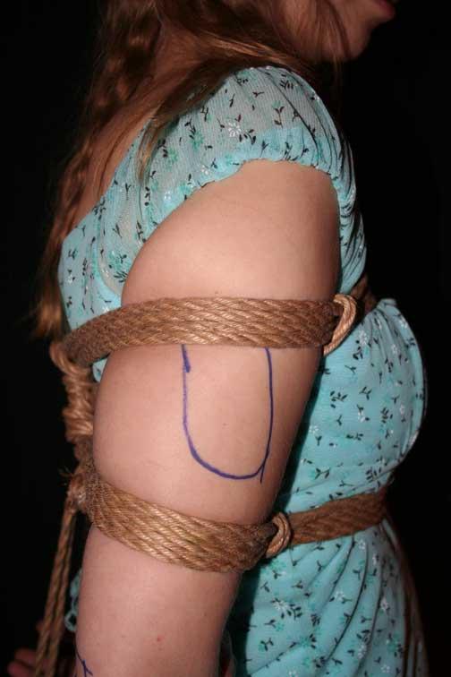
12 Fig 7.
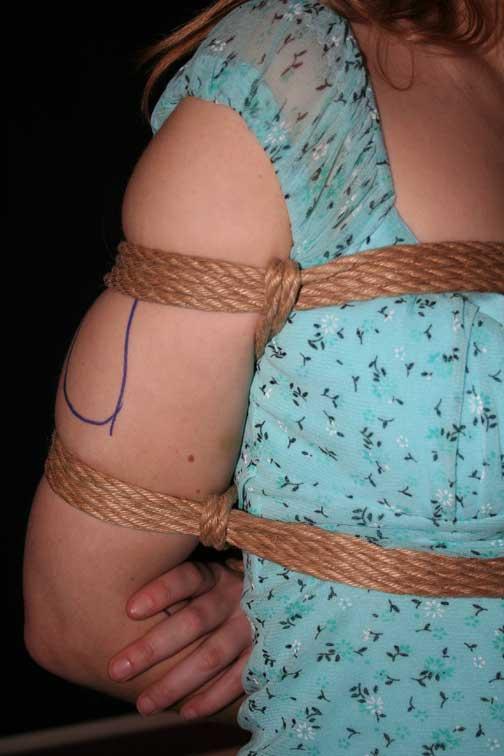
13 Fig 8.
14 Due to the reluctance of people to discuss their experiences, I have little to go on beyond the grapevine and a few individual reports. However, I have noted a preponderance of injuries, usually affecting the radial nerve, and often relating to sideways suspension. In the latter, the tendency is for this to affect the lower arm (nearest the ground), which will be under the greater load. I would like to speculate upon some causes, apart from incorrect construction: 1. The upper binding moving into the danger area (shown above). 2. Failure to run a finger under the upper binding to allow underlying tissue to settle. This ensures nerves are not trapped by muscle and return to their normal position in the protective groove. 3. The cinch rope on the upper binding being pressed into the lower end of the brachial plexus. I believe that this nerve bundle could be pinched between rope, ribs and arm bone. This being a possibility, I would question the wisdom of the upper cinch when using this tie for sideways suspension. Apart from the above, I do not see any problems with the arm/body portion of this tie, when correctly applied. All the other problem areas are well away from the components. However, the position of the hands needs to be watched. It is common for the model to change hand position significantly during a suspension. Even if wrists are placed against each other to protect the sensitive inner sides, they can be rotated. I have often seen an attempt to straighten the arms leading to them ending up in an X position. As can be seen below, the radial artery on the thumb side of the wrist is in risk of pressure. Scissoring of the arms can also increase the tension further. These are all good reasons to ensure sufficient slack in the wrist tie. After all, it is the restriction of the arms, not the wrist tie, which provides most of the immobilisation. Keep an eye out for over-tightness and the consequent signs of discomfort, e.g. flexing fingers, discolouration.

15 With thanks to: Vitimin A, Sluttylatexboy for their medical input and those who have shared their experiences. Please me with any comments or additions: Please circulate this information freely. However, it would be courteous to include a credit and link to More safety information can be found on my site:
Nerve Damage and Bondage
 Nerve Damage and Bondage Overview Anecdotally, nerve damage seems to be the most common serious injury (as opposed to minor injuries like rope marks, bruising, etc) that occurs in bondage scenes. Danger
Lowe Plastic Surgery (LPS)
") Lowe Plastic Surgery (LPS) PATIENT EDUCATION FOR: CUMULATIVE TRAUMA DISORDER THE PROBLEM: There has been a remarkable increase in what is termed cumulative trauma disorder (CTD) in the last 20 years. Other
Lowe Plastic Surgery (LPS) PATIENT EDUCATION FOR: CUMULATIVE TRAUMA DISORDER THE PROBLEM: There has been a remarkable increase in what is termed cumulative trauma disorder (CTD) in the last 20 years. Other
A Patient s Guide to Elbow Anatomy
 A Patient s Guide to Elbow Anatomy Iain is a specialist in musculoskeletal imaging and the diagnosis of musculoskeletal pain. This information is provided with the hope that you can better understand and
Hand Anatomy A Patient's Guide to Hand Anatomy
 Hand Anatomy A Patient's Guide to Hand Anatomy Introduction Few structures of the human anatomy are as unique as the hand. The hand needs to be mobile in order to position the fingers and thumb. Adequate
Physical Sense Activation Programme
 Flexion extension exercises for neck and upper back Sitting on stool Arms hanging by side Bend neck and upper back Breathe out Extend your neck and upper back Lift chest to ceiling Squeeze shoulder blades
Nerve Injury. 1) Upper Lesions of the Brachial Plexus called Erb- Duchene Palsy or syndrome.
 Upper Lesions of the Brachial Plexus called Erb- Duchene Palsy or syndrome.") Nerve Injury - Every nerve goes to muscle or skin so if the nerve is injured this will cause paralysis in the muscle supplied from that nerve (paralysis means loss of function) then other muscles and other
Nerve Injury - Every nerve goes to muscle or skin so if the nerve is injured this will cause paralysis in the muscle supplied from that nerve (paralysis means loss of function) then other muscles and other
The Language of Anatomy. (Anatomical Terminology)
") The Language of Anatomy (Anatomical Terminology) Terms of Position The anatomical position is a fixed position of the body (cadaver) taken as if the body is standing (erect) looking forward with the upper
The Language of Anatomy (Anatomical Terminology) Terms of Position The anatomical position is a fixed position of the body (cadaver) taken as if the body is standing (erect) looking forward with the upper
Traditional Thai Acupressure Points. The anterior aspect of the body THE ANATOMICAL ATLAS
 Traditional Thai Acupressure Points The anterior aspect of the body THE ANATOMICAL ATLAS lines of the SHOULDER BLADES AND POSTERIOR ARM Scapula Line This line runs through landmarks: 1. Above the midpoint
Cubital Tunnel Syndrome
 Disclaimer This movie is an educational resource only and should not be used to manage Orthopaedic Health. All decisions about must be made in conjunction with your Physician or a licensed healthcare provider.
The Elbow and Radioulnar Joints Kinesiology. Dr Cüneyt Mirzanli Istanbul Gelisim University
 The Elbow and Radioulnar Joints Kinesiology Dr Cüneyt Mirzanli Istanbul Gelisim University 1 The Elbow & Radioulnar Joints Most upper extremity movements involve the elbow & radioulnar joints. Usually
A Patient s Guide to Ulnar Nerve Entrapment at the Wrist (Guyon s Canal Syndrome)
") A Patient s Guide to Ulnar Nerve Entrapment at the Wrist (Guyon s Canal Syndrome) Introduction The ulnar nerve is often called the funny bone at the elbow. However, there is little funny about injury to
A Patient s Guide to Ulnar Nerve Entrapment at the Wrist (Guyon s Canal Syndrome) Introduction The ulnar nerve is often called the funny bone at the elbow. However, there is little funny about injury to
ELBOW - 1 FLEXION: ROM (Supine / Sitting)
") ELBOW - 1 FLEXION: ROM (Supine / Sitting) Position (A) Patient: Place arm against side of trunk. Helper: Hold elbow to stabilize. (B) - Lift hand toward shoulder, palm up. - Keep wrist straight. Do sessions
ELBOW - 1 FLEXION: ROM (Supine / Sitting) Position (A) Patient: Place arm against side of trunk. Helper: Hold elbow to stabilize. (B) - Lift hand toward shoulder, palm up. - Keep wrist straight. Do sessions
Body Planes & Positions
 Learning Objectives Objective 1: Identify and utilize anatomical positions, planes, and directional terms. Demonstrate what anatomical position is and how it is used to reference the body. Distinguish
THROWERS TEN EXERCISE PROGRAM
 THROWERS TEN EXERCISE PROGRAM The Thrower s Ten Program is designed to exercise the major muscles necessary for throwing. The Program s goal is to be an organized and concise exercise program. In addition,
Work Related Musculoskeletal Disorders
 Work Related Musculoskeletal Disorders Upper Extremity Disorders Carpel tunnel syndrome Cubital tunnel syndrome Thoracic outlet syndrome Raynaud s syndrome (white finger) Rotator cuff syndrome DeQuervain
EXERCISE INSTRUCTIONS
 EXERCISE INSTRUCTIONS A/ Strength A01 SQUAT Stand on the Power-Plate with feet shoulder width apart. Keeping the back straight and knees slightly bent, gently squeeze the leg muscles. You should feel tension
The Upper Limb III. The Brachial Plexus. Anatomy RHS 241 Lecture 12 Dr. Einas Al-Eisa
 The Upper Limb III The Brachial Plexus Anatomy RHS 241 Lecture 12 Dr. Einas Al-Eisa Brachial plexus Network of nerves supplying the upper limb Compression of the plexus results in motor & sensory changes
Medical Terminology. Anatomical Position, Directional Terms and Movements
 Medical Terminology Anatomical Position, Directional Terms and Movements What we will cover... Content Objectives Students will be able to gain a better understanding and application of medical terminology
PHYSICAL TRAINING INSTRUCTORS MANUAL TABLE OF CONTENT PART 3
 TABLE OF CONTENT PART 3 Exercise No 11: Chest Press... 2 Exercise No 12: Shoulder Press... 3 Exercise No 13: Pull-overs... 5 Exercise No 14: Tricep Extension... 6 informal exercises to develop upper body
region of the upper limb between the shoulder and the elbow Superiorly communicates with the axilla.
 1 region of the upper limb between the shoulder and the elbow Superiorly communicates with the axilla. Inferiorly, a number of important structures pass between arm & forearm through cubital fossa. 2 medial
Stroke: Upper limb exercises. Information for patients Sheffield Teaching Hospitals
 Stroke: Upper limb exercises Information for patients Sheffield Teaching Hospitals Stiffness and stretches After a stroke you may have difficulty moving your arm. If you just leave it, this will make the
Ultimate Personal Training Biceps Exercise Guide
 Ultimate Personal Training Biceps Exercise Guide Major Muscles That Act At The Elbow and Forearm MUSCLE ORIGIN INSERTION Biceps brachii Brachialis Pronator teres Long head from tubercle above glenoid cavity;
Monster Walk Stand with your feet slightly closer than shoulder-width apart in an athletic stance. Loop an elastic band around your ankles.
 Off-season Lower-Body Tennis Exercises Research conducted on elite tennis players shows that lower-body strength is the same on both the left and right sides. Therefore, lower-body training for tennis
MLT Muscle(s) Patient Position Therapist position Stabilization Limb Position Picture Put biceps on slack by bending elbow.
 Patient Position Therapist position Stabilization Limb Position Picture Put biceps on slack by bending elbow.") MLT Muscle(s) Patient Position Therapist position Stabilization Limb Position Picture Put biceps on slack by bending elbow. Pectoralis Minor Supine, arm at side, elbows extended, supinated Head of Table
MLT Muscle(s) Patient Position Therapist position Stabilization Limb Position Picture Put biceps on slack by bending elbow. Pectoralis Minor Supine, arm at side, elbows extended, supinated Head of Table
Hands PA; Obl. Lat.; Norgaard s Thumb AP; Lat. PA. PA; Lat.: Obls.; Elongated PA with ulnar deviation
 Projections Region Basic projections Additional / Modified projections Upper Limbs Hands PA; Obl. Lat.; Norgaard s Thumb ; Lat. PA Fingers PA; Lat. Wrist PA; Lat. Obls. Scaphoid Lunate Trapezium Triquetral
Nerves of the upper limb Prof. Abdulameer Al-Nuaimi. E. mail:
 Nerves of the upper limb Prof. Abdulameer Al-Nuaimi E-mail: a.al-nuaimi@sheffield.ac.uk E. mail: abdulameerh@yahoo.com Brachial plexus Median nerve After originating from the brachial plexus in the axilla,
What is Kinesiology? Basic Biomechanics. Mechanics
 What is Kinesiology? The study of movement, but this definition is too broad Brings together anatomy, physiology, physics, geometry and relates them to human movement Lippert pg 3 Basic Biomechanics the
GENERAL EXERCISES THUMB, WRIST, HAND BMW MANUFACTURING CO. PZ-AM-G-US I July 2017
 GENERAL EXERCISES THUMB, WRIST, HAND BMW MANUFACTURING CO. PZ-AM-G-US I July 2017 Disclosure: The exercises, stretches, and mobilizations provided in this presentation are for educational purposes only
The Golfers Ten Program. 1. Self Stretching of the Shoulder Capsule
 The Golfers Ten Program 1. Self Stretching of the Shoulder Capsule A. Posterior capsular stretch Bring your arm across your chest toward the opposite shoulder. With the opposite arm grasp your arm at your
GOLFERS TEN PROGRAM 1. SELF STRETCHING OF THE SHOULDER CAPSULE
 GOLFERS TEN PROGRAM 1. SELF STRETCHING OF THE SHOULDER CAPSULE POSTERIOR CAPSULAR STRETCH Bring your arm across your chest toward the opposite shoulder. With the opposite arm grasp your arm at your elbow.
The arm: *For images refer back to the slides
 The arm: *For images refer back to the slides Muscles of the arm: deltoid, triceps (which is located at the back of the arm), biceps and brachialis (it lies under the biceps), brachioradialis (it lies
Active-Assisted Stretches
 1 Active-Assisted Stretches Adequate flexibility is fundamental to a functional musculoskeletal system which represents the foundation of movement efficiency. Therefore a commitment toward appropriate
Warm-Up and Stretching Exercises
 Warm-Up and Stretching Exercises Most athletes (swimmers included) use a combination of controlled movement exercises and specific joint/muscle stretching to improve performance potential. The proposed
INTRODUCTION Cubital Tunnel Syndrome
 INTRODUCTION Cubital Tunnel Syndrome Diagram of the ulnar nerve supplying the muscles of forearm and hand Cubital Tunnel is a condition that refers to the ulnar nerve being compressed around the elbow.
Gross Anatomy Questions That Should be Answerable After October 27, 2017
 Gross Anatomy Questions That Should be Answerable After October 27, 2017 1. The inferior angle of the scapula of a woman who was recently in an automobile accident seems to protrude making a ridge beneath
D: Doorway Stretch E: Towel Stretch for Pectoralis Minor Blackburn Exercises: 6 Positions A: Prone Horizontal Abduction (Neutral)
") D: Doorway Stretch Bring your shoulder into a horizontal position out to your side (abduction) and flex your elbow 90û Place your elbow against the edge of a doorway Lead forward and downwards with your
D: Doorway Stretch Bring your shoulder into a horizontal position out to your side (abduction) and flex your elbow 90û Place your elbow against the edge of a doorway Lead forward and downwards with your
2011 EliteSoccerPower.com
 Developing Power for Soccer By Mike Grafstein B.Ph.Ed, RMT, YCS As may or may not know soccer is now a game of power and speed and players of all ages need to train that way. With that in mind I have put
A Patient s Guide to Nursemaid's Elbow in Children. PHYSIO.coza
 A Patient s Guide to Nursemaid's Elbow in Children SANDTON MEDICLINIC 011 706 7495 FAIRWAYS LIFE HOSPITAL 011 875 1827 ST STITHIANS 082 378 9642 JEPPE BOYS HIGH SCHOOL 084 816 5457 JOHANNESBURG, SANDTON@PHYSIO.CO.ZA
A Patient s Guide to Elbow Anatomy. TherAccess - Hand and Upper Extremity Rehab Center
 A Patient s Guide to Elbow Anatomy TherAccess, PLLC is founded by experienced therapy professionals. The purpose of this website was to develop a simple yet comprehensive collection of resources for our
Sign up to receive ATOTW weekly -
 1 SUBCLAVIAN PERIVASCULAR BRACHIAL PLEXUS BLOCK ANAESTHESIA TUTORIAL OF THE WEEK 156 19 th OCTOBER 2009 Dr. Martin Herrick Department of Anaesthesia, Addenbrooke s Hospital, Cambridge, U.K. Correspondence
 www.fitnessfirst-usa.com Chest Fly Shoulders, elbows and wrists aligned in same plane with elbows at 90 degrees Feet should be staggered, and body leaning slightly forward for leverage Step far enough
Location Terms. Anterior and posterior. Proximal and Distal The term proximal (Latin proximus; nearest) describes where the appendage joins the body.
 describes where the appendage joins the body.") HUMAN ANAT OMY Location Terms Anterior and posterior In human anatomical usage, anterior refers to the front of the individual. Similarly, posterior refers to the back of the subject. In standard anatomical
HUMAN ANAT OMY Location Terms Anterior and posterior In human anatomical usage, anterior refers to the front of the individual. Similarly, posterior refers to the back of the subject. In standard anatomical
Medical Terminology. Unit 2
 Medical Terminology Unit 2 Students will apply medical terminology. Objective 1: Identify and utilize anatomical positions, planes, and directional terms. Demonstrate what anatomical position is and how
SHHS Gen Bio Muscular System Lab
 SHHS Gen Bio Muscular System Lab Background: Skeletal muscle is the most abundant tissue in the body of healthy adults, comprising anywhere from about 30 to 50% of total body mass. The amount of muscle
POSTERIOR 1. situated behind: situated at or toward the hind part of the body :
 ANATOMICAL LOCATION Anatomy is a difficult subject with a large component of memorization. There is just no way around that, but we have made every effort to make this course diverse and fun. The first
THROWER S TEN EXERCISE PROGRAM David Andrew Parker, MD
 THROWER S TEN EXERCISE PROGRAM David Andrew Parker, MD The thrower s ten exercise program has been designed to exercise the major muscles necessary to return to throwing. The program s goal is to be an
Nerves of Upper limb. Dr. Brijendra Singh Professor & Head Department of Anatomy AIIMS Rishikesh
 Nerves of Upper limb Dr. Brijendra Singh Professor & Head Department of Anatomy AIIMS Rishikesh 1 Objectives Origin, course & relation of median & ulnar nerves. Motor & sensory distribution Carpal tunnel
Physical Examination of the Shoulder
 General setup Patient will be examined in both the seated and supine position so exam table needed 360 degree access to patient Expose neck and both shoulders (for comparison); female in gown or sports
Only perform through pain free pressure and range of motion. If you are not able to do this pain free, discontinue this exercise.
 Notes: 1- Stretching side bending - / / Hold:30 seconds Lift one arm and bring it up and across your head. Sit straight and place the palm of your hand on your head. Use your hand to pull your head gently
Strength Training for Marathoners
 Strength Training Benefits: Increase Strength Increase Bone Density Increase Metabolism Increase Cardio Fitness Increase Running Performance Decrease Injuries Strength Training for Marathoners General
Throwers Ten Exercise Program
 The Thrower s Ten Program is designed to exercise the major muscles necessary for throwing. The Program s goal is to be an organized and concise exercise program. In addition, all exercises included are
Physical Capability Exam Testing Protocol
 Test Duration: ~ min Physical Capability Exam Testing Protocol Pinch Gauge Grip Dynamometer Inclinometer Stop Watch Lift Box Table Weight Plates (5 lbs., lbs., lbs., 50 lbs., 0 lbs.) Physical Capability
ERI Safety Videos Videos for Safety Meetings. ERGONOMICS EMPLOYEE TRAINING: Preventing Musculoskeletal Disorders. Leader s Guide 2001, ERI PRODUCTIONS
 ERI Safety Videos Videos for Safety Meetings 2120 ERGONOMICS EMPLOYEE TRAINING: Preventing Musculoskeletal Disorders Leader s Guide 2001, ERI PRODUCTIONS ERGONOMICS EMPLOYEE TRAINING: Preventing Musculoskeletal
Dumbbell Bent Over Lateral Rear Delt Raises / Flyes. These are harder than they look! Start with 5lbs (it will feel easy) up to 10lbs week 2.
 up to 10lbs week 2.") Arms(1) Arms, Chest, AP AT P. Begin with 15-20min cardio (any machine). Bosu Ball Chest Dumbbell Press Dumbbell Bent Over Lateral Rear Delt Raises / Flyes Dumbbell Overhead Shoulder Press 2 sets 15 reps
Arms(1) Arms, Chest, AP AT P. Begin with 15-20min cardio (any machine). Bosu Ball Chest Dumbbell Press Dumbbell Bent Over Lateral Rear Delt Raises / Flyes Dumbbell Overhead Shoulder Press 2 sets 15 reps
General Information - Exercise
 General Information - Exercise To maximize the potential for prevention and recovery, it is important to make a commitment to daily stretching and cardiovascular exercise and to perform strengthening exercises
Elbow Elbow Anatomy. Flexion extension. Pronation Supination. Anatomy. Anatomy. Romina Astifidis, MS., PT., CHT
 Elbow Elbow Anatomy Romina Astifidis, MS., PT., CHT Curtis National Hand Center Baltimore, MD October 6-8, 2017 Link between the arm and forearm to position the hand in space Not just a hinge Elbow = 70%
Elbow Exercise Program
 Elbow Exercise Program Name: Date: Diagnosis: Date of Surgery: 1. Deep Friction Massage deep transverse friction across area of elbow that is sore. 5 minutes, several times daily. 2. Grip grip apparatus,
Lying Front Deltoid Stretch Instructions >>>CLICK HERE<<<
 Lying Front Deltoid Stretch Instructions Apr 18, 2015. Muscles Targeted: Lying dumbbell curls, also known as lying supine lying dumbbell curls are the brachioradialis (forearms) and the front deltoids
Elbow. Chapter 2 LISTEN. Mechanism of Injury (If Applicable) Pain
 Pain") Chapter 2 Elbow LISTEN Mechanism of Injury (If Applicable) Patient usually remembers their position at the time of injury Certain mechanisms of injury result in characteristic patterns Fall on outstretched
Chapter 2 Elbow LISTEN Mechanism of Injury (If Applicable) Patient usually remembers their position at the time of injury Certain mechanisms of injury result in characteristic patterns Fall on outstretched
THROWERS TEN EXERCISE PROGRAM
 Throwers Shoulder Home Exercise Program Clayton W. Nuelle, MD THROWERS TEN EXERCISE PROGRAM The throwers ten exercise program has been designed to exercise the major muscles necessary to return to throwing.
Medical Terminology. Anatomical Position, Directional Terms and Movements
 Medical Terminology Anatomical Position, Directional Terms and Movements What we will cover... Content Objectives Students will be able to gain a better understanding and application of medical terminology
Ergonomics Glossary. Force The amount of physical effort a person uses to do a task.
 Ergonomics Glossary Administrative controls Procedures used to reduce the duration, frequency, or severity of exposure to a hazard. They may include training, job rotation, and gradual introduction to
MUSCLES. Anconeus Muscle
 LAB 7 UPPER LIMBS MUSCLES Anconeus Muscle anconeus origin: distal end of dorsal surface of humerus insertion: lateral surface of ulna from distal margin of the semilunar notch to proximal end of the olecranon
Inhibition Associated with somatic dysfunctions, no matter which components are impaired Implies consideration of all components in treatment planning
 Somatic Dysfunction Impaired or altered function of related components of the somatic system including the skeletal, arthrodial, myofascial structures and their related vascular, lymphatic and neural elements.
CUBITAL TUNNEL SYNDROME
 WHAT IS CUBITAL TUNNEL SYNDROME? SYMPTOMS Cubital tunnel syndrome is the second most commonly occurring nerve compression in the upper body It is caused by compression of the ulnar nerve at the elbow The
COMPLETION PROJECT POSITIONING THE PATIENT IN THE OR Source- Alexander s Care of the Patient in Surgery
 COMPLETION PROJECT POSITIONING THE PATIENT IN THE OR Source- Alexander s Care of the Patient in Surgery Name Date 1. The systems involved with anesthesia, positioning and operative procedures are: a. b.
TALLGRASS ORTHOPEDIC & SPORTS MEDICINE THROWING ATHLETE EXERCISE PROGRAM TALLGRASSORTHOPEDICS.COM
 TALLGRASS ORTHOPEDIC & SPORTS MEDICINE THROWING ATHLETE EXERCISE PROGRAM TALLGRASSORTHOPEDICS.COM Patient Name: Date of Surgery: General Principles: The Throwing Athlete Exercise Program is designed to
Summary of exercises included on last page
 Summary of exercises included on last page Anterior Delts Shoulders Anterior Deltoids (Front Delts) Help to raise upper arm forward Arnold Press optional, compound, free Anterior Deltoids, Triceps, Trapezius,
Year 2004 Paper one: Questions supplied by Megan
 QUESTION 47 A 58yo man is noted to have a right foot drop three days following a right total hip replacement. On examination there is weakness of right ankle dorsiflexion and toe extension (grade 4/5).
Lecture 9: Forearm bones and muscles
 Lecture 9: Forearm bones and muscles Remember, the region between the shoulder and the elbow = brachium/arm, between elbow and wrist = antebrachium/forearm. Forearm bones : Humerus (distal ends) Radius
Exercise Report For: Augusta James
 Exercise Report For: Optimizing Sport Performance Provided By: Greg Redman BScPT, BScKin, Wave Physiotherapy Phone: 250-763-9283 Fax:, www.wavephysio.ca Page: 1 Stretch hip flexor kneel w/ball Stretch
Important Safety Instructions 1-2. Maintenance 3. Features 4. Assembly Parts List 5. Assembly Instructions 6-9. Console Operation 10
 Important Safety Instructions 1-2 Maintenance 3 Features 4 Assembly Parts List 5 Assembly Instructions 6-9 Console Operation 10 Moving Machine 12 Exercise Instructions 13-18 Exploded Drawing 19 Parts List
Daily. Workout MOBILITY WARM UP Exercise Descriptions. (See Below)
") MOBILITY WARM UP Pelvic Tilt Lateral Pelvic Tilt Hip Circles Lateral Spine Glide Spinal Flexion and Extension Lateral Spinal Flexion Neck Juts and Tucks Neck Glides Arm Screws Arm Circles Elbow Circles
MOBILITY WARM UP Pelvic Tilt Lateral Pelvic Tilt Hip Circles Lateral Spine Glide Spinal Flexion and Extension Lateral Spinal Flexion Neck Juts and Tucks Neck Glides Arm Screws Arm Circles Elbow Circles
Calisthenic Guidelines
 8 Calisthenics In this chapter you will learn about: Proper form and guidelines for performing calisthenics. Designing a calisthenic exercise program. Abdominal exercise techniques. Calisthenics require
Sets: 3 Time: 30 seconds; ideally performed during cool-down; dynamic stretching for warm-up
 Dan Christoffer, EdD, ATC, ATR Mayo Clinic Sports Medicine Charlton LC 200 First Street SW Rochester, MN 55905 Email: christoffer.daniel@mayo.edu https://sportsmedicine.mayoclinic.org 1. Sleeper Stretch
Beginner and advanced exercises. utilizing a stability ball. Professionally managed by:
 Beginner and advanced exercises utilizing a stability ball Professionally managed by: Mission: The National Institute for Fitness and Sport is committed to enhancing human health, physical fitness and
Key Points for Success:
 SELF WRIST & HAND 1 2 All of the stretches described in this chapter are detailed to stretch the right side. Key Points for Success: Sit comfortably in a position where you can straighten or fully extend
Assessment of the Brachial Plexus EMG Course CNSF Halifax Fraser Moore, Canadian Society of Clinical Neurophysiology McGill University
 Assessment of the Brachial Plexus EMG Course CNSF Halifax 2018 Fraser Moore, Canadian Society of Clinical Neurophysiology McGill University Angela Scott, Association of Electromyography Technologists of
MCQWeek2. All arise from the common flexor origin. The posterior aspect of the medial epicondyle is the common flexor origin.
 MCQWeek2. 1. Regarding superficial muscles of anterior compartment of the forearm: All arise from the common flexor origin. The posterior aspect of the medial epicondyle is the common flexor origin. Flexor
Masters Swimming Dryland Training Program. November-December
 Masters Swimming Dryland Training Program November-December Staggered Stance High Kneeling Chop with Theraband (1 minute each side) Engage core. Pull Theraband down toward knee that is on the ground keeping
Trim and sculpt your body. Celeb Style!
 Bodyfit Book Jan 2012 JimQx_1-17 copy!!!!qx copy 23/01/2012 09:41 Page 28 MEET EPERT RAISING BARRE Trim and sculpt your body Celeb Style! If those dreams, as a child, of pirouetting across a stage in a
Body Organizations Flashcards
 1. What are the two main regions of the body? 2. What three structures are in the Axial Region? 1. Axial Region (Goes down midline of the body) 2. Appendicular Region (limbs) 3. Axial Region (Goes down
The Elbow and the cubital fossa. Prof Oluwadiya Kehinde
 The Elbow and the cubital fossa Prof Oluwadiya Kehinde www.oluwadiya.com Elbow and Forearm Anatomy The elbow joint is formed by the humerus, radius, and the ulna Bony anatomy of the elbow Distal Humerus
Static Flexibility/Stretching
 Static Flexibility/Stretching Points of Emphasis Always stretch before and after workouts. Stretching post-exercise will prevent soreness and accelerate recovery. Always perform a general warm-up prior
Exercises for the Avid Angler
 Exercises for the Avid Angler Key points These exercises are designed to gradually build strength. Progress slowly, start with minimal weight and resistance and gradually increase both as tolerated. Exercises
Pain Assessment Patient Interview (location/nature of symptoms), Body Diagram. Observation and Examination: Tests and Measures
, Body Diagram. Observation and Examination: Tests and Measures") Examination of Upper Quarter Neurogenic Pain Jane Fedorczyk, PT, PhD, CHT Thomas Jefferson University, Philadelphia, PA Center of Excellence for Hand and Upper Limb Rehabilitation I. History Mechanism
Examination of Upper Quarter Neurogenic Pain Jane Fedorczyk, PT, PhD, CHT Thomas Jefferson University, Philadelphia, PA Center of Excellence for Hand and Upper Limb Rehabilitation I. History Mechanism
Figure 1: Bones of the upper limb
 BONES OF THE APPENDICULAR SKELETON The appendicular skeleton is composed of the 126 bones of the appendages and the pectoral and pelvic girdles, which attach the limbs to the axial skeleton. Although the
Body Bar FLEX. Exercises for the Core and Abdominals. by Gordon L. Brown, Jr. for Body Bar, Inc.
 Body Bar FLEX Exercises for the Core and Abdominals by Gordon L. Brown, Jr. for Body Bar, Inc. 1 Exercises for the Core and Abdominals This presentation features stretching and strengthening exercises
Resistance Training Program
 Name: Resistance Training Program How to do Resistance Training: Warm up for 5 minutes before resistance training e.g., walk or cycle slowly o Avoid stretching before resistance training Do all exercises
Routine For: OT Wrist - Assistive/Active
 WRIST - 3 Extension (Assistive) Arm on table with thumb-up. Bend hand back at wrist. Alternate way: Use other hand to bring hand up, then let go. WRIST - 10 Flexion (Assistive) Place forearm on table,
BRACHIAL PLEXUS. DORSAL SCAPULAR NERVE (C5) supraclavicular branch innervates rhomboids (major and minor) and levator scapulae
 supraclavicular branch innervates rhomboids (major and minor) and levator scapulae") THE BRACHIAL PLEXUS DORSAL SCAPULAR NERVE (C5) supraclavicular branch innervates rhomboids (major and minor) and levator scapulae SCHEMA OF THE BRACHIAL PLEXUS THE BRACHIAL PLEXUS PHRENIC NERVE supraclavicular
THE BRACHIAL PLEXUS DORSAL SCAPULAR NERVE (C5) supraclavicular branch innervates rhomboids (major and minor) and levator scapulae SCHEMA OF THE BRACHIAL PLEXUS THE BRACHIAL PLEXUS PHRENIC NERVE supraclavicular
The pectoral region. University of Babylon College of Medicine Dr.HaythemAli Alsayigh M.B.CH.B.-F.I.M.B.S. Surgical Clinical Anatomy
 The pectoral region University of Babylon College of Medicine Dr.HaythemAli Alsayigh M.B.CH.B.-F.I.M.B.S. Surgical Clinical Anatomy Objective Study the Bones and Joints A. Clavicle (collarbone) B. Scapula
WRIST SPRAIN. Description
 WRIST SPRAIN Description Other sports, such as skiing, bowling, pole vaulting Wrist sprain is a violent overstretching and tearing of one Poor physical conditioning (strength and flexibility) or more ligaments
Balance BALANCE BEAM - TANDEM WALK WOBBLE BOARD. Place a half foam roll on the ground in a forward-back direction with the rounded side up.
 The following is a list of the most common exercises in our clinic to be used as a reference for our patients. If one of your prescribed exercises is not listed, please inform us if you have any questions.
Upper Body Exercises
 Lesson Upper Body Exercises Arms & Shoulders By Carone Fitness Anterior (Front) Of Arm Although arms are sometimes focused on too much, they still are an important part of a balanced strength training
Do the same as above, but turn your head TOWARDS the side that you re holding on to the chair.
 Stretch 4-6 times per day and hold each stretch for a minimum of 30 seconds. Perform the stretch gently without bouncing. Discuss any problems with your Chiropractor. Sit upright with your head and shoulder
Clinical examination of the wrist, thumb and hand
 Clinical examination of the wrist, thumb and hand 20 CHAPTER CONTENTS Referred pain 319 History 319 Inspection 320 Functional examination 320 The distal radioulnar joint.............. 320 The wrist.......................
CARPAL TUNNEL SYNDROME (CTS)
") CARPAL TUNNEL SYNDROME (CTS) CTS: why does it matter? Fairly prevalent Early detection may prevent permanent impairment 1 To review: Learning Objectives Signs/symptoms Causes Management prevention treatment
CARPAL TUNNEL SYNDROME (CTS) CTS: why does it matter? Fairly prevalent Early detection may prevent permanent impairment 1 To review: Learning Objectives Signs/symptoms Causes Management prevention treatment