FOOT & ANKLE ORTHOPAEDIC PROCEDURES
|
|
|
- Lindsey Hutchinson
- 5 years ago
- Views:
Transcription


1 FOOT & ANKLE ORTHOPAEDIC FOOT & ANKLE PROCEDURES
 for Hallux Valgus (Bunion) Correction Distal Chevron Osteotomy (Austin) for Hallux Valgus (Bunion) Correction Scarf Osteotomy for Hallux Valgus (Bunion)")
2 FOREFOOT PROCEDURES PIP (Proximal Interphalangeal) Joint Fusion for Hammertoe and DIP (Distal Interphalangeal) Joint Fusion for Mallet Toe Metatarsal Shortening (Weil Osteotomy) for Metatarsalgia Medial Closing Wedge Osteotomy (Akin) for Hallux Valgus (Bunion) Correction Distal Chevron Osteotomy (Austin) for Hallux Valgus (Bunion) Correction Scarf Osteotomy for Hallux Valgus (Bunion) Correction Combination of Reconstructive Forefoot s 5th Metatarsal Rotational Osteotomy for Bunionette Correction Cheilectomy (1 st MTP) for Hallux Rigidus 1 st MTP Joint (MPJ) Fusion for Hallux Rigidus (or Hallux Valgus) 1 st MTP Hemi or Total Joint Replacement for Hallux Rigidus Jones Fracture ORIF for 5 th Metatarsal Fracture MIDFOOT PROCEDURES Midfoot Fusions (TMT, NC, TN, CC) for Arthritis Lisfranc Injury for Lisfranc Joint Dislocation or Fracture Lapidus for Hallux Valgus (Bunion) Correction Cotton Osteotomy (Plantarflexing Opening Wedge of the Medial Cuneiform) for Flatfoot Correction HINDFOOT PROCEDURES Triple Arthrodesis for Arthritis or Acquired Adult Flatfoot Subtalar Fusion for Arthritis Evans Osteotomy (Lateral Column Lengthening) for Flatfoot Correction Calcaneal Osteotomy for Flatfoot or High Arch Correction ANKLE PROCEDURES Ankle Fracture and Syndesmosis Repair Ankle Fusion for Arthritis Total Ankle Arthroplasty/Replacement (TAA/TAR) for Arthritis

3 FOOT & ANKLE FOREFOOT PROCEDURES
, which leads to rigidity of the joint.")

 joint is called a Hammertoe.")


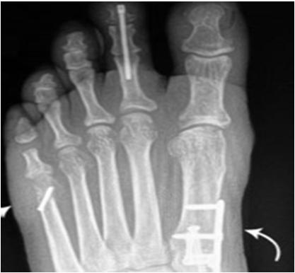
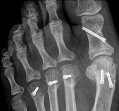
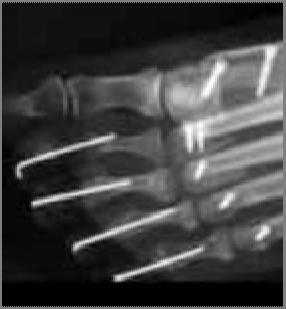
4 PIP (Proximal Interphalangeal) Joint Fusion for Hammertoe Correction Indication The PIP is the first joint of the small toes (digits 2 5). The indication for hammertoe surgery is when the PIP joint has a fixed curved deformity, caused by a contracture of the tendons (shortening), which leads to rigidity of the joint. A deformity producing enough pain or functional limitation may warrant surgery. The deformity develops gradually and cannot be straightened because it isbentandfixedinthispositionforalongperiodof time. The procedure essentially straightens the joint and fuses the proximal and middle phalanges in a straightened position. A deformity of the PIP (proximal interphalangeal) joint is called a Hammertoe. In the DIP (distal interphalangeal) joint, it s called Mallet Toe, and deformity of both the PIP and DIP joints is called Claw Toe. There are a variety of ways that a PIP joint fusion can be performed. The joint can be approached either through a longitudinal or transverse incision on the top of the toe. Once the joint is opened up, a small segment of bone is removed from either side of the joint, which creates enough room for the joint to straighten. The joint is then fixed in the straightened position, usually with a k wire, screw, or hammertoe implant. This procedure may be done in association with other procedures, such as a tendon transfer or tendon lengthening, to help keep the toe in the newly straightened position (e.g. Girdlestone Taylor procedure or Extensor Tendon Lengthening). Additional procedures to address underlying mechanical problems such as a gastrocnemius contracture or hypermobile first ray, which may have caused the small toe deformities, may be corrected in addition to the PIP joint fusion ) K WIRE 2) ARROW LOK 3) SMART TOE 4) PRO TOE


 of bone to help elevate the bone so that the metatarsal head is not as prominent.")

5 Metatarsal Shortening (Weil Osteotomy) A Weil metatarsal shortening osteotomy is performed to decrease pressure on a prominent metatarsal head in the forefoot. The metatarsal head is the portion of the metatarsal bone that articulates with the base of the toe. When the metatarsal is too long or is positioned in such a way that the associated metatarsal head is taking a disproportionate amount of weight, pain can occur. This source of pain in the forefoot is called metatarsalgia. A long or prominent metatarsal bone usually affects the second and, occasionally, the third metatarsal. It is often associated with a claw toe deformity of the involved toe. As the toe claws, it pulls forward the cushioned fat pad normally present in the forefoot, uncovering the metatarsal head and further exposing it to pressure. A Weil osteotomy is performed by making an incision over the base of the second (or other involved) toe. The surgeon exposes the distal aspect of the involved metatarsal, the metatarsal head and the neck. A saw is then used to cut the bone parallel to the sole of the foot. This allows the metatarsal head to be shifted backwards towards the heel, approximately 3 5 mm, though in some cases even farther. It is also possible to remove a small section (1 3 mm) of bone to help elevate the bone so that the metatarsal head is not as prominent. The metatarsal head fragment is then stabilized in the new position with one or two small screws. For speed and convenience, and because of the predictable length of screws needed, it is common for a surgeon to use a snap off or twist off screw. This is a special type of solid screw designed with a wire like shaft attached to the screw head, which allows it to be loaded directly into a wire driver and implanted quickly and easily. The shaft loaded into the wire driver snaps off when the screw head hits the proximal cortex, hence the name. Weil osteotomies are the most common indication for snap off screws. Weil Osteotomy Fixation with Snap Off Screw Weil Second Toe Shortened Above: Snap Off Screw. The most common use for this screw is for a Weil Osteotomy.

 or a scarf osteotomy procedure.")
6 Medial Closing Wedge Osteotomy (Akin) for Hallux Valgus (Bunion) Correction An Akin osteotomy is a medial closing wedge osteotomy of the proximal phalanx of the great toe (hallux) to correct hallux valgus deformity. However, because the procedure only corrects the proximal phalanx, and not the metatarsal (source of the bunion), it is the least powerful procedure, and is usually performed along with a distal chevron (Austin) or a scarf osteotomy procedure. Akin (Closing Wedge) Austin (Distal Chevron) Weil (Metatarsal Shortening) The procedure involves creating an osteotomy on the medial side of the proximal phalanx. Two cuts are made to remove a wedge of bone. The toe is shifted (closing the wedge) and fixed in this position with a small screw or a staple. Top: Screw fixation for various corrective osteotomies. Right: Akin using a nitinol staple for fixation. Most common application for staples.

 in the distal aspect of the first metatarsal, near the metatarsal head.")
7 Distal Chevron Osteotomy (Austin) for Hallux Valgus (Bunion) Correction A Chevron osteotomy is indicated for correction of mild to moderate hallux valgus deformity. This allows for a small reduction of the angle between the first and second metatarsal. It is ideal for bunions that are not particularly pronounced. It is frequently performed along with a medial closing wedge osteotomy (Akin) of the proximal phalanx. The procedure involves a V shaped cut (chevron) in the distal aspect of the first metatarsal, near the metatarsal head. This allows the distal aspect of the bone to be translated in the lateral direction (towards the outside of the foot). The cut bone is then fixed in this position, usually with one or two small screw. Any excess boney prominence on the inside of the foot (medial side) is then resected.


8 Scarf Osteotomy for Hallux Valgus (Bunion) Correction A scarf osteotomy is a type of procedure for hallux valgus (bunion deformity). The term scarf describes the shape of the osteotomy that the surgeon uses. It was popularized in Europe and the term originates from an architectural and carpentry term defined as: a joint made by notching, grooving, or otherwise cutting the ends of two pieces and fastening them together so that they lap over and join firmly into one continuous piece. The scarf osteotomy is preferred by some surgeons because it allows movement of the bone in a number of different planes. The bone segments can be rotated and translated, and can therefore be very versatile. The osteotomy involves a Z shaped cut in the first metatarsal which can then be moved and fixed in a new position, usually with two small screws. The excess bone on the inside of the foot (medial side) is then removed. The procedure to correct hallux valgus may also involves a release of the tight ligaments (lateral release) and tightening of the loose ligaments (medial plication) to balance the joint. Often another osteotomy called an Akin (medial closing wedge) is made in the proximal phalanx to complete the surgical correction. Scarf Lateral View Akin Scarf Weil


9 FOOT & ANKLE Combinations of Reconstructive Forefoot s Hammertoe Weil Bunion
 that has failed non operative management. The deformity is associated with an enlarged angle between the 4 th and 5 th metatarsal bones.")

10 5 th Metatarsal Rotational Osteotomy for Bunionette Correction Indication Arotational5 th metatarsal osteotomy (cutting and rotating the bone) is indicated for patients with a large, painful bunionette deformity (aka Tailor s Bunion) that has failed non operative management. The deformity is associated with an enlarged angle between the 4 th and 5 th metatarsal bones. A5 th metatarsal osteotomy is performed through an incision on the outside of the foot. The dissection is carried down to the bone. The outside of the bone is identified and an incomplete oblique cut is made ¾ of the way through the bone. A drill is then placed from top to bottom through the bone and a screw is positioned, but not tightened, in this hole. The remainder of the bone cut is then made. After the cut has been made, the far end (distal part) of the 5 th metatarsal is rotated inwards towards the 4 th metatarsal, reducing the deformity and allowing for the bunionette correction. The screw is then tightened to stabilize this position, which is reviewed under x ray. Withthebonerotated, thereis nowa prominentarea ofboneontheoutside (laterally). This is cut off and smoothed. The 5 th toe is then straightened up through the capsule on the outside of the base of the 5 th toe, allowing for improved positioning of this toe.
 of the great toe, and spurs act as a mechanical block to motion, which causes pain.")

11 Cheilectomy (1 st MTP) for Hallux Rigidus A cheilectomy removes bone spurs (osteophytes) on the top surface (dorsal aspect) of the great toe joint bones. Bone spurs develop with arthritis (hallux rigidus) of the great toe, and spurs act as a mechanical block to motion, which causes pain. The goal of a cheilectomy is to relieve pain and restore range of motion. An incision is made over the top of the first metatarsophalangeal joint. Care is taken to avoid the tendon that extends the big toe. Any bone spurs are removed. If there is inflamed joint tissue or debris, these are removed as well. The cartilage on the joint surfaces is inspected. Approximately 30 percent of the top portions of the head of the metatarsal bone and corresponding bone spurs are removed. Bone spurs on 1 st MTP joint Cheilectomy to remove bone spurs Pain eliminated, restored range of motion
 together. The goal of surgery is to make the great joint solidly aligned and immobile.")


12 1 st MTP Joint (MPJ) Fusion for Hallux Rigidus (or Hallux Valgus) This procedure involves fusing the great toe joint (first MTP joint) together. The goal of surgery is to make the great joint solidly aligned and immobile. This relieves much of the pain since motion through the arthritic joint is eliminated. Great toe fusion is typically performed in patients who already have significant arthritis of the 1st MTP joint (late stage or severe hallux rigidus). It can also be successfully used, however, as a salvage procedure for patients with severe bunion deformities (hallux valgus). To fuse the great toe joint, any remaining cartilage on the arthritic joint surface is removed and the underlying bone is prepared for fusion, usually by using cup and cone reamers. The joint is positioned in a manner which maximizes walking ability and maintains acceptable clinical alignment. This is traditionally done with the toe positioned so that it just gently touches the ground in a weight bearing position (i.e degrees of dorsiflexion). The fused joint is typically fixated with two cross screws or a plate with screws. MTP Fusion Plate Cross Screws One Third Tubular Plate with Lag Screw MTP Fusion Plate
. The joint is removed and replaced with a metal, plastic, or silicone implant.")
.")


13 1 st MTP Hemi or Total Joint Replacement for Hallux Rigidus A first MTP joint replacement treats arthritis of the great toe (hallux rigidus). The joint is removed and replaced with a metal, plastic, or silicone implant. Implant options include total MTP joint replacements (which replace both sides of the joint) and hemi MTP joint replacements (which replace only one side of the joint either the metatarsal head or the base of the proximal phalanx). The primary goals are to relieve pain and to retain motion (unlike a fusion which eliminates motion of the joint). An incision is made over the first MTP joint and carried down to the join. The joint surface along with a small amount of bone is removed from the arthritic joint. Bone spurs are removed. The canals of the bones are then opened with a special pre drilled or reamer, and the implants are placed. The implants may screw into the shaft of the bone, or may be press fit (e.g. Morse taper). Potential complications include silicone synovitis when a silicone implant is used, as small particles of silicone may wear off. Other complications include implant loosening or displacement, instability of the joint. Hemi (Base of Proximal Phalanx) Hemi (Metatarsal Head) Total MTP Joint Replacement
 are at risk of inadequate healing and re fracture of the bone in the same location.")
 of the metatarsal.")


.")

14 Jones Fracture ORIF Due to a number of mechanical and biologic factors, patients with a base of 5 th metatarsal stress fracture (Jones fractures) are at risk of inadequate healing and re fracture of the bone in the same location. Patients with this injury where it does not adequately heal, or the fracture recurs, may benefit from stabilization of the fracture with a screw placed down the intramedullary canal (shaft) of the metatarsal. Highly active and athletic patients benefit from screw fixation after the injury to accelerate healing and rehabilitation and reduce the risk of refracture. The surgical procedure involves placing an appropriate sized screw, starting at the tip of the base of the 5 th metatarsal, and placing the screw within the bone across the fracture site. This is done by making a small incisionnearthebaseofthe5 th metatarsal. The tip of the 5 th metatarsal is identified on x ray (fluoroscopy). A drill is then used to enter the canal of the 5 th metatarsal, and then placed across the fracture site. This is done under fluoroscopy to ensure the screw is correctly positioned. A screw with an appropriate diameter and length is chosen. The diameter is typically mm and the screw is then positioned across the fracture site so as to stabilizes the bone and compresses the fracture to promote healing of the fracture. Typically a solid core screw is preferred, although some systems use a cannulated approach (instrumentation) before final fixation with a solid core screw. Jones Fracture Screw Fixation
15 FOOT & ANKLE MIDFOOT PROCEDURES
 joints.")


16 Midfoot Fusions (TMT, NC, TN, CC) A midfoot fusion is performed to treat midfoot arthritis. The procedure involves fusion of one or more of the first three tarsometatarsal (TMT) joints. The second TMT joint is most commonly involved, but the first and third may also be affected by arthritis. Typically the 4 th and 5 th TMT joints are not fused, as these joints have more motion than the 1 st,2 nd,or3 rd TMT joints. Other common midfoot fusions include Naviculocuneiform (NC) fusions,talonavicular (TN) fusions, and Calcaneocuboid (CC) fusions, and Intercuneiform fusions. TMT Fusion Plate Fusion of any of these joints begins with removing any remaining cartilage between them. To access the joint for preparation, a special joint distractor or pin distractor, or a lamina spreader may be used. After the joint space is prepared, the bones are stabilized with screws or plates. To help aid in the fusion, bone graft may be used in some instances. These procedure essentially turns a painful, stiff, arthritic joint into a painless, fused joint. Talonavicular (TN) Fusion Naviculocuneiform (NC) Fusion

 to heal. The location of the incision is dictated by both the location of the fracture and the location of the joints that are disrupted.")
 joints are disrupted, then only one or two incisions are made on the top and/or inside aspect of the foot.")

17 Lisfranc Injury FOOT & ANKLE Indication The Lisfranc joints are located in the midfoot. The main indication for this ORIF of the Lisfranc joints is a displaced or unstable Lisfranc fracture, or a dislocation. The purpose of the surgery is to reposition the bones andjointsinthemid part of the foot, allowing the associated torn ligaments (the strong tissues that hold these bones together and support the arch) to heal. The location of the incision is dictated by both the location of the fracture and the location of the joints that are disrupted. If all five of the Lisfranc joints are disrupted, then two or three incisions on the top of the foot may be needed; one on the top inside and inside border of the foot, and one on the top outside of the foot. If the first three tarsometatarsal (TMT) joints are disrupted, then only one or two incisions are made on the top and/or inside aspect of the foot. Once the disrupted tarsometatarsal joints are identified, the dissection is carried down to the involved joints and the debris is cleaned out. The disrupted joints are repositioned back to the position they were in prior to the injury. The joints are then fixed with strong screws. However, if the fragmentation is excessive, a plate may be required. One exception is a disruption of the 4 th and 5 th tarsometatarsal joints; in this case, the bone is provisionally fixed with wires. The wires are then removed after about six weeks so that some movement of these joints can be encouraged. Lisfranc surgery with TMT plates Lisfranc surgery with screws


18 Lapidus for Hallux Valgus (Bunion) Correction The Lapidus procedure is used to correct a moderate to severe hallux valgus deformity. It is also indicated for hallux valgus associated with a hypermobility of the first ray. The procedure involves an incision over the dorsomedial (top and inside) part of the midfoot. The first tarsometatarsal (TMT) joint is exposed. This joint is then prepared for fusion. In the context of fusing this joint, the angle between the first and second metatarsal is decreased. This joint is then typically fused with two screws or a plate. In addition, the bunion is corrected through a medial incision over the great toe, which allows the joint capsule to be tightened, as well as the prominent medial bone associated with the bunion to be removed. It is often necessary to perform a release of the tightened structures on the lateral (outside) part of the great toe joint. Lapidus Plate Cross Screws

.")
.")
19 Cotton Osteotomy (Plantarflexing Opening Wedge of the Medial Cuneiform) for Flatfoot Correction A Cotton Osteotomy is a procedure where the surgeon makes an osteotomy and an opening wedge in the dorsal aspect of the medial cuneiform. The procedure is performed to correct flatfoot deformities, usually in children and young adults. It works by plantarflexing (flexing downwards) the medial column, which are the bones along the inside of the foot). Doing this restores the arch of the foot. The procedure may be done in conjunction with other flatfoot procedures, such as an Evans Osteotomy or a Medial Displacement Calcaneal Osteotomy (also known as an MDCO or Calc Slide ). The osteotomy is made directly across (transverse) the dorsal aspect of the medial cuneiform and then spread open so that a wedge can be inserted, restore the arch of the foot. Various wedge options are available for this procedure. The surgeon may take a bone graft wedge from the patient s own body (autograft). Alternatively, an allograft bone wedge can be used, either pre shaped to perfectly fit the osteotomy, or an allograft ilium tricortical wedge where the surgeon must cut the graft to size. The surgeon may choose to use a specific Cotton plate with a built in titanium wedge. Newer wedge implants made from porous titanium or PEEK are also available. The wedge is usually between 4 7 mm in width, and may be secured with ancillary fixation to hold it in place (screws, staples, or a plate). Wedge being inserted into medial cuneiform Cotton Pre Shaped Allograft Bone Wedge Cotton Titanium Wedge
20 FOOT & ANKLE HINDFOOT PROCEDURES


 to expose the talonavicular joint, and the other is made on the outside to expose the subtalar joint, the calcaneal cuboid joint,")
21 Triple Arthrodesis for Arthritis or Acquired Adult Flatfoot Triple arthrodesis is indicated for patients who have a deformity of the hindfoot, such as acquired adult flatfoot deformity, where there is arthritis or stiffness in the involved joints. This type of procedure sometimes offers a more reliable result than other hindfoot corrective procedures. Because of the importance of preserving all the joints in the foot, this procedure is done a lot less often than it used to be. However, if there is enough arthritis or dysfunction in the involved joints where preservation is improbable, then a triple arthrodesis may be effective. TN Joint Subtalar Joint This surgical procedure involves the fusion of three joints: the talonavicular (TN) joint, the subtalar joint, and the calcaneal cuboid(cc)joint. Twoincisionsaremadeonbothsidesofthefoot;oneismadeontheinside (medial) to expose the talonavicular joint, and the other is made on the outside to expose the subtalar joint, the calcaneal cuboid joint, and the outside (lateral) aspect of the talonavicular joint. Once exposed, all of the remaining cartilage from the joints is removed and prepared for fusion. Typically, the joint is then packed with bone graft, either from other bones, such as the proximal tibia or the iliac crest, or using allograft. Once all the joints have been prepared and the bone graft has been placed, the hindfoot position is corrected to a neutral position. Once repositioned, the foot is first provisionally fixed with wires, and then definitively fixed with screws or plates in each joint. Once everything is in place, the incisions are closed up with sutures. TN Joint CC Joint CC Joint Subtalar Joint
. Arthritis in this joint is commonly seen after heel bone fractures or joint dislocations.")


22 Subtalar Fusion for Arthritis The main indication for a subtalar fusion is to treat painful arthritis in the subtalar joint (the large joint above the heel bone and below the ankle). Arthritis in this joint is commonly seen after heel bone fractures or joint dislocations. Another indication for a subtalar arthrodesis is for patients who need the change to position of the hindfoot in order to distribute load more evenly. For example, a patient with acquired flatfoot deformity, a condition where the heel is offset and load is unevenly distributed, might consider a subtalar fusion. On the lateral (outside front) region of the ankle, a cut is made down to the subtalar joint to expose the joint, particularly the larger posterior portion of the joint, or facet. Once exposed, the joint is ready for fusion. First, the cartilage, or what is left of the cartilage, is removed between the heel bone and the talus. Next, the bone is shaveddowntoapointthatcanfuse. Thejointisthenplacedinitsdesiredpositionandsecuredwithlarge screws. To increase the chance of the joint fusing together, bone graft is sometimes used. 6.5mm Headless / 7.0mm Headed Cannulated Screws


. This wedge comes from the patient s iliac crest which is at the top of the pelvis.")
23 Lateral Column Lengthening (Evans Osteotomy) for Flatfoot Correction A lateral column lengthening procedure (Evans Osteotomy) is indicated for patients with acquired adult flatfoot deformity, where the front part of the foot is splayed out to the side. This procedure is often combined with a medializing calcaneal osteotomy, (often referred to as the All American procedure ), as a technique for adjusting acquired adult flatfoot deformity. The lateral column is made up of the calcaneus, the cuboid, and the fourth and fifth metatarsals. Therefore, the lateral column lengthening procedure involves lengthening this region. There are two general ways of doing a lateral column lengthening, both of which involve inserting a wedge into the lateral column. One way of performing this procedure is by cutting the bone through the front part of the calcaneus. The osteotomy is made right before the calcaneal cuboid joint, which is then spread about 7 10 mm so that a wedge can be inserted, in order to lengthen the column. Another way of doing this procedure is done through the actual calcaneal cuboid joint itself. A bone graft wedge is inserted in the joint, which serves as a joint fusion while also lengthening the lateral column. Various wedge options are available for this procedure. The surgeon may take a bone graft wedge from the patient s own body (autograft). This wedge comes from the patient s iliac crest which is at the top of the pelvis. Alternatively, an allograft bone wedge can be used, either pre shaped to perfectly fit the osteotomy, or an allograft ilium tricortical wedge where the surgeon must cut the graft to size. The surgeon may choose to use a specific Evans plate with a built in titanium wedge. Newer wedge implants made from porous titanium or PEEK are also available. The wedge is usually between 6 12mm in width, and may be secured with ancillary fixation to hold it in place (screws, staples, or a plate). A lateral column lengthening procedure is a very powerful procedure, since it can dramatically change the shape of the foot. The advantages of this procedure include the ability to take a pronounced flatfoot deformity and turn it into a near normal looking foot. However, the disadvantages include the potential of creating a stiffer foot; possibly overcorrecting the foot (which may lead to more symptoms); and a higher rate of specific complications, such as painful hardware, sural nerve irritation, and nonunion. Evans Osteotomy Wedge Plate Evans Plate with Built in Wedge Evans Pre Shaped Allograft Bone Wedge Evans Titanium Wedge

 may be shifted towards the midline of the body (medializing calcaneal osteotomy), or away from the midline of the body")

 portion of the ankle joint.")
 Plate An oblique incision is made on the outside region of the heel.")
24 Calcaneal Osteotomy for Flatfoot or High Arch Correction A calcaneal osteotomy comprises cutting the heel bone and shifting it medially or laterally. A calcaneal osteotomy is indicated for patients whose hindfoot alignment is significantly offset and for whom non operative management has failed. Alignment of the heel influences how weight bearing stress is applied to the foot, ankle, knee and hip. Depending on which way the hindfoot is offset, the heel (calcaneus) may be shifted towards the midline of the body (medializing calcaneal osteotomy), or away from the midline of the body (lateralizing calcaneal osteotomy). For example, a patient with acquired adult flatfoot deformity will often have the heel offset to the outside and may benefit from a medializing calcaneal osteotomy to shift the hindfoot to the inside and change the way load is distributed through the heel. On the other hand, a patient with a high arched foot (cavus foot pattern) often has a heel that is offset to the inside. Individuals with a high arched foot may have symptoms ranging from pain along the lateral or outside half of the foot, to wearing out of the inside (medial) portion of the ankle joint. With this structural alignment, individuals are predisposed to sprain their ankle and may develop ankle instability. In severe forms of ankle instability, a lateralizing calcaneal osteotomy may be beneficial. MDCO (Calc Slide) Plate An oblique incision is made on the outside region of the heel. One precaution while exposing the bone is to avoid cutting or injuring the sural nerve which provides sensation to the outside part of the foot. Once exposed, the back part of the heel bone is cut (osteotomy) into two pieces. The back part of the bone is then either shifted medially or laterally. The bone is shifted between 5 12mm. After the bone is shifted, it is fixed in place usually with one or two large screws or a plate. Any sharp bone edges are smoothed out before closing.

25 FOOT & ANKLE ANKLE PROCEDURES
.")


.")

26 Ankle Fracture and Syndesmosis Repair The upper part of the ankle joint comes from the tibia and the fibula. The tibia forms the front, rear and inner part of the ankle joint. The lower fibula forms the outer part of the ankle joint. The rounded ends of these bones are called malleoli (singular is malleolus). There are two malleoli on the tibia (medial and posterior) and one on the fibula (lateral). Ankle fractures occur when the malleoli are broken. These fractures are very common. Ankle fractures can happen after falls, car accidents or twisting of the ankle. One, two or all three malleoli can be broken. In addition to ankle fractures, injury to the syndesmosis is also common. The syndesmosis is the joint just above the ankle where the tibia and fibula. Although the syndesmosis is technically a joint, there is very little motion between these bones. The main function is to provide stability to the ankle joint and allow motion of the ankle joint. The syndesmosis may become injured with a twisting or rotational injury to the ankle. It is commonly referred to as a high ankle sprain. Distal Fibula Fracture Medial Malleolus Fracture Most ankle fracture surgery involves open reduction and internal fixation (ORIF). An incision is made over the ankle to see the fractured bones. Like a jigsaw puzzle, the pieces of the broken bones are placed back together (open reduction). The broken bones are then held together (internal fixation) in this correct position with metal plates and/or screws. This internal fixation provides stability so movement can begin shortly after surgery as the ankle fracture heals. Plates and screws used to fix an ankle fracture are not removed as long as they are not causing problems. In the case of injury to the syndesmosis, the surgeon will insert one or two screws that go from the fibula into the tibia. The screws may be placed through a plate that sits on the fibula bone. Alternatively, a suture device, like the Arthrex TightRope product, may be used. Distal Fibula (Lateral) Ankle Fractures Left: Arthrex plate with TightRope Right: Synthes plate with syndesmosis screws


27 Ankle Fusion for Arthritis The goal of ankle arthrodesis (ankle fusion) is to relieve pain and maintain or improve function for a patient with ankle arthritis. Ankle arthritis is degeneration of the cartilage that covers the ends of the bones that form the ankle joint. These bones are the tibia, fibula, and talus. Pain typically is made worse with movement of the arthritic ankle. The goal of ankle arthrodesis is to take the ankle bones and fuse them into one bone. This eliminates motion and reduces pain from the arthritic joint. Ankle arthrodesis may be performed through an incision on the outside of the ankle or the front of the ankle. Sometimes a bone graft may be used to aid in fusion. This graft may be taken from the patient s own body (pelvis, heel bone or just below the knee) which is called autograft. Or, allograft DBM bone putty or bone sponge strips may be used. Ankle arthrodesis may be performed through small incisions that allow a camera and tools to be placed into the joint. This is known as arthroscopic surgery. After the joint has been accessed, instruments are used to scrape away remaining cartilage and the joint surface is prepared for fusion. Screws or plates may be used to hold the ankle in the correct position for fusion. If a patient is having his subtalar joint fused at the same time, a nail (a tubular metal implant that goes up into the canal of the bone) may be used to hold the joints in position. The choice of approach and hardware depends on a patient s specific anatomy, condition and the surgeon s preference Ankle Fusion Implant Options 1) Large cannulated screws 2) TTC (Tibiotalocalcaneal ) Nail 3) TTC (Tibiotalocalcaneal ) Anterolateral Plate
, is a surgical procedure that treats severe ankle arthritis.")


28 Total Ankle Arthroplasty/Replacement (TAA/TAR) for Arthritis Total ankle arthroplasty (TAA), also known as total ankle replacement (TAR), is a surgical procedure that treats severe ankle arthritis. Arthritic changes may be a result of normal wear and tear due to aging, or from an injury such as a broken ankle or dislocation. Arthritis eventually leads to loss of cartilage, pain and/or deformity. The goal of an ankle replacement is to improve ankle motion so the patient has less pain during activity. The ankle is approached from the front or the side depending on the type of implant being used. Bone is then cut, allowing for placement of the metal and plastic components that re create the ankle joint. Sometimes the patient will have a tight calf muscle or tight Achilles tendon that needs to be lengthened to improve range of motion of the ankle. Either before or after the ankle replacement is put in place, the surgeon will determine if the calf muscle or Achilles is tight. A tight calf muscle or Achilles tendon is addressed with a lengthening procedure. This is important to improve motion after the surgery, as well as to take stress off the ankle replacement. SALTO TALARIS TOTAL ANKLE Acquired by Integra from Tornier following Wright/Tornier Merger STAR TOTAL ANKLE Scandinavian Total Ankle Replacement. Acquired by Stryker from SBi (Small Bone Innovations) TRABECULAR METAL TOTAL ANKLE Zimmer INFINITY TOTAL ANKLE Wright Medical INBONE TOTAL ANKLE Wright Medical
Index. Clin Podiatr Med Surg 22 (2005) Note: Page numbers of article titles are in boldface type.
 Note: Page numbers of article titles are in boldface type.") Clin Podiatr Med Surg 22 (2005) 309 314 Index Note: Page numbers of article titles are in boldface type. A Abductor digiti minimi muscle, myectomy of, for tailor s bunionette, 243 Achilles tendon, lengthening
Clin Podiatr Med Surg 22 (2005) 309 314 Index Note: Page numbers of article titles are in boldface type. A Abductor digiti minimi muscle, myectomy of, for tailor s bunionette, 243 Achilles tendon, lengthening
Technique Guide. 6.5 mm Midfoot Fusion Bolt. For intramedullary fixation of the medial column of the foot.
 Technique Guide 6.5 mm Midfoot Fusion Bolt. For intramedullary fixation of the medial column of the foot. Table of Contents Introduction 6.5 mm Midfoot Fusion Bolt 2 AO Principles 4 Indications 5 Surgical
Foot and Ankle Systems Coding Reference Guide
 Foot and Ankle Systems Coding Reference Guide Physician Arthrodesis 27870 Arthrodesis, ankle, open 27871 Arthrodesis, tibiofibular joint, proximal or distal 28705 Arthrodesis; pantalar 28715 Arthrodesis;
Use of the 20 Memory Staple in Osteotomies of Fusions of the Forefoot
 168 Forefoot Reconstruction Use of the 20 Memory Staple in Osteotomies of Fusions of the Forefoot Definition, History, Generalities This staple first provides a permanent compression both in the prongs
.org. Tibia (Shinbone) Shaft Fractures. Anatomy. Types of Tibial Shaft Fractures
 Shaft Fractures. Anatomy. Types of Tibial Shaft Fractures") Tibia (Shinbone) Shaft Fractures Page ( 1 ) The tibia, or shinbone, is the most common fractured long bone in your body. The long bones include the femur, humerus, tibia, and fibula. A tibial shaft fracture
Tibia (Shinbone) Shaft Fractures Page ( 1 ) The tibia, or shinbone, is the most common fractured long bone in your body. The long bones include the femur, humerus, tibia, and fibula. A tibial shaft fracture
A Patient s Guide to Adult-Acquired Flatfoot Deformity
 A Patient s Guide to Adult-Acquired Flatfoot Deformity Glendale Adventist Medical Center 1509 Wilson Terrace Glendale, CA 91206 Phone: (818) 409-8000 DISCLAIMER: The information in this booklet is compiled
Midfoot - Reduction & Fixation - ORIF - screw fixation - AO Surgery Reference. ORIF - screw fixation
 Midfoot - TMT (Lisfranc) injury 1. Diagnosis ORIF - screw fixation Authors Mechanism of the injury Tarso-metatarsal (Lisfranc) injuries may be caused by direct or indirect forces. Direct forces include
Midfoot Plating Techniques Surgical Technique
 Midfoot Plating Techniques Surgical Technique Midfoot Plating Techniques Lapidus Plate Designed to provide excellent fixation for a lapidus procedure, the Lapidus Plate offers the foot & ankle surgeon
Index. Note: Page numbers of article titles are in bold face type.
 Index Note: Page numbers of article titles are in bold face type. A Achilles tendon, Zadek osteotomy effects on, 430 Adult acquired flatfoot disorder, 387 403 calcaneal Z osteotomy for, 397 399 historical
6.5 mm midfoot fusion bolt
 6.5 mm midfoot fusion bolt For intramedullary fixation of the medial column of the foot SurgIcal technique Table of Contents Introduction 6.5 mm Midfoot Fusion Bolt 2 AO Principles 4 Indications 5 Surgical
Foot & Ankle Disorders
 Foot & Ankle Disorders Hillingdon PGMC 6-7-2013 Htwe Zaw FRCS (Tr&Orth) Consultant Foot & Ankle and Trauma Surgeon Hillingdon Hospitals NHS Foundation Trust Overview Anatomy: hindfoot-midfoot coupling
ORTHOLOC CROSSCHECK TECHNOLOGY OVERVIEW FOR HOSPITAL VALUE ANALYSIS COMMITTEE
 ORTHOLOC CROSSCHECK TECHNOLOGY OVERVIEW FOR HOSPITAL VALUE ANALYSIS COMMITTEE Indications for Use The ORTHOLOC 3Di Foot Reconstruction System is intended for use in stabilization of fresh fractures, revision
Calcaneus (Heel Bone) Fractures
 Fractures") Page 1 of 8 Calcaneus (Heel Bone) Fractures A fracture of the calcaneus, or heel bone, can be a painful and disabling injury. This type of fracture commonly occurs during a high-energy event such as a
Page 1 of 8 Calcaneus (Heel Bone) Fractures A fracture of the calcaneus, or heel bone, can be a painful and disabling injury. This type of fracture commonly occurs during a high-energy event such as a
CHARLOTTE Lisfranc. Recontruction System SURGICAL TECHNIQUE
 CHARLOTTE Lisfranc Recontruction System SURGICAL TECHNIQUE Contents Preface 3 Chapter 1 4 Chapter 2 4 4 4 4 4 Chapter 3 5 5 5 6 6 6 7 7 8 Chapter 4 8 8 9 10 Appendix 10 Introduction Indications and Contraindications
A Patient s Guide to Hallux Rigidus
 A Patient s Guide to Hallux Rigidus Glendale Adventist Medical Center 1509 Wilson Terrace Glendale, CA 91206 Phone: (818) 409-8000 DISCLAIMER: The information in this booklet is compiled from a variety
Variable Angle LCP Forefoot/Midfoot System 2.4/2.7. Procedure specific plates for osteotomies, arthrodeses and fractures of the foot.
 Variable Angle LCP Forefoot/Midfoot System 2.4/2.7. Procedure specific plates for osteotomies, arthrodeses and fractures of the foot. Compression technology Variable angle locking technology Anatomic and
A Patient s Guide to Hallux Rigidus
 A Patient s Guide to Hallux Rigidus Suite 11-13/14/15 Mount Elizabeth Medical Center 3 Mount Elizabeth Singapore, 228510 Phone: (65) 6738 2628 Fax: (65) 6738 2629 DISCLAIMER: The information in this booklet
.org. Ankle Fractures (Broken Ankle) Anatomy
 Anatomy") Ankle Fractures (Broken Ankle) Page ( 1 ) A broken ankle is also known as an ankle fracture. This means that one or more of the bones that make up the ankle joint are broken. A fractured ankle can range
Ankle Fractures (Broken Ankle) Page ( 1 ) A broken ankle is also known as an ankle fracture. This means that one or more of the bones that make up the ankle joint are broken. A fractured ankle can range
Merete PlantarMAX Lapidus Plate Surgical Technique. Description of Plate
 Merete PlantarMAX Lapidus Plate Surgical Technique Description of Plate Merete Medical has designed the PlantarMax; a special Plantar/Medial Locking Lapidus plate which places the plate in the most biomechanically
19 Arthrodesis of the First Metatarsocuneiform Joint
 19 Arthrodesis of the First Metatarsocuneiform Joint CHARLES GUDAS Abduction of the first metatarsal to correct metatarsus primus varus and hallux valgus was first described by Albrecht in 1911. 1 Lapidus
Contents. Chapter 1 4 Chapter Chapter Chapter Chapter 5 15
 Contents Chapter 1 4 Chapter 2 5 5 Chapter 3 6 6 7 Chapter 4 8 8 8 8 9 10 10 11 12 13 14 14 Chapter 5 15 Introduction Intended Use Indications Device Description Implant Options and Sizing Instrumentation
Zimmer Trabecular Metal Ankle Interpositional Spacer and Trabecular Metal Ankle Fusion Spacer
 Zimmer Trabecular Metal Ankle Interpositional Spacer and Trabecular Metal Ankle Fusion Spacer Surgical Technique 2 Zimmer Trabecular Metal Ankle Interpositional Spacer and Trabecular Metal Ankle Fusion
The Flower Medial Column Fusion Plate
 The Flower Medial Column Fusion Plate PROCEDURE GUIDE www.flowerortho.com The Flower Foot & Ankle Application NC FUSION PLATE 2-HOLE COMPRESSION PLATE TMT FUSION PLATE LAPIDUS FUSION PLATE COMPRESSION
Foot & Ankle. Smart Toe II. Intramedullary Implant. Operative Technique. Foot & Ankle
 Foot & Ankle Smart Toe II Intramedullary Implant Operative Technique Foot & Ankle Smart Toe This publication sets forth detailed recommended procedures for using Stryker Osteosynthesis devices and instruments.
Foot Plating System Product rationale
 Product rationale CONTENTS Introduction 2 Foot plating system 2.7mm Universal Locking Plate 2.7mm 3 Metatarsophalangeal Plate 4 Opening Wedge Locking Plate 5 Opening Wedge Osteotomy Plate 5 Foot plating
A Patient s Guide to Claw Toes and Hammertoes
 A Patient s Guide to Claw Toes and Hammertoes Suite 11-13/14/15 Mount Elizabeth Medical Center 3 Mount Elizabeth Singapore, 228510 Phone: (65) 6738 2628 Fax: (65) 6738 2629 DISCLAIMER: The information
BME. Foot. Procedure Manual
 BME Foot Procedure Manual Table of Contents Hallux Interphalangeal (IP) Joint Arthrodesis 3 Akin Osteotomy 4 First Metatarsophalangeal (MTP) Joint Arthrodesis 5 Chevron Osteotomy/Bunionectomy of the Distal
Physical Examination of the Foot & Ankle
 Inspection Standing, feet straight forward facing toward examiner Swelling Deformity Flatfoot (pes planus and hindfoot valgus) High arch (pes cavus and hindfoot varus) Peek-a-boo heel Varus Too many toes
DAY 1: FRIDAY, 31 st AUGUST Operative Sessions: 8.00 am to 3.30 pm
 DAY 1: FRIDAY, 31 st AUGUST Operative Sessions: 8.00 am to 3.30 pm SURGICAL PROCEDURES / CASES (PROPOSED) 1. Haglund with Tendoachilles Tendinopathy 2. ORIF Calcaneus Fracture 3. OCD talus: Arthroscopic
Rippstein, Trnka, Saragas, Narramore
 THURS 25th MAY 07:45 07:55 Welcome and Introductions Paulo Ferrao Lecture 1: 08:00 10:20 Forefoot I: Hallux Valgus and Lesser Toes Mark Easley 30 mins 08:00 08:30 Surgical Management of Hallux Valgus Saragas,
ORTHOLOC 3Di. Foot Reconstruction System SURGIC AL TECHNIQUE
 ORTHOLOC 3Di Foot Reconstruction System S C R E W TA R G E T I N G G U I D E SURGIC AL TECHNIQUE SURGEON DESIGN TEAM The ORTHOLOC 3Di Foot Reconstruction System was developed in conjuction with: ORTHOLOC
Lesser MPJ Hemi Implant
 Lesser MPJ Hemi Implant Surgical Technique Contents Product The BioPro Lesser MPJ Hemi Implant is a simple, durable, metallic hemiarthroplasty resurfacing prosthesis for the treatment of arthritis, Freiberg
A Patient s Guide to Claw Toes and Hammertoes
 A Patient s Guide to Claw Toes and Hammertoes 20295 NE 29th Place, Ste 300 Aventura, FL 33180 Phone: (786) 629-0910 Fax: (786) 629-0920 admin@instituteofsports.com DISCLAIMER: The information in this booklet
WHAT IS ARTHRITIS OF THE BIG TOE (HALLUX RIGIDUS)?
?") Mr Laurence James BSc MBBS MRCS(Eng) FRCS(Tr&Orth) Consultant Orthopaedic Surgeon Foot, Ankle and Sports Injuries WHAT IS ARTHRITIS OF THE BIG TOE (HALLUX RIGIDUS)? A common term for arthritis of the metatarsophalangeal
Mr Laurence James BSc MBBS MRCS(Eng) FRCS(Tr&Orth) Consultant Orthopaedic Surgeon Foot, Ankle and Sports Injuries WHAT IS ARTHRITIS OF THE BIG TOE (HALLUX RIGIDUS)? A common term for arthritis of the metatarsophalangeal
Flower Medium Headless & Cannulated Screws
 Flower Medium Headless & Cannulated Screws PROCEDURE GUIDE www.flowerortho.com The Flower Foot & Ankle Application NC FUSION PLATE 2-HOLE COMPRESSION PLATE TMT FUSION PLATE LAPIDUS FUSION PLATE COMPRESSION
2017 SAFSA CONGRESS PROGRAMME
 2017 SAFSA CONGRESS PROGRAMME THURSDAY, MAY 25 07h45 07h55: WELCOME & INTRODUCTIONS Forefoot I: Hallux Valgus and Lesser Toes (08h00-10h00 Lectures) 08h00 08h30: Surgical Management of Hallux Valgus Rippstein,
.org. Posterior Tibial Tendon Dysfunction. Anatomy. Cause. Symptoms
 Posterior Tibial Tendon Dysfunction Page ( 1 ) Posterior tibial tendon dysfunction is one of the most common problems of the foot and ankle. It occurs when the posterior tibial tendon becomes inflamed
Surgical Technique Guide
 Guide CAUTION: Federal Law (USA) restricts this device to sale by or on the order of a physician. INDICATIONS FOR USE The Align Anterior Ankle Fusion Plate is intended to facilitate arthrodesis of the
Clinical. Solutions. Synthes Solutions. Foot and Ankle.
 Clinical Solutions Foot and Ankle. Foot and Ankle. Fractures of the tibial shaft Fractures of the distal fibula Fractures of the distal tibia Fractures and osteotomies of the calcaneus Arthrodesis Fractures,
*Rippstein, Trnka, Saragas, Hoffman
 THURS 25th MAY 07:00 07:10 Welcome and Introductions Paulo Ferrao Lecture 1: 07:10 09:45 Forefoot I: Hallux Valgus and Lesser Toes Mark Easley 40 mins 07:10 07:50 Surgical Management of Hallux Valgus 30
Foot Reconstruction System SURGICAL TECHNIQUE
 ORTHOLOC 3Di Foot Reconstruction System SURGICAL TECHNIQUE SURGEON DESIGN TEAM The ORTHOLOC 3Di Foot Reconstruction System was developed in conjuction with: Robert B. Anderson, MD OrthoCarolina Charlotte,
PEDUS-L. Locking Plantar Lapidus Plate
 PEDUS-L Locking Plantar Lapidus Plate Page 1 PEDUS-L - Locking Plantar Lapidus Plate Table of Contents Implants 3 System 4 Operation manual 5 Approach 5 Identification of the TMT 1 joint with a cannula
AcUMEDr. AcUTRAKr. Headless Compression Screw
 AcUMEDr AcUTRAKr Headless Compression Screw AcUTRAKr HEADLESS COMPRESSION SCREW Since 1988 Acumed has been designing innovative solutions for the demanding situations facing orthopedic surgeons, hospitals
Scar Engorged veins. Size of the foot [In clubfoot, small foot]
![Scar Engorged veins. Size of the foot [In clubfoot, small foot]](/thumbs/78/77722241.jpg "Scar Engorged veins. Size of the foot [In clubfoot, small foot]") 6. FOOT HISTORY Pain: Walking, Running Foot wear problem Swelling; tingly feeling Deformity Stiffness Disability: At work; recreation; night; walk; ADL, Sports Previous Rx Comorbidities Smoke, Sugar, Steroid
6. FOOT HISTORY Pain: Walking, Running Foot wear problem Swelling; tingly feeling Deformity Stiffness Disability: At work; recreation; night; walk; ADL, Sports Previous Rx Comorbidities Smoke, Sugar, Steroid
InCoreTM Lapidus System
 InCoreTM Lapidus System Precision Guided Correction Surgical Technique InCore TM Lapidus System Precision Guided Correction Tri-Planar Correction Targeting Guide is intended to aid and stabilize angular/rotational
UNITE Cannulated Screw System
 FOOT & ANKLE INTELLIGENTLY DESIGNED. 1 MEDLINE MEDLINEUNITE Cannulated Screw System Maximum Efficiency. Optimal Functionality. Uncompromising Quality. 2 MEDLINE Medline Unite Cannulated Screw System INTELLIGENT
Columbia/NYOH FOOT and ANKLE ROTATION-SPECIFIC OBJECTIVES
 Updated 2/8/10 Columbia/NYOH FOOT and ANKLE ROTATION-SPECIFIC OBJECTIVES INTERPERSONAL AND COMMUNICATION SKILLS Resident will at all times demonstrate behavior that is beyond reproach. Residents must be
Supplemental Use Guide Standard Triple Arthrodesis
 Acutrak 2 Headless Compression Screw System 4.7 mm and 7.5 mm Screws Supplemental Use Guide Standard Triple Arthrodesis Acumed is a global leader of innovative orthopaedic and medical solutions. We are
Surgical Technique. Customer Service:
 Patent and Patent Pending CAUTION: Federal Law (USA) restricts this device to sale by or on the order of a physician. INDICATIONS FOR USE The Axis Charcot Fixation System in diameters of 5.5, 6.5 and 7.5mm
The American Academy of Foot & Ankle Osteosynthesis. presents
 The American Academy of Foot & Ankle Osteosynthesis presents Comprehensive Course of Internal Fixation for Reconstructive Surgery and Trauma of the Foot & Ankle September 4-6, 2014 Goodlett Farms Innovation
TABLE OF CONTENTS
 TABLE OF CONTENTS 3 4 5 6 7 8 9 10 11 12 13 14 15 16 17 18-24 Hallux Interphalangeal (IP) Joint Arthrodesis Akin Osteotomy First Metatarsophalangeal (MTP) Joint Arthrodesis Chevron Osteotomy/ Bunionectomy
Correction System. Surgical Technique
 Nextra Hammertoe Correction System Surgical Technique Maximized Bone Purchase* Stable and Secure Phalanx Optimized Screw Design Adjustable Bone-to-Bone Apposition Progressive Ratchet Tightening Mechanism
3 section of the Foot
 TERMINOLOGY 101 How many Bones 3 section of the Foot Bilateral Relating to both Plantar Relating to the bottom or sole Lateral Relating to the outside or farther from the median Medial Relating to the
Achilles Tendonitis and Tears
 Achilles Tendonitis and Tears The Achilles tendon is an important structure for normal ankle motion and normal function, even for daily activities such as walking. Achilles tendonitis can occur in patients
Surgical Technique 4.5/8.5MM BEAMING SYSTEM. Customer Service:
 Patent and Patent Pending CAUTION: Federal Law (USA) restricts this device to sale by or on the order of a physician. INDICATIONS FOR USE The 4.5/8.5 screw system is intended for fixation arthrodesis of
radiologymasterclass.co.uk
 http://radiologymasterclass.co.uk Hip X-ray anatomy - Normal AP (anterior-posterior) Shenton's line is formed by the medial edge of the femoral neck and the inferior edge of the superior pubic ramus Loss
Flower Opening Wedge Plate
 Flower Opening Wedge Plate PROCEDURE GUIDE www.flowerortho.com The Flower Foot & Ankle Application NC FUSION PLATE 2-HOLE COMPRESSION PLATE TMT FUSION PLATE LAPIDUS FUSION PLATE COMPRESSION T-PLATE, OBLIQUE
 P R E S E N T S Dr. Mufa T. Ghadiali is skilled in all aspects of General Surgery. His General Surgery Services include: General Surgery Advanced Laparoscopic Surgery Surgical Oncology Gastrointestinal
SURGICAL TECHNIQUE CHI. Cannulated Hemi Implants 1 ST MPJ ARTHROPLASTY
 CHI Cannulated Hemi Implants 1 ST MPJ ARTHROPLASTY SURGICAL TECHNIQUE CHI Cannulated Hemi Implants Design Rationale Pain of the great toe joint often leads to altered lifestyles. The goal of MPJ implant
Section 6: Preoperative Planning
 Clinical Relevance of the PedCat Study: In many ways the PedCat study confirmed radiographic findings. With the measuring tools embedded in the DICOM viewing software it was possible to gauge the thickness
HammerFUZE HAMMERTOE COMPRESSION SYSTEM SURGICAL TECHNIQUE
 HammerFUZE HAMMERTOE COMPRESSION SYSTEM SURGICAL TECHNIQUE HammerFUZE HAMMERTOE COMPRESSION SYSTEM SURGICAL TECHNIQUE Design Rationale The HammerFUZE Hammertoe Compression System was designed to simplify
CARTIVA. Synthetic Cartilage Implant SURGICAL IMPLANTATION TECHNIQUE. First Metatarsal Phalangeal Joint THE DIFFERENCE IS MOVING.
 CARTIVA Synthetic Cartilage Implant SURGICAL IMPLANTATION TECHNIQUE First Metatarsal Phalangeal Joint THE DIFFERENCE IS MOVING. CARTIVA SYNTHETIC CARTILAGE IMPLANT TABLE OF CONTENTS Introduction... 3 Cartiva
Foot and ankle update
 Foot and ankle update Mr Ian Garnham Consultant Foot and Ankle Surgeon Whipps Cross University Hospital Hallux Rigidus Symptoms first ray and 1st MTP pain and swelling worse with push off or forced dorsiflexion
Dr Nabil khouri MD. MSc. Ph.D
 Dr Nabil khouri MD. MSc. Ph.D Foot Anatomy The foot consists of 26 bones: 14 phalangeal, 5 metatarsal, and 7 tarsal. Toes are used to balance the body. Metatarsal Bones gives elasticity to the foot in
CHARLOTTE. 3.0 and 4.3 Multi-Use Compression Screws SURGIC A L T ECHNIQUE
 CHARLOTTE 3.0 and 4.3 Multi-Use Compression Screws SURGIC A L T ECHNIQUE CHARLOTTE 3.0 and 4.3 Multi-Use Compression Screws SURGICAL TECHNIQUE Surgical Technique as described by: Robert Anderson, MD Bruce
Correction System. Surgical Technique
 Nextra Hammertoe Correction System Surgical Technique Maximized Bone Purchase* Stable and Secure Phalanx Optimized Screw Design Adjustable Bone-to-Bone Apposition Progressive Ratchet Tightening Mechanism
Locking Forefoot/Midfoot Plating System
 Locking Forefoot/Midfoot Plating System Locking Forefoot/Midfoot Plating System Acumed is an industry leader in innovative solutions for extremities and trauma. We are dedicated to pioneering solutions
FACTS 1. Most need only Gastro aponeurotic release [in positive Silverskiold test]
![FACTS 1. Most need only Gastro aponeurotic release [in positive Silverskiold test]](/thumbs/83/88335212.jpg "FACTS 1. Most need only Gastro aponeurotic release [in positive Silverskiold test]") FOOT IN CEREBRAL PALSY GAIT IN CEREBRAL PALSY I True Equinus II Jump gait III Apparent Equinus IV Crouch gait Group I True Equinus Extended hip and knee Equinus at ankle II Jump Gait [commonest] Equinus
FOOT IN CEREBRAL PALSY GAIT IN CEREBRAL PALSY I True Equinus II Jump gait III Apparent Equinus IV Crouch gait Group I True Equinus Extended hip and knee Equinus at ankle II Jump Gait [commonest] Equinus
Main Menu. Ankle and Foot Joints click here. The Power is in Your Hands
 1 The Ankle and Foot Joints click here Main Menu Copyright HandsOn Therapy Schools 2009 K.8 http://www.handsonlineeducation.com/classes/k8/k8entry.htm[3/27/18, 1:40:03 PM] Ankle and Foot Joint 26 bones
LOWER EXTREMITIES SINGLE-USE INSTRUMENTATION SURGICAL IMPLANTATION TECHNIQUE. First Metatarsal Phalangeal Joint FOOT THE DIFFERENCE IS MOVING.
 LOWER EXTREMITIES SINGLE-USE INSTRUMENTATION SURGICAL IMPLANTATION TECHNIQUE First Metatarsal Phalangeal Joint FOOT THE DIFFERENCE IS MOVING. CARTIVA SYNTHETIC CARTILAGE IMPLANT SURGICAL IMPLANTATION TECHNIQUE
THE MEMORY STAPLE SURGICAL TECHNIQUE
 THE MEMORY STAPLE SURGICAL TECHNIQUE TABLE OF CONTENTS Introduction PAGE 1 Features and Benefits PAGE 1 Ordering Information PAGE 2-3 Indications PAGE 4 Contra-indications PAGE 4 Surgical Technique PAGE
UNITE Foot Plating System
 FOOT & ANKLE INTELLIGENTLY DESIGNED. 1 MEDLINE MEDLINEUNITE Foot Plating System Clinically Advanced. Optimal Functionality. Intuitive Instrumentation. Medline Unite Foot Plating System INTELLIGENT DESIGN.
Clarification of Terms
 Clarification of Terms The plantar aspect of the foot refers to the role or its bottom The dorsal aspect refers to the top or its superior portion The ankle and foot perform three main functions: 1. shock
DuaFit. Proximal Interphal angeal Impl ant
 DuaFit Proximal Interphal angeal Impl ant DUAFIT - TABLE OF CONTENTS PRODUCT DESCRIPTION 3 INDICATIONS 6 SURGICAL TECHNIQUE #1 7 Without guide wire (DuaFit 0 10 17 ) SURGICAL TECHNIQUE #2 14 With guide
First MPJ Hemi Implant
 First MPJ Hemi Implant Surgical Technique Contents Product The BioPro First MPJ Hemi Implant is a simple, durable, metallic hemiarthroplasty resurfacing prosthesis for the hallux metatarsophalangeal joint
Forefoot/Midfoot Plating System. Surgical Technique
 Forefoot/Midfoot Plating System Surgical Technique Acumed is a global leader of innovative orthopaedic and medical solutions. We are dedicated to developing products, service methods, and approaches that
Implantable K-wire SURGIC A L T ECHNIQUE
 Implantable K-wire SURGIC A L T ECHNIQUE Contents Chapter 1 4 Product Information 4 Device Description 4 Indications 4 Contraindications Chapter 2 5 Surgical Technique 5 Hammertoe Correction 6 MTP Joint
Merete BLP. Surgical Technique. - Bunion Locking Plate - Low Profile Locking Bone Plate System
 Merete BLP - Bunion Locking Plate - Low Profile Locking Bone Plate System Surgical Technique Merete Medical, Inc. 49 Purchase Street Rye, N.Y. 10580 Phone: 914 967-1532 www.merete-medical.com - Surgical
A Patient s Guide to Foot Anatomy
 A Patient s Guide to Foot Anatomy Introduction Our feet are constantly under stress. It's no wonder that 80 percent of us will have some sort of problem with our feet at some time or another. Many things
HemiEDGE. Patent No. 8,845,750. Surgical Technique
 HemiEDGE Patent No. 8,845,750 Surgical Technique Contents Product Based on the clinical success of our First MPJ Hemi Implant, the HemiEDGE, incorporates an overlapping edge extending around the medial,
A Patient s Guide to Bunions. Foot and Ankle Center of Massachusetts, P.C.
 A Patient s Guide to Bunions Welcome to Foot and Ankle Center of Massachusetts, where we believe in accelerating your learning curve with educational materials that are clearly written and professionally
Forefoot/Midfoot Plating System
 Surgical Technique Forefoot/Midfoot Plating System Acumed is a global leader of innovative orthopaedic and medical solutions. We are dedicated to developing products, service methods, and approaches that
Stretch Beyond Your Expectations.
 CLINICIAN INSTRUCTIONS Dynasplint Toe Metatarsophalangeal Extension System Type III Corporate Headquarters: 800.638.6771 toll-free 800.380.3784 fax Canada: 800.668.9139 toll-free 905.851.3494 fax Europe:
Anterior Cruciate Ligament (ACL)
") Anterior Cruciate Ligament (ACL) The anterior cruciate ligament (ACL) is one of the 4 major ligament stabilizers of the knee. ACL tears are among the most common major knee injuries in active people of
Anterior Cruciate Ligament (ACL) The anterior cruciate ligament (ACL) is one of the 4 major ligament stabilizers of the knee. ACL tears are among the most common major knee injuries in active people of
RFS. Resorbable Fixation System SURGICAL TECHNIQUE
 RFS Resorbable Fixation System SURGICAL TECHNIQUE Product Information RFS Resorbable Material - PLGA - What is it? The RFS Pins and Solid/Cannulated Screws are made of PLGA, a bioabsorbable poly lactic/glycolic
CHARLOTTE. 7.0 Multi-Use Compression Screw System SURGICAL TECHNIQUE
 CHARLOTTE 7.0 Multi-Use Compression Screw System SURGICAL TECHNIQUE Contents Chapter 1 1 Chapter 2 2 Chapter 3 3-9 Chapter 4 10-12 Appendix A 13-14 Design Rationale Screw Behavior Surgical Technique Procedure
HEMI IMPLANT ARTHROPLASTY FOR THE SECOND METATARSOPHALANGEAL JOINT
 C H A P T E R 1 5 HEMI IMPLANT ARTHROPLASTY FOR THE SECOND METATARSOPHALANGEAL JOINT Joe T. Southerland, DPM Mickey D. Stapp, DPM INTRODUCTION Hemi-implant arthroplasty of the first metatarsophalangeal
MICA. Minimally Invasive Foot Surgery CHEVRON OSTEOTOMY SURGIC AL TECHNIQUE
 MICA Minimally Invasive Foot Surgery CHEVRON OSTEOTOMY SURGIC AL TECHNIQUE Contents Chapter 1 4 Introduction Chapter 2 5 Indications and Warnings Chapter 3 6 Patient Positioning and Set Up Chapter 4 7
Proper Logging of Surgical Procedures (Effective July 1, 2018)
") Proper Logging of Surgical Procedures (Effective July 1, 2018) GENERAL GUIDELINES: 1) For the procedure codes listed below, the program director must review each entry to determine proper usage. The following
Proper Logging of Surgical Procedures (Effective July 1, 2018) GENERAL GUIDELINES: 1) For the procedure codes listed below, the program director must review each entry to determine proper usage. The following
wave Calcaneal Fracture Plate
 wave Calcaneal Fracture Plate s u r g i c a l t e c h n i q u e Tornier WAVE Calcaneal fracture plate system surgical procedure Indications for Use: The Tornier Calcaneal Fracture Plate System is indicated
Financial Disclosure. Turf Toe
 Seth O Brien, CP, LP Financial Disclosure Mr. Seth O'Brien has no relevant financial relationships with commercial interests to disclose. Turf Toe Common in athletes playing on firm, artificial turf Forceful
SURGICAL TECHNIQUE GUIDE: LAPIDUS ARTHRODESIS USING THE PHANTOM INTRAMEDULLARY NAIL INTRAMEDULLARY NAIL
 LAPIDUS ARTHRODESIS USING THE PHANTOM INTRAMEDULLARY NAIL INTRAMEDULLARY NAIL LAPIDUS ARTHRODESIS USING THE PHANTOM INTRAMEDULLARY NAIL Acknowledgment: Paragon 28 would like to thank James T. Clancy, DPM
DRACO FOOT & ANKLE SYSTEM PRODUCT INFORMATION
 DRACO FOOT & ANKLE SYSTEM PRODUCT INFORMATION 2 DRACO FOOT & ANKLE SYSTEM www.hnmtotal.com DRACO Foot & Ankle System CONTENTS MetaFuse L Plate 3 MetaFuse BLP Plate 4 MetaFuse Midfoot/Lisfranc Plate 5 MetaFuse
Foot & Ankle. EasyClip. Osteosynthesis Compression Staples. Foot & Ankle
 Foot & Ankle EasyClip Osteosynthesis Compression Staples Foot & Ankle Operative Technique EasyClip Osteosynthesis Compression Staples 2 This publication sets forth detailed recommended procedures for using
Surgery-Ortho. Fractures of the tibia and fibula. Management. Treatment of low energy fractures. Fifth stage. Lec-6 د.
 Fifth stage Lec-6 د. مثنى Surgery-Ortho 28/4/2016 Indirect force: (low energy) Fractures of the tibia and fibula Twisting: spiral fractures of both bones Angulatory: oblique fractures with butterfly segment.
MIDFOOT INJURIES-ARE WE UNDERTREATING IT? Mr Rajiv Limaye Mr Prasad Karpe University Hospital of North Tees 3 rd Foot and Ankle Symposium
 MIDFOOT INJURIES-ARE WE UNDERTREATING IT? Mr Rajiv Limaye Mr Prasad Karpe University Hospital of North Tees 3 rd Foot and Ankle Symposium Introduction Increasing sports injuries RTA and traumatic injuries
ORION Foot Osteotomy Wedge System PRODUCT INFORMATION
 ORION Foot Osteotomy Wedge System PRODUCT INFORMATION TITANIUM PLASMA Spray Coating SIMPLE AND SECURE instrument attachment OPTIMIZED TEETH to prevent migration ORION OSTEOTOMY WEDGE SYSTEM Contents HNM
Workshop Outline. Pre-operative planning
 Workshop Objective To build and apply the True/Lok TM circular external fixator frame for correction of the Charcot forefoot deformity (Lisfranc fracture dislocation) Workshop Outline Pre-operative planning
Are you living with toe pain?
 Are you living with toe pain? Unlike a toe fusion, the Arthrosurface Joint Restoration Systems can allow you to resume full activity and preserves the natural anatomy of your joint. Find out if the Arthrosurface