Biomechanical Characterization of a New, Noninvasive Model of Anterior Cruciate Ligament Rupture in the Rat
|
|
|
- Herbert Samson Harmon
- 5 years ago
- Views:
Transcription
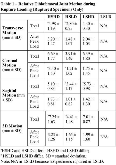
1 Biomechanical Characterization of a New, Noninvasive Model of Anterior Cruciate Ligament Rupture in the Rat Tristan Maerz, MS Eng 1, Michael Kurdziel, MS Eng 1, Abigail Davidson, BS Eng 1, Kevin Baker, PhD 1, Kyle Anderson, MD 1, Howard Matthew, PhD 2. 1 Beaumont Health System, Royal Oak, MI, USA, 2 Wayne State University, Detroit, MI, USA. Disclosures: T. Maerz: None. M. Kurdziel: None. A. Davidson: None. K. Baker: None. K. Anderson: None. H. Matthew: None. Introduction: The rate of posttraumatic osteoarthritis (PTOA) following anterior cruciate ligament (ACL) rupture has been reported as high as 90%. Animal models are important for characterizing physiological events following these injuries. The current standard for ACL injury in the rat is surgical transection, which fails to reproduce high-magnitude trauma and the resultant biologic events following ACL injury. The purpose of this study was to develop and characterize a new model of noninvasive rat ACL rupture to facilitate future PTOA studies. Methods: Lewis rats were euthanized and placed prone on a custom fixture attached to a materials testing system. To induce ACL rupture, both limbs underwent loading by the application of a compressive axial load to the tibia with the knee rested on a stage flexed to 100 (Fig 1A). After preloading and preconditioning, an axial displacement was applied to the tibia at four testing conditions: low and high speed (1 mm/s, 8 mm/s) and low and high displacement (2mm, 3mm). The four testing groups were: high-speed, high-displacement (HSHD); high-speed, low-displacement (HSLD); low-speed, low-displacement (LSLD), and low-speed, high-displacement (LSHD). 3D motion data was quantified using a four-camera motion capture system to track displacement of the tibia relative to the femur via markers placed in the greater trochanter, distal femoral metaphysis, tibial tubercle and distal tibia. Total and net motion of the proximal tibia relative to the distal femur was calculated individually in all three 2D planes (transverse, sagittal, and coronal) as well as total 3D motion. ACL rupture was identified by an audible pop, a drop in load and resultant anterior laxity (Fig 1B,C). Following mechanical loading, whole limbs were disarticulated at the hip joint to quantify the degree of joint laxity in a custom laxity testing fixture. Anterior-Posterior (AP) laxity was measured by applying 2 N axially to the tibia at 90 of knee flexion to induce anterior subluxation. Joint laxity was measured by displacement (mm) and compliance (mm/n) at 2 N, and results were compared between groups. Varus-valgus laxity was measured by applying a 1 N varus or valgus load at the distal tibia at 15 of knee flexion. Displacement (mm) at 1 N was compared between groups. Uninjured limbs were used as laxity controls. One-way ANOVA or Kruskall-Wallis tests were used to compare normal and non-normal variables between groups, respectively. Variables were also grouped by high-speed/low-speed, high-displacement/lowdisplacement, and rupture/non-ruptured. Results: ACL rupture occurred in 100% of rats in HSHD, 33.33% in HSLD, 55.56% in LSHD, and 0% in LSLD. Rupture incidence was significantly associated with both endpoint displacement (P < 0.001) and displacement speed (P = 0.019). Motion-capture data indicates that there are profound differences in joint motion between groups even if rupture occurs (Table 1). HSHD induced the highest amount of transverse, coronal, sagittal, and overall 3D motion. Motion-capture data also shows that total motion
2 after peak load in the coronal plane (medial-lateral and superior-inferior motion) was significantly higher in specimens that ruptured (2.58 mm ± 1.65) compared to specimens that did not rupture (1.25 mm ± 0.97, P = 0.031). There was significantly higher net displacement in the transverse plane (medial-lateral and anterior-posterior motion) in ruptured specimens (2.15 mm ± 0.87) compared to non-ruptured specimens (1.45 mm ± 0.69, P = 0.034). High-speed groups had a significantly more motion after peak load in the coronal plane (2.59 mm ± 1.64) compared to low-speed groups (1.35 mm ± 1.1, P = 0.033). Comparing high- and low-displacement groups, there was a significant difference in total 3D motion (High-displacement: 7.17 mm ± 1.29; low-displacement: 5.19 mm ± 2.38, P = 0.008), 3D motion after peak load (high-displacement: 2.84 mm ± 1.57; low-displacement: 1.70 mm ± 1.19, P = 0.036), motion after peak load in the coronal plane (high-displacement: 2.69 mm ± 1.66; low-displacement: 1.22 mm ± 0.88, P = 0.014), total motion in the coronal plane (high-displacement: 6.58 mm ± 1.37; lowdisplacement: 4.41 mm ± 2.27, P = ), motion after peak load in the transverse plane (highdisplacement: 2.74 mm ± 1.49; low-displacement: 1.43 mm ± 1.04, P = 0.016), total motion in the transverse plane (high-displacement: 4.70 mm ± 1.07; low-displacement: 3.20 mm ± 1.76, P = 0.013), as well as total motion in the sagittal plane (high-displacement: 5.46 mm ± 0.92; low-displacement: 3.72 mm ± 1.76, P = 0.002). Only HSHD exhibited a significant increase in AP and varus laxity compared to uninjured limbs, with corresponding decreases in respective compliance (Table 2). Rupture did not induce valgus laxity in any group. There was a significant difference in AP laxity between high- and lowspeed groups (High-Speed: mm ± 0.456; Low-speed: mm ± 0.61; P = 0.026) and between high- and low-displacement groups (High-displacement: mm ± 0.53; Low-displacement: mm ± 0.42; P = 0.003). There was a significant difference in varus laxity between high- and low-displacement groups (High-displacement: 3.29 mm ± 0.93; Low-displacement: 2.55 mm ± 1.03; P = 0.031) but not highand low-speed groups (High-speed: 3.25 mm ± 0.99; Low-speed: 2.59 mm ± 0.99; P= 0.064). Valgus laxity did not differ as a function of displacement or speed. Discussion: The application of an axial tibial load via the ankle in 100 of knee flexion is able to induce a noninvasive ACL rupture in the rat with concomitant varus laxity. The incidence of ACL rupture is highly dependent upon both speed and endpoint displacement, with endpoint displacement appearing statistically more determinative of rupture. ACL rupture is more repeatable in the high-speed, highdisplacement protocol, and both AP and varus laxity are induced due to ACL rupture. Varus laxity suggests lateral collateral ligament (LCL) injury, a concomitant injury commonly observed in humans in combination with ACL rupture. Motion capture data indicates that joint motion after peak load is most prevalent in the coronal and transverse planes, and this motion is significantly higher in ruptured specimens. This additional motion after rupture may represent translation, rotation, or both, and since LCL laxity is only increased in ruptured specimens, LCL laxity is likely being induced by this post-rupture motion. A clinically-relevant noninvasive ACL rupture model is useful for future studies assessing biologic events after ACL rupture, such as the onset and progression of PTOA. Future studies will utilize this model to assess degenerative changes in the rat joint. Significance: A clinically-relevant noninvasive ACL rupture protocol has been developed and characterized. Since ACL transection does not reproduce high-magnitude joint trauma and introduces confounding variables due to surgical cutting, this noninvasive ACL rupture protocol is able to more closely mimic human biology after ACL rupture.

3
4
5 ORS 2015 Annual Meeting Poster No: 1193
Biomechanics of the Knee. Valerie Nuñez SpR Frimley Park Hospital
 Biomechanics of the Knee Valerie Nuñez SpR Frimley Park Hospital Knee Biomechanics Kinematics Range of Motion Joint Motion Kinetics Knee Stabilisers Joint Forces Axes The Mechanical Stresses to which
RN(EC) ENC(C) GNC(C) MN ACNP *** MECHANISM OF INJURY.. MOST IMPORTANT *** - Useful in determining mechanism of injury / overuse
 ENC(C) GNC(C) MN ACNP *** MECHANISM OF INJURY.. MOST IMPORTANT *** - Useful in determining mechanism of injury / overuse") HISTORY *** MECHANISM OF INJURY.. MOST IMPORTANT *** Age of patient Sport / Occupation - Certain conditions are more prevalent in particular age groups (Osgood Schlaters in youth / Degenerative Joint Disease
HISTORY *** MECHANISM OF INJURY.. MOST IMPORTANT *** Age of patient Sport / Occupation - Certain conditions are more prevalent in particular age groups (Osgood Schlaters in youth / Degenerative Joint Disease
TOTAL KNEE ARTHROPLASTY (TKA)
") TOTAL KNEE ARTHROPLASTY (TKA) 1 Anatomy, Biomechanics, and Design 2 Femur Medial and lateral condyles Convex, asymmetric Medial larger than lateral 3 Tibia Tibial plateau Medial tibial condyle: concave
TOTAL KNEE ARTHROPLASTY (TKA) 1 Anatomy, Biomechanics, and Design 2 Femur Medial and lateral condyles Convex, asymmetric Medial larger than lateral 3 Tibia Tibial plateau Medial tibial condyle: concave
Tensile Forces in Knee Ligaments in Response to Hyperextension
 Tensile Forces in Knee Ligaments in Response to Hyperextension Kei Kimura 1, Hidenori Otsubo 2, Satoshi Yamakawa 1, Toshihiko Yamashita 2, Hiromichi Fujie 1. 1 Tokyo Metropolitan University, hino, Japan,
Physical Examination of the Knee
 History: Pain Traumatic vs. atraumatic? Acute vs Chronic Previous procedures done on the knee? Swelling, catching, instability General Setup Examine standing, sitting and supine Evaluate gait Examine hip
Multiapical Deformities p. 97 Osteotomy Concepts and Frontal Plane Realignment p. 99 Angulation Correction Axis (ACA) p. 99 Bisector Lines p.
 p. 99 Bisector Lines p.") Normal Lower Limb Alignment and Joint Orientation p. 1 Mechanical and Anatomic Bone Axes p. 1 Joint Center Points p. 5 Joint Orientation Lines p. 5 Ankle p. 5 Knee p. 5 Hip p. 8 Joint Orientation Angles
Normal Lower Limb Alignment and Joint Orientation p. 1 Mechanical and Anatomic Bone Axes p. 1 Joint Center Points p. 5 Joint Orientation Lines p. 5 Ankle p. 5 Knee p. 5 Hip p. 8 Joint Orientation Angles
Physical Examination of the Knee
 History: Pain Traumatic vs. atraumatic Acute vs Chronic Mechanism of injury Swelling, catching, instability Previous evaluation and treatment General Setup Examine standing, sitting and supine Evaluate
TKA Gap Planning. Supporting healthcare professionals
 TKA Gap Planning The NAVIO TKA Gap Planning stage helps you adjust the plan based on gap information between the femur and tibia implants. Supporting healthcare professionals Interactive Views Four interactive
Financial Disclosure. Medial Collateral Ligament
 Matthew Murray, M.D. UTHSCSA Sports Medicine Financial Disclosure Dr. Matthew Murray has no relevant financial relationships with commercial interests to disclose. Medial Collateral Ligament Most commonly
Joints of the Lower Limb II
 Joints of the Lower Limb II Lecture Objectives Describe the components of the knee and ankle joint. List the ligaments associated with these joints and their attachments. List the muscles acting on these
ASSESSMENT AND MANAGEMENT OF THE KNEE AND LOWER LIMB.
 ASSESSMENT AND MANAGEMENT OF THE KNEE AND LOWER LIMB www.fisiokinesiterapia.biz Overview History Examination X-rays Fractures and Dislocations. Soft Tissue Injuries Other Knee/Lower limb Problems Anatomy
ACL Athletic Career. ACL Rupture - Warning Features Intensive pain Immediate swelling Locking Feel a Pop Dead leg Cannot continue to play
 FIMS Ambassador Tour to Eastern Europe, 2004 Belgrade, Serbia Montenegro Acute Knee Injuries - Controversies and Challenges Professor KM Chan OBE, JP President of FIMS Belgrade ACL Athletic Career ACL
Biomechanical Risk Factor of Anterior Cruciate Ligament Injury in Adolescent Female Basketball Players: A Prospective Cohort Study
 Biomechanical Risk Factor of Anterior Cruciate Ligament Injury in Adolescent Female Basketball Players: A Prospective Cohort Study kohei koresawa 1, Yumi No 2, Satoshi Kubota 1, Kazuyoshi Gamada 1. 1 Graduate
Knee Injuries. PSK 4U Mr. S. Kelly North Grenville DHS. Medial Collateral Ligament Sprain
 Knee Injuries PSK 4U Mr. S. Kelly North Grenville DHS Medial Collateral Ligament Sprain Result from either a direct blow from the lateral side in a medial direction or a severe outward twist Greater injury
Where Is the Natural Internal-External Rotation Axis of the Tibia?
 Where Is the Natural Internal-External Rotation Axis of the Tibia? Daniel Boguszewski 1, Paul Yang 2, Nirav Joshi 2, Keith Markolf 1, Frank Petrigliano 1, David McAllister 1. 1 University of California
Posterolateral Corner Injuries of the Knee: Pearls and Pitfalls
 Posterolateral Corner Injuries of the Knee: Pearls and Pitfalls Robert A. Arciero,MD,Col,ret Professor, Orthopaedics University of Connecticut Incidence of PLC Injuries with ACL Tears Fanelli, 1995 12%
Lateral knee injuries
 Created as a free resource by Clinical Edge Based on Physio Edge podcast episode 051 with Matt Konopinski Get your free trial of online Physio education at Orthopaedic timeframes Traditionally Orthopaedic
Knee Injury Assessment
 Knee Injury Assessment Clinical Anatomy p. 186 Femur Medial condyle Lateral condyle Femoral trochlea Tibia Intercondylar notch Tibial tuberosity Tibial plateau Fibula Fibular head Patella Clinical Anatomy
Anterior Cruciate Ligament Surgery
 Anatomy Anterior Cruciate Ligament Surgery Roger Ostrander, MD Andrews Institute Anatomy Anatomy Function Primary restraint to anterior tibial translation Secondary restraint to internal tibial rotation
Utility of Instrumented Knee Laxity Testing in Diagnosis of Partial Anterior Cruciate Ligament Tears
 Utility of Instrumented Knee Laxity Testing in Diagnosis of Partial Anterior Cruciate Ligament Tears Ata M. Kiapour, Ph.D. 1, Ali Kiapour, Ph.D. 2, Timothy E. Hewett, Ph.D. 3, Vijay K. Goel, Ph.D. 2. 1
Estimating Total Knee Arthroplasty Joint Loads from Kinematics
 Estimating Total Knee Arthroplasty Joint Loads from Kinematics Clare K. Fitzpatrick, Paul Rullkoetter. University of Denver, Denver, CO, USA. Disclosures: C.K. Fitzpatrick: None. P. Rullkoetter: 5; DePuy
Imaging the Athlete s Knee. Peter Lowry, MD Musculoskeletal Radiology University of Colorado
 Imaging the Athlete s Knee Peter Lowry, MD Musculoskeletal Radiology University of Colorado None Disclosures Knee Imaging: Radiographs Can be performed weight-bearing or non-weight-bearing View options
Biomechanics of Two Reconstruction Techniques for Elbow Ulnar Collateral Ligament Insufficiency
 Biomechanics of Two Reconstruction Techniques for Elbow Ulnar Collateral Ligament Insufficiency Justin E. Chronister, MD 1, Randal P. Morris, BS 2, Clark R. Andersen, MS 2, J. Michael Bennett, MD 3, Thomas
CHAPTER 8: THE BIOMECHANICS OF THE HUMAN LOWER EXTREMITY
 CHAPTER 8: THE BIOMECHANICS OF THE HUMAN LOWER EXTREMITY _ 1. The hip joint is the articulation between the and the. A. femur, acetabulum B. femur, spine C. femur, tibia _ 2. Which of the following is
Discrepancies in Knee Joint Moments Using Common Anatomical Frames Defined by Different Palpable Landmarks
 Journal of Applied Biomechanics, 2008, 24, 185-190 2008 Human Kinetics, Inc. Discrepancies in Knee Joint Moments Using Common Anatomical Frames Defined by Different Palpable Landmarks Dominic Thewlis,
W. Dilworth Cannon, M.D. Professor of Clinical Orthopaedic Surgery University of California San Francisco
 Knee Pain And Injuries In Adults W. Dilworth Cannon, M.D. Professor of Clinical Orthopaedic Surgery University of California San Francisco Pain Control Overview Narcotics rarely necessary after 1 st 1-2
Intra-Articular Tibiofemoral Injection of a Nonsteroidal Anti-Inflammatory Drug has no Detrimental Effects on Joint Mechanics in a Rat Model
 Intra-Articular Tibiofemoral Injection of a Nonsteroidal Anti-Inflammatory Drug has no Detrimental Effects on Joint Mechanics in a Rat Model Corinne N. Riggin, Jennica J. Tucker, Louis J. Soslowsky, PhD,
BIOMECHANICAL MECHANISMS FOR DAMAGE: RETRIEVAL ANALYSIS AND COMPUTATIONAL WEAR PREDICTIONS IN TOTAL KNEE REPLACEMENTS
 Journal of Mechanics in Medicine and Biology Vol. 5, No. 3 (2005) 469 475 c World Scientific Publishing Company BIOMECHANICAL MECHANISMS FOR DAMAGE: RETRIEVAL ANALYSIS AND COMPUTATIONAL WEAR PREDICTIONS
Do Persons with PFP. PFJ Loading? Biomechanical Factors Contributing to Patellomoral Pain: The Dynamic Q Angle. Patellofemoral Pain: A Critical Review
 Biomechanical Factors Contributing to Patellomoral Pain: The Dynamic Q Angle Division Biokinesiology & Physical Therapy Co Director, oratory University of Southern California Movement Performance Institute
Ligamentous and Meniscal Injuries: Diagnosis and Management
 Ligamentous and Meniscal Injuries: Diagnosis and Management Daniel K Williams, MD Franciscan Physician Network Orthopedic Specialists September 29, 2017 No Financial Disclosures INTRODUCTION Overview of
Direct Measurement of Graft Tension in Anatomic Versus Non-anatomic ACL Reconstructions during a Dynamic Pivoting Maneuver
 Direct Measurement of Graft Tension in Anatomic Versus Non-anatomic ACL Reconstructions during a Dynamic Pivoting Maneuver Scott A. Buhler 1, Newton Chan 2, Rikin Patel 2, Sabir K. Ismaily 2, Brian Vial
In the name of god. Knee. By: Tofigh Bahraminia Graduate Student of the Pathology Sports and corrective actions. Heat: Dr. Babakhani. Nov.
 In the name of god Knee By: Tofigh Bahraminia Graduate Student of the Pathology Sports and corrective actions Heat: Dr. Babakhani Nov. 2014 1 Anatomy-Bones Bones Femur Medial/lateral femoral condyles articulate
ChiroCredit.com Presents Biomechanics: Focus on
 ChiroCredit.com Presents Biomechanics: Focus on the Knee Presented by: Ivo Waerlop, DC Shawn Allen, DC 1 Focus on The Knee 2 Pertinent Anatomy Femur Tibia Fibula Patella Prepatellar bursa Infrapatellar
Mohammad Ayati,M.D Department of Orthopaedics, Yazd University of Medical Science.
 IN THE NAME OF GOD Mohammad Ayati,M.D Department of Orthopaedics, Yazd University of Medical Science. Devastating injury resulting from : high-energy usually from MVC or fall from height commonly a dashboard
Exam of the Knee and Ankle I HAVE NO FINANCIAL DISCLOSURES RELEVANT TO THIS PRESENTATION
 Exam of the Knee and Ankle I HAVE NO FINANCIAL DISCLOSURES RELEVANT TO THIS PRESENTATION Disclosures I have no relevant financial relationships with the manufacturers of any commercial products and or
The Effect of Lateral Meniscal Root Injuries on the Stability of the Anterior Cruciate Ligament Deficient Knee
 The Effect of Lateral Meniscal Root Injuries on the Stability of the Anterior Cruciate Ligament Deficient Knee Charles Vega 1, Jebran Haddad 1, Jerry Alexander 2, Jonathan Gold 2, Theodore Shybut 1, Philip
Biomechanical Effects of Femoral Component Axial Rotation in Total Knee Arthroplasty (TKA)
") Biomechanical Effects of Femoral Component Axial Rotation in Total Knee Arthroplasty (TKA) Mohammad Kia, PhD, Timothy Wright, PhD, Michael Cross, MD, David Mayman, MD, Andrew Pearle, MD, Peter Sculco,
Biomechanical Effects of Femoral Component Axial Rotation in Total Knee Arthroplasty (TKA) Mohammad Kia, PhD, Timothy Wright, PhD, Michael Cross, MD, David Mayman, MD, Andrew Pearle, MD, Peter Sculco,
ACL AND PCL INJURIES OF THE KNEE JOINT
 ACL AND PCL INJURIES OF THE KNEE JOINT Dr.KN Subramanian M.Ch Orth., FRCS (Tr & Orth), CCT Orth(UK) Consultant Orthopaedic Surgeon, Special interest: Orthopaedic Sports Injury, Shoulder and Knee Surgery,
The Lower Limb II. Anatomy RHS 241 Lecture 3 Dr. Einas Al-Eisa
 The Lower Limb II Anatomy RHS 241 Lecture 3 Dr. Einas Al-Eisa Tibia The larger & medial bone of the leg Functions: Attachment of muscles Transfer of weight from femur to skeleton of the foot Articulations
ACL Forces and Knee Kinematics Produced by Axial Tibial Compression During a Passive Flexion Extension Cycle
 ACL Forces and Knee Kinematics Produced by Axial Tibial Compression During a Passive Flexion Extension Cycle Keith L. Markolf, Steven R. Jackson, Brock Foster, David R. McAllister Biomechanics Research
Revolution. Unicompartmental Knee System
 Revolution Unicompartmental Knee System While Total Knee Arthroplasty (TKA) is one of the most predictable procedures in orthopedic surgery, many patients undergoing TKA are in fact excellent candidates
Cruciate Ligament. Summary of the Doctoral Thesis
 Study of the Effect of Excessive Tibial Plateau Angle on Degenerative Changes of Canine Cranial Cruciate Ligament Summary of the Doctoral Thesis Tom Ichinohe Graduate School of Veterinary Medicine and
Effects of Posteromedial Vertical Capsulotomy on the Medial Extension Gap in Cruciate-retaining Total Knee Arthroplasty
 Effects of Posteromedial Vertical Capsulotomy on the Medial Extension Gap in Cruciate-retaining Total Knee Arthroplasty Ryutaku Kaneyama, M.D., Hideaki Shiratsuchi, Kazuhiro Oinuma, MD, Yoko Miura, MD,
OrthoMap Express Knee Product Guide. OrthoMap Express Knee Navigation Software 2.0
 OrthoMap Express Knee Product Guide OrthoMap Express Knee Navigation Software 2.0 Product Guide 1 Introduction Introduction The Stryker OrthoMap Express Knee Navigation System is providing surgeons with
Objectives. The BIG Joint. Case 1. Boney Architecture. Presenter Disclosure Information. Common Knee Problems
 3:30 4:15 pm Common Knee Problems SPEAKER Christopher J. Visco, MD Presenter Disclosure Information The following relationships exist related to this presentation: Christopher J. Visco, MD: Speaker s Bureau
The Knee. Two Joints: Tibiofemoral. Patellofemoral
 Evaluating the Knee The Knee Two Joints: Tibiofemoral Patellofemoral HISTORY Remember the questions from lecture #2? Girth OBSERVATION TibioFemoral Alignment What are the consequences of faulty alignment?
On Field Assessment and Management of Acute Knee Injuries: A Physiotherapist s Perspective
 On Field Assessment and Management of Acute Knee Injuries: A Physiotherapist s Perspective Jessica Condliffe Physiotherapist / Clinic Manager TBI Health Wellington Presentation Outline Knee anatomy review
KNEE EXAMINATION. Tips & Tricks from an Emergency Physician Perspective. EM Physicians Less Exposed to MSK Medicine
 KNEE EXAMINATION Tips & Tricks from an Emergency Physician Perspective Dr P O CONNOR Emergency Medicine Physician EUSEM 10/09/2018 EM Physicians Less Exposed to MSK Medicine Musculoskeletal Medicine becoming
Musculoskeletal Examination Benchmarks
 Musculoskeletal Examination Benchmarks _ The approach to examining the musculoskeletal system is the same no matter what joint or limb is being examined. The affected and contralateral region should both
STIFFNESS AFTER TKA PRE, PER AND POST OPERATIVE CAUSING FACTORS
 STIFFNESS AFTER TKA PRE, PER AND POST OPERATIVE CAUSING FACTORS Patrick DJIAN INTRODUCTION Stiffness is one of the most common complications following TKR, causing frustration to both the surgeon and the
What is the most effective MRI specific findings for lateral meniscus posterior root tear in ACL injuries
 What is the most effective MRI specific findings for lateral meniscus posterior root tear in ACL injuries Kazuki Asai 1), Junsuke Nakase 1), Kengo Shimozaki 1), Kazu Toyooka 1), Hiroyuki Tsuchiya 1) 1)
Why does it matter? Patellar Instability 7/23/2018. What is the current operation de jour? Common. Poorly taught. Poorly treated
 Patellar Instability It s Really Not That Difficult! David Shneider MD East Lansing, MI www.patellamdcom Detroit Sports Medicine Foundation July 2018 Why does it matter? Common Poorly taught Poorly treated
The Knee. Prof. Oluwadiya Kehinde
 The Knee Prof. Oluwadiya Kehinde www.oluwadiya.sitesled.com The Knee: Introduction 3 bones: femur, tibia and patella 2 separate joints: tibiofemoral and patellofemoral. Function: i. Primarily a hinge joint,
BIOMECHANICAL EXAMINATION OF THE PEDIATRIC LOWER EXTREMITY
 BIOMECHANICAL EXAMINATION OF THE PEDIATRIC LOWER EXTREMITY B.Resseque, D.P.M. ARCH HEIGHT OFF WEIGHTBEARING Evaluate arch height by placing a ruler from the heel to the first metatarsal head Compare arch
ANATOMIC. Navigated Surgical Technique 4 in 1 TO.G.GB.016/1.0
 ANATOMIC Navigated Surgical Technique 4 in 1 TO.G.GB.016/1.0 SCREEN LAYOUT Take screenshot Surgical step Dynamic navigation zone Information area and buttons 2 SCREEN LAYOUT Indicates action when yellow
2017 Resident Advanced Trauma Techniques Course COMPLICATIONS / CHALLENGES MALUNIONS/DEFORMITY
 2017 Resident Advanced Trauma Techniques Course COMPLICATIONS / CHALLENGES MALUNIONS/DEFORMITY What is a Malunion? Definition: a fracture that has healed in a nonanatomic (i.e. deformed) position Must
Examination of the Knee
 Examination of the Knee Wash your hands & Introduce the exam to the patient Positioning & Draping With the patient supine, make sure both legs are exposed in order to compare each side be sure to use draping
Is There A Difference In Uni- And Multi-compartmental Knee Arthroplasty Kinematics?
 Is There A Difference In Uni- And Multi-compartmental Knee Arthroplasty Kinematics? Toshifumi Watanabe, MD, PhD 1, Stefan Kreuzer, MD, MS 2, Jennifer Amanda Christopher, BS 3, Michael Conditt, PhD 3, Brian
Sports Medicine 15. Unit I: Anatomy. The knee, Thigh, Hip and Groin. Part 4 Anatomies of the Lower Limbs
 Sports Medicine 15 Unit I: Anatomy Part 4 Anatomies of the Lower Limbs The knee, Thigh, Hip and Groin Anatomy of the lower limbs In Part 3 of this section we focused upon 11 of the 12 extrinsic muscles
Imaging the Knee 17/10/2017. Friction syndrome Common in runners or cyclists Fluid between ITB and Lateral femoral condyle
 17/10/2017 Imaging the Knee Alicia M. Yochum RN, DC, DACBR, RMSK Iliotibial Band Syndrome Ligamentous Tears (ACL, PCL, MCL, LCL) Meniscal Tears Cartilage Degeneration Quadriceps/Patellar tendinosis Osteochondral
The examination of the painful knee. Maja K Artandi, MD, FACP Clinical Associate Professor of Medicine Stanford University
 The examination of the painful knee Maja K Artandi, MD, FACP Clinical Associate Professor of Medicine Stanford University Objectives of the talk By the end of this talk you will know The important anatomy
Dorsal surface-the upper area or top of the foot. Terminology
 It is important to learn the terminology as it relates to feet to properly communicate with referring physicians when necessary and to identify the relationship between the anatomical structure of the
Recognizing common injuries to the lower extremity
 Recognizing common injuries to the lower extremity Bones Femur Patella Tibia Tibial Tuberosity Medial Malleolus Fibula Lateral Malleolus Bones Tarsals Talus Calcaneus Metatarsals Phalanges Joints - Knee
Mako Partial Knee Medial bicompartmental
 Mako Partial Knee Medial bicompartmental Surgical reference guide Mako Robotic-Arm Assisted Surgery Table of contents Implant compatibility.... 3 Pre-operative planning.... 4 Intra-operative planning....
BATES VISUAL GUIDE TO PHYSICAL EXAMINATION. OSCE 4: Knee Pain
 BATES VISUAL GUIDE TO PHYSICAL EXAMINATION OSCE 4: Knee Pain This video format is designed to help you prepare for objective structured clinical examinations, or OSCEs. You are going to observe and participate
BIOMECHANICAL EXAMINATION OF THE PEDIATRIC LOWER EXTREMITY 2017
 BIOMECHANICAL EXAMINATION OF THE PEDIATRIC LOWER EXTREMITY 2017 B. RESSEQUE, D.P.M., D.A.B.P.O. Professor, N.Y. College of Podiatric Medicine ARCH HEIGHT OFF WEIGHTBEARING Evaluate arch height by placing
Correlation between findings on stress X-rays (Telos) and MRI in patients with anterior knee instability
 and MRI in patients with anterior knee instability") Correlation between findings on stress X-rays (Telos) and MRI in patients with anterior knee instability Poster No.: C-1687 Congress: ECR 2014 Type: Authors: Scientific Exhibit C. Prieto Santa Cruz 1,
Correlation between findings on stress X-rays (Telos) and MRI in patients with anterior knee instability Poster No.: C-1687 Congress: ECR 2014 Type: Authors: Scientific Exhibit C. Prieto Santa Cruz 1,
Load Response of the Hindfoot Bones in patients with the Ankle Osteoarthritis : in vivo 3D study
 Load Response of the Hindfoot Bones in patients with the Ankle Osteoarthritis : in vivo D study Hara Y, Ikoma K, Kido M, Imai K, Maki M, Takatori R, Tokunaga D, Inoue N,, Kubo T Department of Orthopaedics,
Kineto. Orthopaedics & Rehabilitation Products
 Member of Vincent Medical Holdings Limited Kineto Orthopaedics & Rehabilitation Products Our orthopaedic and rehabilitation products comprise of a variety of adjustable rehabilitation braces for support,
11/4/2018 SUBTLETIES OF LOWER EXTREMITY TRAUMA IMAGING SPEAKER DISCLOSURES
 SUBTLETIES OF LOWER EXTREMITY TRAUMA IMAGING Charles S. Resnik, M.D. Professor of Radiology University of Maryland School of Medicine Upon completion of this presentation, participants will be better able
Evolution. Medial-Pivot Knee System The Bi-Cruciate-Substituting Knee. Key Aspects
 Evolution Medial-Pivot Knee System The Bi-Cruciate-Substituting Knee Key Aspects MicroPort s EVOLUTION Medial-Pivot Knee System was designed to recreate the natural anatomy that is lost during a total
Knee Joint Anatomy 101
 Knee Joint Anatomy 101 Bone Basics There are three bones at the knee joint femur, tibia and patella commonly referred to as the thighbone, shinbone and kneecap. The fibula is not typically associated with
Knee, Ankle, and Foot: Normal and Abnormal Features with MRI and Ultrasound Correlation. Disclosures. Outline. Joint Effusion. Suprapatellar recess
 Knee, Ankle, and Foot: Normal and Abnormal Features with MRI and Ultrasound Correlation Jon A. Jacobson, M.D. Professor of Radiology Director, Division of Musculoskeletal Radiology University of Michigan
Anterolateral Ligament. Bradd G. Burkhart, MD Orlando Orthopaedic Center Sports Medicine
 Anterolateral Ligament Bradd G. Burkhart, MD Orlando Orthopaedic Center Sports Medicine What in the world? TIME magazine in November 2013 stated: In an age filled with advanced medical techniques like
Where Is The Natural Flexion-Extension Axis Of The Knee?
 Where Is The Natural Flexion-Extension Axis Of The Knee? Daniel Boguszewski 1, Paul Yang 2, Keith Markolf 1, Frank Petrigliano 1, David McAllister 1. 1 University of California Los Angeles, Los Angeles,
Applied Functional Science Chain Reaction Skeletal: Real creates Relative. Presented By David Tiberio, Ph.D., PT, FAFS Dean, Gray Institute
 Applied Functional Science Chain Reaction Skeletal: Real creates Relative Presented By David Tiberio, Ph.D., PT, FAFS Dean, Gray Institute 1 How Movement is Created Total body movements are made up of
Please differentiate an internal derangement from an external knee injury.
 Knee Orthopaedic Tests Sports and Knee Injuries James J. Lehman, DC, MBA, DABCO University of Bridgeport College of Chiropractic Knee Injury Strain, Sprain, Internal Derangement Anatomy of the Knee Please
Differential Diagnosis
 Case 31yo M who sustained an injury to L knee while playing Basketball approximately 2 weeks ago. He describes pivoting and hyperextending his knee, which swelled over the next few days. He now presents
HISTORY AND INDICATIONS OF LATERAL TENODESIS IN ATHLETES
 HISTORY AND INDICATIONS OF LATERAL TENODESIS IN ATHLETES Written by Philippe Landreau, Qatar The treatment of anterior cruciate ligament injuries remains challenging in young athletic populations. A residual
Original Report. The Reverse Segond Fracture: Association with a Tear of the Posterior Cruciate Ligament and Medial Meniscus
 Eva M. Escobedo 1 William J. Mills 2 John. Hunter 1 Received July 10, 2001; accepted after revision October 1, 2001. 1 Department of Radiology, University of Washington Harborview Medical enter, 325 Ninth
Overview Ligament Injuries. Anatomy. Epidemiology Very commonly injured joint. ACL Injury 20/06/2016. Meniscus Tears. Patellofemoral Problems
 Overview Ligament Injuries Meniscus Tears Pankaj Sharma MBBS, FRCS (Tr & Orth) Consultant Orthopaedic Surgeon Manchester Royal Infirmary Patellofemoral Problems Knee Examination Anatomy Epidemiology Very
Presented By Dr Vincent VG An MD BSc (Adv) MPhil Dr Murilo Leie MD Mr Joshua Twiggs BEng Dr Brett A Fritsch MBBS FRACS (Orth) FAOrthA.
 MPhil Dr Murilo Leie MD Mr Joshua Twiggs BEng Dr Brett A Fritsch MBBS FRACS (Orth) FAOrthA.") A comparison of kinematic and mechanical alignment with regards to bony resection, soft tissue release, and deformity correction in total knee replacement Presented By Dr Vincent VG An MD BSc (Adv) MPhil
A comparison of kinematic and mechanical alignment with regards to bony resection, soft tissue release, and deformity correction in total knee replacement Presented By Dr Vincent VG An MD BSc (Adv) MPhil
Novel Design of an Anterior Cruciate Ligament (ACL) Injury Prevention Brace
 Injury Prevention Brace") Novel Design of an Anterior Cruciate Ligament (ACL) Injury Prevention Brace Daniel Greenshields, Justin Killewald, Rachel Porter Lawrence Technological University BME Projects II Dr. Mansoor Nasir, Spring
Novel Design of an Anterior Cruciate Ligament (ACL) Injury Prevention Brace Daniel Greenshields, Justin Killewald, Rachel Porter Lawrence Technological University BME Projects II Dr. Mansoor Nasir, Spring
American College of Physicians 2013 Ohio Chapter Scientific Meeting Columbus, OH October 11, 2013
 American College of Physicians 2013 Ohio Chapter Scientific Meeting Columbus, OH October 11, 2013 Paul J. Gubanich, MD, MPH Assistant Professor of Internal Medicine/Sports Medicine Team Physician, Ohio
Rehabilitation of an ACL injury in a 29 year old male with closed kinetic chain exercises: A case study
 Abstract Objective: This paper will examine a rehabilitation program for a healthy 29 year old male who sustained an incomplete tear of the left ACL. Results: Following a 9 week treatment plan focusing
The Knee. Tibio-Femoral
 The Knee Tibio-Femoral Osteology Distal Femur with Proximal Tibia Largest Joint Cavity in the Body A modified hinge joint with significant passive rotation Technically, one degree of freedom (Flexion/Extension)
Disclosures. Outline. The Posterior Cruciate Ligament 5/3/2016
 The Posterior Cruciate Ligament Christopher J. Utz, MD Assistant Professor of Orthopaedic Surgery University of Cincinnati Disclosures I have no disclosures relevant to this topic. Outline 1. PCL Basic
PLAY PLAY. Hard. Smart. Knee Product Guide
 PLAY Hard. PLAY Smart. Knee Product Guide Osteoarthritis/Ligament Bracing M.4 OA Knee Brace Provides exact and effective valgus and varus pressure adjustment exerted close to the knee joint Physioglide
Knee Movement Coordination Deficits. ICD-9-CM: Sprain of cruciate ligament of knee
 1 Knee Movement Coordination Deficits Anterior Cruciate Ligament ACL Tear ICD-9-CM: 844.2 Sprain of cruciate ligament of knee ACL Insufficiency ICD-9-CM: 717.83 Old disruption of anterior cruciate ligament
BIOMECHANICS AND CONTEXT OF ACUTE KNEE INJURIES. Uwe Kersting MiniModule Idræt Biomekanik 2. Objectives
 BIOMECHANICS AND CONTEXT OF ACUTE KNEE INJURIES Uwe Kersting MiniModule 06 2011 Idræt Biomekanik 2 1 Objectives Know about the static and dynamic organisation of the knee joint (anatomy & function) Be
Knee Joint Assessment and General View
 Knee Joint Assessment and General View Done by; Mshari S. Alghadier BSc Physical Therapy RHPT 366 m.alghadier@sau.edu.sa http://faculty.sau.edu.sa/m.alghadier/ Functional anatomy The knee is the largest
ESTIMATION OF ACL FORCES UTILIZING A NOVEL NON-INVASIVE METHODOLOGY THAT REPRODUCES KNEE KINEMATICS BETWEEN SETS OF KNEES. Shon Patrick Darcy
 ESTIMATION OF ACL FORCES UTILIZING A NOVEL NON-INVASIVE METHODOLOGY THAT REPRODUCES KNEE KINEMATICS BETWEEN SETS OF KNEES by Shon Patrick Darcy BS, Walla Walla College, 2000 Submitted to the Graduate Faculty
ORIGINAL ARTICLE. ROLE OF MRI IN EVALUATION OF TRAUMATIC KNEE INJURIES Saurabh Chaudhuri, Priscilla Joshi, Mohit Goel
 ROLE OF MRI IN EVALUATION OF TRAUMATIC KNEE INJURIES Saurabh Chaudhuri, Priscilla Joshi, Mohit Goel 1. Associate Professor, Department of Radiodiagnosis & imaging, Bharati Vidyapeeth Medical College and
CONTROL OF THE BOUNDARY CONDITIONS OF A DYNAMIC KNEE SIMULATOR
 CONTROL OF THE BOUNDARY CONDITIONS OF A DYNAMIC KNEE SIMULATOR J. Tiré 1, J. Victor 2, P. De Baets 3 and M.A. Verstraete 2 1 Ghent University, Belgium 2 Ghent University, Department of Physical Medicine
Abramsohn Retractor 1
 Abramsohn Retractor 1 Calibrated Femoral Tibial Spreaders Small Medium Large Designed to remain in position, with the femur and tibia separated, without the need of an assistant, and to minimize crushing
POSTEROLATERAL CORNER RECONSTRUCTION WHEN AND HOW?
 OTHER KNEE SURGERIES POSTEROLATERAL CORNER RECONSTRUCTION WHEN AND HOW? Written by Jacques Ménétrey, Eric Dromzée and Philippe M. Tscholl, Switzerland Injury of the posterolateral corner (PLC) is relatively
AAP Boot Camp KNEE AND ANKLE EXAM
 AAP Boot Camp KNEE AND ANKLE EXAM Disclosures I have no relevant financial relationships with the manufacturers of any commercial products and or providers of commercial services discussed in this CME
Anatomy and Biomechanics
 Introduction Increased participation= increased injury rates Females were found to be 5.4 times more likely to sustain injury than males. And females injured their ACL ad a rate of 7.8 times more than
PRE & POST OPERATIVE RADIOLOGICAL ASSESSMENT IN TOTAL KNEE REPLACEMENT. Dr. Divya Rani K 2 nd Year Resident Dept. of Radiology
 PRE & POST OPERATIVE RADIOLOGICAL ASSESSMENT IN TOTAL KNEE REPLACEMENT Dr. Divya Rani K 2 nd Year Resident Dept. of Radiology PRE OPERATIVE ASSESSMENT RADIOGRAPHS Radiographs are used for assessment and
This presentation is the intellectual property of the author. Contact them for permission to reprint and/or distribute.
 43 rd Annual Symposium on Sports Medicine UT Health Science Center San Antonio School of Medicine January 22-23, 2016 Intra-articular / Extra-synovial 38 mm length / 13 mm width Fan-shaped structure narrowest-midportion