MATHEMATICAL MODELING OF CEREBRAL CONCUSSION: CORRELATIONS OF REGIONAL BRAIN STRAIN WITH CLINICAL SYMPTOMS
|
|
|
- Samantha Simmons
- 5 years ago
- Views:
Transcription
1 MATHEMATICAL MODELING OF CEREBRAL CONCUSSION: CORRELATIONS OF REGIONAL BRAIN STRAIN WITH CLINICAL SYMPTOMS Liying Zhang 1, King Yang 1, Thomas A. Gennarelli 2 1 Wayne State University, Detroit, MI, USA 2 Medical College of Wisconsin, Milwaukee WI, USA ABSTRACT To address the relationship of clinical symptoms and the location and magnitude of brain strains, the Wayne State University human head finite element model was used with loading conditions predicted to produce either mild (AIS 1) or classical (AIS 2) concussion. Sinusoidal accelerations using 3,000 rad/s 2 at 25 rad/s or 4,500 rad/s 2 at 50 rad/s, respectively were applied in sagittal and coronal planes to evaluate the effect of loading directions on strain magnitudes and distribution. High principal strains began at the surface and later migrated subcortically, eventually maximizing in parietal cortex, basal ganglia, thalamus and parahippocampal areas. Strain magnitude increased as angular velocity increased and peaked 8 ms after the peak of angular velocity. Large principal strains were in caudate, thalamus, midbrain and hippocampus for coronal and corpus callosum, hippocampus and fronto-temporal cortex for sagittal loading. In AIS 1 concussion, peak strain in all brain regions was <0.30 while in AIS 2 concussion, large areas had strains >0.35 (especially the brainstem-thalamic and hippocampal regions). These areas seem to correlate well with observed clinical symptoms of memory dysfunction and altered awareness associated with concussion. Keywords: BRAINS, ACCELERATIONS, FINITE ELEMENT METHOD, TOLERANCES TRAUMATIC BRAIN INJURY (TBI) is one of the leading causes of death and disability worldwide (Murray et al., 1996). In the US, there are approximately 300,000 new cases of TBI admitted to hospitals each year (Kraus and McArthur, 1996; Horn and Scherer, 2000). Currently about 6 million Americans are living with neurobehavioral sequelae or other losses of brain from TBI. Although TBI is a significant health problem, no effective treatments exist which target the underlying pathophysiology to prevent the progression of neural damage initiated by mechanical injury (Faden, 2002). This lack of therapeutic options highlights the importance of more efficacious strategies and equipment to prevent TBI from occurring in the first place. Current regulations use the Head Injury Criterion (HIC) to assess head/brain injury severity. However, the HIC only takes translational acceleration into account, not rotational acceleration, and TBI is attributed to both types of motion. Diffuse brain injury (DBI) forms a broad spectrum of injuries from mild concussion, which is not associated with loss of consciousness, to classical cerebral concussion with transient disturbance of consciousness, to diffuse axonal injury (DAI) with prolonged loss of consciousness of varying duration. Several rotational acceleration limits for diffuse brain injury have been proposed based on the animal, cadaver, or physical model studies (Ommaya et al., 1967; Ommaya and Gennarelli, 1974; Lowenhielm, 1975, 1978; Margulies and Thibault, 1992; Newman et al., 2000; Zhang et al., 2004a). Recently Gennarelli et al. (2003) and Ommaya et al. (2002) reanalyzed previously published thresholds suggested for components of DBI and establish tolerances for the entire spectrum of DBI. Many animal models, in vitro neural tissue models, and finite element (FE) models have been used to estimate neural tissue damage thresholds and their relation to neuropothological outcomes (Margulies and Thibault, 1992; Bandak and Eppinger, 1994; Mendis et al., 1995; Maxwell et al., 1997; Miller et al., 1999; Bain and Meaney, 1999; Willinger et al., 1999; Franklin et al., 2005; Singh et al., 2006; Mao et al., 2006; Elkin and Morrison III, 2007). Recently, validated Wayne State IRCOBI Conference Bern (Switzerland) September
 (Zhang et al., 2003; King et al., 2003; Zhang et al., 2004a; Viano et al., 2005).")
2 University (WSU) human head FE model was utilized to investigate the mechanisms of concussions sustained by American football players using on-field accident data obtained from the National Football League (NFL) (Zhang et al., 2003; King et al., 2003; Zhang et al., 2004a; Viano et al., 2005). Subsequently, the model was used to estimate the brain response of an Indy race car driver during a severe frontal, side and rear crash (Zhang et al., 2004b). The model was further applied to predict various types of brain injury using the data from reconstructions of real-world automotive crashes and to relate the localized tissue strain to the actual injury sustained by the occupant (Franklyn et al., 2005). The correlation of the regional strain with clinical symptoms and injury severity demonstrated the applicability of the current model to predict the risk of DBI caused by given mechanical conditions. The present investigation was performed to relate proposed rotational parameters to localized strain measures for mild to classical concussion injuries using a validated finite element model of the human head. Our hypothesis is that the differences found in the anatomical areas and magnitudes of brain strains for mild and classical concussion levels would correspond to regional symptoms common in humans with those conditions. METHODS The finite element model of the human head developed by Zhang et al. (2001) was exercised to investigate the tissue strain responses at various anatomical regions resulting from a set of applied rotational threshold loadings. This anatomically inspired, high resolution FE model features fine anatomical details including the scalp, skull with an outer table, diploë, and inner table, dura, falx cerebri, tentorium, pia, sagittal sinus, transverse sinus, cerebral spinal fluid (CSF), hemispheres of the cerebrum with distinct white and gray matter, cerebellum, brainstem, lateral ventricles, third ventricles, and bridging veins. The facial model consists of facial bones, nasal cartilage, temporal mandibular joint, ligaments, soft tissue and skin. The entire head model is made up of over 315,000 elements and uses 15 different material properties for various tissues of the head. The model has been subjected to rigorous validation against available cadaveric intracranial and ventricular pressure data, relative displacement data between the brain and the skull, and facial impact data (Zhang et al., 2001; Viano et al., 2005). Fig. 1 - Anatomically Detailed Finite Element Model of Human Head The accelerations used as input to the WSU head model were based on the rotational acceleration thresholds proposed for a spectrum of diffuse brain injury (Gennarelli et al., 2003). As depicted in 124 IRCOBI Conference Bern (Switzerland) September 2008
, moderate diffuse axonal injury (MDAI) and severe diffuse axonal injury (sdai).")
 levels, 4,500 rad/s2 at 50 rad/s and 3,000 rad/s2 at 25 rad/s thresholds for classical concussion (AIS 2) and mild concussion")
 as following: where A is the peak acceleration amplitude and T is the pulse duration.")

3 Figure 2 and Table 1, the entire DBI is divided into six injury categories, namely mild cerebral concussion (mcc), classical cerebral concussion (ccc), severe cerebral concussion (scc), mild diffuse axonal injury (mdai), moderate diffuse axonal injury (MDAI) and severe diffuse axonal injury (sdai). Six sets of peak angular acceleration and angular thresholds were established to describe each injury severity. In the current study, two rotational acceleration (velocity) levels, 4,500 rad/s2 at 50 rad/s and 3,000 rad/s2 at 25 rad/s thresholds for classical concussion (AIS 2) and mild concussion (AIS 1) were simulated and compared. The acceleration-time profile used was based on a standard sinusoidal function α(t) as following: where A is the peak acceleration amplitude and T is the pulse duration. The duration of the pulse was determined based on the magnitude of the acceleration and velocity. Figure 3 shows the rotational acceleration, rotational velocity and angular rotation-time histories. Table 1. DBI Categories and AIS Scale AIS Angular Acceleration Angular Velocity Injury 1 3, mcc 2 4, ccc 3 8, scc 4 12, mdai 5 14, MDAI 6 16, sdai Fig. 2 Angular Tolerances for the Entire Spectrum of Diffuse Brain Injury as listed in Table 1. Fig. 3 - Applied Rotational Acceleration, Rotational Velocity and Angular Displacement Time Histories at the Center of the Gravity of the Head Model IRCOBI Conference Bern (Switzerland) September
4 The two rotational acceleration pulses were applied to the c.g. of the head model in the sagittal (about y-axis), and coronal (about x-axis) planes. The effect of the loading direction on the magnitude and distribution of strains were compared. The severity of affected brain regions were evaluated based on tissue strain damage criterion in order to assess concussion risk. First principal strain of 0.35 previously proposed as the tolerance threshold for mild traumatic brain injury (MTBI) or concussion was used as strain damage threshold to assess the injury severity (Zhang et al., 2003; King et al., 2003; Viano et al., 2005). The locations of neural tissue experiencing strain above this level were then correlated to the anatomic structures associated with common symptoms of concussion (loss of consciousness, amnesia/memory and cognitive dysfunction). In addition, the extent of injury was quantified in the affected regions using the cumulative strain damage measure proposed by Bandak and Eppinger (1994). The cumulative strain damage is the measure of the accumulation of strain response over the period of loading. The measure calculates the volume fraction of the brain that experiences maximum principal strain level greater than a strain damage tolerance level as a consequence of the total event. The strain damage tolerance level used was 0.35 first principal strain. RESULTS The distribution of first principal strain responses in the brain exhibited regionally specific patterns after the two loading conditions were applied. At the same loading severity, the induced strain magnitude did not vary substantially between coronal and sagittal motions. However, the loading direction did produce different strain distributions. Figure 4 shows the composite maps of the cumulative strain predicted by model in the transverse (A and B) and coronal (C) sections throughout the entire acceleration duration. For coronal rotation, the highest strains occurred in the thalamus, midbrain, caudate, hippocampus and temporal lobe regions. In the sagittal loading condition, the hippocampus, corpus callosum and cortex at inter-hemispheric fissure experienced the highest strains. At the higher AIS 2 rotational condition, using a critical strain threshold of 0.35, it was found that the thalamus and the midbrain were affected regions due to a coronal rotation and the hippocampus and cortex region were highly strained due to a sagittal rotation. The overall affected brain volume fraction (ratio of damaged tissue to whole brain tissue) reached to 17% and 15% respectively, for coronal and sagittal loadings. The affected brain volume was only 1.0% and 0.8% for coronal and sagittal direction, respectively, in the mild AIS 1 loading condition. In terms of temporal profiles of the brain response, it was observed that the localized tissue strain was initiated at the surface of the brain earlier in the loading. Later, strains migrated to the white matter and deep gray matter structures with increased magnitude and, eventually accumulated in specific loci of the brain, notable in the midbrain, thalamic pathway and parahippocampal areas. The strain magnitude increased as angular velocity increased. The strain reached peak at about 7-8 ms after the angular velocity reached maximum. Figure 5 shows the timing of the strain predicted by the model in the regions sustained critical strain due to rotation in the coronal and sagittal planes. The strain magnitude at each region was determined by averaging the strain magnitude for a tissue size of 6x6x6 mm at every time increment. 126 IRCOBI Conference Bern (Switzerland) September 2008


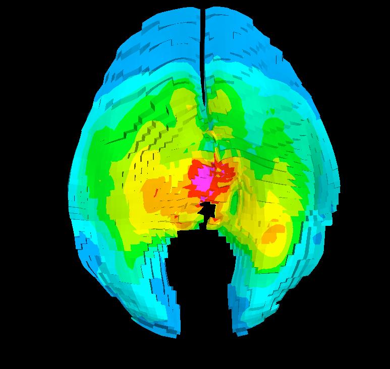

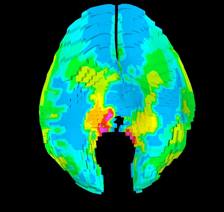
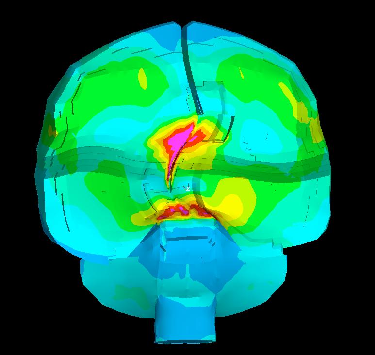
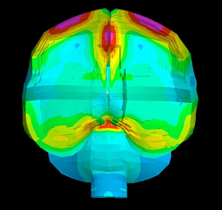
5 Section Location A B B Coronal rotation C First Principal Strain Classical concussion A B Temporal Thalamus Caudate Hipocampus Midbrain Sagittal rotation Hippocampus Fronto-temporal cortex Mild Consussion Coronal rotation Sagittal rotation Fig. 4 - Accumulative Strain Map throughout the Entire Pulse Duration IRCOBI Conference Bern (Switzerland) September
 DISCUSSION The objective of this study was to use the WSU head FE model to relate defined head rotational parameters to localized brain strain patterns for specific grades")


6 Fig. 5 - First Principal Strain Time Histories Experienced by Certain Regions of the Brain During Applied Coronal and Sagittal Rotational Motion at Two Threshold Levels (H For Classical Concussion and L For Mild Concussion) DISCUSSION The objective of this study was to use the WSU head FE model to relate defined head rotational parameters to localized brain strain patterns for specific grades of diffuse brain injury, specifically, mild concussion and classical concussion (Gennarelli and Wodzin, 2005). Many FE head models have been developed and applied to study brain contusion, subdural hematomas, brain edema, and brain deformation (Chu et al., 1994; Mendis et al., 1994; Huang et al., 1999; Willinger et al., 1999; Kleiven et al., 2001; Taktounts et al., 2003). However, there has been limited FE analysis of concussion and diffuse brain injury using human injury data. This is, due in part to lack of a high resolution model that is capable of localizing the injury. The head model applied in this study is constructed with over 300,000 elements and thus has the capability of predicting tissue level response of various brain regions with a level of sophistication not available in other FE models. Recent studies on the mechanisms of the concussion provide first of its kind to correlate the mechanical condition occurring in concussion with the clinical symptoms (Zhang et al., 2003; King et al., 2003; Zhang et al., 2004a; Viano et al., 2005). Model predictions indicated that different regions of the brain were susceptible to higher strain responses after applied rotational acceleration loading. Such regional strain response patterns were further influenced by the direction in which the head rotated. Coronal rotation induced mutifocal high strain in the midbrain, thalamus and caudate regions perhaps indicating an effect of the falx on the strain propagation. In the case of head rotation in the sagittal plane, the critical strain was mainly located in the hippocampus and upper brainstem region. This strain localization was likely dictated by the presence of the tentorium opening and its transverse orientation affecting the tissue deformation in the sagittal plane. The FE model predicted high strain in specific regions of the brain, notably, the midbrain, thalamus and hippocampus. These anatomical structures are important functional units closely associated with memory (hippocampus), and the state of consciousness (midbrain, thalamus), the two most common symptoms in concussion. Our model predicted higher strains in these regions than in other parts of the brain, suggesting that an alteration in cerebral function in these regions is highly associated with tissue distortion in these areas. The severity of such responses exceeds tolerance levels and therefore direct strain damage is certainly possible. Thus, alternative explanations for the observed physiological changes such as a selective vulnerability, diminished injury tolerance, venous hypertension, hyperemia or ischemia need not be considered. The brain function may be directly impaired by injury to that region or may disrupt the neural pathways that communicate between multiple brain structures. Thus, injury to the midbrain could have widespread effects because it is interconnected to diencephalon, the temporal lobe, the limbic system and multiple other areas of cerebrum. In this way, behavioral, cognitive or memory impairments could arise secondarily 128 IRCOBI Conference Bern (Switzerland) September 2008
7 to the affected midbrain. This mechanism may imply the diffuse nature of clinical signs and symptoms for concussion of various severities. The results of the strain locations and associated magnitudes correlate well with the clinical symptoms specified in AIS 1 and AIS 2 concussions. In AIS 1 mild concussion, there is no loss of consciousness and indeed, the model results show low levels of strain in the midbrain-thalamic regions which subserve consciousness. The strains are higher in these areas in the AIS 2 condition, suggestive of loss of consciousness may occur. Similarly, the more severe memory disturbances (including retrograde and anterograde amnesia) that occur in AIS2 concussions are associated with higher strains in the medial temporal lobes (parahippocampal) regions than is the case in AIS 1 concussion where little or no amnesia occurs (Kelly, 1991; 1997). Concussion is a clinical syndrome that may present with a broad spectrum of clinical signs and symptoms including at AIS levels >1, brief loss of consciousness. Reported concussion symptoms observed from NFL players in recent studies were consistent with the common forms of brain dysfunction noted after MTBI that occurred in non-athlete populations (Pellman et al., 2004). It is now generally appreciated that loss of consciousness, previously thought to be the most significant prognosticating factor of brain injury outcome, may have minimal post-injury consequences, and yet a seemingly more minor injury with photophobia and amnesia can lead to a prolonged syndrome. While fewer than 10% of concussed players experienced loss of consciousness, significant abnormalities of memory and cognitive function were observed for over 45% of the NFL players after concussion. The memory/cognitive problems were strongly correlated with delayed return to play (Pellman et al., 2005). FE model predicted strain and strain rate in the fornix, midbrain and corpus callosum showed significant correlations with the memory and cognitive impairments, loss of consciousness and increased intervals for full recovery (Viano et al., 2005). These correlations imply that local strain criteria could be the effective parameters capable of addressing specific symptoms and severity of concussion injury. The locations of the significant strain predicted from the current study were consistent with some of the common signs and symptoms occurred in football-related MTBIs and the general population after mild head injury. Gennarelli et al. (2003) proposed the peak rotational acceleration and velocity based tolerances for various levels of diffuse brain injury in humans and related rotational limits with the abbreviated injury scale (AIS). AIS 2 and AIS 1 are related to the classical concussion and mild concussion with rotational thresholds at 4,500 rad/s 2 with 50 rad/s and 3,000 rad/s 2 with 25 rad/s, respectively. Although these values are estimates based on the literature, they are comparable to thresholds suggested by others. In the literature, several angular acceleration and velocity limits have been proposed for human concussion using the data derived from animal, cadaver, physical models or volunteer tests. Ommaya et al (1967) proposed 4,500 rad/s 2 for the human concussion threshold, on the basis of scaled primate data. Lowenhielm (1975, 1978) suggested 4,500 rad/ s 2 and 30 rad/s as a safe limit in sagittal rotation. Margulies and Thibault (1992) proposed angular acceleration of 16,000 rad/s 2 for moderate to severe DAI in lateral motion. Recent studies on concussive impact to the football players showed that concussed players were exposed to an average rotational acceleration of 6,400 rad/s 2 and angular velocity of 35 rad/s (Pullman et al., 2003; Viano et al., 2005). The typical duration of pulse was around 25 ms. Although this field of science may never achieve consensus about the thresholds for concussions, the magnitude and duration of angular thresholds applied in this study are comparable to the levels in other published concussion thresholds. At applied AIS 2 loading, the model predicted strain levels of consistent with the strain response predicted for the NFL players after concussion. These strain levels along with strain rate response were strongly associated with clinical memory, cognitive and cranial problems. For AIS 1 case, the model prediction implied that the applied rotational loading threshold was insufficient to induce strain above critical level (0.35 threshold) associated clinically severe signs and symptoms. First principal strain of 0.35 used in this study was based on the threshold determined from FE analysis of the NFL MTBI cases (Zhang et al., 2003; King et al., 2003; Viano et al., 2005). Published data on the strain thresholds measured from in vitro or in vivo models of TBI generally fell between IRCOBI Conference Bern (Switzerland) September
8 0.10 and Recently, Tamura et al (2007) examined the relationship of strains measured between the axon and brain tissue. The results revealed that the strain level experienced by each axonal element was only one third of the total strain experienced by the brain tissue. This finding implies that directly incorporating a cellular level axonal threshold into an FE brain model could result in substantial over-prediction of injury occurrence. In terms of the cumulative strain measure, the current study only compared the difference in injury extent between AIS 1 and AIS 2 level injuries. Further studies using FE modeling of animal TBI would be required to establish volumetric strainbased threshold for quantifying injury extent and ultimate neurological outcomes. It should be emphasized that this investigation studied only coronal and sagittal plane rotational accelerations with idealized time histories. The complex multi-planar motions often seen in realworld concussions can induce different distributions of strain in the non-uniform human head. While the contribution of the translational acceleration to the strain response appeared to be minimal, a thorough investigation of tissue strain response from a combination of translational and rotational acceleration in three-dimensional fashion is needed before a generalized mechanical thresholds can be determined with high confidence. In the future, these stain patterns will be compared to actual clinical cases where the concussion biomechanics and the symptoms are known. Traumatic diffuse brain injuries range from damage which involves principally physiological disruption of brain function as in the case of concussion, to severe structural compromise as in axonal injury (Gennarelli, 1993). It can be postulated that the injury parameters affecting physiological function could also cause structural compromise in a continuum manner. The more severe forms of diffuse brain injury, including moderate and severe axonal injury need to be investigated to fully understand the entire spectrum of diffuse brain injury. REFERENCES Bain AC, Meaney DF. Thresholds for mechanical injury to the in vivo white matter. 43rd Stapp Car Crash Conf, SAE Paper 99SC19, Bandak FA, Eppinger RH. A three-dimensional finite element analysis of the human brain under combined rotational and translational accelerations. 37 th Stapp Car Crash Conference, SAE , Chu CS, Lin MS, Huang HM, Lee MC. Finite element analysis of cerebralcontusion. J Biomech 27, 1994, pp Elkin BS, Morrison B. Region-specific tolerance criteria for the living brain. Stapp Car Crash Journal, 51, 2007, pp Faden AI. Neuroprotection and traumatic brain injury: theoretical option and realistic proposition. Current Opinion in Neurology 15, 2002, pp Franklyn M, Fildes B, Zhang L, Yang KH, Sparke L. Analysis of finite element models for head injury investigation: Reconstruction of four real-world impacts. Stapp Car Crash Journal 49, 2005, pp Gennarelli TA, Wodzin E. (eds). Abbreviated Injury Scale 2005, Association for the Advancement of Automotive Medicine, Barrington, IL, p. 51, Gennarelli TA. Cerebral concussion and diffuse brain injury. In: Head Injury (Cooper PR, ed), 1993, pp Batimore: Wiiliams and Wilkins. Gennarelli, TA, Pintar FA, Yognandan N. Biomechanical tolerances for diffuse brain injury and a hypothesis for genotypic variability in response to trauma. Annual Proc Assoc Adv Automotive Med. (Lisbon, Portugal) 47, 2003, pp IRCOBI Conference Bern (Switzerland) September 2008
9 Horn J, Scherer M. Rehabilitation of traumatic brain injury. In: Physical medicine and rehabilitation: the complete approach (Grabois, Hart, Lehmkuhl, eds), 2000, pp Malden (MA): Blackwell Science. Huang HM, Lee MC, Chiu WT, Chen CT, Lee SY. Three-dimensional finite element analysis of subdural hematoma. J Trauma 47, 1999, pp Kelly JP et al. Report of the Sports Medicine Committee. Guidelines for the Management of Concussion in Sports. Denver, CO: Colorado Medical Society, May Kelly JP. Practice parameter: the management of concussion in sports (summary statement). Report of the Quality Standards Subcommittee. Neurology 48, 1997, pp King AI, Yang KY, Zhang L, Hardy WN, Viano DC. Is head injury caused by linear or angular acceleration? Bertil Aldman Lecture, International IRCOBI Conference on the Biomechanics of Impact, Kleiven S, Hardy WN. Correlation of an FE model of the human head with local brain motion consequences for injury prediction. Stapp Car Crash J 46, 2002, pp Kraus JF, McArthur DL. Epidemiologic aspects of brain injury. Neurol Clin 14, 1996, pp Lowenhielm P. Mathematical simulation of gliding contusions. J Biomechanics 81, 1975, pp Löwenhielm P. Tolerance level for bridging vein disruption calculated with a mathematical model. J Bioeng 2, 1978, pp Mao H, Zhang L, Yang KH, King AI. Application of a finite element model of the brain to study traumatic brain injury mechanisms in the rat. Stapp Car Crash J 50, 2006, pp Margulies SS, Thibault LE. A proposed tolerance criterion for diffuse axonal injury in man. J Biomech 25, 1992, pp Maxwell WL, Povlish JT, Graham DL. A mechanistic analysis of nondestructive axonal injury: A review. J Neurotrauma 14(7), 1997, pp Mendis K, Stalnaker RL, Advani AH: A constitutive relationship for large deformation finite element modeling of brain tissue. J Biomech Eng 117, 1995, pp Miller RT, Smith DH, Chen X, Xu BN, Leoni M, Nonaka M, Meaney DF. Comparing experimental data to traumatic brain injury finite element models. 43rd Stapp Car Crash Conf, SAE Paper 99SC20, Murray CJ, Lopez AD. Evidence-based health policy-lesions from the Global Burden of Disease Study. Science 274(5288), 1996, pp Newman J, Shewchenko N, Welbourne E. A proposed new biomechanical head injury assessment function-the maximum power index. Stapp Car Crash Journal 44, 2000, pp Ommaya AK, Gennarelli TA. Cerebral concussion and traumatic unconsciousness: correlation of experimental and clinical observations of blunt head injuries. Brain 97, 1974, pp Ommaya AK, Goldsmith W, Thibault L. Biomechanics and neuropathology of adult and paediatric head injury. British J Neurosurgery 16(3), 2002, pp Ommaya AK, Yarnell P, Hirsch AE, Harris EH. Scaling of experimental data on cerebral concussion in sub-human primates to concussion threshold for man. 11 th Stapp Car Crash Conference, SAE , Pellman EJ, Viano DC, Tucker AM, Casson IR, Waeckerle JF. Concussion in professional football: Reconstruction of game impacts and injuries. Neurosurgery 53(4), 2003, pp Pellman EJ, Viano DC, Tucker AM, Casson IR. Concussion in professional football: Location and direction of helmet impact Part 2. Neurosurgery 53(6), 2003, pp IRCOBI Conference Bern (Switzerland) September
10 Pellman EJ, Powell JW, Viano DC, Casson IR, Tucker AM, Feuer H, Lovell M, Waeckerle JF, Robertson DW. Concussion in professional football: Epidemiological features of game injuries and review of literature Part 3. Neurosurgery 54(1), 2004, pp Pellman EJ, Viano DC, Casson IR, Arfken C, Feuer H. Concussion in professional football:players returning to the same game Part 7. Neurosurgery 56(1), 2005, pp Singh A, Lu Y, Chen C, Kallakuri S, Cavanaugh JM. A new model of traumatic axonal injury to determine the effects of strain and displacement rates. Stapp Car Crash Journal 50, , pp Dearborn, Michigan, USA. Takhounts EG, Eppinger RH, Campbell JQ, Tannous RE, Power ED, Shock LS. On the development of the SIMon finite element head model. Stapp Car Crash J 47, 2004, pp Tamura A, Nagayama K, Matsumoto T, Hayashi S. Variation in nerve fiber strain in brain tissue subjected to uniaxial stretch. Stapp Car Crash J 51, 2007, pp Viano DC, Casson IR Pellman EJ, Zhang L, Yang KH, King AI. Concussion in professional football: Brain responses by finite element analysis Part 9. Neurosurgery 57, 2005, pp Willinger R, Kang HS, Diaw B. Three-dimensional human head finite element model validation against two experimental impacts. Ann Biomed Eng. 27, 1999, pp Zhang L, Yang KH, Dwarampudi R, Omori K, Li T, Chang K, Hardy WN, Khalil TB, King AI Recent advances in brain injury research: A new human head model development and validation. Stapp Car Crash Journal 45, 2001, pp Zhang L, Yang KH, King AI. A proposed injury threshold for mild traumatic brain injury. J Biomechanical Engineering 126(2), 2004a, pp Zhang L, Begeman P, Melvin J. Brain injury prediction for Indy race car drivers using finite element model of the human head. SAE Transactions: J Passenger Cars, 2004b, Society of Automotive Engineers. Zhang L, Yang KH, King AI, Viano DC. A new biomechanical predicator for mild traumatic brain injury A preliminary finding. ASME Bioengineering Conference Proceedings, 2003, Key Biscayne, FL, USA. 132 IRCOBI Conference Bern (Switzerland) September 2008
Mathematical Modeling of Diffuse Brain Injury: Correlations of Foci and Severity of Brain Strain with Clinical Symptoms and Pathology
 Mathematical Modeling of Diffuse Brain Injury: Correlations of Foci and Severity of Brain Strain with Clinical Symptoms and Pathology Liying Zhang 1, Thomas A. Gennarelli 2 Abstract To address the relationships
The UCD community has made this article openly available. Please share how this access benefits you. Your story matters!
 Provided by the author(s) and University College Dublin Library in accordance with publisher policies., Please cite the published version when available. Title Reconstruction of Head Injury Cases Arising
Measuring Head Impact Exposure and Mild Traumatic Brain Injury in Humans
 Measuring Head Impact Exposure and Mild Traumatic Brain Injury in Humans Bryan Cobb Wake Forest University Center for Injury Biomechanics ABSTRACT Helmeted sports such as football offer a unique opportunity
A Comparison between two Methods of Head Impact Reconstruction
 A Comparison between two Methods of Head Impact Reconstruction Arghavan Talebanpour, Lloyd Smith School of mechanical and material engineering Washington State University Abstract Reconstructing head impacts
Investigation of Parameters Affecting Brain Model Validation and Brain Strains Using the SIMon Finite Element Head Model
 Investigation of Parameters Affecting Brain Model Validation and Brain Strains Using the SIMon Finite Element Head Model Aaron M. Drake, Erik G. Takhounts, Vikas Hasija Abstract Finite element (FE) models
Takhounts 1 P age KINEMATIC ROTATIONAL BRAIN INJURY CRITERION (BRIC)
") KINEMATIC ROTATIONAL BRAIN INJURY CRITERION (BRIC) Erik G. Takhounts National Highway Traffic Safety Administration Vikas Hasija Bowhead Systems Management, Inc Stephen A. Ridella National Highway Traffic
KINEMATIC ROTATIONAL BRAIN INJURY CRITERION (BRIC) Erik G. Takhounts National Highway Traffic Safety Administration Vikas Hasija Bowhead Systems Management, Inc Stephen A. Ridella National Highway Traffic
Helmet Construction Influences Brain Strain Patterns for Events Causing Concussion in Youth Ice Hockey
 Helmet Construction Influences Brain Strain Patterns for Events Causing Concussion in Youth Ice Hockey David A. Koncan, Roger Zemek, MD, Michael D. Gilchrist, Thomas B. Hoshizaki Abstract Two conventional
IRC IRCOBI Conference 2012
 A comparison of dynamic impact response and brain deformation metrics within the cerebrum of head impact reconstructions representing three mechanisms of head injury in ice hockey Marshall Kendall 1, Andrew
Of all traumatic brain injuries (TBIs), subdural hematoma
, subdural hematoma") J Neurosurg 120:453 461, 2014 AANS, 2014 The influence of dynamic response and brain deformation metrics on the occurrence of subdural hematoma in different regions of the brain Laboratory investigation
J Neurosurg 120:453 461, 2014 AANS, 2014 The influence of dynamic response and brain deformation metrics on the occurrence of subdural hematoma in different regions of the brain Laboratory investigation
Post, Andrew; Oeur, Anna; Hoshizaki, Thomas Blaine; et al. Materials & Design, 45 :
 Provided by the author(s) and University College Dublin Library in accordance with publisher policies. Please cite the published version when available. Title An examination of American football helmets
V. CENTRAL NERVOUS SYSTEM TRAUMA
 V. CENTRAL NERVOUS SYSTEM TRAUMA I. Concussion - Is a clinical syndrome of altered consiousness secondary to head injury - Brought by a change in the momentum of the head when a moving head suddenly arrested
Concussion: Research Overview
 Concussion: Research Overview September 30, 2013 Hugh J.L. Garton, M.D.,M.HSc. Dept. of Neurosurgery University of Michigan No Disclosures Overview Anatomy / Definitions Biomechanics Cerebral Blood Flow
Peter Halldin. MIPS AB Stockholm Sweden. Royal Institute of Technology (KTH) Stockholm, Sweden
 Stockholm, Sweden") Using an advanced FE model of the human head to understand the risk for brain injuries in helmet impact tests - A Presentation of the MIPS approval test Peter Halldin MIPS AB Stockholm Sweden Royal Institute
Using an advanced FE model of the human head to understand the risk for brain injuries in helmet impact tests - A Presentation of the MIPS approval test Peter Halldin MIPS AB Stockholm Sweden Royal Institute
Almost all high impact sports require some form
 Helmets, Sensors, and More: A Review From football to bicycle, helmets provide defense against traumatic brain injuries. How good are they? BY FRANCIS X. CONIDI, DO, MS Almost all high impact sports require
Medical Neuroscience Tutorial Notes
 Medical Neuroscience Tutorial Notes Blood Supply to the Brain MAP TO NEUROSCIENCE CORE CONCEPTS 1 NCC1. The brain is the body's most complex organ. LEARNING OBJECTIVES After study of the assigned learning
The UCD community has made this article openly available. Please share how this access benefits you. Your story matters!
 Provided by the author(s) and University College Dublin Library in accordance with publisher policies., Please cite the published version when available. Title The dynamic response characteristics of traumatic
The Effects of Skull Thickness Variations on Human Head Dynamic Impact Responses
 SAE TECHNICAL PAPER SERIES 2001-22-0018 The Effects of Skull Thickness Variations on Human Head Dynamic Impact Responses Jesse Ruan and Priya Prasad Ford Motor Co. Reprinted From: Stapp Car Crash Journal,
Slide 1. Slide 2. Slide 3. Tomography vs Topography. Computed Tomography (CT): A simplified Topographical review of the Brain. Learning Objective
: A simplified Topographical review of the Brain. Learning Objective") Slide 1 Computed Tomography (CT): A simplified Topographical review of the Brain Jon Wheiler, ACNP-BC Slide 2 Tomography vs Topography Tomography: A technique for displaying a representation of a cross
Slide 1 Computed Tomography (CT): A simplified Topographical review of the Brain Jon Wheiler, ACNP-BC Slide 2 Tomography vs Topography Tomography: A technique for displaying a representation of a cross
ASPECTS REGARDING THE IMPACT SPEED, AIS AND HIC RELATIONSHIP FOR CAR-PEDESTRIAN TRAFFIC ACCIDENTS
 ASPECTS REGARDING THE IMPACT SPEED, AIS AND HIC RELATIONSHIP FOR CAR-PEDESTRIAN TRAFFIC ACCIDENTS 1 drd.eng. George TOGANEL, 2 Conf.dr.eng. Adrian SOICA Transilvania University of Brasov, Mechanical Engineery
Viscous criterion and its relation with the projectile-thorax energy interactions
 8 th Australasian Congress on Applied Mechanics, ACAM 8 23-26 November 2014, Melbourne, Australia Viscous criterion and its relation with the projectile-thorax energy interactions Narasimha M. Thota 1,2*
TRANSVERSE SECTION PLANE Scalp 2. Cranium. 13. Superior sagittal sinus
 TRANSVERSE SECTION PLANE 1 1. Scalp 2. Cranium 3. Superior sagittal sinus 4. Dura mater 5. Falx cerebri 6. Frontal lobes of the cerebrum 7. Middle meningeal artery 8. Cortex, grey matter 9. Cerebral vessels
An Introduction to Continuum Phenomena in Biomedical Engineering Eric A. Nauman, Ph.D., Director, HIRRT Laboratory
 An Introduction to Continuum Phenomena in Biomedical Engineering Eric A. Nauman, Ph.D., Director, HIRRT Laboratory School of Mechanical Engineering, Weldon School of Biomedical Engineering, Department
Rotational Head Kinematics in Football Impacts: An Injury Risk Function for Concussion
 University of Nebraska - Lincoln DigitalCommons@University of Nebraska - Lincoln Faculty Publications, Department of Psychology Psychology, Department of 2012 Rotational Head Kinematics in Football Impacts:
Biological Bases of Behavior. 3: Structure of the Nervous System
 Biological Bases of Behavior 3: Structure of the Nervous System Neuroanatomy Terms The neuraxis is an imaginary line drawn through the spinal cord up to the front of the brain Anatomical directions are
HEAD AND NECK IMAGING. James Chen (MS IV)
") HEAD AND NECK IMAGING James Chen (MS IV) Anatomy Course Johns Hopkins School of Medicine Sept. 27, 2011 OBJECTIVES Introduce cross sectional imaging of head and neck Computed tomography (CT) Review head
HEAD AND NECK IMAGING James Chen (MS IV) Anatomy Course Johns Hopkins School of Medicine Sept. 27, 2011 OBJECTIVES Introduce cross sectional imaging of head and neck Computed tomography (CT) Review head
Concussion. Concussion is a disturbance of brain function caused by a direct or indirect force to the head.
 Concussion Concussion is a disturbance of brain function caused by a direct or indirect force to the head. Disturbances of brain tissue is largely related to neurometabolic dysfunction rather then structural
A review of head injury and finite element head models
 American Journal of Engineering, Technology and Society 2014; 1(5): 28-52 Published online January 20, 2015 (http://www.openscienceonline.com/journal/ajets) A review of head injury and finite element head
The Effect of Impact Compliance, Velocity, and Location in Predicting Brain Trauma for Falls in Sport. R. Anna Oeur, T.
 The Effect of Impact Compliance, Velocity, and Location in Predicting Brain Trauma for Falls in Sport R. Anna Oeur, T. Blaine Hoshizaki Abstract Peak linear and angular acceleration are commonly used to
2. Subarachnoid Hemorrhage
 Causes: 2. Subarachnoid Hemorrhage A. Saccular (berry) aneurysm - Is the most frequent cause of clinically significant subarachnoid hemorrhage is rupture of a saccular (berry) aneurysm. B. Vascular malformation
The Nervous System PART B
 7 The Nervous System PART B PowerPoint Lecture Slide Presentation by Jerry L. Cook, Sam Houston University ESSENTIALS OF HUMAN ANATOMY & PHYSIOLOGY EIGHTH EDITION ELAINE N. MARIEB Central Nervous System
Richard C. Stabb, D.O. Memorial Symposium. Concussion: A Gray Matter, Understanding Concussion
 Richard C. Stabb, D.O. Memorial Symposium Concussion: A Gray Matter, Understanding Concussion Objectives To review current, traumatic brain injury (TBI), concussion terminology, evaluation and management.
Traumatic Brain Injury TBI Presented by Bill Masten
 1 2 Cerebrum two hemispheres and four lobes. Cerebellum (little brain) coordinates the back and forth ballet of motion. It judges the timing of every movement precisely. Brainstem coordinates the bodies
Department of Cognitive Science UCSD
 Department of Cognitive Science UCSD Verse 1: Neocortex, frontal lobe, Brain stem, brain stem, Hippocampus, neural node, Right hemisphere, Pons and cortex visual, Brain stem, brain stem, Sylvian fissure,
Classical CNS Disease Patterns
 Classical CNS Disease Patterns Inflammatory Traumatic In response to the trauma of having his head bashed in GM would have experienced some of these features. NOT TWO LITTLE PEENY WEENY I CM LACERATIONS.
Traumatic brain injuries are caused by external mechanical forces such as: - Falls - Transport-related accidents - Assault
 PP2231 Brain injury Cerebrum consists of frontal, parietal, occipital and temporal lobes Diencephalon consists of thalamus, hypothalamus Cerbellum Brain stem consists of midbrain, pons, medulla Central
THE ESSENTIAL BRAIN INJURY GUIDE
 THE ESSENTIAL BRAIN INJURY GUIDE Neuroanatomy & Neuroplasticity Section 2 Contributors Erin D. Bigler, PhD Michael R. Hoane, PhD Stephanie Kolakowsky-Hayner, PhD, CBIST, FACRM Dorothy A. Kozlowski, PhD
Brain Injuries. Presented By Dr. Said Said Elshama
 Brain Injuries Presented By Dr. Said Said Elshama Types of head injuries 1- Scalp injuries 2- Skull injuries 3- Intra Cranial injuries ( Brain ) Anatomical structure of meninges Intra- Cranial Injuries
Brain ميهاربا لض اف دمح ا د The Meninges 1- Dura Mater of the Brain endosteal layer does not extend meningeal layer falx cerebri tentorium cerebelli
 .احمد د فاضل ابراهيم Lecture 15 Brain The Meninges Three protective membranes or meninges surround the brain in the skull: the dura mater, the arachnoid mater, and the pia mater 1- Dura Mater of the Brain
Gross Organization I The Brain. Reading: BCP Chapter 7
 Gross Organization I The Brain Reading: BCP Chapter 7 Layout of the Nervous System Central Nervous System (CNS) Located inside of bone Includes the brain (in the skull) and the spinal cord (in the backbone)
Organization of The Nervous System PROF. MOUSAED ALFAYEZ & DR. SANAA ALSHAARAWY
 Organization of The Nervous System PROF. MOUSAED ALFAYEZ & DR. SANAA ALSHAARAWY Objectives At the end of the lecture, the students should be able to: List the parts of the nervous system. List the function
Chapter 3. Structure and Function of the Nervous System. Copyright (c) Allyn and Bacon 2004
 Allyn and Bacon 2004") Chapter 3 Structure and Function of the Nervous System 1 Basic Features of the Nervous System Neuraxis: An imaginary line drawn through the center of the length of the central nervous system, from the
Chapter 3 Structure and Function of the Nervous System 1 Basic Features of the Nervous System Neuraxis: An imaginary line drawn through the center of the length of the central nervous system, from the
Regional, Directional, and Age-Dependent Properties of the Brain Undergoing Large Deformation
 Michael T. Prange e-mail: mprange@exponent.com Susan S. Margulies 1 e-mail: margulies@seas.upenn.edu Department of Bioengineering, University of Pennsylvania, 3320 Smith Walk, Philadelphia, PA 19104-6392
Ventricles, CSF & Meninges. Steven McLoon Department of Neuroscience University of Minnesota
 Ventricles, CSF & Meninges Steven McLoon Department of Neuroscience University of Minnesota 1 Coffee Hour Thursday (Sept 14) 8:30-9:30am Surdyk s Café in Northrop Auditorium Stop by for a minute or an
The significance of traumatic haematoma in the
 Journal of Neurology, Neurosurgery, and Psychiatry 1986;49:29-34 The significance of traumatic haematoma in the region of the basal ganglia P MACPHERSON, E TEASDALE, S DHAKER, G ALLERDYCE, S GALBRAITH
Review of anthropomorphic test dummies for the evaluation of thoracic trauma due to blunt ballistic impacts
 8 th Australasian Congress on Applied Mechanics, ACAM 8 23-26 November 2014, Melbourne, Australia Review of anthropomorphic test dummies for the evaluation of thoracic trauma due to blunt ballistic impacts
Impact Characteristics Describing Concussive Injury in Youth
 Impact Characteristics Describing Concussive Injury in Youth Lauren Dawson Thesis proposal submitted to the Faculty of Graduate and Postdoctoral Studies In partial fulfillment of the requirements For the
Dissection of the Sheep Brain
 Dissection of the Sheep Brain Laboratory Objectives After completing this lab, you should be able to: 1. Identify the main structures in the sheep brain and to compare them with those of the human brain.
ANATOMY & PHYSIOLOGY DISSECTION OF THE SHEEP BRAIN LAB GROUP:
 ANATOMY & PHYSIOLOGY DISSECTION OF THE SHEEP BRAIN LAB GROUP: Introduction The purpose of the sheep brain dissection is to familiarize you with the three dimensional structure of the brain and teach you
Blood supply to the brain Blood brain barrier isolates neural tissue from general circulation
 The Brain and Cranial Nerves Objectives Name the major regions of the brain and describe their functions. Discuss the formation, circulation, and functions of the CSF. List the main components of the medulla
Principles of Anatomy and Physiology
 Principles of Anatomy and Physiology 14 th Edition CHAPTER 14 The Brain and Cranial Nerves Introduction The purpose of the chapter is to: 1. Understand how the brain is organized, protected, and supplied
THE VISUAL PATHWAY FOR DENTAL STUDENTS
 Neuroanatomy Suzanne S. Stensaas, Ph.D. February 16, 2012 Objectives: THE VISUAL PATHWAY FOR DENTAL STUDENTS A. Draw the expected visual fields seen in classic lesions of the nerve, chiasm, thalamus, optic
COMPARISON AND CHARACTERIZATION OF
 COMPARISON AND CHARACTERIZATION OF DIFFERENT CONCUSSIVE BRAIN INJURY EVENTS Marshall Kendall Thesis submitted to the Faculty of Graduate and Postdoctoral Studies in partial fulfillment of the requirements
Chapter 18: The Brain & Cranial Nerves. Origin of the Brain
 Chapter 18: The Brain & Cranial Nerves BIO 218 Fall 2015 Origin of the Brain The brain originates from a structure called the neural tube, which arises during a developmental stage called neurulation.
CEREBRUM. Dr. Jamila EL Medany
 CEREBRUM Dr. Jamila EL Medany Objectives At the end of the lecture, the student should be able to: List the parts of the cerebral hemisphere (cortex, medulla, basal nuclei, lateral ventricle). Describe
LIMBIC SYSTEM. Dr. Amani A. Elfaki Associate Professor Department of Anatomy
 LIMBIC SYSTEM Dr. Amani A. Elfaki Associate Professor Department of Anatomy Learning Objectives Define the limbic system Identify the parts of the limbic system Describe the circulation of the limbic system
IRCOBI. September 9, 2015, Lyon Thomas A. Gennarelli, M.D.
 IRCOBI September 9, 2015, Lyon Thomas A. Gennarelli, M.D. Emeritus Professor of Neurosurgery, Medical College of Wisconsin Clinical Professor of Neurosurgery, George Washington University tgennarelli@att.net
DOSE-RESPONSE MODELS AND EDR DATA FOR ASSESSMENT OF INJURY RISK AND EFFECTIVENESS OF SAFETY SYSTEMS
 DOSE-RESPONSE MODELS AND EDR DATA FOR ASSESSMENT OF INJURY RISK AND EFFECTIVENESS OF SAFETY SYSTEMS Anders Kullgren Folksam Research and Department of Clinical Neuroscience, Section of Personal Injury
KIN 320 Fall 2007 PATHOLOGY OF INJURY. (M-W 10:20-11:40 Room 309 Jenison Field House)
") KIN 320 Fall 2007 PATHOLOGY OF INJURY (M-W 10:20-11:40 Room 309 Jenison Field House) INSTRUCTOR: John W. Powell Ph.D., ATC Office Hours: T-Th: 10:00-11:30 or Office: 105 IM Sports Circle By Appointment
KIN 320 Fall 2007 PATHOLOGY OF INJURY (M-W 10:20-11:40 Room 309 Jenison Field House) INSTRUCTOR: John W. Powell Ph.D., ATC Office Hours: T-Th: 10:00-11:30 or Office: 105 IM Sports Circle By Appointment
The Changing Landscape of Sports Concussions
 The Changing Landscape of Sports Concussions Anthony G. Alessi MD, FAAN Director, UConn NeuroSport Sports Medicine Symposium August 2, 2016 Overview Significance Diagnosis Sideline diagnosis v Management
BIOL Dissection of the Sheep and Human Brain
 BIOL 2401 Dissection of the Sheep and Human Brain Laboratory Objectives After completing this lab, you should be able to: Identify the main structures in the sheep brain and to compare them with those
Principles Arteries & Veins of the CNS LO14
 Principles Arteries & Veins of the CNS LO14 14. Identify (on cadaver specimens, models and diagrams) and name the principal arteries and veins of the CNS: Why is it important to understand blood supply
Effects of striker compliance on dynamic response and brain tissue strain for helmeted ice hockey impacts. Santiago de Grau Amezcua
 Effects of striker compliance on dynamic response and brain tissue strain for helmeted ice hockey impacts Santiago de Grau Amezcua Master s thesis dissertation submitted to The Faculty of Graduate and
INDUCING MILD TRAUMATIC BRAIN INJURY IN THE RODENT THROUGH CORONAL PLANE ANGULAR ACCELERATION
 INDUCING MILD TRAUMATIC BRAIN INJURY IN THE RODENT THROUGH CORONAL PLANE ANGULAR ACCELERATION Ronald J. Fijalkowski, Brian D. Stemper, Benjamin M. Ellingson, Narayan Yoganandan, Frank A. Pintar, Thomas
The Human Brain: Anatomy, Functions, and Injury
 The Human Brain: Anatomy, Functions, and Injury Main Menu Brain Anatomy Brain Functions Injury Mechanisms Brain Anatomy Menu Skull Anatomy Interior Skull Surface Blood Vessels of the Brain Arteries of
DISSECTION OF THE SHEEP'S BRAIN
 Sheep Brain Dissection Guide Page 1 DISSECTION OF THE SHEEP'S BRAIN Introduction The purpose of the sheep brain dissection is to familiarize you with the threedimensional structure of the brain and teach
Meninges and Ventricles
 Meninges and Ventricles Irene Yu, class of 2019 LEARNING OBJECTIVES Describe the meningeal layers, the dural infolds, and the spaces they create. Name the contents of the subarachnoid space. Describe the
FORENSIC SCIENCE NEWSLETTER Forensic Pathology and Neuropathology. William A. Cox, M.D., FCAP.
 NEUROPATHOLOGY FORENSIC SCIENCE NEWSLETTER Forensic Pathology and Neuropathology William A. Cox, M.D., FCAP www.forensicjournals.cm May 15, 2016 This issue of the Forensic Science Newsletter will address
New Frontiers in the Science of Concussion
 New Frontiers in the Science of Concussion Mark R. Lovell, Ph.D., FACPN Chairman and Chief Scientific Officer Founding Director and Professor University of Pittsburgh Sports Concussion Program Copyright
Nervous system, integration: Overview, and peripheral nervous system:
 Nervous system, integration: Overview, and peripheral nervous system: Some review & misc. parts [Fig. 28.11B, p. 573]: - white matter --> looks white due to the myelinated sheaths, which are quite fatty.
The Brain and Cranial Nerves Student Objectives
 The Brain and Cranial Nerves Student Objectives Chapter 14 Textbook and Laboratory Manual Name the major regions of the brain and describe their functions Name the ventricles of the brain and describe
Announcement. Danny to schedule a time if you are interested.
 Announcement If you need more experiments to participate in, contact Danny Sanchez (dsanchez@ucsd.edu) make sure to tell him that you are from LIGN171, so he will let me know about your credit (1 point).
The Dangers of CTE. James Ryan Cox. Skylar Spriggs. Sawyer Solfest. Team THS131
 The Dangers of CTE James Ryan Cox Skylar Spriggs Sawyer Solfest Team THS131 Dec 10th, 2017 The human body works alike to a well-oiled machine, each vital organ working as a valuable gear or cog. Yet if
ACTIVITY 7: NERVOUS SYSTEM HISTOLOGY, BRAIN, CRANIAL NERVES
 ACTIVITY 7: NERVOUS SYSTEM HISTOLOGY, BRAIN, CRANIAL NERVES LABORATORY OBJECTIVES: 1. Histology: Identify structures indicated on three different slides or images of nervous system tissue. These images
Mild Traumatic Brain Injury
 Mild Traumatic Brain Injury Concussions This presentation is for information purposes only, not for any commercial purpose, and may not be sold or redistributed. David Wesley, M.D. Outline Epidemiology
Flawless. Mike Cendoma, MS, ATC. Athletic Trainer. Sports Medicine Concepts. Get Your Coffee and Get Comfy. We ll Start in Just A Few!
 Sports Medicine Concepts Get Your Coffee and Get Comfy. We ll Start in Just A Few! Mike Cendoma, MS, ATC Sports Medicine Concepts, Inc Suite of Services Since 1995 Founder, Program Director In 2Min or
Functional Neuroanatomy and Traumatic Brain Injury The Frontal Lobes
 Functional Neuroanatomy and Traumatic Brain Injury The Frontal Lobes Jessica Matthes, Ph.D., ABN Barrow TBI Symposium March 23, 2019 jessica.matthes@dignityhealth.org Outline TBI Mechanisms of Injury Types
NEURO IMAGING 2. Dr. Said Huwaijah Chairman of radiology Dep, Damascus Univercity
 NEURO IMAGING 2 Dr. Said Huwaijah Chairman of radiology Dep, Damascus Univercity I. EPIDURAL HEMATOMA (EDH) LOCATION Seventy to seventy-five percent occur in temporoparietal region. CAUSE Most likely caused
Unit 12a: The Nervous System The Brain. MDL231 Principle of Anatomy
 Unit 12a: The Nervous System The Brain MDL231 Principle of Anatomy The Brain - Overview Cerebrum T PP H midbrain Cerebellum pons m.o. Brain stem medulla oblongata (M.O.) pons midbrain (mesencephalon) Diencephalon
Student Lab #: Date. Lab: Gross Anatomy of Brain Sheep Brain Dissection Organ System: Nervous Subdivision: CNS (Central Nervous System)
") Lab: Gross Anatomy of Brain Sheep Brain Dissection Organ System: Nervous Subdivision: CNS (Central Nervous System) Student Lab #: Date 1 Objectives: 1. Learn the main components making up a motor neuron.
Lab: Gross Anatomy of Brain Sheep Brain Dissection Organ System: Nervous Subdivision: CNS (Central Nervous System) Student Lab #: Date 1 Objectives: 1. Learn the main components making up a motor neuron.
Head Injury: Classification Most Severe to Least Severe
 Head Injury: Classification Most Severe to Least Severe Douglas I. Katz, MD Professor, Dept. Neurology, Boston University School of Medicine, Boston MA Medical Director Brain Injury Program, HealthSouth
Neuropathology Of Head Trauma. Mary E. Case, M.D. Professor of Pathology St. Louis University Health Sciences Center
 Neuropathology Of Head Trauma Mary E. Case, M.D. Professor of Pathology St. Louis University Health Sciences Center Nothing to disclose Disclosure Introduction 500,000 cases/year of serious head injury
CISC 3250 Systems Neuroscience
 CISC 3250 Systems Neuroscience Levels of organization Central Nervous System 1m 10 11 neurons Neural systems and neuroanatomy Systems 10cm Networks 1mm Neurons 100μm 10 8 neurons Professor Daniel Leeds
Biomechanics of Concussion
 Biomechanics of Concussion David F. Meaney, PhD a, *, Douglas H. Smith, MD b KEYWORDS Biomechanics Tolerance Mild traumatic Brain injury Concussion INTRODUCTION AND SCOPE The recent public awareness of
Brain and Cranial Nerves (Ch. 15) Human Anatomy lecture. caudal = toward the spinal cord)
 Human Anatomy lecture. caudal = toward the spinal cord)") Insight: Some cranial nerve disorders Brain and Cranial Nerves (Ch. 15) Human Anatomy lecture I. Overview (Directional terms: rostral = toward the forehead caudal = toward the spinal cord) A. 3 Major parts
Insight: Some cranial nerve disorders Brain and Cranial Nerves (Ch. 15) Human Anatomy lecture I. Overview (Directional terms: rostral = toward the forehead caudal = toward the spinal cord) A. 3 Major parts
Cerebro-vascular stroke
 Cerebro-vascular stroke CT Terminology Hypodense lesion = lesion of lower density than the normal brain tissue Hyperdense lesion = lesion of higher density than normal brain tissue Isodense lesion = lesion
Introduction to the Central Nervous System: Internal Structure
 Introduction to the Central Nervous System: Internal Structure Objective To understand, in general terms, the internal organization of the brain and spinal cord. To understand the 3-dimensional organization
The dura is sensitive to stretching, which produces the sensation of headache.
 Dural Nerve Supply Branches of the trigeminal, vagus, and first three cervical nerves and branches from the sympathetic system pass to the dura. Numerous sensory endings are in the dura. The dura is sensitive
Central Nervous System (CNS) -> brain and spinal cord. Major Divisions of the nervous system:
 -> brain and spinal cord. Major Divisions of the nervous system:") Central Nervous System (CNS) -> brain and spinal cord Major Divisions of the nervous system: Afferent (sensory input) -> cell bodies outside of the central nervous system (CNS), carry info into the CNS
Central Nervous System (CNS) -> brain and spinal cord Major Divisions of the nervous system: Afferent (sensory input) -> cell bodies outside of the central nervous system (CNS), carry info into the CNS
Five Facts Every Personal Injury Attorney Needs To Know About Head Injuries. A Closer Look at the Long-term Effects of Car Crashes and Head Injuries
 Five Facts Every Personal Injury Attorney Needs To Know About Head Injuries A Closer Look at the Long-term Effects of Car Crashes and Head Injuries By Matthew J. DeGaetano, DC and Steve Baek, DC Certified
INJURY THRESHOLD FOR SAGITTAL PLANE ROTATIONAL INDUCED DIFFUSE AXONAL INJURIES
 INJUY TESOLD FO SAGITTAL PLANE OTATIONAL INDUCED DIFFUSE AXONAL INJUIES J. Davidsson 1, M. Angeria 2 and M. G. isling 2 1 Division of Vehicle Safety, Applied Mechanics, Chalmers University of Technology,
Side Impact Crashworthiness Evaluation. Guidelines for Rating Injury Measures
 Side Impact Crashworthiness Evaluation Guidelines for Rating Injury Measures October 2003 Side Impact Crashworthiness Evaluation Guidelines for Rating Injury Measures Injury measures obtained from instrumented
Neurotrauma. Béla Faludi Dept.. of Neurology University of PécsP
 Neurotrauma Béla Faludi Dept.. of Neurology University of PécsP Emergency!!! Why here? Opened cranial injury visible: neurosurgery Closed injuries sometimes diagnosed by neurologist Masking situation:
SPINAL LOADING ON WHEELCHAIR OCCUPANTS WITH POSTURAL DEFORMITIES IN A REAR IMPACT DURING SURFACE TRANSPORT
 SPINAL LOADING ON WHEELCHAIR OCCUPANTS WITH POSTURAL DEFORMITIES IN A REAR IMPACT DURING SURFACE TRANSPORT J. Walsh 1, C. Simms 1, D. FitzPatrick 2, J. Tiernan 3 1. Trinity Centre for BioEngineering, Trinity
stored information, making decisions, and taking action. 1. It is also the center for intellect, emotions, behavior, and memory.
 Chapter 14 - Outline I. INTRODUCTION A. The brain is the center for registering sensations, correlating them with one another and with stored information, making decisions, and taking action. 1. It is
The Nervous System. Divisions of the Nervous System. Branches of the Autonomic Nervous System. Central versus Peripheral
 The Nervous System Divisions of the Nervous System Central versus Peripheral Central Brain and spinal cord Peripheral Everything else Somatic versus Autonomic Somatic Nerves serving conscious sensations
Protection capability of bicycle helmets under oblique impact assessed with two separate brain FE models
 Protection capability of bicycle helmets under oblique impact assessed with two separate brain FE models Deck C., Bourdet N., Halldin P., DeBruyne G., Willinger R. Abstract The present study proposes a
Department of Human Anatomy GUIDELINES. nuclei. The lateral ventricles. White substance of cerebral hemispheres. course 1
 Department of Human Anatomy GUIDELINES Academic discipline Human Anatomy Module 2 Content module 11 Study subject The olfactory brain. Basal nuclei. The lateral ventricles. White substance of cerebral
Telencephalon (Cerebral Hemisphere)
") Telencephalon (Cerebral Hemisphere) OUTLINE The Cortex - Lobes, Sulci & Gyri - Functional Subdivisions - Limbic Lobe & Limbic System The Subcortex - Basal Ganglia - White Matter (Internal Capsule) - Relations
Telencephalon (Cerebral Hemisphere) OUTLINE The Cortex - Lobes, Sulci & Gyri - Functional Subdivisions - Limbic Lobe & Limbic System The Subcortex - Basal Ganglia - White Matter (Internal Capsule) - Relations
b. The groove between the two crests is called 2. The neural folds move toward each other & the fuse to create a
 Chapter 13: Brain and Cranial Nerves I. Development of the CNS A. The CNS begins as a flat plate called the B. The process proceeds as: 1. The lateral sides of the become elevated as waves called a. The
The human brain weighs roughly 1.5 kg and has an average volume of 1130 cm 3. A sheep s brain weighs in however at kg.
 Sheep Brain Dissection Objectives: 1. List and describe the principal structures of the sheep brain 2. Identify important parts of the sheep brain in a preserved specimen Materials: Dissection tools, lab