IMPACT OF NICOTINE ON NON-TARGETED RADIATION EFFECTS
|
|
|
- James Dixon
- 6 years ago
- Views:
Transcription
1 IMPACT OF NICOTINE ON NON-TARGETED RADIATION EFFECTS By Hedieh Katal Mohseni, M.Sc. A Thesis Submitted to the School of Graduate Studies in Partial Fulfillment of the Requirements for the Degree of Master of Science McMaster University
2 IMPACT OF NICOTINE ON NON-TARGETED RADIATION EFFECTS ii
3 Master of Science McMaster University (Medical Physics) Hamilton, Ontario Title Impact of Nicotine on Non-Targeted Radiation Effects Author Supervisor Hedieh Katal Mohseni Dr. Carmel. E. Mothersill Number of Pages 129 iii
4 Acknowledgements First and foremost, I would like to offer my sincerest gratitude to my supervisors Dr. Carmel Mothersill and Dr. Colin Seymour who have supported me throughout my thesis with their valuable guidance, patience, and knowledge, whilst allowing me the room to work and think freely. Thank you for the opportunities you provided me with, your encouragements and interesting discussions. Indeed, without your efforts this thesis wouldn t have been completed or written. I would like to thank the staff of Medical Physics department for all their help and support during the past two years. A special thanks to the chair of the department, Dr. Michael Farquharson, who provided me with opportunity to teach a course on radiobiology and Dr. Mothersill who gave me the permission to accept this amazing offer. I learned valuable lessons not only in radiobiology during the course of this incredible experience. In my daily work, I have been blessed with a friendly and cheerful group of students. Christine Pinho who never ran out of encouraging words and kept me company in the lab during the late hours and weekends (though my experiments were never as long as hers for me to return the favour). Jenn Fazzari who helped me a lot with my experiments and never left my biology questions unanswered. Manuela who became my first friend at Mc Master and has always been there for me ever since. iv
5 Beyond radiobiology, I have my favourite cousin to thank, without whose help I could not get through the first few months of my new life in Canada. Thank you for sharing your house with me, and always being there when I needed you. I could not have asked for a better cousin. A special thanks to my lovely aunt, Behjat. Thank you for opening your home to me and treating me like your own; I m grateful for all the delicious foods during my exams and busy days. Last but not least, I would like to offer my most heartfelt thanks to my mother and my best friend who kept me going with her warm and comforting words and my father with his kind heart and everlasting faith in me, I love you and thank you for being the best parents one could ever wish for. My gratitude and thanks also go to my supportive and cheerful brother Hooman, and his lovely wife Sahar and their little son Aria, my sister Hoda who I cannot find the words to express my love for, and her loving husband, Amir. Thank you for being there for me throughout my life and supporting my every decision in life. v
6 Abstract Ionizing radiation is without a doubt an invaluable tool in diagnostic imaging as well as radiation therapy. With the growing number of medical and occupational exposures, together with challenges against the LNT model, low dose exposures and non-targeted effects have been subject to intensive research. Additionally, with the advances in the field of radiation therapy and longer life expectancy after the treatment, the risks associated with second malignancies following radiation therapy for various cancers has received a tremendous amount of attention. On the other hand, nicotine, as the addictive component of tobacco has been known for its adverse health effects and its relation to various types of cancers, accounting for one in 10 adult deaths worldwide. Both nicotine and low doses of radiation are amongst the stressors that widely affect the public. Surprisingly, the interactions between low-dose effects and nicotine exposure have not received the proper scientific attention. Our group has been involved in investigation of the non-targeted effects of radiation with a variety of endpoints. Different natural compounds and signalling molecules have also been studied in our lab for their possible role or contribution to bystander signalling. This research involves the study of the impact of nicotine on radiation-induced bystander effects and also radioadaptive responses. Different concentrations of nicotine were used to study the kinetics of the drug as well as any detrimental or modifying effects when used together with radiation. It was shown that nicotine has a protective effect on survival of the cells in certain vi
7 concentrations that follows a biphasic model. Similar bimodal behaviour was observed with bystander effect. No adaptation to a challenge dose of radiation occurred as a result of incubation with varying concentrations of nicotine, nor was such an effect shown with a priming dose of radiation. The results of the present study suggest that nicotine has a complicated effect on the cells which can vary significantly depending on the concentrations used and also the duration of exposure. nachrs may have an important role in the response of the bystander cells when nicotine is involved as the results showed a shift in the response of the receptors to nicotine. This thesis is aimed to shed light on the impact of nicotine and initiate more detailed investigations on pathways through which these effects are mediated. vii
8 Table of Contents Acknowledgements Abstract Table of Contents List of Figures and Tables List of Abbreviations iv vi viii xii xv 1. Introduction Nicotine Drug From and Relevant Numbers Uptake and Distribution Metabolism Nicotinic Acetylcholine Receptors Nicotine-induced Effects on Non-neuronal Cells Neo-angiogenesis Proliferation Apoptosis Radiation Damage Targeted Effects of Radiation Non-targeted Effects of Radiation Low dose Hypersensitivity 17 viii
9 1.6.2 Inverse Dose-rate Effect Genomic Instability Gene Expression Radiation-induced Bystander Effect Mechanisms of Radiation-induced Bystander Effect Radiation-induced Bystander Effects in the Context of Multiple Stressors Adaptive Response Occupational and Environmental Implications of Adaptive Responses Mechanisms of Adaptive Response Aims and Objectives Material and Methods Cell Lines HPV-G Cell Line RT-112 Cell Line Cell Culture Irradiation Nicotine Subculture Nicotine Kinetics Radiation-induced Bystander Effects ix
10 2.8. Nicotine and radiation-induced bystander effects Effect of Sequence of Exposure to Nicotine and Radiation Adaptive Response Viability Assay Immunofluorescence Results Kinetics and Nicotine in HPV-G Cells Prolonged Exposure of HPV-G cells to Nicotine Effect of Sequence of Exposure to Nicotine and Radiation Nicotine and Radiation-induced Bystander Effects Viability Assay Immunofluorescence Nicotine and the Adaptive Response of HPV-G Cells RT-112 Cells and Radiation-induced Bystander Effect Nicotine and Radiation-induced Bystander Effect in RT-112 Cells Discussion and Conclusions Kinetics and Nicotine in HPV-G Cells Prolonged Exposure of HPV-G cells to Nicotine Effect of Sequence of Exposure to Nicotine and Radiation Nicotine and Radiation-induced Bystander Effects Viability Assay Immunofluorescence 83 x
11 4.7. Nicotine and the Adaptive Response of HPV-G Cells Nicotine and Radiation-induced Bystander Effect in RT-112 Cells Conclusions Future Directions Reference List 88 Appendix 102 Raw data xi
12 List of Figures and Tables Figure1.1 Quantitative scheme of nicotine metabolism, based on urinary nicotine data [1] 6 Figure 2.1 Phase contrast image of HPV-G cells in vitro (40X objective) 33 Figure 2.2 Phase contrast image of RT-112 cells in vitro (40X objective) 34 Figure 2.2 Decay scheme of Cs Figure 3.1 Kinetics of nicotine for the control groups i.e. no irradiation. It 54 can be seen that the pattern for 10 nm and 1000 nm are consistent, whereas at 100 nm there exists a remarkable variation in the first 6 hours Figure 3.2 Kinetics of nicotine for the irradiated group. Flasks were 55 irradiated at 0.5 Gy. A deviation from the pattern is clearly observed in the 6 hour time frame for 100 nm of nicotine Figure 3.3 Kinetics of 10 nm of nicotine for control and irradiated 56 conditions. The irradiated group closely followed the control group with a significant decrease in survival Figure 3.4 Kinetics of 100 nm of nicotine for control and irradiated conditions. No significant difference was observed in the irradiated group with respect to the controls 57 Figure 3.5 Kinetics of 1000 nm of nicotine for control and irradiated conditions. A significant decrease in survival for the irradiated flasks which follows the pattern of the control group was observed Figure 3.6 Survival after chronic exposure to various nicotine concentrations. Survival is elevated with respect to control group for concentrations of 100 nm and 10 μm, the increased levels, however, are not significant for 100 nm xii
13 Figure 3.7 Figure 3.8 Figure 3.9 Figure 3.10 Figure 3.11 Figure 3.12 Figure 3.13 Figure 3.14 Figure Figure 3.16 Figure 3.17 Effect of sequence of exposure to nicotine and radiation. For all concentrations the presence of nicotine at the time of irradiation enhanced the survival Impact of nicotine of radiation-induced bystander for various gamma doses. Two distinct patterns were identified. The error bars were omitted to avoid complicating the figure The consistent pattern of response for low doses of radiation indicating a bimodal response The consistent pattern of response for high doses of radiation indicating a bimodal response Schematic of similarities in the patterns of response for 0.1 nm and 1 nm. The rest of the concentrations also follow a common pattern, however less uniform than those of the low concentrations Comparison of the effect of short vs. prolonged exposure to nicotine on radiation-induced bystander signal. Acute exposure decreased the survival of the colonies to a significant extent Comparing the effect of various batches of FBS on the survival of the bystander cells exposed to a wide range of nicotine concentrations. Significant variations were observed for each of nicotine concentrations with an overall consistent response throughout the nicotine range An alternative representation of figure 3.12 to further illustrate the consistent pattern of response for different batches of FBS Schematic of viability assay results. Viability decreased with increasing dose of radiation for nano-molar ranges. In the micromolar ranges no significant correlation between viability and radiation dose was observed. Interestingly, viability increased when cells were exposed to radiation alone compared to the controls Schematic showing the relative fluorescence intensity of cells exposed to nicotine and ICCM. The intensity increased when nicotine cells were exposed to nicotine with a threshold of 100 nm. When ICCM was added this increase shifted to 10 nm and decreased for 1000 nm Illustration of immunofluorescence images of HPV-G cells. Top: cells exposed to nicotine concentration (0 M, 10 nm, 100 nm, and 1000 nm). Bottom: cells exposed to ICCM and nicotine (0 M, 10 nm, 100 nm, and 1000 nm) xiii
14 Figure 3.18 Radiation and nicotine induced adaptive response of HPV-G cells. It was observed that increasing the time between priming and challenge stress had no effect on the survival of the cells. A slight decrease in survival was observed in cells exposed primarily to 100 mgy or nicotine when compared to the survival of the cells which received 5 Gy only and no priming stress 71 Figure 3.19 Figure 3.20 Radiation-induced bystander effect on RT-112 cell. The bystander group showed a significantly lower survival compared to the controls. Interestingly, 0.5 Gy gammas enhanced the survival of this human bladder carcinoma cell line Effect of nicotine on radiation-induced bystander response of RT-112 cells. The results showed that survival of the cells increased with the increasing concentration of nicotine from 10 nm to 1000 nm Table 3.1 Table 3.2 Survival, error, and P values of the kinetic experiment showing the significant changes Survival, error, and P values for 0.1Gy and 0.5 Gy doses for varying concentrations of nicotine. For concentrations of 100 nm and beyond there is a significant reduction in the survival of the cells exposed to 0.5 Gy xiv
15 List of Abbreviations Ach CCCM EC ECM FBS HPV ICCM ITCM LET LGIC LNT LQ model nachr NO ROS Acetylcholine Control Cell Conditioned Medium Endothelial Cells Extra Cellular Matrix Foetal Bovine Serum Human Papilloma Virus Irradiated Cell Conditioned Medium Irradiated Tissue Conditioned Medium Linear Energy Transfer Ligand Gated Ion Channels Linear No-Threshold Linear Quadratic model Nicotinic Acetylcholine Receptor Nitric Oxide Reactive Oxygen Species xv
16 Chapter 1 Background and Motivations 1.1- Nicotine Nicotine is undoubtedly a drug with tremendous cultural, scientific, economical, industrial, and health impacts on the society [2]. The cultural impact of nicotine dates back to at least 2000 years ago when Native Americans used the drug for the purpose of spiritual rituals and held it sacred. The recovery from death-like states induced by nicotine which was in the form of a coma was considered to be a supernatural power. It was also used as a powerful medicine by Shamans for guidance, pain relief, and healing, believing tobacco was given by immortals to humans to guide them from past to present and future [3]. The economical and industrial implications of nicotine are closely tied to tobacco industry. The addictive properties of nicotine maintain the viability of this profitable industry while the cost of smoking remains to be a considerable economical burden on the society. Nicotine is also used in pesticides due to skin absorbance and also recently in pharmacology for manufacturing new drugs [2]. 1
17 Scientifically, nicotine has been widely used to probe receptors and also to identify their subtypes. Indeed, identification of cholinergic receptors and their subtypes and assembly has and will continue to have a major contribution in better characterizing different disorders of the central nervous system such as addiction, depression, and a number of neurodegenerative diseases such as Alzheimer s disease and Parkinson s disease. The recent discovery of non-neuronal nicotinic receptors has also opened the door to exciting areas of research and will be addressed to a greater extent in the following pages of the present research [1, 3]. The health effects of nicotine are those associated with smoking cigarettes, as nicotine itself in most cases is not the cause of smoking-related diseases. However, being the addictive ingredient in cigarettes, it contributes to ongoing use of cigarettes and consequently exposure to a wide range of carcinogens. Therefore, nicotine is not classified as a carcinogen, but there is debate among researchers whether it should be considered as a tumor enhancer. Most of the diseases associated with smoking, and thus nicotine, are well known and include but are not limited to addiction, a number of cancers, such as lung cancer, cancer of the oral cavity, malignant melanoma [4], cardiovascular diseases, and also premature deaths as a result of smoking during pregnancy [2]. There are also tissue and organ specific diseases and complications, a few of which are aging and inflammation [5]. A 2009 report of the world health organization states: currently, tobacco use kills 5.4 million people per year- an average of one person every six seconds- and accounts for one in 10 adult deaths worldwide [6]. Smoking 2
18 alone accounts for 90% of lung cancers which is the leading cancer-related cause of death in the world [4]. Another rare health effect associated with nicotine in poisoning as a result of accidental exposure to pesticides. The regulations in effect to limit public health risks of nicotine are enforced in many countries and include awareness about the detrimental health effects and high taxation [2]. Nicotine is one of the few naturally occurring alkaloids and can be found predominantly in genus Nicotiana plants. They can also be found in variety of other plants, such as horsetail, cauliflower, eggplants, tomatoes, and potatoes [7]; the amount of nicotine in which is small enough not to cause any physiologic effects, but may be traceable in urinary cotinine levels of non-smokers [8] Drug Form and Relevant Numbers In any experiment involving nicotine, it is important to choose the doses that are relevant to human use of tobacco in any form, or the concentrations used in certain medications. As far as the lab experiments are concerned, nicotine can be obtained in two major forms. First is the free base which is in liquid form with molecular weight of 162 g/mol. The other which comes in several different forms is nicotine tartrate with molecular weight of 462 g/mol. investigators of this field strongly suggest the use of nicotine in its free base form for all studies [9]. 3
19 Cigarette tobacco contains on average 1-2% nicotine which translates into mg of nicotine per cigarette. Taking into account the average body weight of 150 lb (1 kg= 2.2 lb), an average cigarette delivers about μg/kg. Dividing this value by the molecular weight of nicotine yields the concentration in molarity. Therefore, the typical nicotine concentration in blood plasma using the above numbers is about 0.31 μm. It has been shown in experimental animals that the nicotine levels in breast milk after chronic exposure is almost 3 times the plasma levels [9] Uptake and Distribution Nicotine is normally consumed through cigarette smoking, oral snuff, pipe tobacco, cigars, and chewing tobacco; the first three contain the same amount of nicotine, where as cigars and chewing tobacco deliver half the concentration of cigarettes [10]. Based on an American study, a smoker consumes 17 cigarettes per day on average [11], resulting in evening plasma levels of μm and minimum concentration ranges of 0.03 to 0.23 μm [12]. The variation in concentration values is the consequence of the complex process of smoking, varying for each individual on puff to puff basis [10]. Once inhaled, nicotine is absorbed through the alveoli in the lungs and from there to arterial blood stream through which it reaches the brain in 8-10 seconds and results in the psychoactive effects of the drug [9]. About 20 minutes after smoking the blood levels drop dramatically, this is the result of the distribution of nicotine throughout the body organs [9]. Nicotine has a high affinity for the liver, kidney, brain, spleen, and lungs. 4
20 Lowest concentrations can be found in the adipose tissue [13]. Nicotine also crosses the placental barrier; studies have shown that nicotine levels in amnionic fluid are even slightly higher than the maternal serum [10] Metabolism Nicotine is metabolized by the liver to a number of metabolites, six of which are considered to be important metabolites (Fig. 1) [10]. The major metabolite is cotinine constituting more than 70% of the primary metabolism. Cotinine is often used as a marker for urine analysis in tobacco addiction tests, as only 5% of nicotine gets excreted by the kidney unmetabolized. Many animal species, including mice, dogs, and rabbit metabolize nicotine in the same way as the humans do; exceptions are rats and guinea pigs which renders them unsuitable as an animal model in nicotine related studies. Nicotine has a plasma elimination half life of about 2 hours; however, nicotine levels of about 0.03 μm are still present in a smokers blood in the morning, assuming overnight abstention [9]. The rate of nicotine metabolism determined through blood level measurements has an average of 1,200 ml/min which is slower after chronic exposures. Non-renal clearance represents about 70% of the blood in the liver. In other words, in each passage of blood through the liver 70% of nicotine is cleared from it [4, 3]. Many different factors are in effect when it comes to nicotine metabolism in humans. Some of these factors include age, sex, diseases and also race. Elderly people metabolize nicotine 5

21 Figure1.1- Quantitative scheme of nicotine metabolism, based on urinary nicotine data [1] 23% slower when compared to younger adult smokers [14], which could be due to slower blood flow and enzymatic changes in liver, as well as slower kidney function [9]. Women metabolize both nicotine and its major metabolite cotinine faster by 13 and 26%, respectively [9]. The same holds true for pregnant women with the rate rising up to 60% and 140% [15]. Asians metabolize nicotine more slowly than Caucasians while African Americans metabolize it faster and Latinos at the same rate [10, 11]. 6
22 1.2- Nicotinic Acetylcholine Receptors In 1921 Otto Loewi and Henry Dale identified acetylcholine (Ach) as a fundamental neurotransmitter. They were awarded the 1936 Nobel prize for physiology and medicine. This discovery was followed over the years by characterizing its two receptors, nicotinic and muscarinic [18]. The adverse effects of nicotine are mediated through nicotinic acetylcholine receptors (nachrs), the function and expression of which has a fundamental role in nicotine addiction. The process of nicotine addiction and tolerance is a complicated one, involving many different pathways and transmitters; however, nicotinic receptors are the main mediators of the effects downstream from the intake of nicotine. Until about a decade ago it was assumed that nachrs were only expressed in the brain and ganglionic receptors of the peripheral nervous system. Recently, various subunits of the nicotinic receptors have been identified in different tissues and organs; some examples are bronchial epithelial cells, keratinocytes, and arterial endothelium, as well as cancer cells [19, 20]. nachr are acetylcholine receptors belonging to ligand-gated ion channel (LGIC) family which have a wide distribution throughout the brain and the rest of the human body [21]. The shape of the receptor represents an assembly of 5 subunits arranged symmetrically around a central pore [22]. There are two main groups of subunits, namely α and β. So far 10 α (α1-α10) and 4 β (β1-β4) subunits have been identified which 7
23 support a wide variety of subunit arrangements, rendering different functions depending on the location of the receptor. The permeability of the receptors depends upon the composition of the subunits; in general, nicotinic receptors are permeable to K +, Na +, and also Ca 2+ ions [6]. Involvement of calcium channels in radiation effects and especially bystander signalling are already shown and accepted [23]. α subunits have the ability to bind to agonists; α7 nachr is one the most abundant subunits and can be activated even with low concentrations of agonist [24]. As the concentration of agonist increases, so do the occupied binding sites, resulting in a rapid, but short-lived desensitization of the receptor [24,25]. Nicotine is an agonist of acetylcholine; the binding of either Ach or its agonists opens the pore of the receptor, allowing the influx of the cations into the cell, thus changing the electrical equilibrium and either initiating or inhibiting an action potential [24,26]. The main difference, however, lies in the degradation process of acetylcholine and nicotine; while acetylcholine is degraded and removed rapidly, nicotine remains in the vicinity of the receptors for longer periods of time. The average residence time of Ach is less than 1ms [27], while nicotine is metabolized through liver enzymes with a half life of 120 minutes [9,24]. Therefore, the effect of nicotine is mediated through desensitization rather than activation [24]. Chronic exposure to nicotine causes the upregulation of nicotine binding sites, both in neuronal and non-neuronal receptors; this upregulation is in fact an increase in the number of nachr which in turn translates into increased sensitivity to nicotine. The pathways to the upregulation in response to nicotine, however, are poorly understood [28]. Considering the wide range of functions 8
24 attributed to nachrs, it is possible that they also play a role in mediating the radiation and bystander effects, which will be investigated further in this thesis. Biological effects of nicotine are generally categorized into effects on neuronal cells and non-neuronal cells, as the cell lines used in the present study are of nonneuronal origin, the emphasis of the effects will be on those related to non-neuronal cells Nicotine-induced Effects on Non-neuronal Cells Since the discovery of non-neuronal nachr, many researchers have focused their studies on characterizing and understanding the effects of nicotine on different cells and tissues. The initial studies were mostly done on lung cancer cell lines which showed that nicotine in fact induced the release of serotonin [28,29]. Interestingly, serotonin is also involved in bystander signalling [30]. To this day, most studies have been essentially focused on three different effects; these are angiogenesis, proliferation, and apoptosis [6,10] Neo-angiogenesis The result of a study published in 2001 showing antigenic properties of nicotine triggered scientists to study the possible correlation between nicotine-induced angiogenesis and diseases such as cancer and cardiovascular diseases which are closely related to tobacco use. Angiogenesis is mediated through nachrs on endothelial cells 9
25 (EC) which may be of physiological or pathological nature; once the balance between the growth factors is lost in favour of angiogenesis, the endothelial cells in the existing vessels are stimulated to proliferate and migrate to form new blood vessels, hence the term neo-angiogenesis [6]. Interesting in vitro research in this field suggests a bimodal response where nicotine concentrations of below 10-8 M in the blood induce EC proliferation and those above 10-6 M cause cytotoxicity [31]. Other studies also support such behaviour which varies depending on concentration and duration of exposure. α7 nachr subunit has shown to have a pivotal role in neo-angiogenesis. The same effects with respect to angiogenesis and tumour growth have been reported in various tumour cells, which were inhibited once nachr antagonists were introduced [6,10] Proliferation Nicotine-induced cell proliferation has important implications both in normal and cancerous tissues and cells, therefore, a large number of studies have focused on these effects in various cell lines, some of which are briefly mentioned in this section. Generally, α7 subunit has been associated with cell proliferation in many tissues and cell lines, as α7 nachr antagonists can attenuate and inhibit the proliferative effects of nicotine. Exposure to nicotine in human keratinocytes causes changes in mrna protein levels of different markers these cells, such as those involved in cell cycle and differentiation; examples are p53 and cyclin D1 [10,32]. The pathways through which nicotine induces its proliferative effects include increasing both the growth factors and 10
26 also the growth factor receptors. The latter is induced by increasing intracellular levels of Ca 2+, this is particularly important in signalling pathways downstream of α7 subunit activation [33]. Chronic exposure to nicotine can alter the composition of subunits in favour of α7; this was shown in human keratinocytes where the composition change observed was α7 in place of α3 nachr. Other subunit composition changes have also been observed in these cells [32]. Human bladder cells synthesize and secrete Ach; nachrs are also found in these cells with a gradient of intensity in their expression depending on the type of cells. In human bladder cancer cell lines, a concentration of 1 μm induced resistance to chemotherapy treatment by upsetting the regulation of the cell cycle through upregulation of cyclin D1. The results of similar studies on other tissues and cells support the proliferation induced by nicotine [6] Apoptosis Apoptosis, or programmed cell death, not only happens during development and aging as a normal mechanism to maintain tissue homeostasis, but also acts as a response to external damaging stimuli, such as drugs and radiation [34]. Nicotine, like radiation, has an influence on apoptosis and has been studied both in vitro and in vivo in many different tissues and cell line by various research groups [21]. 11
27 The first evidence of involvement of nicotine in apoptotic mechanisms was observed before the presence of non-neuronal nachrs was accepted, where chronic exposure to nicotine weakened the effect of anticancer drugs in leukemia cells [6]. Research results support both anti-apoptotic and pro-apoptotic effects depending on the cell line studied, concentrations of the drug, and in vivo versus in vitro investigations. As far as the in vitro studies are concerned, usually primary mammalian or permanent cancer cell lines are used; even though the generally accepted mode of action is through nachrs, very few studies actually investigate the presence or status of these receptors in their model system. A study done by Wright showed that nicotine concentrations of 10 μm, 100 μm, and 1mM inhibited apoptosis induced by several chemotherapeutic drugs, which lead the authors to conclude that nicotine may cause new tumors and reduce the efficacy of cancer treatment [35]. Mai and co-workers also reached the same results after treating lung cancer cells with 1 μm of nicotine, observing that a specific phosphorylation of bcl-2 led to a higher survival rate [36]. Heusch and Maneckjee s work, while confirming the above results, showed that activation of mitogen-activated protein (MAP) kinase signalling pathway after nicotine treatment increased the expression of bcl-2, thus inhibiting apoptosis [37]. Nicotine also increased the number of cultured human umbilical vein endothelial cells by three fold with nicotine concentrations as low as nm, as shown in the results of Heeschen s group. In addition, the number of hypoxia-induced apoptotic cells was decreased. 12
28 There are also a few studies that support the pro-apoptotic effects of nicotine. These pro-apoptotic effects, however, are weak when compared to the results of antiapoptotic studies [21]. One pathway most commonly observed is a significant increase in caspase 3 activity which results in apoptosis [21,38]. A bimodal response was also described in calf pulmonary endothelial cells where concentrations of nicotine as low as 0.1 nm-10 nm stimulated proliferation, while higher concentrations caused a diminished DNA synthesis and apoptosis [31]. To summarize, only a minority of in vitro studies support the pro-apoptotic activities of nicotine, and most of these studies report small effects. Given the heterogeneity of the experimental settings, cell lines used, endpoints, and techniques employed, it is difficult to compare the results and make a solid conclusion about the effects of nicotine Radiation Damage Radiation is with no doubt an invaluable tool in diagnostics and treatment with various clinical applications. Different fates may await a cell after being traversed by radiation tracks. If the damage is significant, the cell loses its functionality, leading to cell death [39]. The second possibility is the loss of reproductive ability and the third is erroneous repair of damage, leading to impaired future copies of the DNA [39 41]. 13
29 Cell death can be induced in different forms; the primary mode of cell death after radiation-induced injury in some cells is apoptosis or programmed cell death, characterized by membrane blebbing, cell shrinkage, and chromatin condensation. On a molecular level, mitochondrial membrane depolarization and rupturing of the plasma membrane are the most prominent characterizations of apoptosis [42,43]. Apoptosis is known as a crucial component of various cellular and physiological functions including normal cell turnover, development of the immune system, embryonic development and chemical-induced cell death [34]. Another mode of cell death, senescence, is characterized by failure to duplicate the DNA while the cell remains active metabolically [44]. Mitotic cell death, another common form of cell death is associated with a failed attempt to complete mitosis. Cells experiencing mitotic death may make it through a couple of mitoses before cell death occurs. Terminal differentiation happens when a cell ceases to divide permanently. It differs from apoptosis in that no membrane blebbing is observed and the process takes much longer (days as opposed to hours) to complete [44] Targeted Effects of Radiation The publication of two books on actions of ionizing radiation on living systems started the era of radiobiology. These were Actions of radiations on living cells in 1947 by Lea and Das Trefferprinzip in der Biologie by Timofeeff-ressovsky and Zimmer in 1947 [45]. There are three historical approaches that attempted to explain the observed effects at the time. The first was Lea s Target Theory. Lea s model was specific to low 14
30 dose radiation and assumed that a cell can have one or more targets that can be hit by one or more radiation tracks. The case that gained applicability in radiobiology was the multitarget-single-hit version. According to this model there are multiple targets in one cell with equal probability of being hit. Each hit would be enough to kill a target but not the whole cell. Having also a single-target single-hit component, this model supported the response of most mammalian systems. The important shortcoming of Lea s model was that it assumed a constant slope with increasing dose in the linear part of the survival plot, whereas the experimental data supported an increasing slope [45,46]. DNA has outstanding damage repair capabilities which Lea s model did not take into consideration. This led to the introduction of an alternative model. In their publication of 1973, Chadwick and Leenhouts explained the theory by the name of molecular model, more widely known as the linear quadratic model (LQ model). Based on this model, double strand breaks (DSB) in the DNA helix was the ultimate damage and took into consideration various damage repair mechanisms. The extent of the repair of the breaks in molecular bonds of the DNA caused by radiation resulted in different radiobiological outcomes [46,47]. Even though this model successfully overcame the issues of the previous model and is in use in radiobiology, it still suffered inconsistencies with the experimental data over the assumption of a proportional correlation between DSBs and induction of lethal lesions [46]. An upgrade to the LQ model was proposed by Kellerer and Rossi in 1973 [46]. This model was the result of an attempt to explain the increased neutron RBE at low doses and also the results of microdosimetry studies done by the authors. This model was based on two assumptions; firstly, radiation causes dose- 15
31 dependent sublesions and secondly the interaction between sublesion within a specific distance may lead to a lesion. This model, however, received criticism by many research groups due to discrepancies with the LQ model [46]. During the past decade a plethora of published research that support non-dna targets have been increasingly questioning the validity of the LNT model. The argument is mainly over extrapolation of risk estimates from high doses for which an actual human data exists to low doses, as different mechanisms are in effect at low doses comparing to high doses. This is discussed in more detail in the following section Non-targeted Effects of Radiation In recent years the standard model for radiation effects has been challenged by a large number of researches showing various cellular responses to ionizing radiation which occur in the absence of direct DNA damage. These effects are referred to as nontargeted effects of radiation and have drawn great interest among physicists and biologists during the last 50 years leading to a paradigm shift in the field of low-dose effects. Kuhn defined paradigm shift as an intellectually violent revolution in which one conceptual world view is replaced by another, and non-targeted effects very well fit to this definition [48]. The linear no-threshold model (LNT model) has been widely challenged by these effects, however, whether it leads to an overestimation or under estimation of cancer risks is subject to debate and may vary depending on the dominant effect. These effects include bystander responses, adaptive responses, low dose 16
32 hypersensitivity, inverse dose-rate effect, gene expression, and genomic instability [49]. The last four will be only briefly mentioned as they were not investigated in this research, while the first two will be discussed in more detail Low Dose Hypersensitivity Low dose hypersensitivity can be defined as excessive cell death in response to extremely low doses (0.1 Gy) of radiation which deviates from the low dose response predicted by standard LQ model. This behaviour usually continues until 0.3 Gy, after which is followed by radioresistance up to 1 Gy. Thereafter, the response follows the standard dose-dependent behaviour. This phenomenon has been observed in many mammalian and non-mammalian cell lines following exposure to both High and low LET radiations [49 51] Inverse Dose-rate Effect Generally, biological effects of radiation weaken as the dose-rate decreases. However it has been shown that at very low dose-rates (0.1-1 cgy/min) there exists an inverse dose-rate effect where more mutations are observed. This is referred to as the inverse dose-rate effect of radiation and has been observed in both somatic and germ-line cells [49,52]. 17
33 Genomic Instability Genomic instability is known to be a key step in cancer which may be inherited or induced. It is defined as genome-wide changes which occur in the progeny of irradiated cells. These changes may comprise chromosomal changes, mutations, and delayed cell death. Genomic instability is one of the well-studied endpoints in radiation research and seems to be dependent on cell line, its genetic background, and radiation type [53] Gene Expression Several studies have reported upregulation or down regulation of some genes in the dose ranges that induce no significant level of damage to cells. Doses as low as 2 cgy, at which no apoptosis or decreased cloning efficiency can be detected, are shown to cause changes in certain genes, which suggest that DNA damage is not necessary in induced changes in gene expressions [54] Bystander Effects Bystander effects are referred to the damages and effects induced in cells that have not been traversed by radiation tracks but are in the vicinity of the directly exposed cells [49,55 59]. The phenomenon was first reported by Nagasawa and Little in 1992 where they observed sister chromatid exchange in 20-40% of the cells whereas only 0.1-1% of the cells were actually hit by α-particle tracks [56,60]. These findings were 18
34 confirmed through the use of different endpoints by different research teams [61]. Using lung epithelial cells from rats, Hickman et al observed increased TP53 protein levels which were higher than what was estimated based on the number of cells hit by alpha radiation [62]. All of the aforementioned studies used α-particles as the source of radiation. An increase in the number of sister chromatid exchange was also reported in the work of Smith et al using x-rays, confirming that the effects were not limited to high LET radiation [63]. Two experimental approaches have been employed to study the bystander effects. First is through irradiation of monolayer of cells with low fluence of alpha particles in a manner that only a small fraction of cells in the population are hit by the radiation tracks [56,64 66]. An alternative to this form of irradiation is using sophisticated microbeams to precisely irradiate single cells which is limited to a few laboratories in the world [67,68]. The second approach uses the cell-conditioned medium harvested from cells exposed to low LET radiation (i.e. x-rays and gamma rays) after a suitable incubation time and replaces it with medium from the unirradiated cells. The current research employs this method of investigation for assessing the impact of nicotine on radiation-induced bystander effects [30,69 72]. Mothersill and Seymour performed extensive studies involving medium transfer from irradiated cells. They established the bystander effect following low-let gamma radiation in vitro, by filtering the supernatants from the irradiated cells and exposing it to unirradiated cells using clonogenic survival as their endpoint. They observed a significant 19
35 decrease in survival. No toxic effect was seen when cells were exposed to irradiated media with no cells. This excluded the possibility of radiation having an effect on the culture medium [73]. In a separate study they exposed the recipient cells to irradiated cell conditioned media (ICCM) for various lengths of time and observed a saturated effect after 30 minutes of exposure. This observation led them to conclude that detrimental effects on cell survival was the results of a signalling cascade rather than cytotoxic factors released in the media [55]. In an effort to show the effect in different cell lines, Mothersill and Seymour observed that unlike epithelial cells, fibroblasts were incapable of producing a cytotoxic signal, nor were they able to receive the signal generated by capable cells [73]. This study was extended to different cell lines and also tissue samples from various irradiated fish, which confirmed the cell line dependent nature of the signal [74 76]. In subsequent experiments, their group showed that the diminished survival was correlated to cell density of the donor flasks and at the same time independent of the cellcell communication. This was concluded based on the observation that the effect was not diminished after administration of an inhibitor of gap junction communication (phorbol myristate acid) [77] Mechanisms of Radiation-induced Bystander Effects The mechanisms underlying the radiation-induced bystander effects are not fully understood and are subject to ongoing debate. However, signal transduction between irradiated and unirradiated cells plays a major role. The research in this area is 20
36 categorized based on whether the irradiated and unirradiated cells are neighbour cells or not. Signal transmission through intercellular junction communication or through interactions between ligands and their specific receptors are the methods that require the two groups of cells to be in contact, i.e. neighbours. The former has been studied by many groups [64,65,78,79]. Azzam et al irradiated human fibroblast cells with 0.3 cgy α- particles where 2% of the cells where hit by radiation and observed a high induction of TP3/CDKN1A signalling pathway which was reduced by administration of lindane, a gap junction inhibitor, thus proving that the effects observed were due to gap junction communication between the irradiated and unirradiated cells [64]. Effect on non-uniform distribution of radioactivity among cells using tritiated thymidine labelling, was assessed via cell survival assay comparing 100% labelling and 50% labelling. Interestingly, the effects observed after 50% labelling could be reversed by lindane, whereas lindane showed no effect on the 100% labelled group, elucidating the role of bystander effect in the survival of V29 cells via gap junction communication [78]. The interaction between ligands and receptors was shown in the work of Albanese and Dainiak. They reported a dose-dependent upregulation of TNFSF6 in a colon cancer cell line induced by ionizing radiation. TNFSF6 is known as death ligand and belongs to the family of plasma membrane-bound growth regulators. Western blot analysis of the vesicles showed a high level of TNFSF6 after a dose of 10 Gy compared to that of the controls [80]. Signal transmission between irradiated and non-irradiated cells through interaction between secreted factors and specific receptors or directly through plasma 21
37 membranes are the two possible mechanisms which are based on the non-adjacent cell approach [60]. Mothersill and Seymour demonstrated a reduction in survival of normal human keratinocytes after exposure to irradiated cell-conditioned medium in low doses. The fact that the response was a temperature dependant one, sensitive to both high and low temperatures (0< and >70 C), led them to suggest that the secreted factors could be proteins [55]. Iyer and Lehnert s work on secreted factors employed exposure of unirradiated lung cancer cells to supernatants of α-irradiated cells. They reported an increase in the growth factor TGF-β1 in the supernatants which subsequently caused the intracellular reactive oxygen species (ROS) to increase and TP53 and CDKN1A to decrease. The authors concluded that increase in intracellular ROS is a key step in mediating the bystander effects following high LET radiation [66]. Narayanan et al also reported intracellular generation of superoxide and hydrogen peroxide in human lung fibroblasts exposed to the serum of α-irradiated cells, further confirming a role of ROS in radiation-induced bystander effects [81]. Direct signalling through plasma membrane was shown through the continuous work of Matsumoto et al. They observed an accumulation of TP53 and showed the importance of the signalling pathway initiated by nitric oxide (NO) in response to bystander signal in human glioblastoma cell lines [60,82,83]. Calcium influx following exposure to ICCM seems to play an important role in the bystander signalling pathways. Many studies have confirmed a rapid spike of calcium measured immediately after addition of ICCM to the reporter cells suggesting that this influx may be one of the initial steps in the signalling cascade [23,71,84,85]. 22
38 Radiation-induced Bystander Effects in the Context of Multiple Stressors Different implications of radiation induced bystander effects with respect to environmental issues, public health, and patient care have driven the attention of some research groups. Mothersill and Seymour have done extensive research in this area and studied many environmental and natural chemicals. The present research also falls into this category of research, studying the effects of nicotine on low dose radiation, radiationinduced bystander effects and adaptive responses. Influence of melanin on RIBE as a natural substance with potential radioprotective properties was tested by Mosse et al. Melanin is more effective in lower dose ranges. Their results showed that melanin enhanced the colony forming ability of the bystander cells when melanin was added prior to irradiation, even though there was no melanin in the filtered medium as proven by absorption spectrum. Adding melanin after irradiation offered less protection in both directly irradiated and bystander cells. The authors concluded that bystander effect may have a physical component based on the fact that melanin is capable of absorbing all types of energy [72]. Poon et al tested small signalling molecules and also drugs such as serotonin, l-dopa, glycine, and nicotine to investigate the hypothesis that these molecules may have an influence on RIBE. Serotonin was depleted following irradiation, an indicator of possible binding of the neurotransmitter to the membrane receptors. This effect was blocked by administration of inhibitors of serotonin and also reserpine, an antagonist of the serotonin. Nicotine in nanomolar ranges enhanced survival and glycine 23
39 also enhanced growth comparing to directly irradiated cells in the nanomolar and micoromolar concentrations [30]. In 2007 Mothersill et al investigated the environmental impact of multiple stressors. They investigated the impact of combination of aluminum, cadmium and low-dose radiation in Atlantic salmon fish. The fish were exposed to 0.5 Gy of gammas in water that contained Al or Cd or both in subtoxic concentrations. Relevant organs were collected and the irradiated tissue conditioned medium (ITCM) induced a bystander effect in all cases. The effects however were not consistent and a significant variation between different organs for different treatments was observed. An important observation of this study was the tissue-specific nature of the effect [86,87] Adaptive Response Radioadaptive response is described as reduced detrimental effects of high doses of irradiation when induced after a low dose priming exposure [50]. Adaptive responses were first reported by Olivieri et al in Human lymphocytes labeled with tritiated thymidine were exposed to 1.5 Gy X-rays at 5, 7, 9, and 11 hours prior to fixation. The number of chromatid aberrations was less than the sum of chromatid aberrations in radiolabeled thymidine or X-rays alone [88]. Since then many studies have reported and reviewed the adaptive responses in vivo and in vitro using different endpoints. The results have been subject to tremendous variability which may be attributed to dose, time between doses, as well as genetic variability among different individuals [50]. Zhou et al (2003) investigated the interaction between adaptive responses and bystander effect by 24
40 exposing the cells to low doses of X-rays 4 hours prior to α-particle irradiation using microbeam technology. They reported a decrease in the bystander mutagenic response and also an elevated sensitivity in the bystander cells which received a challenge dose of X-rays [89]. There have also been studies showing the absence of an adaptive response. Wojcik et al reported no decrease in the number of chromosomal aberrations when isolated mouse lymphocytes were exposed to 0.1 Gy followed by a challenge dose of 1.5 Gy after 48 hours [90]. In 2002 Sorensen et al tested 10 different human lymphocyte cell lines and reported varying results ranging from no adaptive response to adaptive and also synergistic effects [91]. A wide range of dose and dose-rates have been studied in different cell cultures. In general, adaptive responses were observed in Gy and Gy/min as priming doses and dose-rates, respectively [50]. The time interval between the administration of the priming dose and the challenge dose has a high impact on the effect observed and also its magnitude as shown by different groups. The range varies from a few hours up to 40 days. Maguire et al showed a significant effect when the challenge dose was administered 24 hours post the conditioning dose [92]. Cai et al exposed mice to a chronic low dose-rate of X-rays for 40 days and challenged the mice 40 days after with a subsequent large dose of X-rays and saw reduced cytogenetic effects [93]. Animal studies of Radioadaptive responses have been conducted by many research groups as shown above. Ryan et al showed a protective bystander response in three different fish cell lines that were given a 0.1 Gy conditioning dose eight hours 25
41 before the challenge dose. The ICCM derived from the primed fish cells caused an increase in cloning efficiency in unirradiated reporters, when compared to the controls [94]. Moskalev et al in their published work of 2011 used Drosophila Melanogaster flies as their model system and showed that chronic exposure to doses as low as 40 cgy induced a hormetic effect that protected the flies from the subsequent challenge dose of 30 Gy. They also observed strain-specific and also gender-specific differences in their results [95]. Mitchel et al examined radiosensitive and cancer prone mice (heterozygous for Trp53) with a high probability of spontaneous cancers. They exposed the mice to 10 mgy and 100 mgy of Co-60 gammas at a low dose-rate of 0.5 mgy/min. Interestingly, they observed that the exposure had no effect on the spontaneous rate of cancer; however, both 10 mgy and 100 mgy increased the latency period of the cancers, with 100 mgy showing a more significant increase. They concluded that this low dose-rate exposure slows down the malignancy progress in these mice [96] Environmental and Occupational Implications of Adaptive Responses Just like bystander and other non-targeted effects, adaptive responses also challenge the validity of LNT model in the low range region. Implications of adaptive responses has been shown by different groups, however, the focus has mostly been on areas with high background radiation levels. Such areas are found in Brazil, China, India, and Iran [50]. Ramsar in Iran has a background level 5 times higher than normal 26
42 background. In a study done in 2002 by Ghiassi-nejad et al, lymphocytes from local people as well as those of the inhabitants of normal background areas were exposed to a challenge dose of 1.5 Gy gammas. The results showed a significant decrease in the frequency of chromosomal aberrations [97]. Subsequent studies using comet assay showed a higher rate of spontaneous and induced DNA damage in the lymphocytes of the exposed group. Notably, the repair rate in the exposed group was higher as well, provided that the annual exposure was less than 10.2 msv [98]. A more general study done by Tao et al looked at the mortality rate resulting from all cancers in a high background area in China, Yngjiang, which found a lower incident of cancer-related deaths in the mentioned area compared to normal background areas; this reduction however, was not significant [99]. Occupational adaptive responses of the lymphocytes of 12 hospital workers exposed to X and gamma rays investigated through isolation and a subsequent exposure to a 2 Gy irradiation showed lower incidence of dicentrics than the control group as shown in a study performed by Barquinero et al in 1995 [100]. A group of temporary nuclear power plant workers exposed to doses of 0-10 msv showed no increase in the number of micronuclei. However, a Co-60 gamma dose of 3.5 Gy administered in vitro in high and low dose-rates (1 Gy/min and 4 mgy/min, respectively), significantly reduced the micronuclei frequency. Interestingly, the degree of adaptation was more significant after a low dose-rate challenge dose [101]. 27
43 Adaptive Response Mechanisms Ionizing radiation is not the only agent to induce adaptive responses. ROS, hydrogen peroxide, hyperthermia, and many other agents are known to cause adaptation in different living systems and organisms. Considering the fact that all these agents cause DNA damage, the phenomenon has been widely linked to this stimulus [50]. Adaptive responses have also been studied in the context of their connection with other phenomena like radiation-induced bystander effects and hypersensitivity [50,60,75,89, ]. Cellular signalling seems to play an important role in adaptation process which may include stress response and DNA damage repair [50]. There is also evidence for a role of cell cycle and distribution of cells within the cycle on adaptation results. This influence, however, is subject to great variability and therefore debate. A 20 mgy priming dose delayed the cell cycle progression in human ML-1 (Myeloblastic Leukemia) cells as shown by Amundson et al in 2001[105], while Aghamohammadi and Savage had previously shown that a lower priming dose of 10 mgy had no effect on cell cycle delays [106]. Research also suggests the involvement of DNA repair related proteins such as poly (ADP-ribose) polymerase (PARP), DNA dependent protein kinase (DNA-PK), ataxia telangiectasia mutated (ATM) and p53 [60,82,83, ] Aims and Objectives Radiation in low doses and smoking are both among stressors that humans are exposed to, both environmentally and medically. Many studies have looked into the 28
44 effect of nicotine on different cell lines and tissues to demonstrate the detrimental effects of nicotine. A plethora of published research can be found on radiation effects on cells, animals and other biota, many of which have focused on low dose and non-targeted effects. However, there is a considerable debate about the detrimental effects of exposure to ionizing and an ever growing concern about the severe health effects associated with smoking in developing and developed countries. Interestingly, not so many researchers have focused on the interaction between these two widely-affecting sources of public exposure. The work of Mothersill et al on nicotine and bystander effect triggered this study, but no other studies have so far investigated the combination of these effects. This research strives to shed light on the impact of nicotine on the non-targeted effects of ionizing radiation. The focus is on understanding the interactions between nicotine exposure and two most important and well-studied non-targeted phenomena, namely, bystander effect and adaptive responses. This thesis investigates the kinetics of nicotine both alone and in conjunction with low-dose radiation to better understand the behaviour of the drug on the model system used. Furthermore, it focuses on the impact nicotine on bystander effect through clonogenic survival assay, viability assay, and immunofluorescence. The impact of nicotine on adaptive responses is also investigated to explore whether nicotine can activate repair mechanisms to protect the cells against further damage caused by a separate stressor. 29
45 Understanding how these stressors interact with each other and relate to one another can provide an insight into health risks to the public and help towards a better modelling system for environmental and medical risk assessment and protection of the public. 30
46 Chapter 2 Materials and Methods The materials, experimental procedures and protocols used in the course of the presented research are explained individually and in detail in this chapter. These include introducing the cell lines, chemicals and reagents, clonogenic assay technique, kinetics of nicotine in vitro, details of radiation induced bystander and adaptive response experiments, viability and immunofluorescence assay Cell lines HPV-G cell line HPV-G is a non-transformed skin keratinocyte cell line derived from neonatal human foreskin transfected with human papilloma virus [55]. These cells were originally received as a gift from J. Di Paolo, NIH, Bethedsa and maintained and cultured in our lab. Keratinocytes are the predominant cells in the epidermis layer of human skin. Papilloma viruses are small DNA viruses infecting epithelial cells in humans as well as other species, most of which cause benign lesions. Among the more than 70 Human Papilloma Viruses (HPV) identified, only a few are known to be malignant and associated with invasive squamous cell carcinoma and high grade interaepithelial 31
47 neoplasia [110]. HPV16 is in frequently found in cervical carcinomas and has been shown to increase the lifespan of keratinocytes to the extent that renders them immortal, which is more than 200 potential doublings (PD) [111]. This is achieved through coexpression of HPV16 E6 and E7 oncogenes that represses p53 gene without making the cells tumorigenic [112]. The appearance of the cell line grown in culture is in the form of monolayer with typical cobblestone pattern pertaining to epithelial cells as shown in Figure 2.1. The approximate doubling time of the cells is 22 hours [84]. Not all cell lines can produce radiation-induced bystander signal, nor do all cells respond to the signal produced by donor cells. HPV-G cells have been used in many studies as a reporter system because they both produce and respond to the bystander signal. 32

48 Figure 2.1- Phase contrast image of HPV-G cells in vitro (40X objective) RT-112 RT-112 is a Homo sapiens (human) urinary bladder carcinoma cell line from Caucasian ethnicity. These cells belong to the category of epithelial cells which form a monolayer as shown in Figure 2.2. RT-112 is supplied by Cell Line Services, Germany and was a kind gift from Dr. Rub Bristow, Princess Margaret Hospital, Toronto, ON. Frozen cells were thawed and maintained in culture media explained in the following section. Cells were sub-cultured every 6-8 days as suggested by the cell line provider. RT-112 cells were tested for their bystander signal production and response and proved to be suitable for our experiments as shown in the following chapter. 33
 in 40 ml culture media.")
49 Figure 2.2- Phase contrast image of RT-112 cells in vitro (40X objective) 2.2- Cell Culture All the reagents for cell culture were obtained from Gibco (Grand Island, NY) unless otherwise stated. All cell culture work and experiments were performed in a class II laminar flow cabinet. Cell stocks were maintained in T-75 flasks (250 ml) in 40 ml culture media. HPV-G cells were grown in RPMI 1640 media supplemented with 10% fetal bovine serum (FBS) (Invitrigen, Burlington, ON), 5 ml of Penicillian- Streptomycin (Gibco, Burlington, ON), 5 ml of L-Gluthamine (Gibco, Burlington, ON), 0.5 ug/ml hydrocortisone (Sigma-Aldrich, Oakville, ON), and 20 ml of 1M HEPES buffer solution (Gibco, Burlington, ON). All cells were incubated at 37 C and 5% carbon dioxide in air. RT-112 cells were kept in MEM alpha media supplemented with 10% FBS (Invitrigen, 34
50 Burlington, ON), 5 ml of Penicillian- Streptomycin (Gibco, Burlington, ON), 5 ml of L- Gluthamine (Gibco, Burlington, ON), and 20 ml of 1M HEPES buffer solution (Gibco, Burlington, ON) with the same incubation settings as HPV-G cells Irradiation Cells were irradiated in T-25 flasks using McMaster University s Caesium-137 source. Caesium-137 decays to stable Barium-137 and emits kev gamma rays as shown in Figure 2.3. All doses were delivered at the dose rate of 0.15 Gy/min, at the source to flask distance of 40 cm and a minimum field size of cm. Figure 2.3- Decay scheme of Cs
51 2.4- Nicotine Nicotine was purchased from Sigma Aldrich (Mississauga, ON) in the form of one gram of a water permissible liquid with 1.01 kg in 1 litre. The original concentration was calculated to be 6 M which was diluted to 500 mm and was then serially diluted thereafter to achieve 50 mm to 50 nm stock dilutions in 10- fold steps. 0.1 ml of each of the stock concentrations was added to 5 ml of media in the flasks to give the final concentrations which were then used for various experiments Subculture Cells growing on plastic continue dividing until the entire surface of the plastic is covered; at this stage they need to be removed and re-plated in a new flask or dish which is referred to as passaging or sub-culturing. The time between successive passages depends on the cell line and the number of cells seeded in the new flask. For the purpose of experiments, T-75 flasks that were % confluent received a media change on the previous day in order to energize the cells. Cells were dislodged using Trypsin which serves to digest the extracellular matrix (ECM) produced by the growing cells, supplemented by EDTA to help break the calcium dependent intercellular junctions [113]. Cells were rinsed with the aforementioned solution once to remove the traces of medium which interfere with the action of Trypsin, followed by 36
52 incubation with 10 ml of Trypsin/EDTA for no more than 10 minutes to ensure the smooth detachment of the cells from the plastic bottom of the flask. The Trypsin/cell suspension was then neutralized with 10 ml of the culture medium and pipetted gently and repeatedly to obtain a single-cell suspension which in turn would ensure an accurate cell count and seeding. 1 ml of the suspension was added to a new flask filled with 40 ml of fresh culture media while the original flask with the cell suspension residue was kept as backup. To be able to seed the exact number of cells required for each experiment, 1ml of the stock cell suspension was diluted in 10 ml of Isoton II buffer and counted three times using a coulter counter machine preset to gate the size of HPV-G cells, the mean of which, after background subtraction, gave the number of cells in 1 ml of the cell stock. The stock was thereafter serially diluted to allow for more accurate number of cells to be plated. For this purpose, a coulter counter machine was used. The setting of the counter is such that gauges the size of the cells to be counted. In each case, 1 ml of the stock cell suspension was diluted in the Isoton II buffer required for counting. The unit operates on the basis of the changes in electrical impedance. The particles suspended in the buffer solution act as small insulators as they pass through the aperture changing the impedance of the aperture. The result is an electrical charge which the frequency and amplitude of which depends on the number of particles in the sample and the volume of the particles, respectively. To assure a reliable cell count, the aperture size is chosen based on the 37
53 average diameter of the cells to be counted to allow for the passage of only one particle at a time. The occasional coincidence, which is presence of multiple particles within the aperture, is corrected by the instrument through its statistical dependence on sample concentration [114] Nicotine kinetics In order to investigate the kinetics of nicotine in vitro, flasks were set up and divided to two groups of irradiated and controls with varying concentrations of nicotine. In each group nicotine was removed after 2, 6, and 24 hours, or left in the culture media for the entire period of incubation. The irradiated group was irradiated to a low dose of 0.5 Gy gammas one hour post exposure to nicotine. Both groups were then incubated for days at which point they were stained and colonies were scored Radiation-induced Bystander Experiment Donor flasks were set up at the density of 10 5 cells per flask to produce a strong bystander signal. The reporter or recipient flasks were plated at the density of 500 cells per flask. 6 hours post plating, the incubated donor flasks were exposed to gammas of 0.5 Gy and returned immediately to the incubator for 1 hour, after which the irradiated cell conditioned medium (ICCM) or control cell conditioned medium (CCCM) was harvested from the donor cells and transferred to the recipient flasks, the medium from which was discarded right before the transfer. The recipient flasks were then incubated for
54 days, followed by staining and scoring of the colonies. In order to be certain that the effect observed is solely due to the ICCM and CCCM and not the donor cells, the harvested media from the donor flask was filtered using a 0.22 μm filter. This sterilizes the media and insures that no donor cells are present in the transferred media. A 1 hour incubation time was chosen based on the study that showed incubation times between 30 minutes to 24 hours did not alter or diminish the bystander signal [73] Nicotine and Radiation-induced Bystander Effect The details of this experiment are similar to the bystander protocol explained above with the exception that one hour prior to irradiation varying concentrations of nicotine were added to the donor flasks. In a separate experiment, after following the steps explained in this and also the previous section, the ICCM and CCCM were removed one hour after transfer to the recipient flasks and replaced with fresh media to study the effect of short term exposure of the bystander flasks to nicotine. Both experiments were incubated for days; colonies were stained and counted, subsequently. 39
55 2.9- Effect of the Sequence of Exposure to Nicotine and Radiation To be able to study and compare the effect of presence of nicotine at the time of exposure to radiation, two experimental groups were set up with identical number of cells and incubated for 6 hours. After the incubation period, the first group was exposed to nicotine concentrations and then exposed to radiation doses of Gy after 18 hours of incubation. The second group was irradiated to the same doses of radiation post initial incubation and nicotine was added to the flasks immediately after irradiation. Both groups were incubated for days and the colonies were stained and scored Adaptive Response In this experiment cells were plated with the density of 1000 cells per flask and incubated for 6 hours to allow the cells to adhere to the bottom of the flasks. The number of cells plated was decided based on the radiation dose delivered. After 6 hours, varying concentrations of nicotine were added to the flasks as the priming stressor. Controls for low a dose of radiation (0.1 Gy) as the primary stress and also the effect of challenge dose alone were included. After exposure to the priming stressor, be it radiation or nicotine concentration, the cells were returned to the incubator and incubated at 37 degrees for 3 and 12 hours prior to exposure to the challenge dose of 5 Gy. Thereafter, the flasks were incubated for days to allow the colonies to form, stained and counted subsequently. 40
56 2.11- Viability Assay Alamar Blue (Gibco, Burlington, ON) was used to stain the cells for viability. Live cells maintain a reducing environment within the cytosol. Alamar Blue takes this fact to advantage. The active ingredient of the dye, Resazurin, is non-toxic, cell permeable and blue in nature with no fluorescence. When this compound is taken up by the viable cells, it is reduced to resorufin. Resorufin is fluorescent and red in color. Viable cells continuously convert resazurin to resorufin which can be measured through fluorescence or absorption [115]. Recipient and donor cells were plated in 24 and 96-well plates with suitable densities, followed by exposure to nicotine and subsequently radiation. The bystander protocol was followed as explained in previous sections. One hour post media transfer, Alamar Blue was added to the wells without further dilution. The reagent was added at 10% of the sample volume (10 μl Alamar Blue for 100 μl of sample), wrapped in aluminum foil to protect the cells from light and incubated for 4 hours in 37 C. The absorbance was measured using a plate reader (Molecular Devices, model: spectra MAX 340PC) at 570 nm. A reading was also made as 600 nm to be used as reference. Using only media in the wells, a background absorbance measurement was also made and deducted from the sample measurements. 41
57 2.12- Immunofluorescence Assay Donor cells were plated in standard 24-well plates (Falcon, Franklin lanes, NJ) and recipients in the glass bottom 96-well plates (Greiner bio-one, Germany) to provide better images. Recipient cells were incubated for 24 hours as opposed to the usual 6 hours to avoid losing cells during immunofluorescence preparation due to multiple rinsing. Following the nicotine bystander experiment, the recipient cells were fixed for 3 minutes with 4% fresh depolymerised paraformadehyde. To visualize the membrane-associated nachr subunits 7% sucrose was added to the fixing solution to avoid cell permeabilization. Cells were then rinsed twice and incubated over night at 4 C with rat monoclonal antibody against nicotinic acetylcholine receptor subunits α1, α3, and α5 (Abcam, USA) as the primary anti-nachr subunit antibody. Binding of the primary antibody was visualized by incubating the cells with FITC-conjugated goat anti-rabbit IgG antibody (Invitrogen, Oakville, ON) for one hour in room temperature. The specificity of the antibody was confirmed by omitting the primary antibody. After rinsing, the cells were examined using a fluorescence microscope (Olympus 1X81) using ImagePro software and images were taken. The images were then imported and intensity values of the regions of interest were extracted using ImageJ software. The intensity of fluorescence was calculated pixel by pixel by dividing the summation of fluorescence intensity of all pixels by the area occupied by the pixel for each region of interest. Samples of cell-free areas were also measured for the purpose of background subtraction. 42
58 3 different cells were chosen from each of the triplicate wells and also 3 background areas were picked for each sample. 43
59 CHAPTER 3 RESULTS 3.1- Kinetics of Nicotine in HPV-G Cells Nicotine kinetics can vary to a remarkable extent depending on the cell line studied. In order to investigate the time-dependent effects of nicotine on HPV-G cells and also any modifying effects if used with radiation, a total of 78 T-25 flasks were set up with a density of 500 cells per flask. Flasks were divided into two groups and incubated for 6 hours to allow the cells to adhere to the bottom of the flasks. After the first incubation period, nicotine was added to the flasks in both groups in concentrations of 10 nm, 100 nm, and 1 μm (consistent with the reported blood and plasma nicotine levels [9]). At this point, group one was left in the incubator and triplicates from each concentration were taken out after 2, 6, and 24 hours and received a media change as to remove the nicotine and returned to the incubator subsequently. The last set of concentrations in this group received no media change and maintained the nicotine for the entire period of incubation. The second group was irradiated to a low dose of 0.5 Gy one hour post exposure to nicotine. Upon return, this group was also incubated and received the same media change regimen as the first group. Both groups were then incubated for days at which point they were stained and colonies were scored. 44
60 Figures 3.1 and 3.2 show the results of the experiment for zero and 0.5 Gy doses for different concentrations. As can be seen in the figures, in both cases (i.e. irradiated and control) the pattern was consistent for 10 nm and 1000 nm as survival dropped in the first 6 hours and picked up again until it peaked at 24 hours and then decreased as the duration of exposure increased. For 100 nm concentration, however, both 0.5 Gy and zero doses, exhibited a different pattern, where survival increased up to 6 hours and then gradually decreased. Another interesting point is that the overall survival except for long exposures was higher than the control indicating that proliferation was enhanced. Figures 3.3, 3.4, and 3.5 represent the same data in terms of control versus irradiation for each of nicotine concentrations. As depicted in the figures, the patterns for control and 0.5 Gy dose are completely consistent. Survival significantly decreased when cells were exposed to radiation for 10 and 1000 nm (table 3.1). Surprisingly, at 100 nm, exposure to radiation had no significant effect on survival of the colonies Prolonged Exposure of HPV-G Cells to Nicotine Even though nicotine is cleared by the liver with a half life of approximately 120 minutes [9,10], a regular smoker is still chronically exposed to low concentrations of nicotine due to the boost provided by each smoking session. To investigate the effect of chronic exposure to nicotine, HPV-G cells were exposed to a wide range of nicotine concentrations 6 hours post plating and incubated for days. The results of the stained and scored colonies are illustrated in Figure 3.6. An increase in the number of 45
61 colonies above the control level was observed for 100 nm and 10 μm in a bimodal fashion. This shows that at least in certain concentrations, nicotine protected the cells against death Effect of the Sequence of Exposure to Nicotine and Radiation To investigate the possible correlation between the sequence of exposure to nicotine and radiation and the observed effect, a limited range of concentrations was chosen. Two experimental groups were set up with identical number of cells (500 cells per flask) and incubated for 6 hours. After the incubation period, the first group was exposed to nicotine in concentrations of 1 nm, 10 nm, and 100 nm and then exposed to radiation doses of 0, 0.1, 0.5, 1, 2, 3, and 5 Gy after 18 hours of incubation. The second group was irradiated to the same doses of radiation post initial incubation and nicotine was added to the flasks immediately after irradiation. Both groups were incubated for days and the colonies were stained and scored. The results of this experiment are shown in Figure 3.7. For all concentrations of nicotine investigated in this experiment, the presence of nicotine at the time of irradiation enhanced the growth and survival of the colonies. For each of the concentrations, the pattern of response of the two groups was quite similar. 46
62 3.4- Nicotine and Radiation-induced Bystander Effect The main body of the present research focuses on the impact of nicotine on radiation-induced bystander effects. A total of 78 T-25 flasks were plated for each of the radiation doses (0.1, 0.5, 1, 2, 3, and 5 Gy), half of which were plated as donor flasks with the density of 500 cells per flasks with the density of 100,000 cells and the rest as reporters or recipients. Cells were exposed to nicotine one hour pre irradiation and the ICCM or CCCM was transferred from donors to reporter cells one hour post irradiation. The results are shown in Figures 3.8 to The observed effect was of a bimodal nature as shown in Figure 3.8, increasing and decreasing depending on the dose of radiation and concentrations used. Two distinguishable patterns were observed following high and low doses of radiation in Figure 3.8 and these are shown separately in Figures 3.9 and Figure 3.9 depicts the pattern for low doses for which the survival was clearly decreased for 0.5 Gy when compared to 0.1 Gy. This effect was more prominent at higher concentrations of nicotine. The differences were especially significant for concentrations of 100 nm, 1 μm, 10 μm, 100 μm (P<0.04). Also shown in this figure is the bimodal effect of the combination of nicotine and RIBE. Doses of 1 Gy and higher exhibited the same bimodal response (Figure 3.10); however, the shape of the response was different than that of the low doses, with the rise and falls being more pronounced. When data was arranged in the form of dose-response curves for all concentrations, two separate patterns for the two lowest concentrations (0.1 nm and 1 nm) and higher concentrations (> 10 nm) was observed which is shown in Figure
63 In a separate experiment, the effect of duration of exposure to nicotine in on RIBE was tested to see whether removing nicotine from the culture media of the donor cells prior to irradiation influenced the response of the cells when compared to prolonged exposure to nicotine previously investigated. To accomplish this, nicotine concentrations of 1, 10, and 100 nm were added to the donor flasks 5 hours post plating and subsequently removed after an hour, at which point the donors were irradiated. Figure 3.12 compares the results of short and prolonged exposure to nicotine in the context of radiation-induced bystander effect. It was shown that a short exposure to nicotine inhibited the survival and subsequent growth of the colonies. This inhibition was significant for all concentrations tested (P<0.02). Small differences in the detailed analysis of serum batches seem to have a significant impact on the behaviour of cells and their response to bystander signal in vitro. Three different batches of FBS were tested to study the presence of such differences, the results of which is depicted in Figure Clearly, significant differences were observed with respect to clonogenic survival of the cells. Furthermore, the consistency of the patterns of the responses was confirmed as illustrated in Figure Viability Assay Alamar Blue is a simple and convenient method for assessing the viability of cells after a certain treatment. Donor cells plated (16,000 cells per well) in triplicates in 24-48
64 well plates were exposed to nicotine after reaching confluency and were incubated for 24 hours, washed with PBS and received new media, after which they were irradiated to doses of 0.5 and 5 Gy as well as control for dose. One hour post-irradiation the ICCM or CCCM was transferred to the recipient cells plated with the density of 10,000 cells per well. Alamar blue was added to the wells at a volume of 10 μl for 100 μl of culture media without any dilution. No-cell controls were also included which served as background reading. Cells were then incubated in a cell culture incubator for 4 hours to reach maximum absorbance after which the absorbance was measured using a plate reader at 570 nm. Reading was also done at 600 nm as a reference for normalization. The results of this experiment are shown in Figure The viability of cells decreased significantly with increasing dose of radiation when cells were exposed to nano-molar ranges of nicotine and increased with increasing dose of radiation in micro-molar ranges with one exception being 100 μm, for which the viability of cells increased when exposed to 5 Gy gammas comparing to 0.5 Gy. Interestingly, simple exposure to radiation exhibited an increasing trend as the dose of radiation increased Immunofluorescence Assay Immunofluorescence is probably the most reliable way for observing and quantifying the expression of different receptors. Quantifying the expression of receptors is of vital importance when studying the impact of nicotine on a certain model system. To achieve this goal, the recipient cells were set up in special glass-bottom 96-well plates to achieve better image quality with density of and 500. Donor cells were plated in standard 49
65 24-well plates with the density of 1000 cells per well. These numbers were determined based on trial and error to achieve the necessary number of cells for visualization under the microscope. The recipient cells were incubated for 24 hours before treatment. This was done to ensure that cells had adhered to the bottom of the wells as the previous trial had shown that a majority of cells were removed during numerous washings required by the protocol. 100 μl of 4% paraformadehyde containing 7% sucrose was added to the recipient cell for 3 minutes to fix the cells. Sucrose was added in order to avoid cell permeabilization as nachrs are extracellular receptors with the epitope being on the extracellular side. The fixed samples were washed and incubated over night at 4 C with 50 μl of rat monoclonal nicotinic acetylcholine receptor antibody against alpha 1, 3, and 5 subunits (Abcam, USA). The choice of antibody was based on the published studies [5,20]. To visualize the binding of the receptor FITC-conjugated goat anti-rabbit IgG antibody was used as the secondary antibody. 50 μl of the secondary antibody was added to the cells, one hour after which the samples were examined under the microscope. Nicotine caused an over expression of the receptor at concentrations of 100 nm and 1000 nm (P=0.038 and P=0.015, respectively) with no significant increase at 10 nm compared to the control (P=0.805). The number of receptors increased significantly for 10 nm (P=0.033) and 100 nm (P=0.032) when cells received the ICCM from the donor cells. The effect, however, was opposite for 1000 nm where a statistically significant decrease was observed. This decrease however was only significant when compared to nicotine data and not the control (P= and P=0.922, respectively). When compared to nicotine-only intensities, the bystander group showed a significant increase at 10 nm 50
66 (P=0.042) followed by a drop at 1000 nm (P= ) and 100 nm which failed to reach significance (P= 0.558) (figure 3.16). Figure 3.17 is an illustration of immunofluorescence images of HPV-G cells taken with the Olympus 1X81 microscope at 20X magnification. The images were tinted and enhanced for the purpose of illustration Nicotine and the Adaptive Response of HPV-G cells To investigate whether or not nicotine can act as a priming stressor to induce an adaptive response, T-25 flasks were set up at a density of 1000 cells per flask and received either 100 mgy or varying concentrations of nicotine ( nm) as a priming stressor; this was followed by 3 and 12 hours of incubation after which all the flasks were exposed to a challenge dose of 5 Gy to investigate whether the length of time gap between the priming and challenge stressor had an effect of the response of the cells. The results are shown in Figure 3.18 for 3 hours and 12 hours of time gap, respectively. It was observed that increasing the time between priming and challenge stress had no effect on the survival of the cells. A slight decrease in survival was observed in cells exposed primarily to 100 mgy or nicotine when compared to the survival of the cells which received 5 Gy only and no priming stress. This decrease was not significant for 12 hours (P>0.22). In the 3 hours group however, this decrease was significant for 100 nm and 1000 nm (P=0.035 and P=0.027). The survival of the cells that received nicotine was lower than those exposed to 100 mgy, but this decrease failed to reach statistical 51
67 significance in the 12 hour experiment (P>0.39), however, it was statistically significant for 100 nm and 1000 nm in the 3 hour group (P=0.022 for both) RT-112 Cells and Radiation-induced Bystander Effect The same protocol used with HPV-G cells was employed to investigate whether or not RT-112 cells showed bystander signalling following irradiation. The results as depicted in Figure 3.19 indicated that RT-112 cells generate and respond to bystander signal as the bystander survival was significantly lower than the controls and also 0.5 Gy irradiated flasks (P=0.0002). The controls for sham irradiation and also the irradiated media remained at the control levels, confirming that results observed were solely due to the factors released from the cells into the culture media and not the media or other external factors Nicotine and Radiation-induced Bystander Effect in RT-112 Cells In an experiment similar in details to HPV-G cells previously explained in section 3-4, the impact of nicotine on RIBE on RT-112 cells was studied. The concentrations of nicotine, however, were limited to nm, controls for sham irradiation (absolute control), media irradiation were also included as usual. Results of this experiment showed that survival of the cells increased with the increasing concentration of nicotine from 10 52
68 nm to 1000 nm (Figure 3.20). Zero concentration of nicotine remained at the same level as the control. Sham irradiation and also media were also in agreement with the controls. Surprisingly, exposure to 0.5 Gy gammas had no significant effect on the average survival of the cells, however, the errors were relatively high (±13%). 53
69 Survival (%) nm- 0Gy 100 nm- 0Gy 1000 nm- 0Gy Time (hour) Figure 3.1- Kinetics of nicotine for the control group i.e. no irradiation. It can be seen that the pattern for 10 nm and 1000 nm are consistent, whereas at 100 nm there exists a remarkable variation in the first 6 hours 54
70 Survival (%) nm- 0.5 Gy 100 nm- 0.5 Gy 1000 nm- 0.5 Gy Time (hour) Figure 3.2- Kinetics of nicotine for the irradiated group. Flasks were irradiated at 0.5 Gy. A deviation from the pattern is clearly observed in the 6 hour time frame for 100 nm of nicotine 55
71 Survival (%) nm- 0Gy 10 nm- 0.5 Gy Time (hour) Figure 3.3- Kinetics of 10 nm of nicotine for control and irradiated conditions. The irradiated group closely followed the control group with a significant decrease in survival 56
72 Survival (%) nm- 0Gy 100 nm- 0.5 Gy Time (hour) Figure 3.4- Kinetics of 100 nm of nicotine for control and irradiated conditions. No significant difference was observed in the irradiated group with respect to the controls 57
73 Survival (%) nm- 0Gy 1000 nm- 0.5 Gy Time (hour) Figure 3.5- Kinetics of 1000 nm of nicotine for control and irradiated conditions. A significant decrease in survival for the irradiated flasks which follows the pattern of the control group was observed 58
74 Survival (%) * Survival Control 0.1 nm 1 nm 10 nm 100 nm 1 µm Concentration (M) 10 µm 100 µm 1mM Figure 3.6- Survival after chronic exposure to various nicotine concentrations. Survival is elevated with respect to control group for concentrations of 100 nm and 10 μm, the increased levels, however, are not significant for 100 nm 59
75 Survival (%) Survival (%) Survival (%) 100 A Nicotine before irradaition Nicotine after irradiation Nicotine concentration= 1 nm Dose (Gy) 100 B Nicotine before irradaition Nicotine after irradiation Nicotine concentration= 10 nm Dose (Gy) 100 C Nicotine before irradaition Nicotine after irradiation Nicotine concentration= 100 nm Dose (Gy) Figure 3.7- The effect of sequence of exposure to nicotine and radiation. For all concentrations the presence of nicotine at the time of irradiation enhanced the survival 60
76 Survival (%) Gy 0.5 Gy 1 Gy 2 Gy 3 Gy 5 Gy Control 0 M 0.1 nm 1 nm 10 nm 100 nm 1 µm 10 µm 100 µm 1 mm Concentration (M) Figure 3.8- Impact of nicotine of radiation-induced bystander for various gamma doses. Two distinct patterns were identified. The error bars were omitted to avoid complicating the figure 61
77 Survival (%) 0.1 Gy 0.5 Gy Control 0 M 0.1 nm 1 nm 10 nm 100 nm 1 µm 10 µm 100 µm 1 mm Concentration (M) Figure 3.9- The consistent pattern of response for low doses of radiation indicating a bimodal response 62
78 Survival (%) Gy 2 Gy 3 Gy 5 Gy Control 0 M 0.1 nm 1 nm 10 nm 100 nm 1 µm 10 µm 100 µm 1 mm Concentration (M) Figure The consistent pattern of response for high doses of radiation indicating a bimodal response 63
79 Survival (%) Control 0 M 0.1 nm 1 nm 10 nm 100 nm 1 µm 10 µm 100 µm 1 mm Dose (Gy) Figure Schematic of similarities in the patterns of response for 0.1 nm and 1 nm. The rest of the concentrations also follow a common pattern, however less uniform than those of the low concentrations 64
80 Survival 1 (%) Long nic exposure 1 hour nic exposure * * * * * Control Media Abs control 0.5 Gy 0 M 1 nm Concentration (M) 10 nm 100 nm Figure Comparison of the effect of short vs. prolonged exposure to nicotine on radiation-induced bystander signal. Acute exposure decreased the survival of the colonies to a significant extent 65
81 Survival (%) Serum 1 Serum 2 Serum Control Media Sham irradiation 0.5 Gy 0 M 0.1 nm 1 nm10 nm 100 nm 1 µm10 µm 100 µm 1 mm Concentration (M) Figure Comparing the effect of various batches of FBS on the survival of the bystander cells exposed to a wide range of nicotine concentrations. Significant variations were observed for each of nicotine concentrations with an overall consistent response throughout the nicotine range 66
82 Survival (%) Serum 1 Serum 2 Serum M 0.1 nm 1 nm 10 nm 100 nm 1 µm 10 µm 100 µm 1 mm Concentration (M) An alternative representation of Figure 3.12 to further illustrate the consistent pattern of response for different batches of FBS 67
83 Absorption at 570 nm Gy 0.5 Gy 5 Gy Control Media Abs Control Dose 1 nm Concentration (M) 10 nm 100 nm 1 µm 10 µm 100 µm Figure Schematic of viability assay results. Viability decreased with increasing dose of radiation for nano-molar ranges. In the micro-molar ranges no significant correlation between viability and radiation dose was observed. Interestingly, viability increased when cells were exposed to radiation alone compared to the controls 68
84 Relative intensity of fluorescence Nicotine Nicotine+BS M 10 nm 100 nm Concentration (nm) 1000 nm Figure Schematic showing the relative fluorescence intensity of cells exposed to nicotine and ICCM. The intensity increased when nicotine cells were exposed to nicotine with a threshold of 100 nm. When ICCM was added this increase shifted to 10 nm and decreased for 1000 nm 69


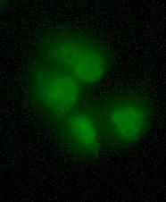
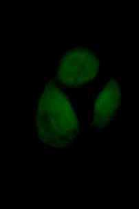
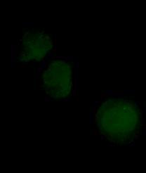

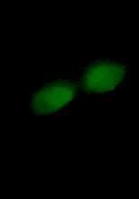
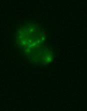
85 Figure Illustration of immunofluorescence images of HPV-G cells. Top: cells exposed to nicotine concentration (0 M, 10 nm, 100 nm, and 1000 nm). Bottom: cells exposed to ICCM and nicotine (0 M, 10 nm, 100 nm, and 1000 nm) 70
nuclear science and technology
 EUROPEAN COMMISSION nuclear science and technology The role of intercellular communication and DNA double-strand breaks in the induction of bystander effects (INTERSTANDER) Contract N o FIGH-CT2002-00218
Radiation induced bystander effects adaptive responses and low dose risk
 Radiation induced bystander effects adaptive responses and low dose risk Carmel Mothersill and Colin Seymour Medical Physics and Applied Radiation Sciences McMaster University, Hamilton, Ontario Canada
CHAPTER TWO MECHANISMS OF RADIATION EFFECTS
 10-2 densely ionizing radiation CHAPTER TWO MECHANISMS OF RADIATION EFFECTS 2.0 INTRODUCTION Cell survival curves describe the relationship between the fractional survival, S, of a population of radiated
Radiation Oncology. Initial Certification Qualifying (Computer-based) Examination: Study Guide for Radiation and Cancer Biology
 Examination: Study Guide for Radiation and Cancer Biology") Radiation Oncology Initial Certification Qualifying (Computer-based) Examination: Study Guide for Radiation and Cancer Biology This exam tests your knowledge of the principles of cancer and radiation biology
Radiation Oncology Initial Certification Qualifying (Computer-based) Examination: Study Guide for Radiation and Cancer Biology This exam tests your knowledge of the principles of cancer and radiation biology
The Impact of Bystander Effects and Adaptive Responses in the Health Risks of Low Dose Ionizing Radiation
 The Impact of Bystander Effects and Adaptive Responses in the Health Risks of Low Dose Ionizing Radiation Edouard Azzam New Jersey Medical School Newark, USA Two phenomena have been recently implicated
Radiation induced bystander effects What? Why? And How? Dose-Response Carmel Mothersill Colin Seymour McMaster University
 Radiation induced bystander effects What? Why? And How? Carmel Mothersill Colin Seymour McMaster University Outline Bystander effects and genomic instability In vivo relevance and evidence The fish model
UNC-Duke Biology Course for Residents Fall
 UNC-Duke Biology Course for Residents Fall 2018 1 UNC-Duke Biology Course for Residents Fall 2018 2 UNC-Duke Biology Course for Residents Fall 2018 3 UNC-Duke Biology Course for Residents Fall 2018 4 UNC-Duke
Neurotransmitter Systems II Receptors. Reading: BCP Chapter 6
 Neurotransmitter Systems II Receptors Reading: BCP Chapter 6 Neurotransmitter Systems Normal function of the human brain requires an orderly set of chemical reactions. Some of the most important chemical
LET, RBE and Damage to DNA
 LET, RBE and Damage to DNA Linear Energy Transfer (LET) When is stopping power not equal to LET? Stopping power (-de/dx) gives the energy lost by a charged particle in a medium. LET gives the energy absorbed
Chapter 7. What is Radiation Biology? Ionizing Radiation. Energy Transfer Determinants 09/21/2014
 Chapter 7 Molecular & Cellular Radiation Biology What is Radiation Biology? A branch of biology concerned with how ionizing radiation effects living systems. Biological damage that occurs from different
Determination of Radon Concentration in Some Types of Cigarettes
 Determination of Radon Concentration in Some Types of Cigarettes Tarfa.H.Alsheddi 1, Amal Mohamed 2 and Shaffa.Al.Mansour 3 1 Department of physics, King Faisal University, Saudi Arabia. 2 Department of
William F. Morgan. Ph.D., D.Sc.
 Biological Responses at Low Radiation Doses: Advances in Radiation Biology and Potential ti Implications for Radiation Exposure Regulations. William F. Morgan. Ph.D., D.Sc. Pacific Northwest National Laboratory
Cancer. The fundamental defect is. unregulated cell division. Properties of Cancerous Cells. Causes of Cancer. Altered growth and proliferation
 Cancer The fundamental defect is unregulated cell division. Properties of Cancerous Cells Altered growth and proliferation Loss of growth factor dependence Loss of contact inhibition Immortalization Alterated
U.S. Low Dose Radiation Research Program
 U.S. Low Dose Radiation Research Program Update November 2010 ISCORS NF Metting, ScD, Program Manager Office of Science Office of Biological and Environmental Research The Department of Energy Office of
Cancer. The fundamental defect is. unregulated cell division. Properties of Cancerous Cells. Causes of Cancer. Altered growth and proliferation
 Cancer The fundamental defect is unregulated cell division. Properties of Cancerous Cells Altered growth and proliferation Loss of growth factor dependence Loss of contact inhibition Immortalization Alterated
609G: Concepts of Cancer Genetics and Treatments (3 credits)
") Master of Chemical and Life Sciences Program College of Computer, Mathematical, and Natural Sciences 609G: Concepts of Cancer Genetics and Treatments (3 credits) Text books: Principles of Cancer Genetics,
Master of Chemical and Life Sciences Program College of Computer, Mathematical, and Natural Sciences 609G: Concepts of Cancer Genetics and Treatments (3 credits) Text books: Principles of Cancer Genetics,
Introduction. Cancer Biology. Tumor-suppressor genes. Proto-oncogenes. DNA stability genes. Mechanisms of carcinogenesis.
 Cancer Biology Chapter 18 Eric J. Hall., Amato Giaccia, Radiobiology for the Radiologist Introduction Tissue homeostasis depends on the regulated cell division and self-elimination (programmed cell death)
PHRM20001: Pharmacology - How Drugs Work!
 PHRM20001: Pharmacology - How Drugs Work Drug: a chemical that affects physiological function in a specific way. Endogenous substances: hormones, neurotransmitters, antibodies, genes. Exogenous substances:
Chronic cell death may play a crucial role in mutagenesis and carcinogenesis due to radon exposure
 Chronic cell death may play a crucial role in mutagenesis and carcinogenesis due to radon exposure Balázs G. Madas, Imre Balásházy MTA Centre for Energy Research,, Hungary balazs.madas@energia.mta.hu Low
Biological Effects of Radiation KJ350.
 Biological Effects of Radiation KJ350 deborah.oughton@nmbu.no 2111 2005 Radiation Biology Interaction of radiation with biological material Doses (Gy, Sv) and effects Scientific Controversy Radiation Protection
Biochemistry of Carcinogenesis. Lecture # 35 Alexander N. Koval
 Biochemistry of Carcinogenesis Lecture # 35 Alexander N. Koval What is Cancer? The term "cancer" refers to a group of diseases in which cells grow and spread unrestrained throughout the body. It is difficult
2015 AP Biology Unit #4 Quiz 1 Cell Communication, Cancer and The Cell Cycle Week of November
 Name: Class: Date: 2015 AP Biology Unit #4 Quiz 1 Cell Communication, Cancer and The Cell Cycle Week of 16-20 November Multiple Choice Identify the choice that best completes the statement or answers the
LOW DOSES OF RADIATION REDUCE RISK IN VIVO
 Dose-Response: An International Journal Volume 5 Issue 1 ADAPTIVE BIOLOGICAL RESPONSES FOLLOWING EXPOSURES TO IONIZING RADIATION Article 4 3-2007 LOW DOSES OF RADIATION REDUCE RISK IN VIVO REJ Mitchel
Principles of chemotherapy
 Principles of chemotherapy Chemotherapy first coined by Paul Ehrlich Aim to selectively destroy cancer cells whilst relatively sparing tumours cells Growth characteristics of cancer cells allows for selective
Introduction to pathology lecture 5/ Cell injury apoptosis. Dr H Awad 2017/18
 Introduction to pathology lecture 5/ Cell injury apoptosis Dr H Awad 2017/18 Apoptosis = programmed cell death = cell suicide= individual cell death Apoptosis cell death induced by a tightly regulated
Genomic Instability Induced by Ionizing Radiation
 Genomic Instability Induced by Ionizing Radiation Christian Streffer Universitätsklinikum Essen, 45122 Essen, Germany INTRODUCTION In contrast to general assumptions it has frequently been shown that DNA
Molecular Radiobiology Module 4 Part #3
 Molecular Radiobiology Module 4 Part #3 Bushong - Chapter 31 10-526-197 - Rhodes Interaction & damage is a matter of chance Energy deposited rapidly 10-17 seconds Interactions are non-selective in tissue
Transformation of Normal HMECs (Human Mammary Epithelial Cells) into Metastatic Breast Cancer Cells: Introduction - The Broad Picture:
 into Metastatic Breast Cancer Cells: Introduction - The Broad Picture:") Transformation of Normal HMECs (Human Mammary Epithelial Cells) into Metastatic Breast Cancer Cells: Introduction - The Broad Picture: Spandana Baruah December, 2016 Cancer is defined as: «A disease caused
Transformation of Normal HMECs (Human Mammary Epithelial Cells) into Metastatic Breast Cancer Cells: Introduction - The Broad Picture: Spandana Baruah December, 2016 Cancer is defined as: «A disease caused
Radiation Protection in the World of Modern Radiobiology: Time for A New Approach. R. E. J. Mitchel and D. R Boreham
 Radiation Protection in the World of Modern Radiobiology: Time for A New Approach R. E. J. Mitchel and D. R Boreham Radiation Biology and Health Physics Branch, AECL, Chalk River Laboratories, Chalk River
Direct evidence for a bystander effect of ionizing radiation in primary human fibroblasts
 doi: 10.1054/ bjoc.2000.1665, available online at http://www.idealibrary.com on http://www.bjcancer.com Direct evidence for a bystander effect of ionizing radiation in primary human fibroblasts OV Belyakov,
TFY4315 STRÅLINGSBIOFYSIKK
 Norges teknisk-naturvitenskaplige universitet Institutt for fysikk EKSAMENSOPPGÅVER med løysingsforslag Examination papers with solution proposals TFY4315 STRÅLINGSBIOFYSIKK Biophysics of Ionizing Radiation
Cancer and Gene Alterations - 1
 Cancer and Gene Alterations - 1 Cancer and Gene Alteration As we know, cancer is a disease of unregulated cell growth. Although we looked at some of the features of cancer when we discussed mitosis checkpoints,
DEFINITIONS. Pharmacokinetics. Pharmacodynamics. The process by which a drug is absorbed, distributed, metabolized and eliminated by the body
 PHARMACOLOGY BASICS DEFINITIONS Pharmacokinetics The process by which a drug is absorbed, distributed, metabolized and eliminated by the body Pharmacodynamics The interactions of a drug and the receptors
5.1. KEY CONCEPT Cells have distinct phases of growth, reproduction, and normal functions. 68 Reinforcement Unit 2 Resource Book
 5.1 THE CELL CYCLE KEY CONCEPT Cells have distinct phases of growth, reproduction, and normal functions. Cells have a regular pattern of growth, DNA duplication, and division that is called the cell cycle.
Problem Set 8 Key 1 of 8
 7.06 2003 Problem Set 8 Key 1 of 8 7.06 2003 Problem Set 8 Key 1. As a bright MD/PhD, you are interested in questions about the control of cell number in the body. Recently, you've seen three patients
Fundamentals of Pharmacology
 Fundamentals of Pharmacology Topic Page Receptors 2 Ion channels / GABA 4 GPCR s 6 TK receptors 8 Basics of PK 11 ADR s / Clinical study design 13 Introduction to the ANS 16 Cholinergic Pharmacology 20
Psych 181: Dr. Anagnostaras
 Psych 181: Dr. Anagnostaras Lecture 5 Synaptic Transmission Introduction to synaptic transmission Synapses (Gk., to clasp or join) Site of action of most psychoactive drugs 6.5 1 Synapses Know basic terminology:
Chapter 12. Regulation of Cell Division. AP Biology
 Chapter 12. Regulation of Cell Division Coordination of cell division! Multicellular organism " need to coordinate across different parts of organism! timing of cell division! rates of cell division "
ENVIRONMENTAL TOXICOLOGY
 ENVIRONMENTAL TOXICOLOGY Chapter 4 Toxicokinetics Mohd Amir Bin Arshad Toxicokinetics study on how a substance gets into the body and what happens to it in the body" The kinetics (movement) of substances
7.012 Quiz 3 Answers
 MIT Biology Department 7.012: Introductory Biology - Fall 2004 Instructors: Professor Eric Lander, Professor Robert A. Weinberg, Dr. Claudette Gardel Friday 11/12/04 7.012 Quiz 3 Answers A > 85 B 72-84
Chapter 2: Cellular Mechanisms and Cognition
 Chapter 2: Cellular Mechanisms and Cognition MULTIPLE CHOICE 1. Two principles about neurons were defined by Ramón y Cajal. The principle of connectional specificity states that, whereas the principle
Modelling the induction of cell death and chromosome damage by therapeutic protons
 Modelling the induction of cell death and chromosome damage by therapeutic protons M.P. Carante 1,2 and F. Ballarini 1,2, * 1 University of Pavia, Physics Department, Pavia, Italy 2 INFN, Sezione di Pavia,
Are non-targeted effects important in radiation protection? Carmel Mothersill and Colin Seymour McMaster University Canada
 Are non-targeted effects important in radiation protection? Carmel Mothersill and Colin Seymour McMaster University Canada Non-targeted radiation effects Inter-animal/plant signaling Effects in neighbouring
Mitosis and the Cell Cycle
 Mitosis and the Cell Cycle Chapter 12 The Cell Cycle: Cell Growth & Cell Division Where it all began You started as a cell smaller than a period at the end of a sentence Getting from there to here Cell
Chemistry 106: Drugs in Society Lecture 19: How do Drugs Elicit an Effect? Interactions between Drugs and Macromolecular Targets 11/02/17
 Chemistry 106: Drugs in Society Lecture 19: How do Drugs Elicit an Effect? Interactions between Drugs and Macromolecular Targets 11/02/17 Targets for Therapeutic Intervention: A Comparison of Enzymes to
Nature of Radiation and DNA damage
 Nature of Radiation and DNA damage Index 1. What is radiation? 2. Ionizing Radiation 3. Interaction of Gamma-radiation with Matter 4. Radiobiology 5. Direct and Indirect action of radiation 6. Steps of
Acquisition of Radiation Resistant Ability in Non- Irradiated Cells by Secreted Factors from Low Dose Irradiated Cells
 Nagoya University Acquisition of Radiation Resistant Ability in Non- Irradiated Cells by Secreted Factors from Low Dose Irradiated Cells Jun Kumagai a *, Akane Oohashi b and Genro Kashino c a Institute
Nicotine: Everything You Need to Know but Have Never Been Told
 Bernd Mayer Pharmacology & Toxicology University of Graz, Austria Nicotine: Everything You Need to Know but Have Never Been Told Bernhard-Michael (Bernd) Mayer, PhD Professor & Chair Department of Pharmacology
Synaptic Communication. Steven McLoon Department of Neuroscience University of Minnesota
 Synaptic Communication Steven McLoon Department of Neuroscience University of Minnesota 1 Course News The first exam is next week on Friday! Be sure to checkout the sample exam on the course website. 2
Ligand-Gated Ion Channels
 Ligand-Gated Ion Channels The Other Machines That Make It Possible... Topics I Introduction & Electrochemical Gradients Passive Membrane Properties Action Potentials Voltage-Gated Ion Channels Topics II
Ionizing Radiation. Nuclear Medicine
 Ionizing Radiation Nuclear Medicine Somatic Deterministic Effect Erythema Somatic Stochastic Effect Leukemia Genetic Effects DNA BIOLOGICAL EFFECTS OF IONIZING RADIATION ON TISSUES, ORGANS AND SYSTEMS
Pharmacokinetics Dr. Iman Lec. 3
 Pharmacokinetics r. Iman Lec. 3 Pharmacokinetics A dequate drug doses must be delivered to the target organ to get therapeutic but not toxic levels. So, pharmacokinetic examines the movement of drug over
- Neurotransmitters Of The Brain -
 - Neurotransmitters Of The Brain - INTRODUCTION Synapsis: a specialized connection between two neurons that permits the transmission of signals in a one-way fashion (presynaptic postsynaptic). Types of
María José Mesa López
 María José Mesa López q Radiobiology. q Ionizing Radiations. q Mutations. q Stochastic Effects Vs Deterministic Effects. q Cellular Radiosensitivity. q Bibliography. Science which combines the basic principles
HUMAN LUNG CANCER RISKS FROM RADON PART II INFLUENCE FROM COMBINED ADAPTIVE RESPONSE AND BYSTANDER EFFECTS A MICRODOSE ANALYSIS
 Dose-Response: An International Journal Volume 9 Issue 4 Article 8 12-2011 HUMAN LUNG CANCER RISKS FROM RADON PART II INFLUENCE FROM COMBINED ADAPTIVE RESPONSE AND BYSTANDER EFFECTS A MICRODOSE ANALYSIS
Modelling of Biological Processes
 Modelling of Biological Processes WHAT HAPPENS AFTER EARLY MOLECULAR DAMAGE? Stephen McMahon Queen s University, Belfast, Northern Ireland 3 rd August 2016 1 Do we need biology? The Linear-quadratic relationship
Regulators of Cell Cycle Progression
 Regulators of Cell Cycle Progression Studies of Cdk s and cyclins in genetically modified mice reveal a high level of plasticity, allowing different cyclins and Cdk s to compensate for the loss of one
SPI Determine the relationship between cell growth and cell reproduction. EQ-How do you determine SA/V of a cell?
 SPI 3210.1.6 Determine the relationship between cell growth and cell reproduction. EQ-How do you determine SA/V of a cell? Click on a lesson name to select. Chapter 9 Cellular Reproduction Section 1: Cellular
RADIATION RISK ASSESSMENT
 RADIATION RISK ASSESSMENT EXPOSURE and TOXITY ASSESSMENT Osipova Nina, associated professor, PhD in chemistry, Matveenko Irina, Associate professor, PhD in philology TOMSK -2013 The contents 1.What is
Cellular Reproduction
 Cellular Reproduction How do we know that cells divide to form other cells? Growth Chapter 9 Cuts or wounds heal New blood is produced Hair and fingernails grow back Do cells grow? 1 Hollywood Limits to
Biological Effects of Radiation
 Radiation and Radioisotope Applications EPFL Doctoral Course PY-031 Biological Effects of Radiation Lecture 09 Rafael Macian 23.11.06 EPFL Doctoral Course PY-031: Radioisotope and Radiation Applications
Basics of Pharmacology
 Basics of Pharmacology Pekka Rauhala Transmed 2013 What is pharmacology? Pharmacology may be defined as the study of the effects of drugs on the function of living systems Pharmacodynamics The mechanism(s)
Solution key Problem Set
 Solution key- 7.013 Problem Set 6-2013 Question 1 a) Our immune system is comprised of different cell types. Complete the table below by selecting all correct cell types from the choices provided. Cells
Ch. 45 Continues (Have You Read Ch. 45 yet?) u Central Nervous System Synapses - Synaptic functions of neurons - Information transmission via nerve
 u Central Nervous System Synapses - Synaptic functions of neurons - Information transmission via nerve") Ch. 45 Continues (Have You Read Ch. 45 yet?) u Central Nervous System Synapses - Synaptic functions of neurons - Information transmission via nerve impulses - Impulse may be blocked in its transmission
Ch. 45 Continues (Have You Read Ch. 45 yet?) u Central Nervous System Synapses - Synaptic functions of neurons - Information transmission via nerve impulses - Impulse may be blocked in its transmission
Regulation of Cell Division. AP Biology
 Regulation of Cell Division 2006-2007 Coordination of cell division A multicellular organism needs to coordinate cell division across different tissues & organs critical for normal growth, development
CANCER AND LOW DOSE RESPONSES IN VIVO: IMPLICATIONS FOR RADIATION PROTECTION
 CANCER AND LOW DOSE RESPONSES IN VIVO: IMPLICATIONS FOR RADIATION PROTECTION Ron Mitchel Radiation Biology and Health Physics Branch Atomic Energy of Canada Limited, Chalk River Laboratories, Chalk River,
Part-4. Cell cycle regulatory protein 5 (Cdk5) A novel target of ERK in Carb induced cell death
 A novel target of ERK in Carb induced cell death") Part-4 Cell cycle regulatory protein 5 (Cdk5) A novel target of ERK in Carb induced cell death 95 1. Introduction The process of replicating DNA and dividing cells can be described as a series of coordinated
Part-4 Cell cycle regulatory protein 5 (Cdk5) A novel target of ERK in Carb induced cell death 95 1. Introduction The process of replicating DNA and dividing cells can be described as a series of coordinated
COURSE: Medical Microbiology, PAMB 650/720 - Fall 2008 Lecture 16
 COURSE: Medical Microbiology, PAMB 650/720 - Fall 2008 Lecture 16 Tumor Immunology M. Nagarkatti Teaching Objectives: Introduction to Cancer Immunology Know the antigens expressed by cancer cells Understand
Regulation of Cell Division (Ch. 12)
") Regulation of Cell Division (Ch. 12) Coordination of cell division A multicellular organism needs to coordinate cell division across different tissues & organs critical for normal growth, development &
Regulation of Cell Division (Ch. 12) Coordination of cell division A multicellular organism needs to coordinate cell division across different tissues & organs critical for normal growth, development &
Cell Cycle. Cell Cycle the cell s life cycle that extends from one division to the next G1 phase, the first gap phase. S phase, synthesis phase
 Cell Cycle the cell s life cycle that extends from one division to the next G1 phase, the first gap phase Cell Cycle interval between cell division and DNA replication accumulates materials needed to replicate
Radiobiology of fractionated treatments: the classical approach and the 4 Rs. Vischioni Barbara MD, PhD Centro Nazionale Adroterapia Oncologica
 Radiobiology of fractionated treatments: the classical approach and the 4 Rs Vischioni Barbara MD, PhD Centro Nazionale Adroterapia Oncologica Radiobiology It is fundamental in radiation oncology Radiobiology
BIT 120. Copy of Cancer/HIV Lecture
 BIT 120 Copy of Cancer/HIV Lecture Cancer DEFINITION Any abnormal growth of cells that has malignant potential i.e.. Leukemia Uncontrolled mitosis in WBC Genetic disease caused by an accumulation of mutations
ANATOMY & PHYSIOLOGY - CLUTCH CH. 6 - CELL COMMUNICATION.
 !! www.clutchprep.com CONCEPT: CELL-TO-CELL CONNECTIONS AND SIGNALING Gap and Tight Junctions: Adjacent cells communicate and hold on to each other via junctions. Two important kinds: Gap Junctions are
Radioactivity. Lecture 8 Biological Effects of Radiation
 Radioactivity Lecture 8 Biological Effects of Radiation Studies of impact of ionizing radiation on the human body - Hiroshima - US-Japanese teams medical tests, autopsies, human organ analysis, on-site
2401 : Anatomy/Physiology
 Dr. Chris Doumen Week 11 2401 : Anatomy/Physiology Autonomic Nervous System TextBook Readings Pages 533 through 552 Make use of the figures in your textbook ; a picture is worth a thousand words! Work
Drug Receptor Interactions and Pharmacodynamics
 Drug Receptor Interactions and Pharmacodynamics Dr. Raz Mohammed MSc Pharmacology School of Pharmacy 22.10.2017 Lec 6 Pharmacodynamics definition Pharmacodynamics describes the actions of a drug on the
Possible Consequences of Inhomogeneous Suborgan Distribution of Dose and the Linear No-Threshold Dose-Effect Relationship
 Possible Consequences of Inhomogeneous Suborgan Distribution of Dose and the Linear No-Threshold Dose-Effect Relationship Balázs G. Madas, Imre Balásházy Centre for Energy Research, Hungarian Academy of
Review of Neurochemistry What are neurotransmitters?
 Review of Neurochemistry What are neurotransmitters? In molecular terms, neurotransmitters are molecules that ( ) and of neurons by, for example, increasing or decreasing enzymatic activity or altering
Receptors. Dr. Sanaa Bardaweel
 Receptors Types and Theories Dr. Sanaa Bardaweel Some terms in receptor-drug interactions Agonists: drugs that mimic the natural messengers and activate receptors. Antagonist: drugs that block receptors.
Regulation of Cell Division
 Regulation of Cell Division Two HeLa cancer cells are just completing cytokinesis. Explain how the cell division of cancer cells like these is misregulated. Identify genetic and other changes that might
The Nervous System Mark Stanford, Ph.D.
 The Nervous System Functional Neuroanatomy and How Neurons Communicate Mark Stanford, Ph.D. Santa Clara Valley Health & Hospital System Addiction Medicine and Therapy Services The Nervous System In response
BL 424 Test pts name Multiple choice have one choice each and are worth 3 points.
 BL 424 Test 3 2010 150 pts name Multiple choice have one choice each and are worth 3 points. 1. The plasma membrane functions as a a. selective barrier to the passage of molecules. b. sensor through which
HORMONES (Biomedical Importance)
") hormones HORMONES (Biomedical Importance) Hormones are the chemical messengers of the body. They are defined as organic substances secreted into blood stream to control the metabolic and biological activities.
hormones HORMONES (Biomedical Importance) Hormones are the chemical messengers of the body. They are defined as organic substances secreted into blood stream to control the metabolic and biological activities.
Chapter 3 Neurotransmitter release
 NEUROPHYSIOLOGIE CELLULAIRE CONSTANCE HAMMOND Chapter 3 Neurotransmitter release In chapter 3, we proose 3 videos: Observation Calcium Channel, Ca 2+ Unitary and Total Currents Ca 2+ and Neurotransmitter
STEIN IN-TERM EXAM -- BIOLOGY FEBRUARY 18, PAGE
 STEIN IN-TERM EXAM -- BIOLOGY 3058 -- FEBRUARY 18, 2016 -- PAGE 1 of 8 There are 25 questions in this Biology 3058 exam. All questions are "A, B, C, D, E, F, G, H" questions worth one point each. There
Cells communicate with each other via signaling ligands which interact with receptors located on the surface or inside the target cell.
 BENG 100 Frontiers of Biomedical Engineering Professor Mark Saltzman Chapter 6 SUMMARY In this chapter, cell signaling was presented within the context of three physiological systems that utilize communication
Genetics and Cancer Ch 20
 Genetics and Cancer Ch 20 Cancer is genetic Hereditary cancers Predisposition genes Ex. some forms of colon cancer Sporadic cancers ~90% of cancers Descendants of cancerous cells all cancerous (clonal)
HIBBING COMMUNITY COLLEGE COURSE OUTLINE
 HIBBING COMMUNITY COLLEGE COURSE OUTLINE COURSE NUMBER & TITLE: BIOL 2151: Human Physiology CREDITS: 4 (Lecture 3 / Lab 1) PREREQUISITES: Human Anatomy or Integrated Science recommended CATALOG DESCRIPTION:
The Potential Impact of Bystander Effects on Radiation Risks in a Mars Mission
 RADIATION RESEARCH 156, 612 617 (2001) 0033-7587/01 $5.00 2001 by Radiation Research Society. All rights of reproduction in any form reserved. The Potential Impact of Bystander Effects on Radiation Risks
Ameen Alsaras. Ameen Alsaras. Mohd.Khatatbeh
 9 Ameen Alsaras Ameen Alsaras Mohd.Khatatbeh Nerve Cells (Neurons) *Remember: The neural cell consists of: 1-Cell body 2-Dendrites 3-Axon which ends as axon terminals. The conduction of impulse through
Synaptic transmission
 Outline Synaptic transmission Sompol Tapechum M.D., Ph.D. Department of Physiology Faculty of Medicine Siriraj Hospital, Bangkok, Thailand. sisth@mahidol.ac.th 2 Structure of synapse Modes of synaptic
Zool 3200: Cell Biology Exam 4 Part II 2/3/15
 Name:Key Trask Zool 3200: Cell Biology Exam 4 Part II 2/3/15 Answer each of the following questions in the space provided, explaining your answers when asked to do so; circle the correct answer or answers
3) Most of the organelles in a neuron are located in the A) dendritic region. B) axon hillock. C) axon. D) cell body. E) axon terminals.
 Most of the organelles in a neuron are located in the A) dendritic region. B) axon hillock. C) axon. D) cell body. E) axon terminals.") Chapter 48 Neurons, Synapses, and Signaling Multiple-Choice Questions 1) A simple nervous system A) must include chemical senses, mechanoreception, and vision. B) includes a minimum of 12 ganglia. C) has
Chapter 48 Neurons, Synapses, and Signaling Multiple-Choice Questions 1) A simple nervous system A) must include chemical senses, mechanoreception, and vision. B) includes a minimum of 12 ganglia. C) has
Industrial Toxicology
 Industrial Toxicology Learning Objectives Know the assumptions of the doseresponse and time-course curves Be able to define and label key points of a curve Know the difference between potency and efficacy
What is radiation quality?
 What is radiation quality? Dudley T Goodhead Medical Research Council, UK DoReMi Radiation Quality workshop Brussels. 9-10 July 2013 What is radiation quality? Let s start at the very beginning. A very
Neuropharmacology NOTES
 Neuropharmacology NOTES Contents Topic Page # Lecture 1- Intro to Neurochemical Transmission & Neuromodulation 2 Lecture 2- Serotonin & Noradrenaline 7 Lecture 3- Acetylcholine & Dopamine 14 Lecture 4-
RADIOBIOLOGICAL PRINCIPLES IN INTRAVASCULAR IRRADIATION
 Cardiovascular Radiation Medicine 1:1 (1999) 42 47 BIOLOGY/PATHOLOGY ORIGINAL ARTICLE RADIOBIOLOGICAL PRINCIPLES IN INTRAVASCULAR IRRADIATION Eric J. Hall, D.Sc.,* Richard C. Miller, Ph.D., and David J.
STEIN IN-TERM EXAM -- BIOLOGY FEBRUARY 15, PAGE
 STEIN IN-TERM EXAM -- BIOLOGY 3058 -- FEBRUARY 15, 2018 -- PAGE 1 of 8 There are 25 questions in this Biology 3058 exam. All questions are "A, B, C, D, E, F, G, H" questions worth one point each. There
Dania Ahmad. Tamer Barakat + Dania Ahmad. Faisal I. Mohammed
 16 Dania Ahmad Tamer Barakat + Dania Ahmad Faisal I. Mohammed Revision: What are the basic types of neurons? sensory (afferent), motor (efferent) and interneuron (equaled association neurons). We classified
Lecture 14 - The cell cycle and cell death
 02.17.10 Lecture 14 - The cell cycle and cell death The cell cycle: cells duplicate their contents and divide The cell cycle may be divided into 4 phases The cell cycle triggers essential processes (DNA