Review Article SPECT/CT for Lymphatic Mapping of Sentinel Nodes in Early Squamous Cell Carcinoma of the Oral Cavity and Oropharynx
|
|
|
- Evelyn McKinney
- 5 years ago
- Views:
Transcription
1 International Molecular Imaging Volume 2011, Article ID , 6 pages doi: /2011/ Review Article SPECT/CT for Lymphatic Mapping of Sentinel Nodes in Early Squamous Cell Carcinoma of the Oral Cavity and Oropharynx Haerle Stephan K. 1 and Stoeckli Sandro J. 2 1 Department of Otolaryngology-Head and Neck Surgery, University Hospital Zurich, Frauenklinikstrasse 24, 8091 Zurich, Switzerland 2 Department of Otolaryngology-Head and Neck Surgery, Kantonsspital St. Gallen, Rorschacherstraβe 95, 9000 St. Gallen, Switzerland Correspondence should be addressed to Stoeckli Sandro J., sandro.stoeckli@kssg.ch Received 15 July 2010; Accepted 24 August 2010 Academic Editor: Domenico Rubello Copyright 2011 H. Stephan K. and S. Sandro J. This is an open access article distributed under the Creative Commons Attribution License, which permits unrestricted use, distribution, and reproduction in any medium, provided the original work is properly cited. Adequate staging and treatment of the neck in squamous cell carcinoma of the oral cavity and oropharynx (OSCC) is of paramount importance. Elective neck dissection (END) of the clinical N0-neck is widely advocated as neck treatment. With regard to the prevalence of 20 40% of occult neck metastases found in the ND specimens, the majority of patients undergo surgery of the lymphatic drainage basin without therapeutic benefit. Sentinel node biopsy (SNB) has been shown to be a safe, reliable and accurate alternative treatment modality for selected patients. Using this technique, lymphatic mapping is crucial. Previous reports suggested a benefit of single photon emission computed tomography with CT (SPECT/CT) over dynamic planar lymphoscintigraphy (LS) alone. SPECT/CT allows the surgeon for better topographical orientation and delineation of sentinel lymph nodes (SLN s) against surrounding structures. Additionally, SPECT/CT has the potential to detect more SLN s which might harbour occult disease, than LS. SPECT/CT is recommended to be used routinely, although SPECT/CT is not indispensable for successful SNB. 1. Background Squamous cell carcinoma of the oral cavity and oropharynx (OSCC) accounts for one of the most common cancers worldwide, with more than a quarter million new cases annually [1].The presence or absence of lymph node involvement is of paramount importance for prognosis and therapy decision [2, 3]. Therefore, an adequate staging and management of the neck is needed. The most challenging issue remains the treatment of the clinically and radiologically negative neck. Most centers throughout the world advocate elective neck dissection (END) for histopathologic staging and removal of microscopic disease in this situation. With regard to the prevalence of 20% 40% of occult neck metastases found in the neck dissection specimens, the majority of patients undergo surgery of the lymphatic drainage basin without therapeutic benefit. Sentinel node biopsy (SNB) has been shown to be very accurate in selecting patients who benefit from elective neck treatment and sparing the costs and morbidity to the others. Detection of the sentinel nodes by lymphatic mapping is crucial with this technique. Single-photon emission computed tomography with CT (SPECT/CT) has been recently introduced to enhance the diagnostic accuracy of preoperative lymphoscintigraphy. 2. Sentinel Node Biopsy By definition, the sentinel lymph node (SLN) is the first draining lymph node to receive lymphatic drainage from a primary tumor of a specific site [4]. In case of lymphatic spread, the lymphatic drain will first pass the SLN. All following nodes may be reached only subsequently by the disease. Therefore, selective excision of the SLN with subsequent thorough histopathologic work-up reflects adequately the nodal status of the remaining neck [5]. Since Alex and Krag [6] have described their first experience with SNB for OSCC, the technique has gained large popularity and many centers followed with validation and observational studies [7 9]. Lymphatic mapping of the SLN in the complex head and neck areahasbeenshown tobe essential [10]. The problems
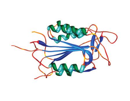
2 2 International Molecular Imaging in the head and neck area are threefold: first, there is a high density of lymph nodes, second, the structure of these nodes shows an unique complexity of lymphatic pathways, and third, the SLNs are located in close proximity to the primary tumor. Therefore, sophisticated lymphatic mapping techniques are required. During the preoperative setting, a dynamic lymphoscintigraphy (LS) assesses the individual draining pattern after injection of radiolabeled particles around the primary tumor. The intraoperative use of a handheld gamma probe helps the surgeon to localise and excise the first echelon lymph nodes. The success of this technique has been abundantly reported in the literature and welldocumented guidelines do exist [11]. As with breast cancer, preliminary reports showed a new imaging technology with promising results: SPECT/CT. 3. Patient s Selection For SNB of the OSCC, patients with stages I and II (T1 and T2) disease and no clinical and radiological evidence of cervical lymph node involvement are eligible. Absence of suspicious or metastatic lymph nodes is based on palpation, ultrasound with fine needle aspiration cytology (FNAC), or contrast-enhanced computed tomography (CT), or magnetic resonance imaging (MRI), or 18 F-fluoro-2-deoxy-Dglucose positron emission tomography ( 18 F-FDG-PET)/CT. With regard to conventional imaging, lymph nodes greater than 1.5 cm in level II and greater than 1 cm in all other levels, or lymph nodes with round shape, central necrosis, and peripheral contrast enhancement are considered pathologic. In metabolic imaging, a lymph node with a clearly higher FDG-uptake compared to the background and anatomically corresponding to a lymph node in the low-dose CT scan is considered pathologic. 4. Tracer To assess the individual lymphatic drainage pattern, a peritumoral injection of radiolabeled particles is performed. The particles will enter the lymphatic capillaries and accumulate in the first draining node. There is a variety of colloidal and soluble tracers available although most trials report using Tc-99m-labeled human serum albumin colloid (Nanocoll, GE Healthcare). With its particle size of 8 30 nm, Nanocoll migrates to the sentinel node within minutes and remains there until the next day [11, 12]. This allows a flexible way for planning surgery to take place. 5. Imaging A standard technique for preoperative imaging is the use of a gamma camera for lymphoscintigraphy to assess the individual drainage pattern of the injected radiolabeled tracer via the capillaries to the larger collector lymphatics [13]. Imaging will be performed either the day before or the day of surgery. According to the joint practice guidelines for radionuclide lymphoscintigraphy, the setting of the camera is proposed to be as follows. A large-field-of-view gamma camera provided with a high- or ultrahigh-resolution lowenergy collimator should be used, with a 10% 20% window centered on the 140-keV energy peak of Tc-99m [11]. The gamma camera should be routinely checked for quality control as proposed in published protocols [14]. Immediately after the injection of the radiotracer, the lymphatic drainage is monitored dynamically with the gamma camera in the anteroposterior projection (1 image/3 minutes). The lymphatic drainage is then observed by the nuclear medicine specialist and the HN-surgeon at the monitor. When accumulation of the radiotracer in the first echelon node(s) occurs, the dynamic imaging can be interrupted and static imaging in the anterior-posterior, lateral and, if necessary, anterior oblique view can be performed. For the different projections, a three-headed camera is recommended. To be able to localize the nodes in a three-dimensional view, static images in at least two projections are needed. The patient is imaged in the supine position with head up [11]. Most reports in the literature use the term sentinel node interchangeably for lymphoscintigraphy and SNB. As most tracer accumulations or hot spots detected by lymphoscintigraphy or SPECT/CT correspond to more than one ultimately excised sentinel lymph node, the term hot spot should be used in the context of planar imaging or fused imaging whereas sentinel lymph node should be used in the context of surgical SNB. Intraoperatively, the surgeon will be guided to the sentinel nodes by a hand-held gamma probe containing a radiation detector with surrounding metal shielding and a collimated tip. The response related to the detected count rate is provided by a connected analyzer [11]. Using this technique, SNB has become a safe and reliable method to detect SLNs with a previously published SLN detection rate of 96% [5]. 6. SPECT/CT for Sentinel Node Mapping in HNSCC: A Comparison in the Literature Besides the previously described imaging technique using a preoperative lymphoscintigraphy, novel systems composed of a gamma camera and a CT scan combined in the same device have been recently introduced into clinical practice. Single photon emission CT (SPECT) and CT data are acquired at the same clinical setting without changing the patient s positioning, thus allowing for generation of accurate fused images combining the functional data of SPECT with the anatomical data of the CT scan (Figure 1). Different centers have already reported on their experience with SPECT/CT for SLN mapping in early OSCC, however, with contradictory results [11, 15 24]. In 2000, Even-Sapir et al. described the fusion of the SPECT lymphoscintigraphy data with CT using a hybrid gamma-camera and a lowdose CT system that allows SPECT and CT to be performed at the same time without changing the patient s position [25]. Three years later, the same author introduced the hybrid SPECT/CT system into sentinel node mapping of HNSCC [15]. In 2004, two feasibility studies using planar
 Axial volume 1 Se: 1 I: 790.")
 Figure 1: 64-year-old female suffering from a left-sided tongue cancer.")
.")
 shows the axial SPECT image with the corresponding cross hair in the sentinel node with a clear delineation from the injection site.")
3 International Molecular Imaging 3 3D volume 2 Se: HD MIP no cut DF0V 56.6 cm Coronal volume 2/volume 1 Se: A: 0 Im: 15 DF0V: 57.9 cm 39 No VOI 8.8 mm /8.8 sp 08:48:05 AM m = 4M= 168 V = %PET 8.8/ 8.8 mm /8.8 sp 08:48:05 AM m = 4M= 39 V = 37 Axial volume 2 Se: I: Im: 15 DF0V: 13 cm (a) Axial volume 1 Se: 1 I: Im: 30 DF0V: 13 cm (b) 8.8/ 8.8 mm /8.8 sp 08:48:05 AM m = 4M= 39 V = 39 (c) 4.4/ kv N/A ma mm /4.4 sp Tilt: 0 09:00:41 AM W = 602 L = 1065 V = 857 (d) Figure 1: 64-year-old female suffering from a left-sided tongue cancer. (a) shows the MIP (maximum intensity projection) image of SPECT acquisition in anteroposterior view. A large uptake is seen at the injection site with a small, focal uptake of the left lower boarder (cross hair). (b) shows a fused coronal SPECT/CT image that localises the small focal uptake in the neck region level IIA/B. (c) shows the axial SPECT image with the corresponding cross hair in the sentinel node with a clear delineation from the injection site. (d) shows the corresponding low-dose CT scan localising the uptake by linked cross hair into the neck level IIA/B. lymphoscintigraphy and SPECT/CT were published [16, 17]. Lopez et al. included ten patients stating that they believe that SPECT/CT will become a useful tool for sentinel node mapping [16]. Wagner et al. found an additional value by using SPECT/CT for sentinel node mapping than lymphoscintigraphy alone [17]: they have found an additional lymph node nearby the submandibular gland which has only been detected by SPECT/CT. This lymph node has been overlooked by planar lymphoscintigraphy and the intraoperative gamma probe as the radioactive scattering from the primary obscured the location of the radiolabeled SLNs. The same problem has been shown by other authors in the earlier period of radioguided imaging [26, 27] and was thought be resolved by introducing SPECT/CT. Thomsen et al. found SLNs close to the primary difficult to detect. Therefore, added oblique planar images and/or tomographic scans would help to overcome this problem [18]. Terada et al. also performed a feasible study on SPECT/CT and HN mucosal carcinoma and concluded that they were able to extract all the SLNs based on the fusioned images and to confirm its radioactivity with the gamma probe without the adverse effect of overlapping radioactivity from the primary site [19]. Khafif et al. included 22 patients with biopsy proven OSCC and found an improved identification of the SLNs of 30% compared to planar imaging [20]. Bilde et al. included 34 consecutive patients with stages I and II OSCC undergoing planar lymphoscintigraphy and SPECT/CT. After all, SPECT/CT demonstrated an extra SLN in 47% compared to lymphoscintigraphy alone [21]. In the same year, Keski- Säntti et al. were the first and only authors who found
4 4 International Molecular Imaging Study group Even-Sapir et al. [15] Table 1: An overview of various studies using SPECT/CT in the context of lymphatic mapping for SNB in OSCC. Number of patients (n) 6 SPECT/CT and the reported detection of SLN s 3 additional nodes detected in 6 patients compared to lymphoscintigraphy alone Lopez et al. [16] % visualization of the SLN s by SPECT/CT Wagner et al. [17] Thomsen et al. [18] Terada et al. [19] Khafifetal.[20] 20 Bilde et al. [21] 34 Keski-Säntti et al. [22] 11 additional nodes out of 49 SLNs detected compared to lymphoscintigraphy alone SPECT/CT and/or added oblique images revealed extra nodes in 15/40 patients % visualization of the SLN s by SPECT/CT 15 Haerle et al. [23] 58 SPECT/CT improved SLN identification and/or localization compared with planar images in 6 patients (30%) SPECT/CT demonstrated extra SLN s compared to planar imaging in 15 out of 32 patients (47%) 1 additional SLN located in the jugular chain detected compared to lymphoscintigraphy alone 11 additional hot spots could be revealed by SPECT/CT compared to lymphoscintigraphy alone. In one case even with additional occult disease. The value of SPECT/CT according to the authors SPECT/CT adds data that is of clinical relevance to SNB in patients with mucosal HNSCC SPECT/CT is shown to be an effective method for anatomic localization of the SLN s in N0 OSCC SPECT/CT adds additional information regarding nodes that are adjacent to the primary lesion SPECT/CT has added information which could not have been obtained from planar lymphoscintigraphy SPECT/CT proved to be an easy, accurate, and reliable method SPECT/CT provides additional preoperative data of clinical relevance to SNB in patients with OSCC SPECT/CT detects more SLN s than lymphoscintigraphy and provides additional anatomical and spatial information about their localization. SPECT/CT enables more accurate localization of the SLN s, but it rarely reveals SLN s, that are not detected on planar images. SPECT/CT has the potential to detect more SLN s, which might harbour occult disease, than lymphoscintigraphy alone. SNB: sentinel node biopsy; OSCC: oral/oropharyngeal squamous cell carcinoma; HNSCC: head and neck squamous cell carcinoma; SLN: sentinel lymph node. no additionally revealed SLNs by SPECT/CT compared to planar imaging [22]. They concluded that despite the better topographical orientation achieved by SPECT/CT, it is not necessarily needed for preoperative lymphatic mapping. The largest single institutional cohort study was done by Haerle et al. A total of 58 patients undergoing SNB with preoperative lymphoscintigraphy and SPECT/CT have been described. Lymphoscintigraphy showed full concordance with SPECT/CT in 81% of the cases. SPECT/CT was able to detect additional HS in eleven patients, in one case even with additional metastatic disease. Therefore, in conclusion, SPECT/CT has the potential to detect more SLNs, which might harbour occult disease, than Lymphoscintigraphy alone. However, with regard to the excellent results achieved with LS and the intraoperative use of the gamma probe, SPECT/CT is not indispensable for successful SNB. The additional hot spots have all been detected in the same levels or in levels close to those in which lymphoscintigraphy has already shown hot spots. Therefore, both imaging modalities have difficulties in detecting level I sentinel nodes close to the injection site [23]. In summary, the reported series looking at preoperative sentinel node mapping for OSCC were all smaller series apart from the latter series [23]. An overview of the different series is shown in Table In the Future Wherever applicable and affordable, SPECT/CT might become a routine preoperative imaging solution in the context of SNB for OSCC. As technical developments in SPECT/CT are ongoing [28] and high-resolution multislice CT scanners and the use of intravenous contrast will be integrated in SPECT/CT systems, even further improved spatial resolution of CT images and a better delineation of tumor adjacent structures are available. Together with the use of novel portable gamma-camera systems tested recently [29], possibly the future will provide an integrated system that combines fused imaging with an intraoperative handheld gamma-camera enabling three-dimensional visualization [30]. Apart from imaging development, new tracers may be integrated in the process of more accurate detection of the sentinel lymph nodes. 8. Conclusions The technique of SNB offers a reliable, safe, and individual treatment plan for each patient s unique lymphatic drainage pattern causing low morbidity. The use of this successful technique allows the surgeon to select patients for elective
5 International Molecular Imaging 5 neck dissection of the N0-neck. The use of preoperative planar lymphoscintigraphy and an intraoperative hand-held gamma probe results in excellent sentinel node-detection rates. SPECT/CT allows the surgeon for better topographical orientation and delineation of SLNs against surrounding structures, for example, muscles, vessels, and bones. Additionally, the surgical time may be reduced with regard to better spatial resolution. SPECT/CT has the potential to detect more SLNs, which might harbour occult disease, than lymphoscintigraphy alone. Therefore, we recommend using SPECT/CT routinely, although SPECT/CT is not indispensable for successful SNB. References [1] D.M.Parkin,F.Bray,J.Ferlay,andP.Pisani, Globalcancer statistics, 2002, CA: Cancer Journal for Clinicians, vol. 55, no. 2, pp , [2] D. M. Don, Y. Anzai, R. B. Lufkin, Y.-S. Fu, and T. C. Calcaterra, Evaluation of cervical lymph node metastases in squamous cell carcinoma of the head and neck, Laryngoscope, vol. 105, no. 7, pp , [3]C.G.Gourin,B.T.Conger,E.S.Porubsky,W.C.Sheils,P. A. Bilodeau, and T. A. Coleman, The effectofoccultnodal metastases on survival and regional control in patients with head and neck squamous cell carcinoma, Laryngoscope, vol. 118, no. 7, pp , [4] L. H. Sobin and C. H. Wittekind, Eds., TNM Classification of Malignant Tumors, John Wiley & Sons, New York, NY, USA, 6th edition, [5] S. J. Stoeckli, Sentinel node biopsy for oral and oropharyngeal squamous cell carcinoma of the head and neck, Laryngoscope, vol. 117, no. 9, pp , [6] J. C. Alex and D. N. Krag, The gamma-probe-guided resection of radiolabeled primary lymph nodes, Surgical Oncology Clinics of North America, vol. 5, no. 1, pp , [7]J.C.Alex,C.T.Sasaki,D.N.Krag,B.Wenig,andP.B. Pyle, Sentinel lymph node radiolocalization in head and neck squamous cell carcinoma, Laryngoscope, vol. 110, no. 2, pp , [8] T.Shoaib,D.S.Soutar,J.E.Prosseretal., Asuggestedmethod for sentinel node biopsy in squamous cell carcinoma of the head and neck, Head and Neck, vol. 21, no. 8, pp , [9] S. J. Stoeckli, H. Steinert, M. Pfaltz, and S. Schmid, Sentinel lymph node evaluation in squamous cell carcinoma of the head and neck, Otolaryngology Head and Neck Surgery, vol. 125, no. 3, pp , [10] G. L. Ross, D. S. Soutar, T. Shoaib et al., The ability of lymphoscintigraphy to direct sentinel node biopsy in the clinically N0 neck for patients with head and neck squamous cell carcinoma, British Radiology, vol. 75, no. 900, pp , [11] L. W. T. Alkureishi, Z. Burak, J. A. Alvarez, et al., Joint practice guidelines for radionuclide lymphoscintigraphy for sentinel node localization in oral/oropharyngeal squamous cell carcinoma, Annals of Surgical Oncology, vol. 16, no. 11, pp , [12] A. J. Wilhelm, G. S. Mijnhout, and E. J. F. Franssen, Radiopharmaceuticals in sentinel lymph-node detection an overview, European Nuclear Medicine, vol. 26, pp. S36 S42, [13] S. Modi, A. W. B. Stanton, P. S. Mortimer, and J. R. Levick, Clinical assessment of human lymph flow using removal rate constants of interstitial macromolecules: a critical review of lymphoscintigraphy, Lymphatic Research and Biology, vol. 5, no. 3, pp , [14] IEC/TR Ed. 1.0, Nuclear medicine instrumentation- Routine tests-part 2: scintillation cameras and single photon emission computed tomography imaging, Tech. Rep., International Electrotechnical Commission, Geneva, Switzerland, [15] E. Even-Sapir, H. Lerman, G. Lievshitz et al., Lymphoscintigraphy for sentinel node mapping using a hybrid SPECT/CT system, Nuclear Medicine, vol. 44, no. 9, pp , [16] R. Lopez, P. Payoux, P. Gantet, J. P. Esquerré, F. Boutault, and J. R. Paoli, Multimodal image registration for localization of sentinel nodes in head and neck squamous cell carcinoma, JournalofOralandMaxillofacialSurgery, vol. 62, no. 12, pp , [17] A. Wagner, K. Schicho, C. Glaser et al., SPECT-CT for topographic mapping of sentinel lymph nodes prior to gamma probe-guided biopsy in head and neck squamous cell carcinoma, Cranio-Maxillofacial Surgery, vol. 32, no. 6, pp , [18] J. B. Thomsen, J. A. Sørensen, P. Grupe, and A. Krogdahl, Sentinel lymph node biopsy in oral cancer: validation of technique and clinical implications of added oblique planar lymphoscintigraphy and/or tomography, Acta Radiologica, vol. 46, no. 6, pp , [19] A. Terada, Y. Hasegawa, M. Goto et al., Sentinel lymph node radiolocalization in clinically negative neck oral cancer, Head and Neck, vol. 28, no. 2, pp , [20] A. Khafif, S. Schneebaum, D. M. Fliss et al., Lymphoscintigraphy for sentinel node mapping using a hybrid single photon emission CT (SPECT)/CT system in oral cavity squamous cell carcinoma, Head and Neck, vol. 28, no. 10, pp , [21] A. Bilde, C. Von Buchwald, J. Mortensen et al., The role of SPECT-CT in the lymphoscintigraphic identification of sentinel nodes in patients with oral cancer, Acta Oto- Laryngologica, vol. 126, no. 10, pp , [22] H. Keski-Säntti, S. Mätzke, T. Kauppinen, J. Törnwall, and T. Atula, Sentinel lymph node mapping using SPECT-CT fusion imaging in patients with oral cavity squamous cell carcinoma, European Archives of Oto-Rhino-Laryngology, vol. 263, no. 11, pp , [23] S. K. Haerle, T. F. Hany, K. Strobel, D. Sidler, and S. J. Stoeckli, Is there an additional value of spect/ct over planar lymphoscintigraphy for sentinel node mapping in oral/oropharyngeal squamous cell carcinoma? Annals of Surgical Oncology, vol. 16, no. 11, pp , [24]L.Vermeeren,W.M.C.Klop,M.W.M.vandenBrekel,A. J.M.Balm,O.E.Nieweg,andR.A.Valdés Olmos, Sentinel node detection in head and neck malignancies: innovations in radioguided surgery, Oncology, vol.2009, ArticleID , 10 pages, [25] E. Even-Sapir, Z. Keidar, J. Sachs et al., The new technology of combined transmission and emission tomography in evaluation of endocrine neoplasms, Nuclear Medicine, vol. 42, no. 7, pp , [26] W. M. Koch, M. A. Choti, A. C. Civelek, D. W. Eisele, and J. R. Saunders, Gamma probe-directed biopsy of the sentinel node in oral squamous cell carcinoma, Archives of Otolaryngology Head and Neck Surgery, vol. 124, no. 4, pp , 1998.
6 6 International Molecular Imaging [27] D.R.Colnot,E.J.C.Nieuwenhuis,M.W.M.vandenBrekel et al., Head and neck squamous cell carcinoma: us-guided fine-needle aspiration of sentinel lymph nodes for improved staging initial experience, Radiology, vol. 218, no. 1, pp , [28] A. K. Buck, S. Nekolla, S. Ziegler et al., SPECT/CT, Nuclear Medicine, vol. 49, no. 8, pp , [29] L. Vermeeren, R. A. ValdésOlmos,W.M.Klop,A.J.Balm, and M. W. van den Brekel, A portable gamma-camera for intraoperative detection of sentinel nodes in the head and neck region, Nuclear Medicine, vol. 51, no. 5, pp , [30] T. Wendler, A. Hartl, T. Lasser et al., Towards intra-operative 3D nuclear imaging: reconstruction of 3D radioactive distributions using tracked gamma probes, Medical Image Computing and Computer-Assisted Intervention, vol. 10, no. 2, pp , 2007.


7 MEDIATORS of INFLAMMATION The Scientific World Journal Gastroenterology Research and Practice Diabetes Research International Endocrinology Immunology Research Disease Markers Submit your manuscripts at BioMed Research International PPAR Research Obesity Ophthalmology Evidence-Based Complementary and Alternative Medicine Stem Cells International Oncology Parkinson s Disease Computational and Mathematical Methods in Medicine AIDS Behavioural Neurology Research and Treatment Oxidative Medicine and Cellular Longevity
Radionuclide detection of sentinel lymph node
 Radionuclide detection of sentinel lymph node Sophia I. Koukouraki Assoc. Professor Department of Nuclear Medicine Medicine School, University of Crete 1 BACKGROUND The prognosis of malignant disease is
The GOSTT concept. (radio)guided intraoperative Scintigraphic Tumor Targeting. Emmanuel Deshayes. GOSTT = Radioguided Surgery
guided intraoperative Scintigraphic Tumor Targeting. Emmanuel Deshayes. GOSTT = Radioguided Surgery") IAEA WorkShop, November 2017 Emmanuel Deshayes With the kind help of Pr Francesco Giammarile The GOSTT concept GOSTT = Radioguided Surgery (radio)guided intraoperative Scintigraphic Tumor Targeting 1 Radioguided
IAEA WorkShop, November 2017 Emmanuel Deshayes With the kind help of Pr Francesco Giammarile The GOSTT concept GOSTT = Radioguided Surgery (radio)guided intraoperative Scintigraphic Tumor Targeting 1 Radioguided
SPECT/CT Imaging of the Sentinel Lymph Node
 IAEA Regional Training Course on Hybrid Imaging SPECT/CT Imaging of the Sentinel Lymph Node Giuliano Mariani Regional Center of Nuclear Medicine, University of Pisa Medical School, Pisa, Italy Vilnius,
The Concept of GOSTT
 IAEA Regional Training Course on Sentinel Lymph Node Mapping and Radioguided Surgery The Concept of GOSTT Giuliano Mariani Regional Center of Nuclear Medicine, University of Pisa Medical School, Pisa,
SENTINEL LYMPH NODE BIOPSY IN ORAL CAVITY SQUAMOUS CELL CARCINOMA WITHOUT CLINICALLY EVIDENT METASTASIS
 SENTINEL LYMPH NODE BIOPSY IN ORAL CAVITY SQUAMOUS CELL CARCINOMA WITHOUT CLINICALLY EVIDENT METASTASIS Risto Kontio, MD, 1 I. Leivo, MD, PhD, 2 E. Leppänen, MD, 3 T. Atula, MD, PhD 4 1 Department of Maxillofacial
GOSTT General concept
 GOSTT General concept Francesco GIAMMARILE «Aut tace aut loquere meliora silentio» Presentation Outline Introduction: GOSTT and radioguided surgery The Sentinel Node Concept Latest technological knowledge
Case 1: 79 yr-old woman with a lump in upper outer quadrant of left breast.
 Case 1: 79 yr-old woman with a lump in upper outer quadrant of left breast. Giuliano Mariani Regional Center of Nuclear Medicine, University of Pisa Medical School, Pisa (Italy) Relevant history 79-yr
LYMPHATIC DRAINAGE IN THE HEAD & NECK
 LYMPHATIC DRAINAGE IN THE HEAD & NECK Like other parts of the body, the head and neck contains lymph nodes (commonly called glands). Which form part of the overall Lymphatic Drainage system of the body.
SQUAMOUS CELL CARCINOMA
 ORIGINAL ARTICLE Sentinel Lymph Node Biopsy in N0 Squamous Cell Carcinoma of the Oral Cavity and Oropharynx Robert D. Hart, MD; Joseph G. Nasser, MD, DDS; Jonathan R. Trites, MD; S. Mark Taylor, MD; Martin
Case Report PET/CT Imaging in Oncology: Exceptions That Prove the Rule
 Case Reports in Oncological Medicine Volume 2013, Article ID 865032, 4 pages http://dx.doi.org/10.1155/2013/865032 Case Report PET/CT Imaging in Oncology: Exceptions That Prove the Rule M. Casali, 1 A.
Nuclear Medicine and PET. D. J. McMahon rev cewood
 Nuclear Medicine and PET D. J. McMahon 150504 rev cewood 2018-02-15 Key Points Nuclear Medicine and PET: Imaging: Understand how Nuc Med & PET differ from Radiography & CT by the source of radiation. Be
Citation for published version (APA): Vermeeren, L. (2011). Sentinel nodes in complex areas: innovating radioguided surgery
: Vermeeren, L. (2011). Sentinel nodes in complex areas: innovating radioguided surgery") UvA-DARE (Digital Academic Repository) Sentinel nodes in complex areas: innovating radioguided surgery Vermeeren, L. Link to publication Citation for published version (APA): Vermeeren, L. (2011). Sentinel
UvA-DARE (Digital Academic Repository) Sentinel nodes in complex areas: innovating radioguided surgery Vermeeren, L. Link to publication Citation for published version (APA): Vermeeren, L. (2011). Sentinel
Sentinel Node Localisation of Melanoma
 Sentinel Node Localisation of Melanoma V Bongers, Diakonessenhuis, Utrecht 1. Introduction A melanoma is mostly a malignancy of the skin. The sentinel lymph node (SLN) concept of sequential progression
Molecular Imaging and Breast Cancer
 Molecular Imaging and Breast Cancer Breast cancer forms in tissues of the breast usually in the ducts, tubes that carry milk to the nipple, and lobules, the glands that make milk. It occurs in both men
A preliminary study on sentinel lymph node biopsy: feasibility and predictive ability in oral cavity cancer
 ORIGINAL ARTICLE Annals of Nuclear Medicine Vol. 18, No. 3, 257 262, 2004 A preliminary study on sentinel lymph node biopsy: feasibility and predictive ability in oral cavity cancer Kazuaki CHIKAMATSU,*
Canadian Scientific Journal. Intraoperative color detection of lymph nodes metastases in thyroid cancer
 Canadian Scientific Journal 2 (2014) Contents lists available at Canadian Scientific Journal Canadian Scientific Journal journal homepage: Intraoperative color detection of lymph nodes metastases in thyroid
performed to help sway the clinician in what the appropriate diagnosis is, which can substantially alter the treatment of management.
 Hello, I am Maura Polansky at the University of Texas MD Anderson Cancer Center. I am a Physician Assistant in the Department of Gastrointestinal Medical Oncology and the Program Director for Physician
Austin Radiological Association Nuclear Medicine Procedure LYMPHOSCINTIGRAPHY (Tc-99m-Sulfur Colloid [Filtered])
![Austin Radiological Association Nuclear Medicine Procedure LYMPHOSCINTIGRAPHY (Tc-99m-Sulfur Colloid [Filtered])](/thumbs/87/95258373.jpg "Austin Radiological Association Nuclear Medicine Procedure LYMPHOSCINTIGRAPHY (Tc-99m-Sulfur Colloid [Filtered])") Austin Radiological Association Nuclear Medicine Procedure LYMPHOSCINTIGRAPHY (Tc-99m-Sulfur Colloid [Filtered]) Overview Indications The Lymphoscintigraphy Study demonstrates the flow of lymph from the
Austin Radiological Association Nuclear Medicine Procedure LYMPHOSCINTIGRAPHY (Tc-99m-Sulfur Colloid [Filtered]) Overview Indications The Lymphoscintigraphy Study demonstrates the flow of lymph from the
General Nuclear Medicine
 General Nuclear Medicine What is General Nuclear Medicine? What are some common uses of the procedure? How should I prepare? What does the equipment look like? How does the procedure work? How is the procedure
Case Report Internal Jugular Vein Thrombosis in Isolated Tuberculous Cervical Lymphadenopathy
 Volume 2016, Article ID 5184196, 4 pages http://dx.doi.org/10.1155/2016/5184196 Case Report Internal Jugular Vein Thrombosis in Isolated Tuberculous Cervical Lymphadenopathy Sanjay Khaladkar, Avadhesh
Appendix 1: Regional Lymph Node Stations for Staging Esophageal Cancer
 Appendix 1: Regional Lymph Node Stations for Staging Esophageal Cancer Locoregional (N stage) disease was redefined in the seventh edition of the AJCC Cancer Staging Manual as any periesophageal lymph
Case Report Metastatic Malignant Melanoma of Parotid Gland with a Regressed Primary Tumor
 Case Reports in Otolaryngology Volume 2016, Article ID 5393404, 4 pages http://dx.doi.org/10.1155/2016/5393404 Case Report Metastatic Malignant Melanoma of Parotid Gland with a Regressed Primary Tumor
Clinical Study Sentinel Lymph Node Detection Using Laser-Assisted Indocyanine Green Dye Lymphangiography in Patients with Melanoma
 International Surgical Oncology Volume 2013, Article ID 904214, 4 pages http://dx.doi.org/10.1155/2013/904214 Clinical Study Sentinel Lymph Node Detection Using Laser-Assisted Indocyanine Green Dye Lymphangiography
UvA-DARE (Digital Academic Repository)
") UvA-DARE (Digital Academic Repository) In search of the sentinel node : validation and sophistication of lymphatic mapping and sentinel node biopsy in breast cancer and melanoma van der Ploeg, I.M.C. Link
UvA-DARE (Digital Academic Repository) In search of the sentinel node : validation and sophistication of lymphatic mapping and sentinel node biopsy in breast cancer and melanoma van der Ploeg, I.M.C. Link
THE ROLE OF CONTEMPORARY IMAGING AND HYBRID METHODS IN THE DIAGNOSIS OF CUTANEOUS MALIGNANT MELANOMA(CMM) AND MERKEL CELL CARCINOMA (MCC)
 AND MERKEL CELL CARCINOMA (MCC)") THE ROLE OF CONTEMPORARY IMAGING AND HYBRID METHODS IN THE DIAGNOSIS OF CUTANEOUS MALIGNANT MELANOMA(CMM) AND MERKEL CELL CARCINOMA (MCC) I.Kostadinova, Sofia, Bulgaria CMM some clinical facts The incidence
THE ROLE OF CONTEMPORARY IMAGING AND HYBRID METHODS IN THE DIAGNOSIS OF CUTANEOUS MALIGNANT MELANOMA(CMM) AND MERKEL CELL CARCINOMA (MCC) I.Kostadinova, Sofia, Bulgaria CMM some clinical facts The incidence
ENHANCED SENTINEL LYMPHOSCINTIGRAPHIC MAPPING IN BREAST TUMOR USING THE GRADED SHIELD TECHNIQUE
 ENHANCED SENTINEL LYMPHOSCINTIGRAPHIC MAPPING IN BREAST TUMOR USING THE GRADED SHIELD TECHNIQUE Yu-Wen Chen, Yung-Chang Lai, Chien-Chin Hsu, and Ming-Feng Hou 1 Departments of Nuclear Medicine and 1 Gastroenteric
SENTINEL LYMPH NODE RADIOLOCALIZATION IN CLINICALLY NEGATIVE NECK ORAL CANCER
 SENTINEL LYMPH NODE RADIOLOCALIZATION IN CLINICALLY NEGATIVE NECK ORAL CANCER Akihiro Terada, MD, 1 Yasuhisa Hasegawa, MD, 1 Mitsuo Goto, DDS, 2 Eiju Sato, DDS, 3 Ikuo Hyodo, MD, 1 Tetsuya Ogawa, MD, 1
Lymphoscintigraphy is a special type of nuclear medicine imaging that provides pictures called scintigrams of the lymphatic system.
 Scan for mobile link. Lymphoscintigraphy Lymphoscintigraphy helps evaluate your body s lymphatic system for disease using small amounts of radioactive materials called radiotracers that are typically injected
declipse SPECT Imaging Probe Worldwide first registration-free ultrasound fusion with high-resolution 3D SPECT images
 declipse SPECT Imaging Probe Worldwide first registration-free ultrasound fusion with high-resolution 3D SPECT images high-resolution 3D SPECT live ultrasound realtime fusion of SPECT / ultrasound Hybrid
Case Report A Case of Primary Submandibular Gland Oncocytic Carcinoma
 Case Reports in Otolaryngology Volume 2013, Article ID 384238, 4 pages http://dx.doi.org/10.1155/2013/384238 Case Report A Case of Primary Submandibular Gland Oncocytic Carcinoma Kunihiko Tokashiki, Kiyoaki
Molecular Imaging and Cancer
 Molecular Imaging and Cancer Cancer causes one in every four deaths in the United States, second only to heart disease. According to the U.S. Department of Health and Human Services, more than 512,000
Mandana Moosavi 1 and Stuart Kreisman Background
 Case Reports in Endocrinology Volume 2016, Article ID 6471081, 4 pages http://dx.doi.org/10.1155/2016/6471081 Case Report A Case Report of Dramatically Increased Thyroglobulin after Lymph Node Biopsy in
Index. Surg Oncol Clin N Am 16 (2007) Note: Page numbers of article titles are in boldface type.
 Note: Page numbers of article titles are in boldface type.") Surg Oncol Clin N Am 16 (2007) 465 469 Index Note: Page numbers of article titles are in boldface type. A Adjuvant therapy, preoperative for gastric cancer, staging and, 339 B Breast cancer, metabolic
Surg Oncol Clin N Am 16 (2007) 465 469 Index Note: Page numbers of article titles are in boldface type. A Adjuvant therapy, preoperative for gastric cancer, staging and, 339 B Breast cancer, metabolic
Sentinel Lymph Node Radiolocalization with 99m Tc Filtered Tin Colloid in Clinically Node-Negative Squamous Cell Carcinomas of the Oral Cavity
 J Korean Med Sci 2006; 21: 865-70 ISSN 1011-8934 Copyright The Korean Academy of Medical Sciences Sentinel Lymph Node Radiolocalization with 99m Tc Filtered Tin Colloid in Clinically Node-Negative Squamous
Sentinel lymph node (SLN) biopsy is a wellestablished
 biopsy is a wellestablished") ORIGINAL ARTICLE DISCORDANT LYMPHATIC DRAINAGE PATTERNS REVEALED BY SERIAL LYMPHOSCINTIGRAPHY IN CUTANEOUS HEAD AND NECK MALIGNANCIES Alliric I. Willis, MD, John A. Ridge, MD, PhD Department of Surgical
ORIGINAL ARTICLE DISCORDANT LYMPHATIC DRAINAGE PATTERNS REVEALED BY SERIAL LYMPHOSCINTIGRAPHY IN CUTANEOUS HEAD AND NECK MALIGNANCIES Alliric I. Willis, MD, John A. Ridge, MD, PhD Department of Surgical
Technical Considerations. Imaging Considerations
 354 CUTANEOUS MALIGNANCY OF THE HEAD AND NECK desmoplastic melanomas are characterized by a uniform desmoplasia that is prominent throughout the entire tumor (termed pure desmoplastic melanoma), whereas
SENTINEL NODE BIOPSY IN SQUAMOUS CELL CARCINOMA OF THE ORAL CAVITY
 SENTINEL NODE BIOPSY IN SQUAMOUS CELL CARCINOMA OF THE ORAL CAVITY M.G. Vigili, G. Tartaglione, S. Rahimi E.N.T. Dept. San Carlo Hospital, Rome Italy Head and Neck Squamous Cell Carcinoma (SCC) spreads
CONTRAST-ENHANCED 18 F-FDG-PET/CT FOR THE ASSESSMENT OF NECROTIC LYMPH NODE METASTASES
 ORIGINAL ARTICLE CONTRAST-ENHANCED 18 F-FDG-PET/CT FOR THE ASSESSMENT OF NECROTIC LYMPH NODE METASTASES Stephan K. Haerle, MD, 1 Klaus Strobel, MD, 2 Nader Ahmad, MD, 1 Alex Soltermann, MD, 3 Daniel T.
Nonvisualization of sentinel node by lymphoscintigraphy in advanced breast cancer
 Radiology Case Reports Nonvisualization of sentinel node by lymphoscintigraphy in advanced breast cancer Volume 5, Issue 3, 2010 Brian Wosnitzer, MD; Rosna Mirtcheva, MD; and Munir Ghesani, MD Previous
PET-MRI in malignant bone tumours. Lars Stegger Department of Nuclear Medicine University Hospital Münster, Germany
 PET-MRI in malignant bone tumours Lars Stegger Department of Nuclear Medicine University Hospital Münster, Germany Content From PET to PET/MRI General considerations Bone metastases Primary bone tumours
Management of Neck Metastasis from Unknown Primary
 Management of Neck Metastasis from Unknown Primary.. Definition Histologic evidence of malignancy in the cervical lymph node (s) with no apparent primary site of original tumour Diagnosis after a thorough
Comparative evaluation of oral cancer staging using PET-CT vs. CECT
 International Journal of Current Microbiology and Applied Sciences ISSN: 2319-7706 Volume 4 Number 5 (2015) pp. 1168-1175 http://www.ijcmas.com Original Research Article Comparative evaluation of oral
Molecular Imaging and the Brain
 Molecular imaging technologies are playing an important role in neuroimaging, a branch of medical imaging, by providing a window into the living brain. Where CT and conventional MR imaging provide important
Case Report Tortuous Common Carotid Artery: A Report of Four Cases Observed in Cadaveric Dissections
 Case Reports in Otolaryngology Volume 2016, Article ID 2028402, 4 pages http://dx.doi.org/10.1155/2016/2028402 Case Report Tortuous Common Carotid Artery: A Report of Four Cases Observed in Cadaveric Dissections
I. Cancer staging problem
 Instrumentation Lab (Craig Levin) Angela Craig Peter Jin Frezghi Guillem Billie Garry Research interests (by imaging modality) I. High resolution radionuclide imaging : positron emission tomography (PET)
Optimized. clinical pathway. propels high utilization of PET/MR at Pitié-Salpêtrière Hospital
 Optimized propels high utilization of PET/MR at Pitié-Salpêtrière Hospital clinical pathway As one of Europe s largest teaching hospitals, Pitié-Salpêtrière Hospital is renowned for its innovative research
Positron Emission Tomography Computed Tomography (PET/CT)
") Positron Emission Tomography Computed Tomography (PET/CT) What is Positron Emission Tomography Computed Tomography (PET/CT) Scanning? What are some common uses of the procedure? How should I prepare for
Positron Emission Tomography Computed Tomography (PET/CT) What is Positron Emission Tomography Computed Tomography (PET/CT) Scanning? What are some common uses of the procedure? How should I prepare for
HISTOPATHOLOGIC VALIDATION OF THE SENTINEL NODE CONCEPT IN ORAL AND OROPHARYNGEAL SQUAMOUS CELL CARCINOMA
 HISTOPATHOLOGIC VALIDATION OF THE SENTINEL NODE CONCEPT IN ORAL AND OROPHARYNGEAL SQUAMOUS CELL CARCINOMA Eline J. C. Nieuwenhuis, MD, 1 Isaac van der Waal, MD, PhD, 2 C. René Leemans, MD, PhD, 1 Alain
Los Angeles Radiological Society 62 nd Annual Midwinter Radiology Conference January 31, 2010
 Los Angeles Radiological Society 62 nd Annual Midwinter Radiology Conference January 31, 2010 Self Assessment Module on Nuclear Medicine and PET/CT Case Review FDG PET/CT IN LYMPHOMA AND MELANOMA Submitted
Negative axillary sentinel node (SN) biopsy (SNB) Improved Sentinel Node Identification by SPECT/CT in Overweight Patients with Breast Cancer
 biopsy (SNB) Improved Sentinel Node Identification by SPECT/CT in Overweight Patients with Breast Cancer") Improved Sentinel Node Identification by SPECT/CT in Overweight Patients with Breast Cancer Hedva Lerman 1, Gennady Lievshitz 1, Osnat Zak 2, Ur Metser 1,3, Shlomo Schneebaum 3,4, and Einat Even-Sapir
Improved Sentinel Node Identification by SPECT/CT in Overweight Patients with Breast Cancer Hedva Lerman 1, Gennady Lievshitz 1, Osnat Zak 2, Ur Metser 1,3, Shlomo Schneebaum 3,4, and Einat Even-Sapir
Positron emission tomography/computer tomography in the evaluation of head and neck cancer treatment
 Positron emission tomography/computer tomography in the evaluation of head and neck cancer treatment Severina Šedienė 1, Ilona Kulakienė 1, Viktoras Rudžianskas 2 1 Lithuanian University of Health Sciences,
Department of Nuclear Medicine with Positron Emission Tomography
 (PET) Unit [1] Contact information: Registration: +48 41 367 4850 Main office: +48 41 367 4860 Fax: +48 41 367 4887 e-mail: zmnsco@onkol.kielce.pl [2] Head of the Department: Professor Janusz Braziewicz
Case Report Intracranial Capillary Hemangioma in the Posterior Fossa of an Adult Male
 Case Reports in Radiology Volume 2016, Article ID 6434623, 4 pages http://dx.doi.org/10.1155/2016/6434623 Case Report Intracranial Capillary Hemangioma in the Posterior Fossa of an Adult Male Jordan Nepute,
Clinical Study Mucosal Melanoma in the Head and Neck Region: Different Clinical Features and Same Outcome to Cutaneous Melanoma
 ISRN Dermatology Volume 2013, Article ID 586915, 5 pages http://dx.doi.org/10.1155/2013/586915 Clinical Study Mucosal Melanoma in the Head and Neck Region: Different Clinical Features and Same Outcome
Research Article Comparison of Colour Duplex Ultrasound with Computed Tomography to Measure the Maximum Abdominal Aortic Aneurysmal Diameter
 International Vascular Medicine, Article ID 574762, 4 pages http://dx.doi.org/10.1155/2014/574762 Research Article Comparison of Colour Duplex Ultrasound with Computed Tomography to Measure the Maximum
Research Article Prevalence of Clinically Significant Extraosseous Findings on Unenhanced CT Portions of 18 F-Fluoride PET/CT Bone Scans
 The Scientific World Journal Volume 2012, Article ID 979867, 5 pages doi:10.1100/2012/979867 The cientificworldjournal Research Article Prevalence of Clinically Significant Extraosseous Findings on Unenhanced
Bone PET/MRI : Diagnostic yield in bone metastases and malignant primitive bone tumors
 Bone PET/MRI : Diagnostic yield in bone metastases and malignant primitive bone tumors Lars Stegger, Benjamin Noto Department of Nuclear Medicine University Hospital Münster, Germany Content From PET to
FISABIO HOSPITAL UNIVERSITARIO DOCTOR PESET NUCLEAR MEDICINE
 HOSPITAL UNIVERSITARIO DOCTOR PESET HEAD OF RESEARCH SECTION ELISA CABALLERO CALABUIG CABALLERO_ELI@GVA.ES SAN LÁZARO, S/N, 1ª PLANTA / 46017 VALENCIA / SPAIN The research activity of the Department of
Kentaro Tanaka, 1 Hiroki Mori, 1 Mutsumi Okazaki, 1 Aya Nishizawa, 2 and Hiroo Yokozeki Introduction. 2. Case Presentation
 Case Reports in Oncological Medicine Volume 2013, Article ID 259326, 4 pages http://dx.doi.org/10.1155/2013/259326 Case Report Long-Term Treatment Outcome after Only Popliteal Lymph Node Dissection for
Original article. Lymphatic mapping to tailor selective lymphadenectomy in cn0 tongue carcinoma: beyond the sentinel node concept
 Original article Lymphatic mapping to tailor selective lymphadenectomy in cn0 tongue carcinoma: beyond the sentinel node concept C. De Cicco 1, G. Trifirò 1, L. Calabrese 2, R. Bruschini 2, M. E. Ferrari
Case Report Overlap of Acute Cholecystitis with Gallstones and Squamous Cell Carcinoma of the Gallbladder in an Elderly Patient
 Case Reports in Surgery Volume 2015, Article ID 767196, 4 pages http://dx.doi.org/10.1155/2015/767196 Case Report Overlap of Acute Cholecystitis with Gallstones and Squamous Cell Carcinoma of the Gallbladder
Solitary Contralateral Adrenal Metastases after Nephrectomy for Renal Cell Carcinoma
 Original Report ISSN 1537-744X; DOI 10.1100/tsw.2004.39 Solitary Contralateral Adrenal after Nephrectomy for Renal Cell Carcinoma Nikolaos Antoniou, M.D. and Demetrios Karanastasis, M.D. General Hospital
POSITRON EMISSION TOMOGRAPHY (PET)
") Status Active Medical and Behavioral Health Policy Section: Radiology Policy Number: V-27 Effective Date: 08/27/2014 Blue Cross and Blue Shield of Minnesota medical policies do not imply that members should
Status Active Medical and Behavioral Health Policy Section: Radiology Policy Number: V-27 Effective Date: 08/27/2014 Blue Cross and Blue Shield of Minnesota medical policies do not imply that members should
Advances in diagnostic modalities to detect occult lymph node metastases in head and neck squamous cell carcinoma
 CLINICAL REVIEW David W. Eisele, MD, Section Editor Advances in diagnostic modalities to detect occult lymph node metastases in head and neck squamous cell carcinoma Remco de Bree, MD, PhD, 1* Robert P.
Cover Page. The handle holds various files of this Leiden University dissertation
 Cover Page The handle http://hdl.handle.net/1887/44147 holds various files of this Leiden University dissertation Author: Berg, Nynke van den Title: Advancing surgical guidance : from (hybrid) molecule
CLINICAL PRESENTATION AND RADIOLOGY QUIZ QUESTION
 Donald L. Renfrew, MD Radiology Associates of the Fox Valley, 333 N. Commercial Street, Suite 100, Neenah, WI 54956 4/30/2011 Radiology Quiz of the Week # 18 Page 1 CLINICAL PRESENTATION AND RADIOLOGY
Accepted 13 November 2009 Published online in Wiley InterScience (www.interscience.wiley.com). DOI: /hed.21345
. DOI: /hed.21345") ORIGINAL ARTICLE PATHOLOGIC EVALUATION OF SENTINEL LYMPH NODES IN ORAL SQUAMOUS CELL CARCINOMA Nirav P. Trivedi, MS, MCH, 1 Hiran Kattilaparambil Ravindran, MD, DNB, 2 Shanmugham Sundram, MD, 3 Subramania
ORIGINAL ARTICLE PATHOLOGIC EVALUATION OF SENTINEL LYMPH NODES IN ORAL SQUAMOUS CELL CARCINOMA Nirav P. Trivedi, MS, MCH, 1 Hiran Kattilaparambil Ravindran, MD, DNB, 2 Shanmugham Sundram, MD, 3 Subramania
KYAMC Journal Vol. 4, No.-2, January 2014
 Orginal Artical Detection of Cervical Lymphnode Metastasis in Oral Squamous Cell Carcinoma by Ultrasonogram Guided Fine Needle Aspiration Cytology (FNAC) and Comparison with Computed Tomographic (CT) Findings
WProbe: Exploring new frontiers in radioguided cancer surgery
 Wireless gamma probe for radioguided Sentinel lymph Node (SLN) Localization WProbe: Exploring new frontiers in radioguided cancer surgery Precise. Comfortable. Easy Since 2007 ONCOVISION has been fully
Hybrid Imaging SPECT/CT PET/CT PET/MRI. SNMMI Southwest Chapter Aaron C. Jessop, MD
 Hybrid Imaging SPECT/CT PET/CT PET/MRI SNMMI Southwest Chapter 2014 Aaron C. Jessop, MD Assistant Professor, Department of Nuclear Medicine UT MD Anderson Cancer Center, Houston, Texas Complimentary role
Case Report Two Cases of Small Cell Cancer of the Maxillary Sinus Treated with Cisplatin plus Irinotecan and Radiotherapy
 Case Reports in Otolaryngology Volume 2013, Article ID 893638, 4 pages http://dx.doi.org/10.1155/2013/893638 Case Report Two Cases of Small Cell Cancer of the Maxillary Sinus Treated with Cisplatin plus
The EANM practical guidelines for sentinel lymph node localisation in oral cavity squamous cell carcinoma
 European Journal of Nuclear Medicine and Molecular Imaging (2019) 46:623 637 https://doi.org/10.1007/s00259-018-4235-5 GUIDELINES The EANM practical guidelines for sentinel lymph node localisation in oral
Clinical Study Can Axial-Based Nodal Size Criteria Be Used in Other Imaging Planes to Accurately Determine Enlarged Head and Neck Lymph Nodes?
 ISRN Otolaryngology Volume 2013, Article ID 232968, 7 pages http://dx.doi.org/10.1155/2013/232968 Clinical Study Can Axial-Based Nodal Size Criteria Be Used in Other Imaging Planes to Accurately Determine
Melanoma Surgery Update James R. Ouellette, DO FACS Premier Health Cancer Institute Wright State University Chief, Surgical Oncology Division
 Melanoma Surgery Update 2018 James R. Ouellette, DO FACS Premier Health Cancer Institute Wright State University Chief, Surgical Oncology Division Surgery for Melanoma Mainstay of treatment for potentially
Case Report Five-Year Survival after Surgery for Invasive Micropapillary Carcinoma of the Stomach
 Case Reports in Surgery Volume 2013, Article ID 560712, 4 pages http://dx.doi.org/10.1155/2013/560712 Case Report Five-Year Survival after Surgery for Invasive Micropapillary Carcinoma of the Stomach Shigeo
Nuclear Medicine in the Diabetic Foot
 26.11.2015, Uniklinik Balgrist Nuclear Medicine in the Diabetic Foot Martin Hüllner Nuklearmedizin und Neuroradiologie, USZ / UZH Outline A. Imaging modalities brief technical overview B. Nuclear medicine
Introduction. Mitsuru Koizumi 1 Masamichi Koyama 1
 https://doi.org/10.1007/s12149-018-1319-z ORIGINAL ARTICLE Comparison between single photon emission computed tomography with computed tomography and planar scintigraphy in sentinel node biopsy in breast
Nuclear Medicine Head and Neck Region. Bán Zsuzsanna, MD University of Pécs, Department of Nuclear Medicine
 Nuclear Medicine Head and Neck Region Bán Zsuzsanna, MD University of Pécs, Department of Nuclear Medicine Thyroid scintigraphy Parathyroid scintigraphy F18-FDG PET examinations in head and neck cancer
«Aut tace aut loquere meliora silentio» Francesco GIAMMARILE
 «Aut tace aut loquere meliora silentio» Francesco GIAMMARILE Presentation Outline Introduction: the GOSTT Concept The EANM Guideline in Melanoma Future trends and concluding remarks I sort of thought your
Breast Cancer Diagnosis, Treatment and Follow-up
 Breast Cancer Diagnosis, Treatment and Follow-up What is breast cancer? Each of the body s organs, including the breast, is made up of many types of cells. Normally, healthy cells grow and divide to produce
Dr Sneha Shah Tata Memorial Hospital, Mumbai.
 Dr Sneha Shah Tata Memorial Hospital, Mumbai. Topics covered Lymphomas including Burkitts Pediatric solid tumors (non CNS) Musculoskeletal Ewings & osteosarcoma. Neuroblastomas Nasopharyngeal carcinomas
Case Report PET/MRI for Preoperative Planning in Patients with Soft Tissue Sarcoma: A Technical Report of Two Patients
 Volume 2013, Article ID 791078, 4 pages http://dx.doi.org/10.1155/2013/791078 Case Report PET/MRI for Preoperative Planning in Patients with Soft Tissue Sarcoma: A Technical Report of Two Patients Annika
Imaging in gastric cancer
 Imaging in gastric cancer Gastric cancer remains a deadly disease because of late diagnosis. Adenocarcinoma represents 90% of malignant tumors. Diagnosis is based on endoscopic examination with biopsies.
Osman Ilkay Ozdamar, 1 Gul Ozbilen Acar, 1 Cigdem Kafkasli, 1 M. Tayyar Kalcioglu, 1 Tulay Zenginkinet, 2 and H. Gonca Tamer 3. 1.
 Case Reports in Otolaryngology Volume 2015, Article ID 79658, 4 pages http://dx.doi.org/10.1155/2015/79658 Case Report Papillary Thyroid Microcarcinoma with a Large Cystic Dilated Lymph Node Metastasis
Children's (Pediatric) Nuclear Medicine
 Nuclear Medicine") Scan for mobile link. Children's (Pediatric) Nuclear Medicine Children s (pediatric) nuclear medicine imaging uses small amounts of radioactive materials called radiotracers, a special camera and a computer
Scan for mobile link. Children's (Pediatric) Nuclear Medicine Children s (pediatric) nuclear medicine imaging uses small amounts of radioactive materials called radiotracers, a special camera and a computer
Principles of nuclear metabolic imaging. Prof. Dr. Alex Maes AZ Groeninge Kortrijk and KULeuven Belgium
 Principles of nuclear metabolic imaging Prof. Dr. Alex Maes AZ Groeninge Kortrijk and KULeuven Belgium I. Molecular imaging probes A. Introduction - Chemical disturbances will precede anatomical abnormalities
Case Report Tubular Carcinoma of the Breast: Advantages and Limitations of Breast Tomosynthesis
 Case Reports in Radiology Volume 2016, Article ID 3906195, 4 pages http://dx.doi.org/10.1155/2016/3906195 Case Report Tubular Carcinoma of the Breast: Advantages and Limitations of Breast Tomosynthesis
Cervical Cancer: 2018 FIGO Staging
 Cervical Cancer: 2018 FIGO Staging Jonathan S. Berek, MD, MMS Laurie Kraus Lacob Professor Stanford University School of Medicine Director, Stanford Women s Cancer Center Senior Scientific Advisor, Stanford
ANNEX 1 OBJECTIVES. At the completion of the training period, the fellow should be able to:
 1 ANNEX 1 OBJECTIVES At the completion of the training period, the fellow should be able to: 1. Breast Surgery Evaluate and manage common benign and malignant breast conditions. Assess the indications
How many sentinel nodes should be harvested in oral squamous cell carcinoma?
 DOI 10.1007/s00405-007-0548-x HEAD AND NECK How many sentinel nodes should be harvested in oral squamous cell carcinoma? Timo Atula Taimur Shoaib Gary L. Ross Henry W. Gray David S. Soutar Received: 26
The nodal neck level of sentinel lymph nodes in mucosal head and neck cancer
 British Journal of Plastic Surgery (2005) 58, 790 794 The nodal neck level of sentinel lymph nodes in mucosal head and neck cancer T. Shoaib a, *, D.S. Soutar a, D.G. MacDonald b, H.W. Gray c, G.L. Ross
An Introduction to PET Imaging in Oncology
 January 2002 An Introduction to PET Imaging in Oncology Janet McLaren, Harvard Medical School Year III Basics of PET Principle of Physiologic Imaging: Allows in vivo visualization of structures by their
R. F. Falkenstern-Ge, 1 S. Bode-Erdmann, 2 G. Ott, 2 M. Wohlleber, 1 and M. Kohlhäufl Introduction. 2. Histology
 Case Reports in Oncological Medicine Volume 2013, Article ID 167585, 4 pages http://dx.doi.org/10.1155/2013/167585 Case Report Late Lung Metastasis of a Primary Eccrine Sweat Gland Carcinoma 10 Years after
Case Report A Unique Case of Left Second Supernumerary and Left Third Bifid Intrathoracic Ribs with Block Vertebrae and Hypoplastic Left Lung
 Volume 2013, Article ID 620120, 4 pages http://dx.doi.org/10.1155/2013/620120 Case Report A Unique Case of Left Second Supernumerary and Left Third Bifid Intrathoracic Ribs with Block Vertebrae and Hypoplastic
PET IMAGING (POSITRON EMISSION TOMOGRAPY) FACT SHEET
 FACT SHEET") Positron Emission Tomography (PET) When calling Anthem (1-800-533-1120) or using the Point of Care authorization system for a Health Service Review, the following clinical information may be needed to
Positron Emission Tomography (PET) When calling Anthem (1-800-533-1120) or using the Point of Care authorization system for a Health Service Review, the following clinical information may be needed to
Research Article Predictions of the Length of Lumbar Puncture Needles
 Computational and Mathematical Methods in Medicine, Article ID 732694, 5 pages http://dx.doi.org/10.1155/2014/732694 Research Article Predictions of the Length of Lumbar Puncture Needles Hon-Ping Ma, 1,2
LYMPHOSCINTIGRAPHY: WHY DO IT?
 OVERVIEW Lymphoscintigraphy refers to the scintigraphic visualization of lymphatic drainage of a specific body site following intradermal injection of a radiolabeled colloid. It is a valuable procedure
Case Report Crossed Renal Ectopia without Fusion An Unusual Cause of Acute Abdominal Pain: A Case Report
 Case Reports in Urology Volume 2012, Article ID 728531, 4 pages doi:10.1155/2012/728531 Case Report Crossed Renal Ectopia without Fusion An Unusual Cause of Acute Abdominal Pain: A Case Report D. P. Ramaema,
UvA-DARE (Digital Academic Repository)
") UvADARE (Digital Academic Repository) In search of the sentinel node : validation and sophistication of lymphatic mapping and sentinel node biopsy in breast cancer and melanoma van der Ploeg, I.M.C. Link
UvADARE (Digital Academic Repository) In search of the sentinel node : validation and sophistication of lymphatic mapping and sentinel node biopsy in breast cancer and melanoma van der Ploeg, I.M.C. Link
Evaluation of Whole-Field and Split-Field Intensity Modulation Radiation Therapy (IMRT) Techniques in Head and Neck Cancer
 Techniques in Head and Neck Cancer") 1 Charles Poole April Case Study April 30, 2012 Evaluation of Whole-Field and Split-Field Intensity Modulation Radiation Therapy (IMRT) Techniques in Head and Neck Cancer Abstract: Introduction: This study
1 Charles Poole April Case Study April 30, 2012 Evaluation of Whole-Field and Split-Field Intensity Modulation Radiation Therapy (IMRT) Techniques in Head and Neck Cancer Abstract: Introduction: This study