, , 2011 HODGKIN LYMPHOMA
|
|
|
- Briana Freeman
- 6 years ago
- Views:
Transcription
1 European Federation of Cytology Societies 4tu Annual Tutorial in Cytopathology Trieste, June 6-10, 2011 HODGKIN LYMPHOMA
2 Classification The World Health Organization Classification of Lymphomas (2001) HODGKIN LYMPHOMA (HODGKIN DISEASE) (WHO 2001) Nodular N d l lymphocyte-predominant t Hodgkin lymphoma Classical Hodgkin lymphoma Nodular sclerosis classical Hodgkin lymphoma Mixed cellularity classical Hodgkin lymphoma Lymphocyte-rich y classical Hodgkin lymphoma Lymphocyte-depleted classical Hodgkin lymphoma


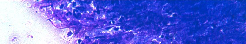
3 Power Doppler US pattern and FNC guide in Hodgkin lymphoma Lymph nodes may be partially involved from HL. Power Doppler may reveal an abnormal vascular pattern in a defined area of the lymph node that may be used as target in the FNC guide.

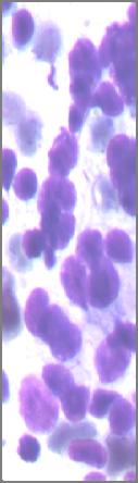
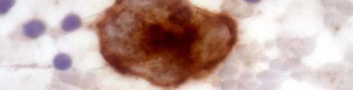

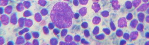
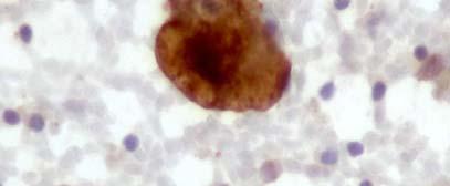
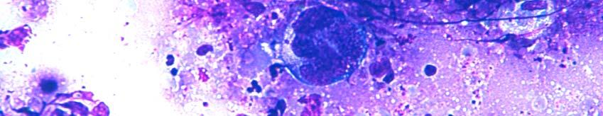
4 HODGKIN LYMPHOMA: diagnostic cells CD30 RS cells with lobated nuclei, clumped chromatin and large nucleoli, CD30+. Hodgkin cell in a polymorphus background with numerous eosinophils.

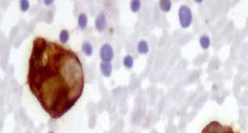
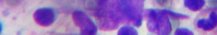

5 HODGKIN LYMPHOMA: diagnostic cells CD30 CD15 RS cells, CD15 and CD30 positives, with lobated nuclei, irregular chromatin and numerous large nucleoli.

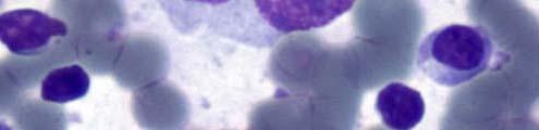
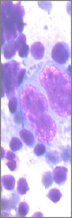
6 HODGKIN LYMPHOMA: diagnostic cells CD30 RS mirror cells, CD30 positive, note the extremely large nucleoli.


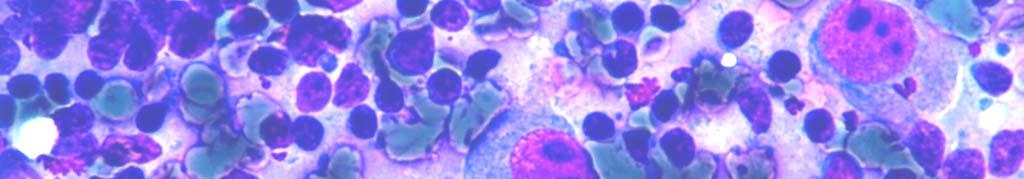
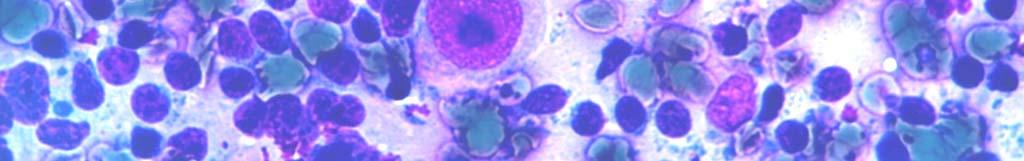
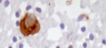
7 HODGKIN LYMPHOMA: diagnostic cells CD15 CD30 Mononucleated HC cells with irregular chromatin and extremely large nucleoli. CD15 and CD30 positive, mononucleated HC cells.


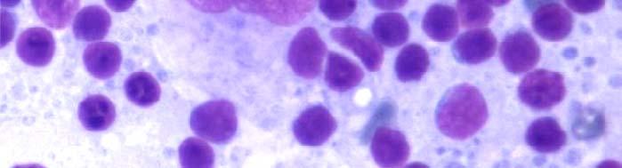
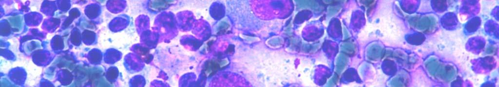
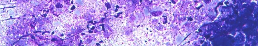
8 HODGKIN LYMPHOMA: diagnostic cells A multinucleated cell with clumping chromatin A polyploid, l multinucleated, l t nucleolated l and hypercromasia. This cell may be RSH cells in a lymphoid background. considered a mummified RSH cells.
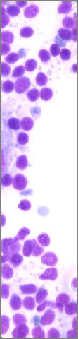
9 Nodular lymphocyte-predominant Hodgkin lymphoma NLHL: Cytological findings NLHL: diffuse lymphoid background with a variable number of large neoplastic monucleated cells known as lymphocytic and/or histiocytic Reed- Sternberg cell variants (L&H). The nuclei are often folded or multilobated, the chromatin is mostly vesicular, the nucleoli are usually multiple and smaller than those seen in classical HRS cells. Small lymphocytes, histiocytes and some polyclonal plasma cells are present in the background. ICC: L&H cells: CD15-, CD30-, CD20+, CD79a+, BCL6+ and CD45+. FC: not contributive, CD4/CD8 co-expression has been reported.


.")


10 Nodular lymphocyte predominant, Hodgkin lymphoma CD20 Lymphocytic and/or histiocytic Reed-Sternberg cell variants (L&H). The nuclei may be round, folded or multilobated, the chromatin is mostly vesicular, the nucleoli are usually multiple and smaller than those seen in classical HRS cells. L&H cells are CD20+, CD15-,CD30-.
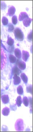
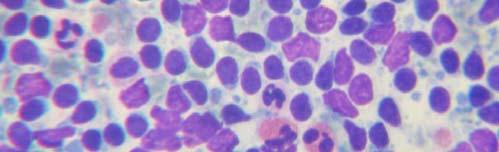
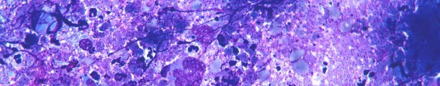
11 Classical Hodgkin lymphoma: Cytological findings Classical l Hodgkin lymphoma: HRS cells are large and show abundant, slightly basophilic, cytoplasm and two or more nuclear lobes or nuclei. Diagnostic RS cells have at least two nucleoli in two separate nuclear lobes. Mononuclear variants are termed Hodgkin cells, and the cells represent only a minority of the cellular infiltrate. The background is composed by reactive cellular infiltrate that varies according to the histological subtype. HRS cells are positive for CD30 in nearly all cases, for CD15 in the majority of cases and usually negative for CD45. Most HRS cells express the proliferation-associated nuclear antigen Ki-67. Classical Hodgkin lymphoma is classified as: Nodular sclerosis, Lymphocyte-rich, Mixed cellularity and Lymphocyte-depleted Nodular sclerosis contains a variable number of HRS cells in a background of small lymphocytes and other non-neoplastic inflammatory cells. Diagnostic cells may be rare or extremely numerous as in the syncitial variant. The HRS cells of this subtype tend to have more lobated nuclei with smaller lobes, and less prominent nucleoli. Lacunar HRS, determined by formalin fixation, which characterize this entity on histological sections are not observed on the smears. Eosinophils may be numerous. Mixed cellularity HRS cells are typical; the background cells consist of a mixture of eosinophils, neutrophils, histiocytes and plasma cells. One cell type may prevail over the others. The histiocytes may show pronounced epithelioid differentiation and may form granuloma-like like clusters. Lymphocyte-rich: scattered HRS cells in a background characterized by an abundance of small lymphocytes. Eosinophils and neutrophils may be present in small numbers. The small lymphocytes may be admixed with a large number of histiocytes with or without epithelioid features. Lymphocyte-depleted: rich in HRS cells and/or depleted in non-neoplastic neoplastic lymphocytes. In some cases pleomorphic HRS cells may predominate, producing a sarcomatous appearance. ICC: HC and HRS cells: CD15+, CD30+, CD20-, CD45-. DD: DLBCL: all types or metastasis in syncitial variant.


 and")
")

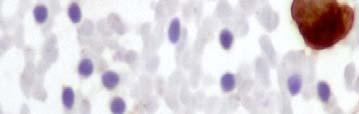
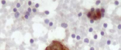
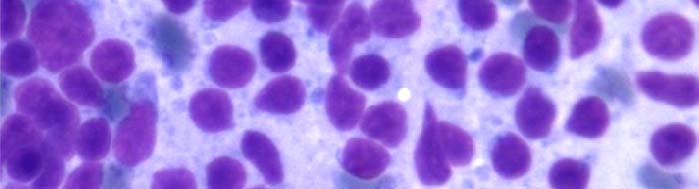
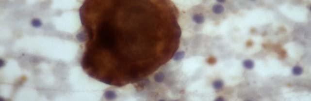
12 Classical Hodgkin lymphoma CD30 HL: One RSH cell (lower left) and two HC (center) in a lymphoid background. HL: A CD 30 positive, RSH cell.


 and two")


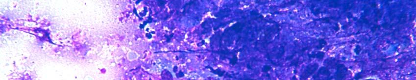
13 Classical Hodgkin lymphoma CD15 HL: One RSH cell (lower left) and two HC (upper HL: one mirror cell and two, CD15 positive, HC. right) in an hematic and scantily lymphoid background.


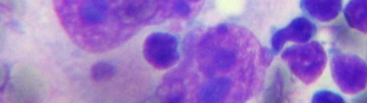
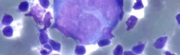
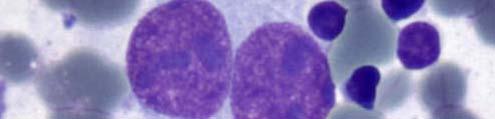
14 Classical Hodgkin lymphoma HL: One HC with a large nucleolus in a lymphoid background. HL: RSH cells and numerous HC in a lymphoid background. Two eosinophils are present (lower left corner).


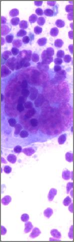
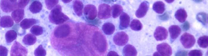
15 Classical Hodgkin lymphoma CD30 Extremely numerous and/or atypical RSH cell in a background of granulocytes. This picture may suggest a lymphocyte depletion. Extremely numerous CD30 positive RSH cell may suggest a lymphocyte depletion.
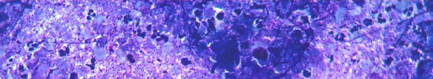
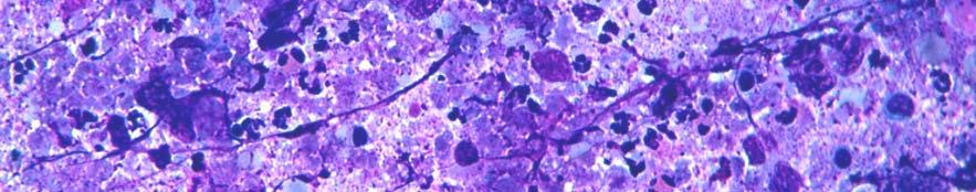
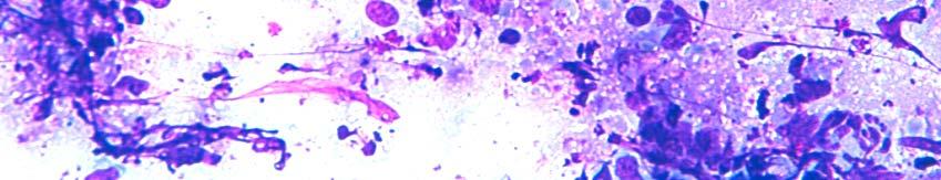
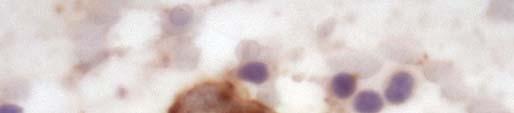
16 suppurative Hodgkin lymphoma RSH cells in a necrotic and acute inflammatory background


17 microscopic indirect signs Fibrosis fragments with intermingled lymphiod cells and nuclear crushes may be observed in HL smears. More than occasional or numerous eosinophils may suggest HL.


18 microscopic indirect signs An atypical and ill-preserved mononuclated cell in a back ground of granulocytes and an eosinophil. This image is suspect for HL. An atypical and ill- preserved multinucleated cell suspected for RSH cell. An sole atypical binucleated cell with large nucleoli suspected for RSH cell.


19 microscopic indirect signs An ill-preserved drs cell llin a necrotic and A granulomatous pattern without t a inflammatory background. This image is consistent clinical context might hide a HL. suspect for HL. At the same time granulomatous pattern may be observed in the follow-up of HL and NHL.
Incidence. Bimodal age incidence 15-40, >55 years Childhood form (0-14) more common in developing countries M:F=1.5:1; in all subtypes except NS
 more common in developing countries M:F=1.5:1; in all subtypes except NS") Hodgkin Lymphoma Hodgkin Lymphoma 30% of all lymphomas Absolute incidence unchanged Arise in lymph node, cervical region Neoplastic tissues usually contain a small number of tumor cells Incidence Bimodal
Hodgkin Lymphoma Hodgkin Lymphoma 30% of all lymphomas Absolute incidence unchanged Arise in lymph node, cervical region Neoplastic tissues usually contain a small number of tumor cells Incidence Bimodal
DETERMINATION OF A LYMPHOID PROCESS
 Chapter 2 Applications of Touch Preparation Cytology to Intraoperative Consultations: Lymph Nodes and Extranodal Tissues for Evaluation of Hematolymphoid Disorders INTRODUCTION As discussed in Chap. 1,
Non-Hodgkin lymphomas (NHLs) Hodgkin lymphoma )HL)
 Hodgkin lymphoma )HL)") Non-Hodgkin lymphomas (NHLs) Hodgkin lymphoma )HL) Lymphoid Neoplasms: 1- non-hodgkin lymphomas (NHLs) 2- Hodgkin lymphoma 3- plasma cell neoplasms Non-Hodgkin lymphomas (NHLs) Acute Lymphoblastic Leukemia/Lymphoma
Non-Hodgkin lymphomas (NHLs) Hodgkin lymphoma )HL) Lymphoid Neoplasms: 1- non-hodgkin lymphomas (NHLs) 2- Hodgkin lymphoma 3- plasma cell neoplasms Non-Hodgkin lymphomas (NHLs) Acute Lymphoblastic Leukemia/Lymphoma
Nodular lymphocyte predominant Hodgkin lymphoma. Lymphoma Tumor Board. January 5, 2018
 Nodular lymphocyte predominant Hodgkin lymphoma Lymphoma Tumor Board January 5, 2018 Etiology Subtypes of Classical Hodgkin Lymphoma (chl)* Nodular sclerosing HL Most common subtype Composed of large tumor
Lymphoma: What You Need to Know. Richard van der Jagt MD, FRCPC
 Lymphoma: What You Need to Know Richard van der Jagt MD, FRCPC Overview Concepts, classification, biology Epidemiology Clinical presentation Diagnosis Staging Three important types of lymphoma Conceptualizing
Anaplastic Large Cell Lymphoma (of T cell lineage)
") Anaplastic Large Cell Lymphoma (of T cell lineage) Definition T-cell lymphoma comprised of large cells with abundant cytoplasm and pleomorphic, often horseshoe-shaped nuclei CD30+ Most express cytotoxic
Anaplastic Large Cell Lymphoma (of T cell lineage) Definition T-cell lymphoma comprised of large cells with abundant cytoplasm and pleomorphic, often horseshoe-shaped nuclei CD30+ Most express cytotoxic
Hematopathology Lab. Third year medical students
 Hematopathology Lab Third year medical students Objectives Identify the lesion Know the specific name of the lesion Know associated disease Know relevant pathologic background Spherocytes: appear small,
Pathology of the Lymphoid System
 Pathology of the Lymphoid System Learning Objectives: Define lymphadenitis and enumerate its types. Briefly describe the morphological appearance of reactive lymph node. Describe the microscopic picture
HODGKIN LYMPHOMA DR. ALEJANDRA ZARATE OSORNO HOSPITAL ESPAÑOL DE MEXICO
 HODGKIN LYMPHOMA DR. ALEJANDRA ZARATE OSORNO HOSPITAL ESPAÑOL DE MEXICO HODGKIN LYMPHOMA CLASSIFICATION Lukes & Butler Rye WHO-2016 Linphocytic and/or histiocytic Nodular & diffuse Nodular Sclerosis Lymphocyte
Pearls and pitfalls in interpretation of lymphoid lesions in needle biopsies
 Pearls and pitfalls in interpretation of lymphoid lesions in needle biopsies Megan S. Lim MD PhD University of Pennsylvania October 8, 2018 Objectives To understand how the trend toward less invasive lymph
Pathology of the Lymphoid System
 Pathology of the Lymphoid System Learning Objectives: Define lymphadenitis and enumerate its types. Briefly describe the morphological appearance of reactive lymph node. Describe the microscopic picture
MECHANISMS OF HUMAN DISEASE: LABORATORY SESSIONS LYMPHOMA. April 16, 2008
 MECHANISMS OF HUMAN DISEASE: LABORATORY SESSIONS LYMPHOMA April 16, 2008 FACULTY COPY GOAL: Learn the appearance of normal peripheral blood elements and lymph nodes. Recognize abnormal peripheral blood
Differential cell counts in the histiocytic variant of lymphocytic predominance subtype of Hodgkin's
 Journal of Clinical Pathology, 1978, 31, 1234-1238 Differential cell counts in the histiocytic variant of lymphocytic predominance subtype of Hodgkin's disease FIONA I. SUTHERLAND, J. B. MAcGILLIVRAY,
QUALITY ASSURANCE PROGRAM CYTOLOGY CYCLE 01/2018 (TRIAL)
") [Pick the Date] FINAL REPORT QUALITY ASSURANCE PROGRAM CYTOLOGY CYCLE 01/2018 (TRIAL) NOTES FROM THE COORDINATOR 1. For this cycle 01/2018, a total of 32 pen drives had been circulated. Twenty-eight institutions
[Pick the Date] FINAL REPORT QUALITY ASSURANCE PROGRAM CYTOLOGY CYCLE 01/2018 (TRIAL) NOTES FROM THE COORDINATOR 1. For this cycle 01/2018, a total of 32 pen drives had been circulated. Twenty-eight institutions
From Morphology to Molecular Pathology: A Practical Approach for Cytopathologists Part 1-Cytomorphology. Songlin Zhang, MD, PhD LSUHSC-Shreveport
 From Morphology to Molecular Pathology: A Practical Approach for Cytopathologists Part 1-Cytomorphology Songlin Zhang, MD, PhD LSUHSC-Shreveport I have no Conflict of Interest. FNA on Lymphoproliferative
Case Report A case of EBV positive diffuse large B-cell lymphoma of the adolescent
 Int J Clin Exp Med 2014;7(1):307-311 www.ijcem.com /ISSN:1940-5901/IJCEM1311029 Case Report A case of EBV positive diffuse large B-cell lymphoma of the adolescent Qilin Ao 2, Ying Wang 1, Sanpeng Xu 2,
Differential diagnosis of hematolymphoid tumors composed of medium-sized cells. Brian Skinnider B.C. Cancer Agency, Vancouver General Hospital
 Differential diagnosis of hematolymphoid tumors composed of medium-sized cells Brian Skinnider B.C. Cancer Agency, Vancouver General Hospital Lymphoma classification Lymphoma diagnosis starts with morphologic
Burkitt lymphoma. Sporadic Endemic in Africa associated with EBV Translocations involving MYC gene on chromosome 8
 Heme 8 Burkitt lymphoma Sporadic Endemic in Africa associated with EBV Translocations involving MYC gene on chromosome 8 Most common is t(8;14) Believed to be the fastest growing tumor in humans!!!! Morphology
Case Report PAX5-Negative Classical Hodgkin Lymphoma: A Case Report of a Rare Entity and Review of the Literature
 Hindawi Case Reports in Hematology Volume 2017, Article ID 7531729, 4 pages https://doi.org/10.1155/2017/7531729 Case Report PAX5-Negative Classical Hodgkin Lymphoma: A Case Report of a Rare Entity and
Lách
 Lách Lách Lách Lách Splenogonadal fusion. Splenic tissue is attached to testicular tissue. Pseudocyst (false or secondary cyst). A, Outer aspect. Pseudocyst (false or secondary cyst). B, Inner surface.
Primary Cutaneous CD30-Positive T-cell Lymphoproliferative Disorders
 Primary Cutaneous CD30-Positive T-cell Lymphoproliferative Disorders Definition A spectrum of related conditions originating from transformed or activated CD30-positive T-lymphocytes May coexist in individual
Thomas Hodgkin and Hodgkin lymphoma
 J Hematopathol (2014) 7:123 138 DOI 10.1007/s12308-014-0214-3 REVIEW ARTICLE Thomas Hodgkin and Hodgkin lymphoma Judith A. Ferry Received: 26 June 2014 /Accepted: 31 July 2014 /Published online: 12 August
A clinicopathological study of nodular sclerosing Hodgkin's disease
 A clinicopathological study of nodular sclerosing Hodgkin's disease R. M. CROSS J. clini. Path. (1968), 21, 0-10 From the Royal Air Force Institute ofpathology and Tropical Medicine, Halton, Buckinghamshire
Dr Rodney Itaki Lecturer Anatomical Pathology Discipline
 Lymphoid Neoplasia & Hodgkin s Disease Dr Rodney Itaki Lecturer Anatomical Pathology Discipline Lymphomas Are neoplasms of lymphoid cells. Classified as: Hodgkin Disease (30%) Non-Hodgkin lymphoma (70%).
Plasma cell myeloma (multiple myeloma)
") Plasma cell myeloma (multiple myeloma) Common lymphoid neoplasm, present at old age (70 years average) Remember: plasma cells are terminally differentiated B-lymphocytes that produces antibodies. B-cells
Plasma cell myeloma (multiple myeloma) Common lymphoid neoplasm, present at old age (70 years average) Remember: plasma cells are terminally differentiated B-lymphocytes that produces antibodies. B-cells
Diagnostic Cytology of Cancer Cases
 Diagnostic Cytology of Cancer Cases Somporn Techangamsuwan Companion Animal Cancer Research Unit (CAC-RU) Department of Pathology, Faculty of Veterinary Science, Chulalongkorn University 1 Tumor or Non-tumor
Thyroid master class. Thyroid Fine needle aspiration cytology and liquid-based techniques: Hologic and Becton Dickinson
 Thyroid master class Thyroid Fine needle aspiration cytology and liquid-based techniques: Hologic and Becton Dickinson Principle of LBC Collection of cells in liquid medium Immediate fixation Processor-prepared
Mimics of Lymphoma in Routine Biopsies. Mixed follicular and paracortical hyperplasia. Types of Lymphoid Hyperplasia
 Mimics of Lymphoma in Routine Biopsies Patrick Treseler, MD, PhD Professor of Pathology University of California San Francisco Types of Lymphoid Hyperplasia Follicular hyperplasia (B-cells) Paracortical
Mimics of Lymphoma in Routine Biopsies. I have nothing to disclose regarding the information to be reported in this talk.
 Mimics of Lymphoma in Routine Biopsies Patrick Treseler, MD, PhD Professor of Pathology University of California San Francisco I have nothing to disclose regarding the information to be reported in this
Immunopathology of Lymphoma
 Immunopathology of Lymphoma Noraidah Masir MBBCh, M.Med (Pathology), D.Phil. Department of Pathology Faculty of Medicine Universiti Kebangsaan Malaysia Lymphoma classification has been challenging to pathologists.
Medullary Thyroid Carcinoma. This case was provided by Treant Hospital, Bethesda, Hoogeveen, The Netherlands
 Medullary Thyroid Carcinoma This case was provided by Treant Hospital, Bethesda, Hoogeveen, The Netherlands ADS-01504 Rev. 001 2016 Hologic, Inc. All rights reserved. Overview Medullary Thyroid Carcinoma
Case Presentation. Maha Akkawi, MD, Fatima Obeidat, MD, Tariq Aladily, MD. Department of Pathology Jordan University Hospital Amman, Jordan
 Case Presentation Maha Akkawi, MD, Fatima Obeidat, MD, Tariq Aladily, MD Department of Pathology Jordan University Hospital Amman, Jordan The 25th Annual Congress of the ADIAP The 8/11/2013 1 5th International
Case 3. Ann T. Moriarty,MD
 Case 3 Ann T. Moriarty,MD Case 3 59 year old male with asymptomatic cervical lymphadenopathy. These images are from a fine needle biopsy of a left cervical lymph node. Image 1 Papanicolaou Stained smear,100x.
Leukaemia Section Short Communication
 Atlas of Genetics and Cytogenetics in Oncology and Haematology OPEN ACCESS JOURNAL INIST-CNRS Leukaemia Section Short Communication Classification of Hodgkin lymphoma over years Antonino Carbone, Annunziata
Evening specialty conference: Liver
 Evening specialty conference: Liver Joseph Misdraji, M.D. Disclosure of Relevant Financial Relationships Disclosure of Relevant Financial Relationships USCAP requires that all planners (Education Committee)
Megakaryocyte or Precursor, Normal
 Precursor, Normal SYNONYMS none VITAL STATISTICS size...20-160 µm in diameter N:C ratio...varible, depending on maturation of cell; early forms have a high N:C rato which decreases as cell matures and
HISTOPATHOLOGY. Shannon Martinson
 HISTOPATHOLOGY Shannon Martinson March 2013 Case #1 History: 8 year old beagle Neck pain for the past couple of weeks Paresis, followed by paralysis developed over the past few days Gross Description courtesy
Cytopathological Study of Lymph Node Lesions - A 2 Years Retrospective Study
 Original Article Print ISSN: 2321-6379 Online ISSN: 2321-595X DOI: 10.17354/ijss/2018/24 Cytopathological Study of Lymph Node Lesions - A 2 Years Retrospective Study K Florence, K Suresh, K Lavanya Department
HENATOLYMPHOID SYSTEM THIRD YEAR MEDICAL STUDENTS- UNIVERSITY OF JORDAN AHMAD T. MANSOUR, MD. Parts 2 and 3
 HENATOLYMPHOID SYSTEM THIRD YEAR MEDICAL STUDENTS- UNIVERSITY OF JORDAN AHMAD T. MANSOUR, MD Parts 2 and 3 NEOPLASTIC LYMPHOID DISEASES Introduction o The bone marrow is the source of all cells in the
Cytology of Follicular Dendritic Cell Sarcoma on Intraoperative Touch Imprint Smears
 The Korean Journal of Pathology 2009; 43: 589-93 DOI: 10.4132/KoreanJPathol.2009.43.6.589 Cytology of Follicular Dendritic Cell Sarcoma on Intraoperative Touch Imprint Smears - Case Report - Ju Young Song
Many of the hematolymphoid disorders are derived
 REVIEW ARTICLE Practical Immunohistochemistry in Hematopathology: A Review of Useful Antibodies for Diagnosis Ji Lu, MD and Karen L. Chang, MD Abstract: This review article offers some useful panels of
VETERINARY HEMATOLOGY ATLAS OF COMMON DOMESTIC AND NON-DOMESTIC SPECIES COPYRIGHTED MATERIAL SECOND EDITION
 VETERINARY HEMATOLOGY ATLAS OF COMMON DOMESTIC AND NON-DOMESTIC SPECIES SECOND EDITION COPYRIGHTED MATERIAL CHAPTER ONE HEMATOPOIESIS GENERAL FEATURES All blood cells have a finite life span, but in normal
Case year female. Routine Pap smear
 Case 1 57 year female Routine Pap smear Diagnosis? 1. Atypical glandular cells of unknown significance (AGUS) 2. Endocervical AIS 3. Endocervical adenocarcinoma 4. Endometrial adenocarcinoma 5. Adenocarcinoma
Thyroid follicular neoplasms in cytology. Ulrika Klopčič Institute of Oncology, Department of Cytopathology, Ljubljana, Slovenia
 Thyroid follicular neoplasms in cytology Ulrika Klopčič Institute of Oncology, Department of Cytopathology, Ljubljana, Slovenia Lecture overview importance of FNAB in assessing thyroid lesions follicular
Case Report Nodular Lymphocyte Predominant Hodgkin Lymphoma versus T-Cell/Histiocyte-Rich Large B-Cell Lymphoma: A Diagnostic Challenge
 Case Reports in Pathology, Article ID 956217, 5 pages http://dx.doi.org/10.1155/2014/956217 Case Report Nodular Lymphocyte Predominant Hodgkin Lymphoma versus T-Cell/Histiocyte-Rich Large B-Cell Lymphoma:
A Report of a Rare Case of Anaplastic Large Cell Lymphoma of the Oral Cavity
 AJMS Al Ameen J Med Sci (2 0 1 2 )5 (1 ):9 8-1 0 2 (A US National Library of Medicine enlisted journal) I S S N 0 9 7 4-1 1 4 3 C O D E N : A A J M B G CASE REPORT A Report of a Rare Case of Anaplastic
Unknown Case 6. Ann T. Moriarty, MD
 Unknown Case 6 Ann T. Moriarty, MD Unknown Case 6 61 year old male with an enlarged cervical lymph node. He has a history of lung carcinoma, renal cell carcinoma and lymphoma. Case 6 Image 1: Fine needle
ADx Bone Marrow Report. Patient Information Referring Physician Specimen Information
 ADx Bone Marrow Report Patient Information Referring Physician Specimen Information Patient Name: Specimen: Bone Marrow Site: Left iliac Physician: Accession #: ID#: Reported: 08/19/2014 - CHRONIC MYELOGENOUS
Note: The cause of testicular neoplasms remains unknown
 - In the 15- to 34-year-old age group, they are the most common tumors of men. - Tumors of the testis are a heterogeneous group of neoplasms that include: I. Germ cell tumors : 95%; all are malignant.
A case of giant cell tumour of soft parts in a horse Francesco Cian 1, Sarah Whiteoak 2, Jennifer Stewart 1
 A case of giant cell tumour of soft parts in a horse Francesco Cian 1, Sarah Whiteoak 2, Jennifer Stewart 1 1 Animal Health Trust, Newmarket, UK 2 608 Equine and Farm Vets, Rowington, UK Signalment: Horse,
ECP meeting, Lisbon, september 2012 Slide seminar New and old challenges in the diagnosis of peripheral T-cell lymphomas
 ECP meeting, Lisbon, september 2012 Slide seminar New and old challenges in the diagnosis of peripheral T-cell lymphomas Philippe Gaulard, Dept of Pathology, INSERM U955, Hôpital Henri Mondor, 94010 -
Abstract. Anatomic Pathology / DIAGNOSIS AND SUBCLASSIFICATION OF PRIMARY AND RECURRENT LYMPHOMA
 Anatomic Pathology / DIAGNOSIS AND SUBCLASSIFICATION OF PRIMARY AND RECURRENT LYMPHOMA Diagnosis and Subclassification of Primary and Recurrent Lymphoma The Usefulness and Limitations of Combined Fine-Needle
Contents. vii. Preface... Acknowledgments... v xiii
 Contents Preface... Acknowledgments... v xiii SECTION I 1. Introduction... 3 Knowledge-Based Diagnosis... 4 Systematic Examination of the Lymph Node... 7 Cell Type Identification... 9 Cell Size and Cellularity...
This is the second learning component (Learning Component 2) in our first learning module (Learning Module 1). In this component we review a very
 in our first learning module (Learning Module 1). In this component we review a very") This is the second learning component (Learning Component 2) in our first learning module (Learning Module 1). In this component we review a very basic response to injury inflammation. We ll look at examples
This is the second learning component (Learning Component 2) in our first learning module (Learning Module 1). In this component we review a very basic response to injury inflammation. We ll look at examples
Lymph node cytopathology : A practical approach to lymphoproliferative disorders
 Lymph node cytopathology : A practical approach to lymphoproliferative disorders Koray Ceyhan, M.D Department of Pathology Faculty of Medicine Ankara University Ankara, Turkey Diagnostic use of FNA in
Gray Zones and Double Hits Distinguishing True Burkitt Lymphoma from Other High-Grade B-NHLs Burkitt Lymphoma Burkitt-Like Lymphoma DLBCL Patrick Tres
 Gray Zones and Double Hits Distinguishing True Burkitt Lymphoma from Other High-Grade B-NHLs Burkitt Lymphoma Burkitt-Like Lymphoma DLBCL Patrick Treseler, MD, PhD University of California San Francisco
Dispersed Cell Population & Lymphoglandular Bodies: Features of lymphoid aspirates FNA OF LYMPH NODES OF THE HEAD AND NECK
 William C. Faquin, MD, PhD Professor of Pathology Harvard Medical School Director of Head and Neck Pathology Massachusetts Eye and Ear Massachusetts General Hospital FNA OF LYMPH NODES OF THE HEAD AND
2012 by American Society of Hematology
 2012 by American Society of Hematology Common Types of HIV-Associated Lymphomas DLBCL includes primary CNS lymphoma (PCNSL) Burkitt Lymphoma HIV-positive patients have a 60-200 fold increased incidence
PROBABLE HODGKIN'S DISEASE IN A DOG: REPORT OF A CASE 1
 PROBABLE HODGKIN'S DISEASE IN A DOG: REPORT OF A CASE 1 LEONARD K. STALKER, M.D. Fellow in Surgery, The Mayo Foundation CARL F. SCHLOTTHAUER, D.V.M. AND WILLIAM H. FELDMAN, D.V.M., M.S. Division of Experimental
Clinicopathologic features of 112 cases with mantle cell lymphoma
 Cancer Biol Med 2015;12:46-52. doi: 10.7497/j.issn.2095-3941.2015.0007 ORIGINAL ARTICLE Clinicopathologic features of 112 cases with mantle cell lymphoma Dong-Mei Zhou, Gang Chen, Xiong-Wei Zheng, Wei-Feng
Assessing the lung and mediastinum in cancer-is tissue the issue? George Santis
 1 Assessing the lung and mediastinum in cancer-is tissue the issue? George Santis Optimal management of Cancer Histological diagnosis & accurate staging at presentation Molecular analysis of primary tumour
Megakaryoblastic Leukemia in a Dog A. Hillström 1, H. Tvedten 1, M. Kiupel 2.
 Megakaryoblastic Leukemia in a Dog A. Hillström 1, H. Tvedten 1, M. Kiupel 2. 1 University Animal Hospital, Swedish University of Agricultural Sciences and Strömsholm Referral Animal Hospital, Sweden 2
Lymphoma and Pseudolymphoma
 Lymphoma and Pseudolymphoma Laura B. Pincus, MD Co-Director, Cutaneous Lymphoma Clinic Associate Professor Dermatology and Pathology University of California, San Francisco I HAVE NO RELEVANT RELATIONSHIPS
Neoplastic proliferation arising from white blood cells. Introductory remarks. Classification
 Neoplastic proliferation arising from white blood cells Lymphoproliferative and myeloproliferative diseases and syndromes Oliver Rácz, 2012-2017 1 Introductory remarks Leukemia and lymphoma are old descriptive
Hemopoiesis and Blood
 Hemopoiesis and Blood Blood Cells o o o Erythrocytes Leukocytes Thrombocytes Function o Transport nutrients and wastes throughout the bloodstream, fight foreign antigens and blood coagulation. Location
CINtec p16 INK4a Staining Atlas
 CINtec p16 INK4a Staining Atlas Rating Rating Positive The rating positive will be assigned if the p16 INK4a -stained slide shows a continuous staining of cells of the basal and parabasal cell layers of
EQA SCHEME CIRCULATION 33 EDUCATIONAL SLIDES DR GRAEME SMITH MONKLANDS DGH
 EQA SCHEME CIRCULATION 33 EDUCATIONAL SLIDES DR GRAEME SMITH MONKLANDS DGH CASE E1 M: 68 yrs Left destructive sinonasal lesion.?lymphoma?adenocarcinoma CD20 CD10 BCL6 MIB1 Answers Diffuse large B cell
Update in Lymphoma Imaging
 Update in Lymphoma Imaging Victorine V. Muse, MD Lymphoma Update in Lymphoma Imaging Victorine V Muse, MD Heterogeneous group of lymphoid neoplasms divided into two broad histological categories Hodgkin
BOSNIAN-TURKISH CYTOPATHOLOGY SCHOOL June 18-19, 2016 Sarajevo. Case Discussions. 60 year old woman Routine gynecologic control LBC
 BOSNIAN-TURKISH CYTOPATHOLOGY SCHOOL June 18-19, 2016 Sarajevo Case Discussions Prof Dr Sıtkı Tuzlalı Tuzlalı Pathology Laboratory 60 year old woman Routine gynecologic control LBC 1 2 Endometrial thickening
7 Omar Abu Reesh. Dr. Ahmad Mansour Dr. Ahmad Mansour
 7 Omar Abu Reesh Dr. Ahmad Mansour Dr. Ahmad Mansour -Leukemia: neoplastic leukocytes circulating in the peripheral bloodstream. -Lymphoma: a neoplastic process in the lymph nodes, spleen or other lymphatic
FINE NEEDLE ASPIRATION (FNAC) AS A DIAGNOSTIC TOOL IN PAEDIATRIC LYMPHADENOPATHY.
 AS A DIAGNOSTIC TOOL IN PAEDIATRIC LYMPHADENOPATHY.") IJCRR Vol 06 issue 01 Section: Healthcare Category: Research Received on: 16/10/13 Revised on: 18/11/13 Accepted on: 20/12/13 FINE NEEDLE ASPIRATION (FNAC) AS A DIAGNOSTIC TOOL IN PAEDIATRIC Heming Agrawal,
IJCRR Vol 06 issue 01 Section: Healthcare Category: Research Received on: 16/10/13 Revised on: 18/11/13 Accepted on: 20/12/13 FINE NEEDLE ASPIRATION (FNAC) AS A DIAGNOSTIC TOOL IN PAEDIATRIC Heming Agrawal,
SQUAMOUS CELLS: Atypical squamous cells (ASC) - of undetermined significance (ASC-US) - cannot exclude HSIL (ASC-H)
 - of undetermined significance (ASC-US) - cannot exclude HSIL (ASC-H)") SQUAMOUS CELLS: Atypical squamous cells (ASC) - of undetermined significance (ASC-US) - cannot exclude HSIL (ASC-H) ASC refers to cytologic changes suggestive of SIL, which are qualitativley or quantitatively
SQUAMOUS CELLS: Atypical squamous cells (ASC) - of undetermined significance (ASC-US) - cannot exclude HSIL (ASC-H) ASC refers to cytologic changes suggestive of SIL, which are qualitativley or quantitatively
3/24/2017 DENDRITIC CELL NEOPLASMS: HISTOLOGY, IMMUNOHISTOCHEMISTRY, AND MOLECULAR GENETICS. Disclosure of Relevant Financial Relationships
 DENDRITIC CELL NEOPLASMS: HISTOLOGY, IMMUNOHISTOCHEMISTRY, AND MOLECULAR GENETICS Jason L. Hornick, M.D., Ph.D. Director of Surgical Pathology and Immunohistochemistry Brigham and Women s Hospital Professor
Chapter 12 The Role of Fine Needle Aspiration Biopsy in the Diagnosis and Management of Hematopoietic Neoplasms
 Chapter 12 The Role of Fine Needle Aspiration Biopsy in the Diagnosis and Management of Hematopoietic Neoplasms Rana S. Hoda FINE NEEDLE ASPIRATION Introduction Fine needle aspiration (FNA) is a well-established
Almost any suspected tumor can be aspirated easily and safely. Some masses are more risky to aspirate including:
 DOES THIS PATIENT HAVE CANCER? USING IN-HOUSE CYTOLOGY TO HELP YOU MAKE THIS DIAGNOSIS. Joyce Obradovich, DVM, Diplomate, ACVIM (Oncology) Animal Cancer & Imaging Center, Canton, Michigan Almost every
The patient had a mild splenomegaly but no obvious lymph node enlargement. The consensus phenotype obtained from part one of the exercise was:
 Case History An 86 year old male was admitted to hospital with chest infection. Haematological examination subsequently revealed the following: Hb- 11.0 g/dl; WBC- 67.1 x 10^9/l; PLT- 99 x10^9/l; RBC-
Mantle Cell Lymphoma
 Mantle Cell Lymphoma Clinical Case A 56 year-old woman complains of pain and fullness in the left superior abdominal quadrant for the last 8 months. She has lost 25 kg, and lately has had night sweats.
Participants Identification No. % Evaluation. Mitotic figure Educational Erythrocyte precursor, abnormal 1 0.
 Cell Identification Mitotic figure 212 99.5 Educational Erythrocyte precursor, abnormal BMD-02 The arrowed cell is a mitotic figure. It was correctly identified by 99.5% of the participants. A cell containing
Introduction. 23 rd Annual Seminar in Pathology. FLUIDS, Part 1. Pittsburgh, PA Gladwyn Leiman UVMMC, VT
 23 rd Annual Seminar in Pathology Pittsburgh, PA Gladwyn Leiman UVMMC, VT FLUIDS, Part 1 "Blue walls", Claudia Hansen, 2009 Introduction o Challenging to everyone o Almost any benign or malignant process
Primary Spinal T-Cell Rich B-Cell Lymphoma: A Case Report
 Primary Spinal T-Cell Rich B-Cell Lymphoma: A Case Report Pages with reference to book, From 148 To 149 Suhail Muzaffar,Irshad Nabi Soomro,Naila Kayani,Shahid Siddiqui ( Departments of Pathology, The Aga
Shades of gray between large B-cell lymphomas and Hodgkin lymphomas: differential diagnosis and biological implications
 & 2013 USCAP, Inc. All rights reserved 0893-3952/13 $32.00 S57 Shades of gray between large B-cell lymphomas and Hodgkin lymphomas: differential diagnosis and biological implications Nancy L Harris Department
Ascitic Fluid and Use of Immunocytochemistry. Mercè Jordà, University of Miami
 Ascitic Fluid and Use of Immunocytochemistry Mercè Jordà, University of Miami Is It Malignant? Yes? No Ascitic Fluid Cytomorphologic Useful Findings Tight clusters with smooth borders Cellular and nuclear
Nuclear morphometric study of Non- Hodgkin's Lymphoma (NHL)
") Original Research Article Nuclear morphometric study of Non- Hodgkin's Lymphoma (NHL) Sridhar Reddy Erugula 1, P. Sujatha 2, Ayesha Sameera 3, B. Suresh Reddy 4, Jesudass Govada 5, G. Sudhakar 6, Kandukuri
Original Research Article Nuclear morphometric study of Non- Hodgkin's Lymphoma (NHL) Sridhar Reddy Erugula 1, P. Sujatha 2, Ayesha Sameera 3, B. Suresh Reddy 4, Jesudass Govada 5, G. Sudhakar 6, Kandukuri
FNA of Thyroid. Toward a Uniform Terminology With Management Guidelines. NCI NCI Thyroid FNA State of the Science Conference
 FNA of Thyroid NCI NCI Thyroid FNA State of the Science Conference Toward a Uniform Terminology With Management Guidelines Thyroid Thyroid FNA Cytomorphology NCI Thyroid FNA State of the Science Conference
EPIDEMIOLOGICAL AND MORPHOLOGICAL CHARACTERISTICS OF CUTANEOUS ROUND CELL TUMORS DIAGNOSED USING ASPIRATIVE CYTOLOGY IN DOGS
 Scientific Works. Series C. Veterinary Medicine. Vol. LXIII (1) ISSN 2065-1295; ISSN 2343-9394 (CD-ROM); ISSN 2067-3663 (Online); ISSN-L 2065-1295 Abstract EPIDEMIOLOGICAL AND MORPHOLOGICAL CHARACTERISTICS
Title. Author(s)SONODA, Mitsuo; KOBAYASHI, Kosaku. CitationJapanese Journal of Veterinary Research, 18(3): 125- Issue Date DOI. Doc URL.
SONODA, Mitsuo; KOBAYASHI, Kosaku. CitationJapanese Journal of Veterinary Research, 18(3): 125- Issue Date DOI. Doc URL.") Title PLASMACYTOID CELLS OF CANINE PERIPHERAL BLOOD IN ELE Author(s)SONODA, Mitsuo; KOBAYASHI, Kosaku CitationJapanese Journal of Veterinary Research, 18(3): 125- Issue Date 1970-09 DOI 10.14943/jjvr.18.3.125
Title PLASMACYTOID CELLS OF CANINE PERIPHERAL BLOOD IN ELE Author(s)SONODA, Mitsuo; KOBAYASHI, Kosaku CitationJapanese Journal of Veterinary Research, 18(3): 125- Issue Date 1970-09 DOI 10.14943/jjvr.18.3.125
Aggressive B-cell Lymphomas
 Neoplastic Hematopathology Update 2018 Aggressive B-cell Lymphomas Raju K. Pillai City of Hope National Medical Center I do not have any disclosures Disclosures Outline New entities and changes in WHO
Concurrent malignant melanoma and cutaneous involvement by classical hodgkin lymphoma (CHL) in a 63 year-old man
 in a 63 year-old man") Gru and Lu Diagnostic Pathology 2013, 8:135 CASE REPORT Open Access Concurrent malignant melanoma and cutaneous involvement by classical hodgkin lymphoma (CHL) in a 63 year-old man Alejandro A Gru 1* and
Gru and Lu Diagnostic Pathology 2013, 8:135 CASE REPORT Open Access Concurrent malignant melanoma and cutaneous involvement by classical hodgkin lymphoma (CHL) in a 63 year-old man Alejandro A Gru 1* and
Mantle Cell Lymphoma
 HEMATOPATHOLOGY Original Article Mantle Cell Lymphoma Morphologic Findings in Bone Marrow Involvement JAY WASMAN, MD, 1 NANCY S. ROSENTHAL, MD,' AND DIANE C. FARHI, MD 2 Although mantle cell lymphoma (MCL),
Region: 11. Clinic6patholo cal Studies
 Tumors of the Thvmus and ThVIIliC Region: 11. Clinic6patholo cal Studies on Hodgkin s Disease of Re Thymus N. P. Bergh, M.D., P. Gatzinsky, M.D., S. Larsson, M.D., P. Lundin, M.D., and B. Ridell, M.D.
Methotrexate-associated Lymphoproliferative Disorders
 Methotrexate-associated Lymphoproliferative Disorders Definition A lymphoid proliferation or lymphoma in a patient immunosuppressed with methotrexate, typically for treatment of autoimmune disease (rheumatoid
Hodgkin s lymphoma: the pathologist s viewpoint
 162 REVIEW Hodgkin s lymphoma: the pathologist s viewpoint S A Pileri, S Ascani, L Leoncini, E Sabattini, P L Zinzani, P P Piccaluga, A Pileri Jr, M Giunti, B Falini, G B Bolis, H Stein... Despite its
Prepared By Jocelyn Palao and Layla Faqih
 Prepared By Jocelyn Palao and Layla Faqih The structure of the suspected atypical cell should always be compared to the structure of other similar, benign, cells which are present in the smears. The diagnosis
UNUSUAL OCULAR HODGKINʼS-LIKE LYMPHOMA IN A DOG
 Bulgarian Journal of Veterinary Medicine, 2019 ONLINE FIRST ISSN 1311-1477; DOI: 10.15547/bjvm.2241 Case report UNUSUL OCULR HODGKINʼS-LIKE LYMPHOM IN DOG Summary M. KHORDDMEHR 1, S. JROLMSJED 2, J. SHRFI-HELN
Presentation material is for education purposes only. All rights reserved URMC Radiology Page 1 of 98
 Presentation material is for education purposes only. All rights reserved. 2011 URMC Radiology Page 1 of 98 Radiology / Pathology Conference February 2011 Brooke Koltz, Cytopathology Resident Presentation
International Journal of Pharma and Bio Sciences CHROMOPHOBE VARIANT OF RENAL CELL CARCINOMA MASQUARDING AS RENAL ONCOCYTOMA ON CYTOLOGY.
 Case Report Pathology International Journal of Pharma and Bio Sciences ISSN 0975-6299 CHROMOPHOBE VARIANT OF RENAL CELL CARCINOMA MASQUARDING AS RENAL ONCOCYTOMA ON CYTOLOGY. DR.MAMATHA K*, DR. ARAKERI
Objectives. Atypical Glandular Cells. Atypical Endocervical Cells. Reactive Endocervical Cells
 2013 California Society of Pathologists 66 th Annual Meeting San Francisco, CA Atypical Glandular Cells to Early Invasive Adenocarcinoma: Cervical Cytology and Histology Christina S. Kong, MD Associate
الفتوي الاصفر الحبيبوم = Xanthogranuloma_Juvenile JUVENILE XANTHOGRANULOMA 1 / 9
 JUVENILE XANTHOGRANULOMA 1 / 9 Clinical Findings CUTANEOUS LESIONS JXG is a benign, self-healing disorder that is characterized by asymptomatic yellowish papulonodular lesions of the skin and other organs
Tinh hoàn
 Tinh hoàn Tinh hoàn Tinh hoàn Tiền liệt tuyến Tiền liệt tuyến Mào tinh hoàn Mào tinh hoàn Túi tinh Túi tinh Túi tinh Túi tinh So-called cystadenoma of seminal vesicle. Gross appearance of granulomatous
MULTISYSTEMIC LANGERHANS CELL HISTIOCYTOSIS IN ADULT AN UNCOMMON INCIDENCE POSING A DIAGNOSTIC CHALLENGE. Monal Trisal
 Pathology Case Report International Journal of Clinical And Diagnostic Research ISSN 2395-3403 Volume 4, Issue 4, July-Aug 2016. Glorigin Lifesciences Private Limited. MULTISYSTEMIC LANGERHANS CELL HISTIOCYTOSIS