Handling of Upper GI Cancer Specimens
|
|
|
- Esmond Ellis
- 5 years ago
- Views:
Transcription
1 Handling of Upper GI Cancer Specimens Prof Ray McMahon Histopathology Department Manchester Royal Infirmary Bryan Warren School Sarajevo November 2016
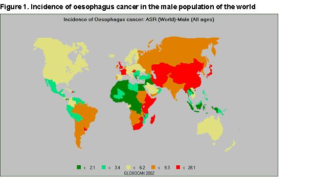
2 Pathology of oesophageal cancer Epidemiology Classification Role of Barrett s oesophagus Handling of specimens Resections EMRs Newer techniques
3
4 Oesophageal Cancer: epidemiology Majority are SCC Adenocarcinoma on the increase associated with Barrett s oesophagus Variants Prognosis related to stage (TNM)
5 Oesophageal Cancer (SCC) Alcohol: spirits, maize-based beer, mate Tobacco: smoke or chew Diet: Iran bread and tea, lack of vitamins Chemoprevention (Linxian): no benefit Infections: fungal, HPV Exogenous factors: lye, radiation Genetic factors: tylosis (17q25), ALDH2 polymorphism, MTHF reductase Associations: achalasia, Plummer-Vinson syndrome, coeliac disease
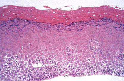
6 Precancerous lesions Tylosis
7 Oesophageal Cancer: clinical Early cancers asymptomatic Dysphagia from invasion into wall with narrowing of lumen Pain from involvement of adjacent structures
8

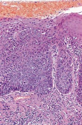
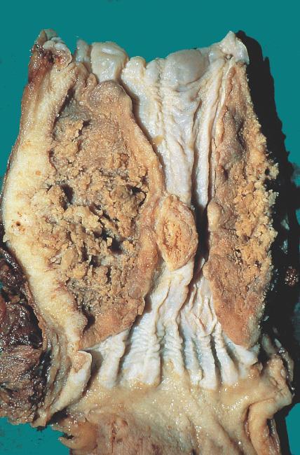
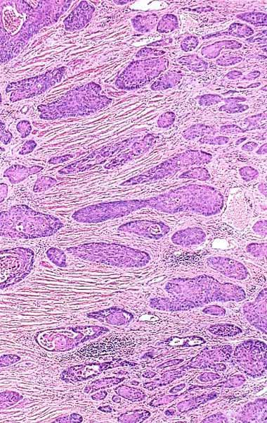
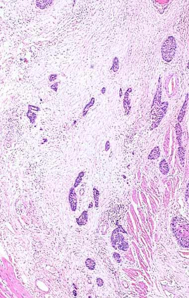
9 Squamous Cell Carcinoma

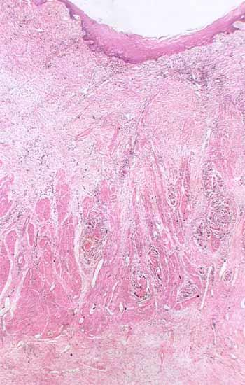
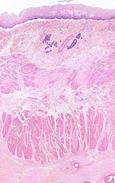
10 Verrucous Carcinoma Well differentiated Slowly growing Rarely metastatic Extensive local spread
11 Superficial Spreading Squamous Carcinoma In situ or invasive carcinoma confined to mucosa and submucosa > 20mm High frequency of lymph node involvement Poor prognosis Soga J et al 1982 Cancer 50:

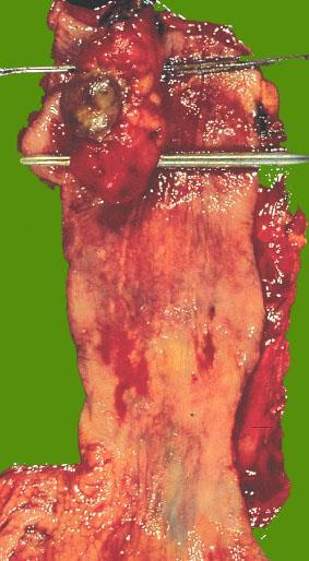
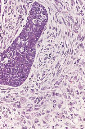
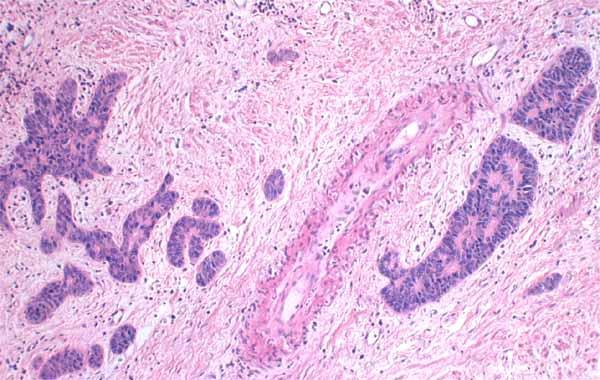
12 Basaloid Squamous Carcinoma Elderly males Solid, discrete nests Small, mitotically active cells Microcystic spaces or necrosis Stromal hyalinisation May be confused with adenoid cystic carcinoma Poor prognosis Abe K et al 1996 Am J Surg Pathol 20:
13 Spindle Cell Carcinoma Polypoid carcinoma, carcinosarcoma, pseudosarcoma Biphasic pattern Mesenchymal metaplasia Present early Better prognosis
14
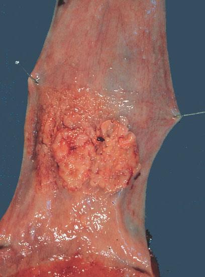
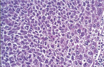
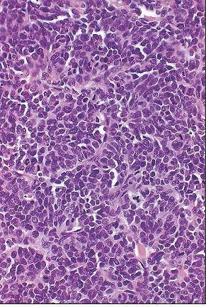
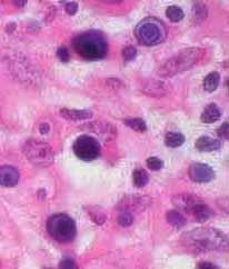
15 Small Cell Carcinoma Uncommon Identical to lung counterpart TTF-1 positive Rarely associated with ectopic hormone syndrome May be combined with squamous carcinoma or adenocarcinoma
16
17 Adenocarcinoma Increasing in frequency Males > females Smoking, alcohol, obesity Barrett s oesophagus p53, c-erbb-2, COX-2 upregulation Heterotopic gastric mucosa - inlet patch
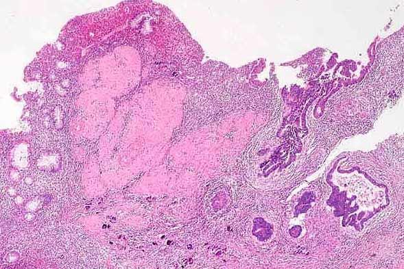
18 Adenocarcinoma arising in CLO
19

20 Intestinal type Tubular or papillary Adenocarcinoma

21 CK7 and CK20 Intestinal metaplasia Barrett s CK7++/CK20+sup Gastric CK7-/CK20++ Adenocarcinoma Oesophageal CK7+/CK20-90% Gastric CK7+/CK20-20%


22 Oesophageal Cancer TNM TX T0 Tis T1 T1a T1b T2 T3 T4 T4a T4b GX G1 G2 G3 G4 Primary tumor cannot be assessed No evidence of primary tumor High-grade dysplasia Tumor invades lamina propria, muscularis mucosae, or submucosa Tumor invades lamina propria or muscularis mucosae Tumor invades submucosa Tumor invades muscularis propria Tumor invades adventitia Tumor invades adjacent structures Resectable tumor invading pleura, pericardium, or diaphragm Unresectable tumor invading other adjacent structures, such as the aorta, vertebral body, and trachea Grade cannot be assessed stage grouping as G1 Well differentiated Moderately differentiated Poorly differentiated Undifferentiated stage grouping as G3 squamous

23 Oesophageal Cancer TNM NX N0 N1 N2 N3 Regional lymph node(s) cannot be assessed No regional lymph node metastasis Metastasis in 1-2 regional lymph nodes Metastasis in 3-6 regional lymph nodes Metastasis in 7 or more regional lymph nodes
24 Type I 1-5cm above GOJ Low oesophageal carcinoma
25 Type II 1cm above to 2cm below GOJ True cardia carcinoma

26 Type III 2-5cm below the endoscopic cardia Sub-cardiac carcinoma
27 Siewert s Classification Type I Type II Type III adenocarcinomas of the distal third of the oesophagus (1-5cm above cardia) low oesophageal carcinoma adenocarcinomas straddling the gastrooesophageal junction (1cm above to 2 cm below cardia) true cardiac tumour subcardial gastric adenocarcinomas that grow proximally to involve the GOJ (2-5 cm below cardia) subcardiac carcinoma
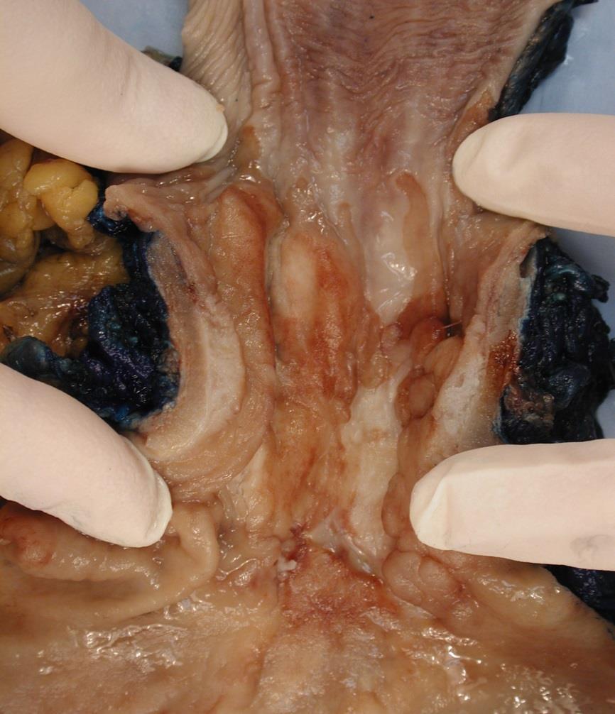
28 Siewert s Classification Type I Type II Type III adenocarcinomas of the distal third of the oesophagus (1-5cm above GOJ) staged by oesophageal rules adenocarcinomas straddling the gastro-oesophageal junction (1cm above to 2 cm below cardia) staged by oesophageal rules subcardial gastric adenocarcinomas that grow proximally to involve the GOJ (2-5 cm below cardia) staged by gastric rules In TNM7, tumours with an epicentre within 5cm of the OGJ which extend into the oesophagus are classified according to the oesophageal carcinoma scheme

29 Gastric Cancer TNM TX T0 Tis T1 T1a Primary tumor cannot be assessed No evidence of primary tumor Carcinoma in situ: intraepithelial tumor without invasion of the lamina propria Tumor invades lamina propria, muscularis mucosae, or submucosa Tumor invades lamina propria or muscularis mucosae T1b Tumor invades submucosa T2 T3 T4 Tumor invades muscularis propria Tumor penetrates subserosal connective tissue without invasion of visceral peritoneum or adjacent structures Tumor invades serosa (visceral peritoneum) or adjacent structures T4a Tumor invades serosa (visceral peritoneum) T4b Tumor invades adjacent structures
30 Gastric Cancer TNM NX N0 N1 N2 N3 N3a N3b Regional lymph node(s) cannot be assessed No regional lymph node metastasis Metastasis in 1-2 regional lymph nodes Metastasis in 3-6 regional lymph nodes Metastasis in seven or more regional lymph nodes Metastasis in 7-15 regional lymph nodes Metastasis in 16 or more regional lymph nodes

31 Gastric Cancer TNM M0 M1 No distant metastasis Distant metastasis
32
33
34

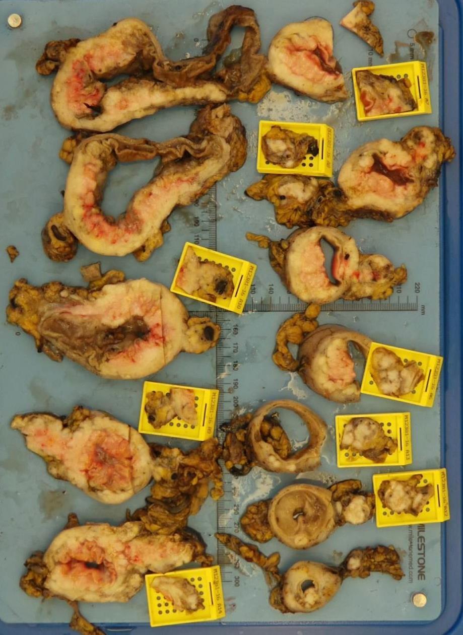
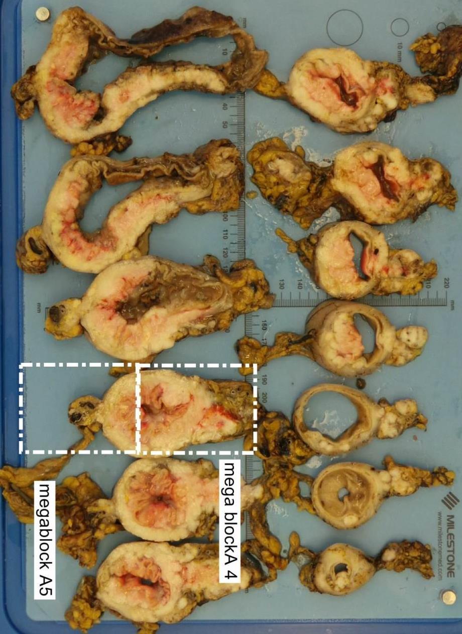
35 Open and ink CRM Leave unopened with a foam or paper wick and ink CRM Slice transversely first Look for lymph nodes first Specimen Choices
36
37
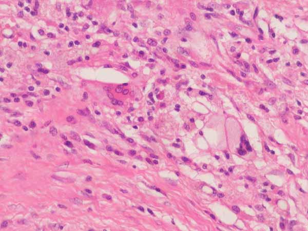
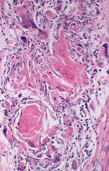
38 Effects of Chemoradiotherapy on Epithelial Tumours Tumour Vacuolation and eosinophilia Nuclear pyknosis, apoptosis Necrosis Stromal Fibrosis, hyalinisation Inflammation Giant cell granulomatous reaction Calcification, malakoplakia Residual acellular elements keratin mucus pools
39
40
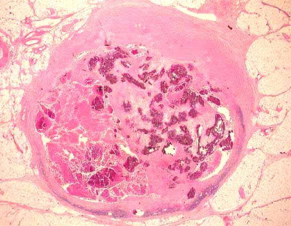
41

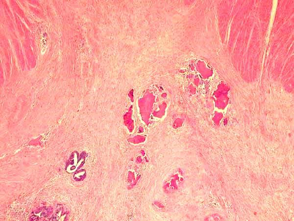
42 Mucin Pools Better prognosis No effect on outcome when at resection margin Hornick JL et al 2006 Am J Surg Pathol 30:28-35
43 yptnm staging e.g. ypt2 N0 Categorises the extent of tumour actually present at the time of examination. Ignore residual acellular elements eg mucus pools, keratin NOT an estimate of disease prior to neoadjuvant therapy
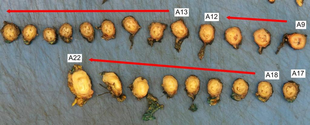
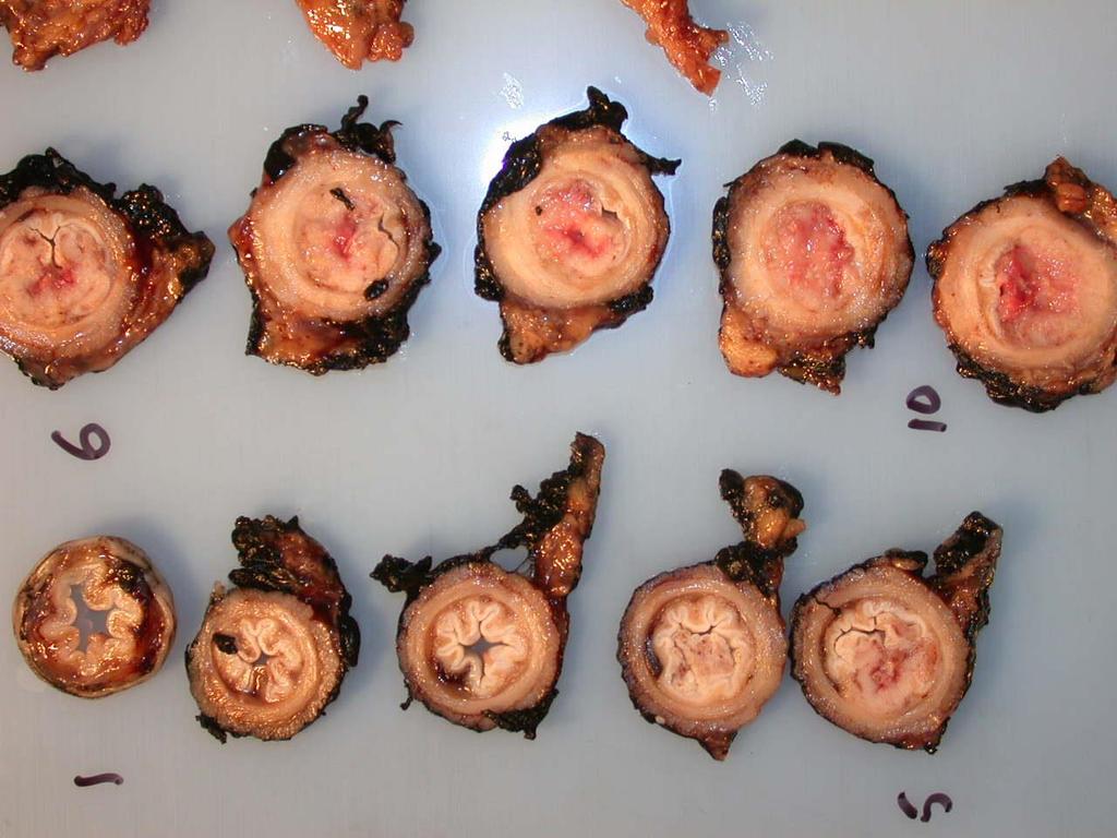
44 Pathological Response Complete or partial Quantitation if partial Requires widespread sampling Serial slices of site of the previous tumour

45 Complete Response
46
47 Partial Response Reduction in tumour volume Requires comparison with untreated tumour Pre-treatment biopsy? representative of the whole lesion Microscopic foci only Relative proportions of tumour and fibrosis
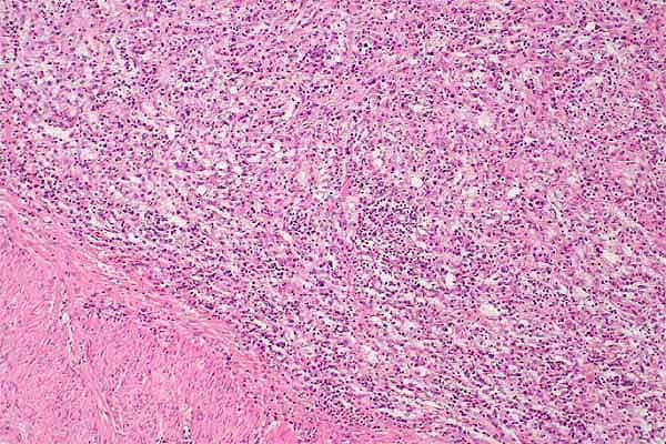
48 Mandard Classification Relative Proportion of Tumour: Fibrosis TRG1 No residual cancer TRG2 Rare residual cancer cells TRG3 Fibrosis outgrowing cancer TRG4 Cancer outgrowing fibrosis TRG5 Absence of regression Significant correlation with disease free survival for TRG1-3 vs TRG4-5 Mandard AM et al 1994 Cancer 73:2680-6
49 TRG3 TRG4
50 Pathological Response Mandard Classification? Too complex? Reproducible Simpler System No residual tumour Minimal residual disease only occasional microscopic tumour foci are identified with difficulty No marked regression
51 Wu TT et al 2007 Am J Surg Pathol 31:58-64
52 Complete response disappearance of the primary tumour in the postoperative specimen Partial response microscopic evidence of residual tumour in the postoperative specimen Stable disease less than 50% decrease or less than a 25% increase in tumour volume Progressive disease no significant change in tumour mass or more than a 25% increase in tumour volume Japanese Society for Esophageal Disease
53 More than 10 grading systems available (Mandard, Japanese, Dworak, Wheeler, Becker, Junker and Mueller, Rubbia-Brandt, Ryan, Le Sodan, Schneider, Lowy, Mansourd) This tells us none is entirely reliable Relies on complete embedding of abnormal area (presumed tumour bed) As there is no national or international consensus, (RCPath) cannot be prescriptive and suggest that regression system to be used should be determined locally by MDT involving pathologist RCPath Dataset; Grabsch, Mapstone and Novelli, in preparation
54
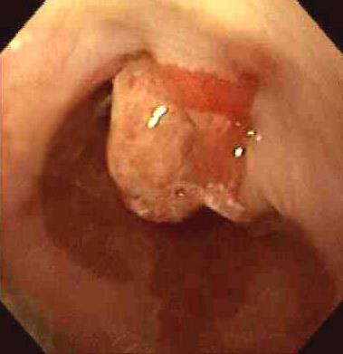
55 Margin Involvement Proximal margin involvement Predicts local recurrence May be discontinuous with the main tissue mass Always sample histologically Distal margin involvement Circumferential margin involvement
56 Margin Involvement Number median survival R months R months R months p< Number 5 year survival R1/ R
57 Circumferential Margins 50 patients Circumferential margin involved in 20 (40%) Median follow up 36 months Local recurrence: CRM positive 11/20 CRM negative 4/30
58 135 patients Circumferential Margins Circumferential margin involved in 64 (47%) (tumour within 1mm) Median survival: CRM positive 21 months CRM negative 39 months Effect only seen when low metastatic burden (<25% nodes positive)
59 Circumferential Margins 329 patients Circumferential margin involved in 20% 5 year survival: CRM positive 22% CRM negative 29% difference not significant
60 249 patients Circumferential Margins Circumferential margin involved in 32% Median survival: CRM positive 18 months CRM negative 37 months p< CRM status had a greater prognostic effect in T3 tumours with a low metastatic lymph node burden (p=0.04). Griffiths EA et al 2006 Eur J Surg Oncol 32: 413-9
61 Oesophageal Cancer: clinical Surveillance is the application of a test that allows detection at a stage when intervention may improve outcome Benefit from surveillance is limited Endoscopy with quadrantic biopsies every 2cm + macroscopically abnormal areas Patches of dysplasia easily missed Annual progression of 0.6% to adenocarcinoma
62 Oesophageal Cancer: endoscopy White light can detect nodules, ulcers or strictures (features of early cancers) Often early cancers are macroscopically normal Chromoendoscopy: Lugol s iodine, methylene blue Trimodal imaging: white-light, autofluorescence, narrow-band imaging Vasculature and mucosal pit patterns

63 Lugol s iodine
64 Oesophageal Cancer: endoscopy magnification and high resolution endoscopy chromo-endoscopy auto-fluorescence endoscopy narrow band imaging microscopic tools: confocal microscopy; multiphoton microscopy in situ molecular analysis: FISH spectroscopic analysis: fluorescence, light scattering, optical coherence, Raman (inelastic) spectroscopy
65 Oesophageal Cancer: endoscopy Confocal fluorescence microscopy: high negative predictive value but poor sensitivity Elastic scattering spectroscopy: changes in subcellular components during malignant transformation; high sensitivity but poor specificity Optical coherence tomography: similar to ultrasound (3mm depth): in vivo studies not convincing

66 Standard endoscopic view of CLO with HGD: no lesion identified

67 Auto-fluorescence image: purple is abnormal

68 Narrow band image of same abnormal area

69 Chromo-endoscopy with indigo carmine dye-spray: HGD on histology
70

71 Low grade dysplasia in EMR/ER makes histological assessment easier

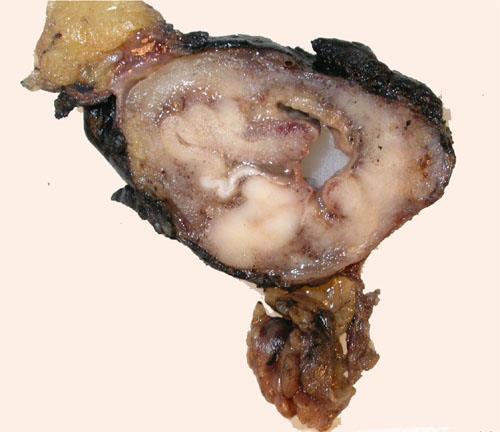
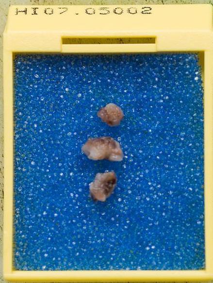
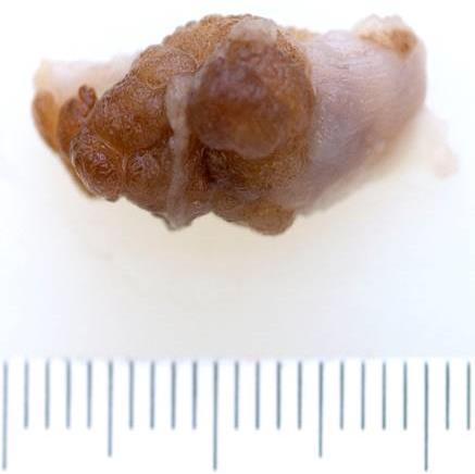
72 69F. Long history of achalasia. Nodule close to OGJ. Multiple biopsies at three endoscopic sittings had provided equivocal results. EMR.
73 Oesophageal EMR Olympus/Keymed inject submucosa and form pseudopolyp, direct snare Cook Duette band EMR/ER band ligation and snare methodology and subsequent therapy likely influences how important margins are
74 Practicalities Oesophageal EMR size and number depends on preferred technique of endoscopist specimen preparation band still attached! orientation adequate fixation specimen dissection depends on size we are more interested in the deep margin than peripheral margins

")
75 close collaboration with endoscopist and assistant to identify important landmarks resect en bloc if possible keep specimen(s) intact pin out on cork mark margins embed whole specimen(s) for histology
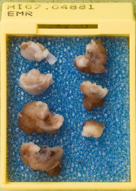
76 Oesophageal EMRs


77 Histological assessment of EMR The rationale for EMR/ER in CLO Intramucosal pathology Submucosal involvement by carcinoma Endoscopic treatment and/or survelliance Referral for surgery
78 The rationale for EMR/ER in CLO: risk of lymph node metastatic disease intramucosal disease submucosal disease adenocarcinoma 2.0% 24.6% squamous cell carcinoma 3.6% 26% For mucosal disease, the surgical mortality outweighs the risk of metastasis Bergman, 2007

79 What are the diagnostic pathological issues in oesophageal EMR?

80 HGD versus intramucosal carcinoma

81 Entrapped and submucosal glands mimicking submucosal adenocarcinoma
82 Reporting oesophageal EMRs is it Barrett s? are there treatment effects? neoplasia diagnosis depth of spread lymphovascular spread peripheral margins status deep margin status
83 Oesophageal EMR/ER diagnosis and complete excision CLO only 21 Squamous only 9 Gastric mucosa only 7 LGD excised 5 LGD at margins 9 HGD excised 9 HGD at margins 36 IMC excised 4 IMC at margins 12 Ca into SM excised 8 Ca into SM at margins 17 Total 137 Shepherd and Barr, Gloucester

84 Low grade dysplasia at margins
85 Oesophageal EMR/ER in Gloucestershire number of normals reflects endoscopic difficulties benign nodules in CLO, hiatus hernia, etc LGD and HGD often at margins reflects endoscopic difficulties matters less because of subsequent ablative therapy IMC often at margins don t know the implications of this but one suspects that this, too, will be successfully ablated submucosal adenocarcinoma (ironically) more often clear of margins but doesn t matter much as this is an indication for radical surgery Shepherd and Barr, Gloucester

86 CLO and the double muscularis mucosae and entrapment of dysplastic epithelium Takubo et al, 1991, Lewis et al, 2008
87 Oesophageal EMR pathology is important, mainly to confirm or refute: the presence of malignancy if present, depth of malignancy margins matter less (but this does depend on subsequent management strategy)

88 THANK YOU FOR YOUR ATTENTION ANY QUESTIONS?
Greater Manchester & Cheshire Guidelines for Pathology Reporting for Oesophageal and Gastric Malignancy
 Greater Manchester & Cheshire Guidelines for Pathology Reporting for Oesophageal and Gastric Malignancy Authors: Dr Gordon Armstrong, Dr Sue Pritchard 1. General Comments 1.1 Cancer reporting: Biopsies
Tumours of the Oesophagus & Gastro-Oesophageal Junction Histopathology Reporting Proforma
 Tumours of the Oesophagus & Gastro-Oesophageal Junction Histopathology Reporting Proforma Mandatory questions (i.e. protocol standards) are in bold (e.g. S1.01). S1.01 Identification Family name Given
Greater Manchester & Cheshire Guidelines for Pathology Reporting of Oesophageal and Gastric Malignancy
 Greater Manchester & Cheshire Guidelines for Pathology Reporting of Oesophageal and Gastric Malignancy Authors: Dr Stephen Hayes, Dr David Bisset, Dr Gordon Armstrong, Dr Sue Pritchard 1. General Comments
Oesophagus and Stomach update dysplasia and early cancer
 Oesophagus and Stomach update dysplasia and early cancer Dr Tim Bracey STR teaching 13/4/16 Please check pathkids.com for previous talks One of the biggest units in the country (100 major resections per
Histopathology of Endoscopic Resection Specimens from Barrett's Esophagus
 Histopathology of Endoscopic Resection Specimens from Barrett's Esophagus Br J Surg 38 oct. 1950 Definition of Barrett's esophagus A change in the esophageal epithelium of any length that can be recognized
Earlyoesophagealcancer. dr. Nina Zidar Institute of Pathology Faculty ofmedicine University of Ljubljana Slovenia
 Earlyoesophagealcancer dr. Nina Zidar Institute of Pathology Faculty ofmedicine University of Ljubljana Slovenia Early carcinoma of oesophagus = tumor limited to mucosa or submucosa, not extending into
Gastric Cancer Histopathology Reporting Proforma
 Gastric Cancer Histopathology Reporting Proforma Mandatory questions (i.e. protocol standards) are in bold (e.g. S1.01). S1.01 Identification Family name Given name(s) Date of birth Sex Male Female Intersex/indeterminate
Gastric Cancer Staging AJCC eighth edition. Duncan McLeod Westmead Hospital, NSW
 Gastric Cancer Staging AJCC eighth edition Duncan McLeod Westmead Hospital, NSW Summary of changes New clinical stage prognostic groups, ctnm Postneoadjuvant therapy pathologic stage groupings, yptnm -
The Pathologist s Role in the Diagnosis and Management of Neoplasia in Barrett s Oesophagus Cian Muldoon, St. James s Hospital, Dublin
 The Pathologist s Role in the Diagnosis and Management of Neoplasia in Barrett s Oesophagus Cian Muldoon, St. James s Hospital, Dublin 24.06.15 Norman Barrett Smiles [A brief digression - Chair becoming
CLINICAL EFFECTIVENESS
 Re-audit of gastrointestinal tract specimens with respect to compliance with RCPath guidelines Dr Manisha Ram Dr Moina Kadri Background epidemiology and aetiology Over the past 20 years there has been
Barrett s Esophagus: Old Dog, New Tricks
 Barrett s Esophagus: Old Dog, New Tricks Stuart Jon Spechler, M.D. Chief, Division of Gastroenterology, VA North Texas Healthcare System; Co-Director, Esophageal Diseases Center, Professor of Medicine,
Quiz Adenocarcinoma of the distal stomach has been increasing in the last 20 years. a. True b. False
 Quiz 1 1. Which of the following are risk factors for esophagus cancer. a. Obesity b. Gastroesophageal reflux c. Smoking and Alcohol d. All of the above 2. Adenocarcinoma of the distal stomach has been
Gastric and Oesophageal Neuroendocrine tumours. Dr Tim Bracey, Consultant Pathologist MBChB PhD MRCS FRCPath
 Gastric and Oesophageal Neuroendocrine tumours Dr Tim Bracey, Consultant Pathologist MBChB PhD MRCS FRCPath Intestinal (and BO) endocrine cells in crypt bases NE cell (granules towards vessels) Paneth
Rectal Cancer Cookbook Update. A. JOURET-MOURIN with the collaboration of A Hoorens,P Demetter, G De Hertogh,C Cuvelier and C Sempoux
 Rectal Cancer Cookbook Update A. JOURET-MOURIN with the collaboration of A Hoorens,P Demetter, G De Hertogh,C Cuvelier and C Sempoux Prof Dr A Jouret-Mourin, Department of Pathology, UCL, St Luc, Brussels
Upper GIT IV Gastric cancer
 Upper GIT IV Gastric cancer Luigi Tornillo PathoBasic 23.10.2014 Pathology Introduction Classification Morphogenesis Problems Intraepithelial neoplasia Surveillance EGJ Predictive factors Gastric cancer
Gastrointestinal pathology 2018 lecture 2. Dr Heyam Awad FRCPath
 Gastrointestinal pathology 2018 lecture 2 Dr Heyam Awad FRCPath Eosinophilic esophagitis Incidence of eosinophilic gastritis is increasing. Symptoms: food impaction and dysphagia. Histology: infiltration
American Journal of Gastroenterology. Volumetric Laser Endomicroscopy Detects Subsquamous Barrett s Adenocarcinoma
 Volumetric Laser Endomicroscopy Detects Subsquamous Barrett s Adenocarcinoma Journal: Manuscript ID: AJG-13-1412.R1 Manuscript Type: Letter to the Editor Keywords: Barrett-s esophagus, Esophagus, Endoscopy
Management of Barrett s: From Imaging to Resection
 Management of Barrett s: From Imaging to Resection Michael Wallace, MD, MPH, FACG Professor of Medicine Mayo Clinic Florida Goals of Endoscopic Evaluation in Barrett s Detect Barrett s and dysplasia Reduce/eliminate
Barrett s Esophagus. Abdul Sami Khan, M.D. Gastroenterologist Aurora Healthcare Burlington, Elkhorn, Lake Geneva, WI
 Barrett s Esophagus Abdul Sami Khan, M.D. Gastroenterologist Aurora Healthcare Burlington, Elkhorn, Lake Geneva, WI A 58 year-old, obese white man has had heartburn for more than 20 years. He read a magazine
New Developments in the Endoscopic Diagnosis and Management of Barrett s Esophagus
 New Developments in the Endoscopic Diagnosis and Management of Barrett s Esophagus Prateek Sharma, MD Key Clinical Management Points: Endoscopic recognition of a columnar lined distal esophagus is crucial
Oncology SpR training day 23 November Oesophageal cancer
 Oncology SpR training day 23 November 2015 Oesophageal cancer Squamous Adenocarcinoma Adenosquamous Basaloid squamous Spindle cell squamous Verrucous Mucoepidermoid Adenoid cystic Undifferentiated WHO
Esophageal Cancer Staging Essentials: The New TNM Staging System (7th edition) and Clinicoradiologic Implications
 and Clinicoradiologic Implications") Esophageal Cancer Staging Essentials: The New TNM Staging System (7th edition) and Clinicoradiologic Implications Poster No.: E-0060 Congress: ESTI 2012 Type: Scientific Exhibit Authors: K. Lee, T. J.
Esophageal Cancer Staging Essentials: The New TNM Staging System (7th edition) and Clinicoradiologic Implications Poster No.: E-0060 Congress: ESTI 2012 Type: Scientific Exhibit Authors: K. Lee, T. J.
Carcinoma of the Urinary Bladder Histopathology
 Carcinoma of the Urinary Bladder Histopathology Reporting Proforma (Radical & Partial Cystectomy, Cystoprostatectomy) Includes the International Collaboration on Cancer reporting dataset denoted by * Family
COLLECTING CANCER DATA: STOMACH AND ESOPHAGUS
 COLLECTING CANCER DATA: STOMACH AND ESOPHAGUS 2017 2018 NAACCR WEBINAR SERIES Q&A Please submit all questions concerning webinar content through the Q&A panel. Reminder: If you have participants watching
AGA SECTION. Gastroenterology 2016;150:
 Gastroenterology 2016;150:1026 1030 April 2016 AGA Section 1027 Procedural intervention (3) Upper endoscopy indications 3 6 Non-response of symptoms to a 4 8 week empiric trial of twice-daily PPI Troublesome
Pathology of the oesophagus and the stomach. Neil A Shepherd Gloucester, UK. Bristol Pathology 1 st Year Training School, The layers of the GI tract
 Pathology of the oesophagus and the stomach Neil A Shepherd Gloucester, UK Bristol Pathology 1 st Year Training School, The layers of the GI tract 1 Some facts about Histopathology and the upper GI tract
Pathology of Treated GI Neoplasia
 Pathology of Treated GI Neoplasia Pathology section seminar Liverpool DDF meeting Tuesday 19 th June 2012 at14:00-15:15 Venue: Hall 11c in the ACC Presenters: Phillip Kaye, Adrian Bateman, Shaun Walsh,
Carcinoma of the Renal Pelvis and Ureter Histopathology
 Carcinoma of the Renal Pelvis and Ureter Histopathology Reporting Proforma (NEPHROURETERECTOMY AND URETERECTOMY) Includes the International Collaboration on Cancer reporting dataset denoted by * Family
Esophageal cancer: Biology, natural history, staging and therapeutic options
 EGEUS 2nd Meeting Esophageal cancer: Biology, natural history, staging and therapeutic options Michael Bau Mortensen MD, Ph.D. Associate Professor of Surgery Centre for Surgical Ultrasound, Upper GI Section,
ACG Clinical Guideline: Diagnosis and Management of Barrett s Esophagus
 ACG Clinical Guideline: Diagnosis and Management of Barrett s Esophagus Nicholas J. Shaheen, MD, MPH, FACG 1, Gary W. Falk, MD, MS, FACG 2, Prasad G. Iyer, MD, MSc, FACG 3 and Lauren Gerson, MD, MSc, FACG
7/20/2017. Esophageal Cancer: A Less Common But Deadly Cancer. Objectives. Disclosure Statement NYNPA Conference October Saratoga New York
 Esophageal Cancer: A Less Common But Deadly Cancer 2017 NYNPA Conference October 18-22 Saratoga New York Mary McGreal DNP, RN, ANP-C, CCRN, CMC, Adjunct Professor at Stony Brook University School of Nursing
Colorectal Cancer Structured Pathology Reporting Proforma DD MM YYYY
 Colorectal Cancer Structured Pathology Reporting Proforma Mandatory questions (i.e. protocol standards) are in bold (e.g. S1.03). Family name Given name(s) Date of birth DD MM YYYY S1.02 Clinical details
Barrett Esophagus - RadioFrequency Ablation (BE-RFA) - Project manual + FAQ
 - Project manual + FAQ") Barrett Esophagus - RadioFrequency Ablation (BE-RFA) - Project manual + FAQ Table of contents 1 General project information...3 1.1 Inclusion criteria...3 1.2 Registration time points...3 1.3 Project variable
Barrett Esophagus - RadioFrequency Ablation (BE-RFA) - Project manual + FAQ Table of contents 1 General project information...3 1.1 Inclusion criteria...3 1.2 Registration time points...3 1.3 Project variable
A218 : Esophagus cancer tissues. (formalin fixed)
") (formalin fixed) For research use only Specifications: No. of cases: 40 Tissue type: Esophagus cancer tissues No. of spots: 2 spots from each cancer case (80 spots) 4 non-neoplastic spots (4 spots) Total
(formalin fixed) For research use only Specifications: No. of cases: 40 Tissue type: Esophagus cancer tissues No. of spots: 2 spots from each cancer case (80 spots) 4 non-neoplastic spots (4 spots) Total
11/21/13 CEA: 1.7 WNL
 Case Scenario 1 A 70 year-old white male presented to his primary care physician with a recent history of rectal bleeding. He was referred for imaging and a colonoscopy and was found to have adenocarcinoma.
Staging Challenges in Lower GI Cancers. Disclosure of Relevant Financial Relationships. AJCC 8 th edition and CAP protocol updates
 Staging Challenges in Lower GI Cancers Sanjay Kakar, MD University of California, San Francisco March 05, 2017 Disclosure of Relevant Financial Relationships USCAP requires that all planners (Education
Case Scenario year-old white male presented to personal physician with dyspepsia with reflux.
 Case Scenario 1 57-year-old white male presented to personal physician with dyspepsia with reflux. 7/12 EGD: In the gastroesophageal junction we found an exophytic tumor. The tumor occupies approximately
LARYNGEAL DYSPLASIA. Tomas Fernandez M; 3 rd year ENT resident, Son Espases University Hospital
 LARYNGEAL DYSPLASIA Tomas Fernandez M; 3 rd year ENT resident, Son Espases University Hospital INTRODUCTION Laryngeal cancer constitutes 1-2% of all malignancies diagnosed worldwide Survival is related
Update on staging colorectal carcinoma, the 8 th edition AJCC. General overview of staging. When is staging required? 11/1/2017
 Update on staging colorectal carcinoma, the 8 th edition AJCC Dale C. Snover, MD November 3, 2017 General overview of staging Reason for uniform staging Requirements to use AJCC manual and/or CAP protocols
Abstracting Upper GI Cancer Incidence and Treatment Data Quiz 1 Multiple Primary and Histologies Case 1 Final Pathology:
 Abstracting Upper GI Cancer Incidence and Treatment Data Quiz 1 Multiple Primary and Histologies Case 1 A 74 year old male with a history of GERD presents complaining of dysphagia. An esophagogastroduodenoscopy
Frozen Section Analysis of Esophageal Endoscopic Mucosal Resection Specimens in the Real-Time Management of Barrett s Esophagus
 CLINICAL GASTROENTEROLOGY AND HEPATOLOGY 2006;4:173 178 Frozen Section Analysis of Esophageal Endoscopic Mucosal Resection Specimens in the Real-Time Management of Barrett s Esophagus GANAPATHY A. PRASAD,*
National Oesophago-Gastric Cancer Audit New Patient Registration sheet Patients with Oesophageal High Grade Glandular Dysplasia
 National Oesophago-Gastric Cancer Audit New Patient Registration sheet Patients with Oesophageal High Grade Glandular Dysplasia Patient Details Surname: NHS number: Forename: Postcode: Sex: Male Female
Definition of Synoptic Reporting
 Definition of Synoptic Reporting The CAP has developed this list of specific features that define synoptic reporting formatting: 1. All required cancer data from an applicable cancer protocol that are
Towards a more personalized approach in the treatment of esophageal cancer focusing on predictive factors in response to chemoradiation Wang, Da
 University of Groningen Towards a more personalized approach in the treatment of esophageal cancer focusing on predictive factors in response to chemoradiation Wang, Da IMPORTANT NOTE: You are advised
PATHOLOGY GROUP GUIDELINES FOR THE EXAMINATION AND REPORTING OF COLORECTAL CANCER SPECIMENS
 PATHOLOGY GROUP GUIDELINES FOR THE EXAMINATION AND REPORTING OF COLORECTAL CANCER SPECIMENS Produced by: Address: Yorkshire Cancer Network Pathology Group Arthington House, Cookridge Hospital, Hospital
Case Scenario 1. The patient has now completed his neoadjuvant chemoradiation and has been cleared for surgery.
 Case Scenario 1 July 10, 2010 A 67-year-old male with squamous cell carcinoma of the mid thoracic esophagus presents for surgical resection. The patient has completed preoperative chemoradiation. This
Learning Objectives:
 Crescent City GI Update 2018 Ochsner Clinic, NOLA Optimizing Endoscopic Evaluation of Barrett s Esophagus What Should I Do in My Practice? Gregory G. Ginsberg, M.D. Professor of Medicine University of
Small Intestine. Protocol revision date: January 2005 Based on AJCC/UICC TNM, 6 th edition
 Small Intestine Protocol applies to all invasive carcinomas of the small intestine, including those with focal endocrine differentiation. Excludes carcinoid tumors, lymphomas, and stromal tumors (sarcomas).
A916: rectum: adenocarcinoma
 General facts of colorectal cancer The colon has cecum, ascending, transverse, descending and sigmoid colon sections. Cancer can start in any of the r sections or in the rectum. The wall of each of these
History. Prevalence at Endoscopy. Prevalence and Reflux Sx. Prevalence at Endoscopy. Barrett s Esophagus: Controversy and Management
 Barrett s Esophagus: Controversy and Management History Norman Barrett (1950) Chronic Peptic Ulcer of the Oesophagus and Oesophagitis Allison and Johnstone (1953) The Oesophagus Lined with Gastric Mucous
Endoscopic UltraSound (EUS) Endoscopic Mucosal Resection (EMR) Moishe Liberman Director C.E.T.O.C.
 Endoscopic Mucosal Resection (EMR) Moishe Liberman Director C.E.T.O.C.") Endoscopic UltraSound (EUS) Endoscopic Mucosal Resection (EMR) Moishe Liberman Director C.E.T.O.C. Division of Thoracic Surgery Centre Hospitalier de l Université de Montréal Research Grants: Disclosures
Endoscopic UltraSound (EUS) Endoscopic Mucosal Resection (EMR) Moishe Liberman Director C.E.T.O.C. Division of Thoracic Surgery Centre Hospitalier de l Université de Montréal Research Grants: Disclosures
Case Report Intramucosal Signet Ring Cell Gastric Cancer Diagnosed 15 Months after the Initial Endoscopic Examination
 Hindawi Publishing Corporation Case Reports in Medicine Volume 2015, Article ID 479625, 5 pages http://dx.doi.org/10.1155/2015/479625 Case Report Intramucosal Signet Ring Cell Gastric Cancer Diagnosed
Neoplasia 2018 Lecture 2. Dr Heyam Awad MD, FRCPath
 Neoplasia 2018 Lecture 2 Dr Heyam Awad MD, FRCPath ILOS 1. List the differences between benign and malignant tumors. 2. Recognize the histological features of malignancy. 3. Define dysplasia and understand
Changes to the diagnosis and management of Barrett s Oesophagus
 Changes to the diagnosis and management of Barrett s Oesophagus A review of the new BSG and NICE guidelines and best practice Anjan Dhar DM, MD, FRCPE, AGAF, MBBS (Hons.), Cert. Med. Ed Senior Lecturer
Philip Chiu Associate Professor Department of Surgery, Prince of Wales Hospital The Chinese University of Hong Kong
 Application of Chromoendoscopy, NBI and AFI in Esophagus why, who, and how? Philip Chiu Associate Professor Department of Surgery, Prince of Wales Hospital The Chinese University of Hong Kong Cancer of
ESOPHAGEAL CANCER AND GERD. Prof Salman Guraya FRCS, Masters MedEd
 ESOPHAGEAL CANCER AND GERD Prof Salman Guraya FRCS, Masters MedEd Learning objectives Esophagus anatomy and physiology Esophageal cancer Causes, presentations of esophageal cancer Diagnosis and management
The International Association for the Study of Lung Cancer (IASLC) Lung Cancer Staging Project, Data Elements
 Lung Cancer Staging Project, Data Elements") Page 1 Contents 1.1. Registration... 2 1.2. Patient Characteristics... 3 1.3. Laboratory Values at Diagnosis... 5 1.4. Lung Cancers with Multiple Lesions... 6 1.5. Primary Tumour Description... 10 1.6.
Page 1 Contents 1.1. Registration... 2 1.2. Patient Characteristics... 3 1.3. Laboratory Values at Diagnosis... 5 1.4. Lung Cancers with Multiple Lesions... 6 1.5. Primary Tumour Description... 10 1.6.
Colon and Rectum. Protocol revision date: January 2005 Based on AJCC/UICC TNM, 6th edition
 Colon and Rectum Protocol applies to all invasive carcinomas of the colon and rectum. Carcinoid tumors, lymphomas, sarcomas, and tumors of the vermiform appendix are excluded. Protocol revision date: January
Faculty Disclosure. Objectives. State of the Art #3: Referrals for Gastroscopy (focus on common esophagus problems) 24/11/2014
 24/11/2014") State of the Art #3: Referrals for Gastroscopy (focus on common esophagus problems) Dr. Amy Morse November 2014 Faculty: Amy Morse Faculty Disclosure Relationships with commercial interests: Grants/Research
State of the Art #3: Referrals for Gastroscopy (focus on common esophagus problems) Dr. Amy Morse November 2014 Faculty: Amy Morse Faculty Disclosure Relationships with commercial interests: Grants/Research
32 Adenocarcinoma of the oesophagogastric junction
 Adenocarcinoma of the oesophagogastric junction S.J. Spechler P. Hainaut M.F. Dixon R. Lambert R. Genta R. Siewert Definition Adenocarcinomas that straddle the junction of the oesophagus and stomach are
8. The polyp in the illustration can be described as (circle all that apply) a. Exophytic b. Pedunculated c. Sessile d. Frank
 a. Exophytic b. Pedunculated c. Sessile d. Frank") Quiz 1 Overview 1. Beginning with the cecum, which is the correct sequence of colon subsites? a. Cecum, ascending, splenic flexure, transverse, hepatic flexure, descending, sigmoid. b. Cecum, ascending,
Quiz 1 Overview 1. Beginning with the cecum, which is the correct sequence of colon subsites? a. Cecum, ascending, splenic flexure, transverse, hepatic flexure, descending, sigmoid. b. Cecum, ascending,
Vulvar Carcinoma. Definition: Cases should be classified as carsinoma of the vulva when the primary site growth is in the vulva Malignant melanoma sho
 Carcinoma Vulva & Vagina Subdivisi Onkologi Ginekologi Bagian Obgin FK USU Vulvar Carcinoma. Definition: Cases should be classified as carsinoma of the vulva when the primary site growth is in the vulva
Paris classification (2003) 삼성의료원내과이준행
 삼성의료원내과이준행") Paris classification (2003) 삼성의료원내과이준행 JGCA classification - Japanese Gastric Cancer Association - Type 0 superficial polypoid, flat/depressed, or excavated tumors Type 1 polypoid carcinomas, usually attached
Paris classification (2003) 삼성의료원내과이준행 JGCA classification - Japanese Gastric Cancer Association - Type 0 superficial polypoid, flat/depressed, or excavated tumors Type 1 polypoid carcinomas, usually attached
CT PET SCANNING for GIT Malignancies A clinician s perspective
 CT PET SCANNING for GIT Malignancies A clinician s perspective Damon Bizos Head, Surgical Gastroenterology Charlotte Maxeke Johannesburg Academic Hospital Case presentation 54 year old with recent onset
A215- Urinary bladder cancer tissues
 A215- Urinary bladder cancer tissues (formalin fixed) For research use only Specifications: No. of cases: 45 Tissue type: Urinary bladder cancer tissues No. of spots: 2 spots from each cancer case (90
Factors for Endoscopic Submucosal Dissection in Early Colorectal Neoplasms: A Single Center Clinical Experience in China
 ORIGINAL ARTICLE Clin Endosc 2015;48:405-410 http://dx.doi.org/10.5946/ce.2015.48.5.405 Print ISSN 2234-2400 On-line ISSN 2234-2443 Open Access Factors for Endoscopic Submucosal Dissection in Early Colorectal
GUIDELINES ON NON-MUSCLE- INVASIVE BLADDER CANCER
 GUIDELINES ON NON-MUSCLE- INVASIVE BLADDER CANCER (Limited text update December 21) M. Babjuk, W. Oosterlinck, R. Sylvester, E. Kaasinen, A. Böhle, J. Palou, M. Rouprêt Eur Urol 211 Apr;59(4):584-94 Introduction
Definition of GERD American College of Gastroenterology
 Definition of GERD American College of Gastroenterology GERD is defined as chronic symptoms or mucosal damage produced by the abnormal reflux of gastric contents into the esophagus DeVault et al. Am J
Protocol for the Examination of Specimens From Patients With Carcinoma of the Esophagus
 Protocol for the Examination of Specimens From Patients With Carcinoma of the Esophagus Version: Protocol Posting Date: June 2017 Includes ptnm requirements from the 8 th Edition, AJCC Staging Manual For
VULVAR CARCINOMA. Page 1 of 5
 VULVAR CARCINOMA EXAMPLE OF A VULVAR CARCINOMA USING PROPOSED TEMPLATE Case: Invasive squamous cell carcinoma arising in D-VIN Tumor in left labia major Left partial vaginectomy and sentinel lymph node
Expert panel observations
 Expert panel observations Professor Neil A Shepherd Gloucester and Cheltenham, UK Gloucestershire Cellular Pathology Laboratory Three big issues in BCSP pathology serrated pathology & what do we do about
Catholic University of Louvain, St - Luc University Hospital Head and Neck Oncology Programme. Anatomopathology. Pathology 1 Sept.
 Anatomopathology Pathology 1 Anatomopathology Biopsies Frozen section Surgical specimen Peculiarities for various tumor site References Pathology 2 Biopsies Minimum data, which should be given by the pathologist
malignant polyp Daily Challenges in Digestive Endoscopy for Endoscopists and Endoscopy Nurses BSGIE Annual Meeting 18/09/2014 Mechelen
 Plan Incidental finding of a malignant polyp 1. What is a polyp malignant? 2. Role of the pathologist and the endoscopist 3. Quantitative and qualitative risk assessment 4. How to decide what to do? Hubert
PRINCESS MARGARET CANCER CENTRE CLINICAL PRACTICE GUIDELINES GYNECOLOGIC CANCER VULVAR
 PRINCESS MARGARET CANCER CENTRE CLINICAL PRACTICE GUIDELINES GYNECOLOGIC CANCER VULVAR Last Revision Date July 2015 1 Site Group: Gynecologic Cancer Vulvar Author: Dr. Stephane Laframboise 1. INTRODUCTION
Kidney Case 1 SURGICAL PATHOLOGY REPORT
 Kidney Case 1 Surgical Pathology Report February 9, 2007 Clinical History: This 45 year old woman was found to have a left renal mass. CT urography with reconstruction revealed a 2 cm medial mass which
Management of pt1 polyps. Maria Pellise
 Management of pt1 polyps Maria Pellise Early colorectal cancer Malignant polyp Screening programmes SM Invasive adenocar cinoma Advances in diagnostic & therapeutic endoscopy pt1 polyps 0.75 5.6% of large-bowel
Histopathology: gastritis and peptic ulceration
 Histopathology: gastritis and peptic ulceration These presentations are to help you identify, and to test yourself on identifying, basic histopathological features. They do not contain the additional factual
University Mainz. Early Gastric Cancer. Ralf Kiesslich. Johannes Gutenberg University Mainz, Germany. Early Gastric Cancer 15.6.
 Ralf Kiesslich Johannes Gutenberg University Mainz, Germany DIAGNOSIS Unmask lesions - Chromoendoscopy -NBI Red flag technology - Autofluorescence Surface and detail analysis - Magnifying endoscopy - High
Gastric Cancer in a Young Postpartum Female. Kings County Hospital Center SUNY Downstate Case Conference May 24, 2012
 Gastric Cancer in a Young Postpartum Female Kings County Hospital Center SUNY Downstate Case Conference May 24, 2012 Case HPI: 31 yo F, G5P3, 3 weeks s/p C-section, with gastric outlet obstruction. Pt
Non Small Cell Lung Cancer Histopathology ד"ר יהודית זנדבנק
 Non Small Cell Lung Cancer Histopathology ד"ר יהודית זנדבנק 26.06.09 Lecture outlines WHO histological classification Macro/Micro assessment Early diagnosis Minimal pathology Main subtypes SCC, AdCa, LCLC
References. GI Biopsies. What Should Pathologists Assistants Know About Gastrointestinal Histopathology? James M Crawford, MD, PhD
 What Should Pathologists Assistants Know About Gastrointestinal Histopathology? James M Crawford, MD, PhD jcrawford1@nshs.edu Executive Director and Senior Vice President for Laboratory Services North
Barrett esophagus. Bible class Inselspital
 Barrett esophagus Bible class Inselspital 2015.08.10 Guidelines Definition? BSG: ACG: Definition? BSG: ACG: What are the arguments for and against IM as prerequisite for the Dg? What are the arguments
Q&A. Fabulous Prizes. Collecting Cancer Data: Bladder, Renal Pelvis, and Ureter 5/2/13. NAACCR Webinar Series
 Collecting Cancer Data Bladder & Renal Pelvis NAACCR 2012 2013 Webinar Series Q&A Please submit all questions concerning webinar content through the Q&A panel. Reminder: If you have participants watching
UICC TNM 8 th Edition Errata
 UICC TNM 8 th Edition Errata ions are in italics Page 28 Oropharynx p16 positive Pathological Stage II,T2 N2 M0 T3 N0,N1 M0 Stage II,T2 N2 M0 T3,T4 N0,N1 M0 Page 61 Oesophagus Adenocarcinoma Pathological
Dysplasia 4/19/2017. How do I practice Chromoendoscopy for Surveillance of Colitis? SCENIC: Polypoid Dysplasia in UC. Background
 SCENIC: Polypoid in UC Definition How do I practice for Surveillance of Colitis? Themos Dassopoulos, M.D. Director, BSW Center for IBD Themistocles.Dassopoulos@BSWHealth.org Tel: 469-800-7189 Cell: 314-686-2623
Professor, Department of Endoscopic Diagnostic and Therapeutics, Chiba University School of Medicine
 Gastrointestinal Diseases Esophageal Disorders JMAJ 47(): 41 47, 24 Teruo KOUZU Professor, Department of Endoscopic Diagnostic and Therapeutics, Chiba University School of Medicine Abstract: Recent trends
Treatment Strategy for Non-curative Resection of Early Gastric Cancer. Jun Haneg Lee. Sungkyunkwan University, Samsung Medical Center, Seoul Korea
 Treatment Strategy for Non-curative Resection of Early Gastric Cancer Jun Haneg Lee. Sungkyunkwan University, Samsung Medical Center, Seoul Korea Classic EMR/ESD data analysis style Endoscopic resection
Question: If in a particular case, there is doubt about the correct T, N or M category, what do you do?
 Exercise 1 Question: If in a particular case, there is doubt about the correct T, N or M category, what do you do? : 1. I mention both categories that are in consideration, e.g. pt1-2 2. I classify as
Squamous Cell Carcinoma of the Head and Neck (SCCHN)
") Squamous Cell Carcinoma of the Head and Neck (SCCHN) Part 1 Bruce M. Wenig, M.D. Dept. of Pathology & Laboratory Medicine Continuum Health Partners New York, NY College of American Pathologists 2004. Materials
Squamous Cell Carcinoma of the Head and Neck (SCCHN) Part 1 Bruce M. Wenig, M.D. Dept. of Pathology & Laboratory Medicine Continuum Health Partners New York, NY College of American Pathologists 2004. Materials
Large Colorectal Adenomas An Approach to Pathologic Evaluation
 Anatomic Pathology / LARGE COLORECTAL ADENOMAS AND PATHOLOGIC EVALUATION Large Colorectal Adenomas An Approach to Pathologic Evaluation Elizabeth D. Euscher, MD, 1 Theodore H. Niemann, MD, 1 Joel G. Lucas,
Novel endoscopic observation in Barrett s oesophagus using high resolution magnification endoscopy and narrow band imaging
 Alimentary Pharmacology & Therapeutics Novel endoscopic observation in Barrett s oesophagus using high resolution magnification endoscopy and narrow band imaging G. K. ANAGNOSTOPOULOS*, K. YAO*, P. KAYE,
Hong Kong Society of Upper Gastrointestinal Surgeons CLINICAL MEETING 29 NOV 2012
 Hong Kong Society of Upper Gastrointestinal Surgeons CLINICAL MEETING 29 NOV 2012 Esophageal Leiomyoma Introduction Case presentation Operative video Discussion Esophageal Leiomyoma Benign tumors of the
Burning Issues in the Esophagus
 Burning Issues in the Esophagus Elizabeth Montgomery, MD Johns Hopkins Medical Institutions Dr. Montgomery reports no relevant financial relationships with commercial interests. Squamous Epithelium Muscularis
A Proposed Strategy for Treatment of Superficial Carcinoma. in the Thoracic Esophagus Based on an Analysis. of Lymph Node Metastasis
 Kitakanto Med J 2002 ; 52 : 189-193 189 A Proposed Strategy for Treatment of Superficial Carcinoma in the Thoracic Esophagus Based on an Analysis of Lymph Node Metastasis Susumu Kawate,' Susumu Ohwada,'
UICC TNM 8 th Edition Errata
 UICC TNM 8 th Edition Errata ions are in italics Head and Neck Tumours Pages 20, p27, p34, p38, p41, and p49 ly pn2a Metastasis in a single ipsilateral lymph node, less than 3cm in greatest dimension with
Advances in Endoscopic Imaging
 Advances in Endoscopic Imaging SGNA meeting February 20, 2010 Amar R. Deshpande, MD Asst Professor of Medicine Division of Gastroenterology University of Miami Miller School of Medicine Objectives To recognize
MUSCLE-INVASIVE AND METASTATIC BLADDER CANCER
 MUSCLE-INVASIVE AND METASTATIC BLADDER CANCER (Text update March 2008) A. Stenzl (chairman), N.C. Cowan, M. De Santis, G. Jakse, M. Kuczyk, A.S. Merseburger, M.J. Ribal, A. Sherif, J.A. Witjes Introduction
BLADDER CANCER EPIDEMIOLOGY
 BLADDER CANCER WHAT IS NEW AND CLINICALLY RELEVANT Canadian Geese - Geist Reservoir (my backyard), Indianapolis, USA BLADDER CANCER EPIDEMIOLOGY Urinary bladder 17,960 2% Urinary bladder 4,390 1.6% Siegel
MUSCLE - INVASIVE AND METASTATIC BLADDER CANCER
 10 MUSCLE - INVASIVE AND METASTATIC BLADDER CANCER Recommendations from the EAU Working Party on Muscle Invasive and Metastatic Bladder Cancer G. Jakse (chairman), F. Algaba, S. Fossa, A. Stenzl, C. Sternberg