A New Method to Rapidly Evaluate LVEF from a Contractility Polar Map. Lebeau et al.
|
|
|
- Conrad Nichols
- 6 years ago
- Views:
Transcription
1 A New Method to Rapidly Evaluate LVEF from a Contractility Polar Map Lebeau et al.
2 Good afternoon It is my pleasure to present to you a new method to rapidly evaluate LVEF from a contractility polar map in trans thoracic Echocardiography
3
4 2 This method was developped as a tool to teach cardiology resident and technician how to quantitate LVEF in TTE
5 Part 1 Scientific data supporting LVEF from WMSI A new tool for estimating left ventricular ejection fraction derived from wall motion score index Ref.: Lebeau R. and all, CJC Vol. 19 No 4 March 2003 Part 2 Semi quantitative visual estimation of LVEF from WMSI map - How to do it - Study and results
6 3 The first part of my presentation will concentrate on the scientific data supporting the evaluation of the LVEF from WMSI as published in the CJC in March 2003 The second part will focus on the visual estimation of the LVEF from the WMSI map, the data behind this rapid method and how to do it
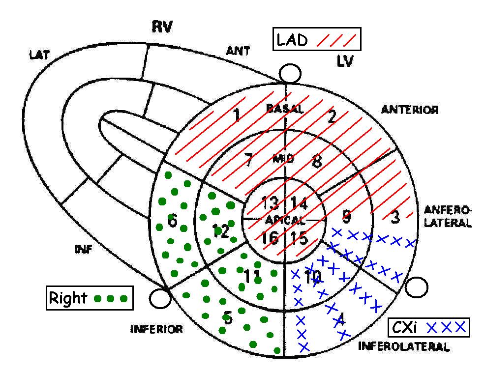
7 LVEF = systolic function Commonly use in management of cardiac patients. Direct correlation between survival and number of hospitalisations post myocardial infarction. Important in decision making for therapeutics - Treatment (ICD) - Cardiac insufficiency - Post myocardial infarction
8 4 The LV systolic function is commonly used in evaluation of cardiac patient There is a direct correlation between number of hospitalisation post MI, survival and LVEF Knowledge of LVEF is necessary in therapeutic decision making such as ICD candidate or therapy for heart failure
9 LVEF / ECHO 1. Mmode: - Teichholz - Quinones 2. 2D : - Planimetric Simpson biplane - Visual estimate 3. Doppler and Mmode : Dumesnil 4. Wall motion score index (Rifkin 1990, r:0.91) (Berning 1994, r:0.93)
10 5 Several methods can be use to assess LVEF, Some using the m-mode such as the Teichholz, Quinones, Other using 2D data such as the Simpson biplane. and visual estimate. The doppler and m-mode allow us to use the Dumesnil technique. As will be shown, the WMSI and the visual method from the WMSI can also be useful

11 Long axis 4 chambers 2 chambers 4 chambers 2 chambers
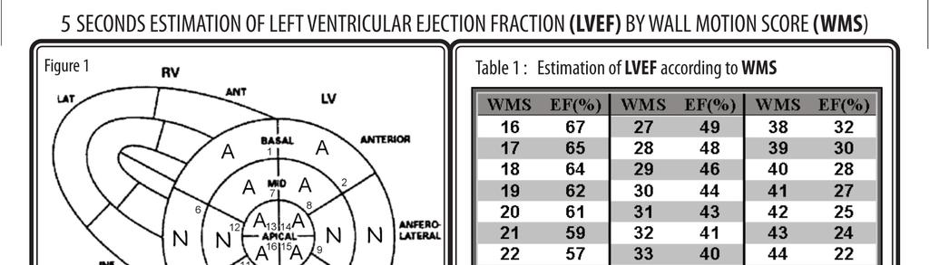
12 6 The polar map is done by evaluating the wall motion in short axis at 3 differents level : the mitral valve, the papillary muscle level and at the apical level. It can all be done in the short axis view but the long axis view and apical (4C - 2C) can also be used.
13
14 7 This graph represent the 16 segments of the polar map and the coronary blood supply to each segment The territory supplie by the LAD, by the Cx and by the RCA are described here.
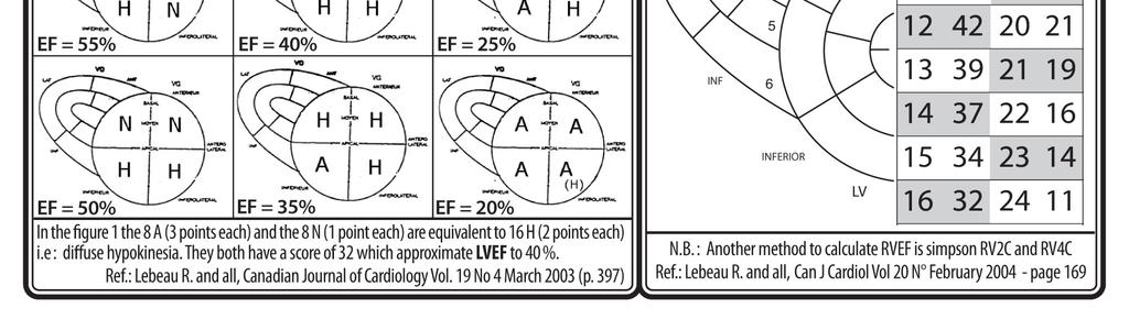
15 Normal(N) = 1 x 8 = 8 Hypokinesia(H) = 2 Akinesia(A) = 3 x 8 = 24 Dyskinesia(D) = 4 Aneuvrysm(AN) = 5 Total = 2
16 8 The WM score is calculated from this polar map by adding all 16 segments. Each segment gets : 1 point if it is normokinetic, 2 point for HK, 3 for AK, 4 for DK and 5 if aneurysmal In this example there are 8 normal segments so we have 8 points, 8 akinetic segments, therefore 24 points. The WMS is therefore = 32 and the WMSI 32/16 = 2.
17 Clinical characteristics of the patients Age (years) Sex 21 to 88 (mean 69) 140 men 103 women 243 pts LV dimension Diastolic 40 to 79 (mean 56) Systolic 20 to 70 (mean 42)
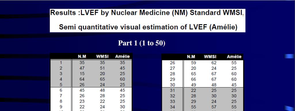
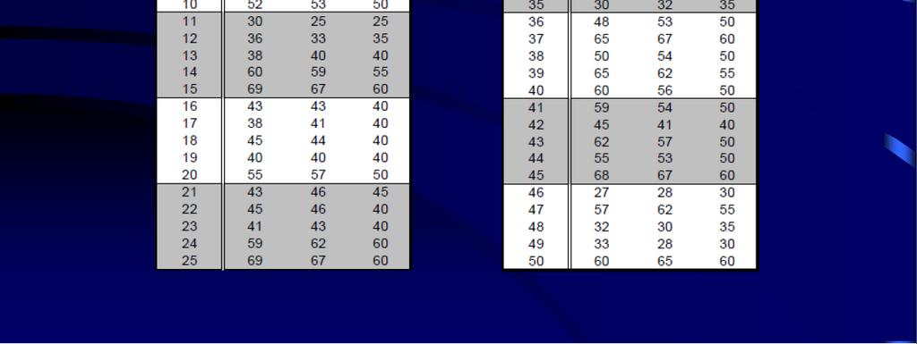
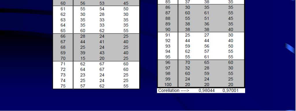
18 9 In our study, we compared the LVEF mesured by RNA and to the WMSI obtained by echo. 243 patients were studied, 140 men and 103 women ranging from 21 to 88 years old. The LV dimension in diastole range from 40 to 79 mm and from 20 to 70 mm in systole. The polar map by echo and the LVEF in nuclear medicine were obtained within 3 days of each other in all 243 patients.
19 Methods 150 pts WMSI vs LVEF-RNA Regression Equation: LVEF-RNA = 92,8 25,8 * WMSI
20 Using the LVEF mesured by RNA and the WMSI from the echo, a regression equation was established. This was done with the data from the first 150 patients.
21 Regression Equation LVEF-RNA = 92,8 25,8 x WMSI 92,8-25,8 x 1,0 = 67 (WMS=16) 92,8-25,8 x 1,6 = 53 (WMS=25) 92,8-25,8 x 2,1 = 40 (WMS=33) 92,8-25,8 x 2,6 = 27 (WMS=41) 92,8-25,8 x 2,9 = 15 (WMS=48) WMS WMSI EF (%) WMS WMSI EF (%) 16 1, , , , , , , , , , , , , , , , , , , , , , , , , , , , , , , , , ,0 15
22 This regression equation allowed us to derive a LVEF from the WMSI and to design this EF table, where each WMSI correspond to a LVEF
23 Methods 150 pts LVEF -WMSI vs LVEF-RNA Regression Equation: 243 pts LV function Echo - NM LVEF-RNA = 92,8 25,8 * WMSI 93pts Regression Equation vs LVEF-RNA 243pts
24 In the susbsequent 93 patients, the LVEF obtained with regression equation (EF Table) was compared to the RNA LVEF
25 Results
26 In the first 150 patients, the correlation between WMSI and RNA EF was 0.82 The correlation between the LVEF obtained by regression equation, in the subsequent 93 patients and RNA was 0.86 Correlation for the whole group of 243 patients was 0.88 If we exclude 20 outliers. (Difference of EF more than 12 % between the two techniques). The correlation between WMSI and RNA EF was 0.92
27 Results
28 In various subgroup including patients with dyskinesis (DK), aneurysm (AN), atrial fibrillation (AF), the correlation remains good. Interobserver and intraobserver variability was good.
29 LVEF-RNA vs other technics LVEF-RNA Teichholz Quinones Simpson Dumesnil Standard score Score plus : 9pts % 9pts % 9pts % 9pts % 9pts % 9pts > 61 % 54 pts mild HK = 1,5 moderate HK = 2,0 severe HK = 2,5
30 We compared the WMSI with other techniques in 54 pts with LVEF ranging from 10-61% We also compared the LVEF by RNA to the LVEF obtained in echo using the Teichholz, Quinones, Simpson,Dumesnil and our score technique. We also use the "score plus" technique where mild HK got 1.5 point, moderate HK 2 points and severe HK 2.5 points
31 WMSI-LVEF vs RNA (54 pts) Results Teichholz Quinones Dumesnil Simpson (r = 0,43) (r = 0,50) (r = 0,83) (r = 0,82) Standard score (r = 0,85) Score plus (r = 0,83) mild HK = 1,5 moderate HK = 2,0 severe HK = 2,5
32 The result in 54 patients showed the best correlation with the Simpsons and Dumesnil technique. The correlation with our score (the standard and the modified "score plus") was equally good with a r value of 0.85 and 0.83
33 Normal(N) = 1 x 8 = 8 Hypokinesia(H) = 2 Akinesia(A) = 3 x 8 = 24 Dyskinesia(D) = 4 Aneuvrysm(AN) = 5 Total = 2
34 This is a typical LAD infarct The WMS score is equal 32 WMSI is equal 2.0
35 Estimation of left ventricular ejection fraction according to wall motion score index WMS WMSI EF (%) WMS WMSI EF (%) 16 1, , , , , , , , , , , , , , , , , , , , , , , , , , , , , , , , , ,0 15
36 The WMSI is 2 and the LVEF is 41% using our regression equation and EF table
37 Normal(N) = 1 x 8 = 8 Hypokinesia(H) = 2 Akinesia(A) = 3 x 8 = 24 Dyskinesia(D) = 4 Aneuvrysm(AN) = 5 Total = Normal(N) = 0 Hypokinesia(H) = 1 Akinesia(A) = 2 Dyskinesia(D) = 3 Aneuvrysm(AN) = 4
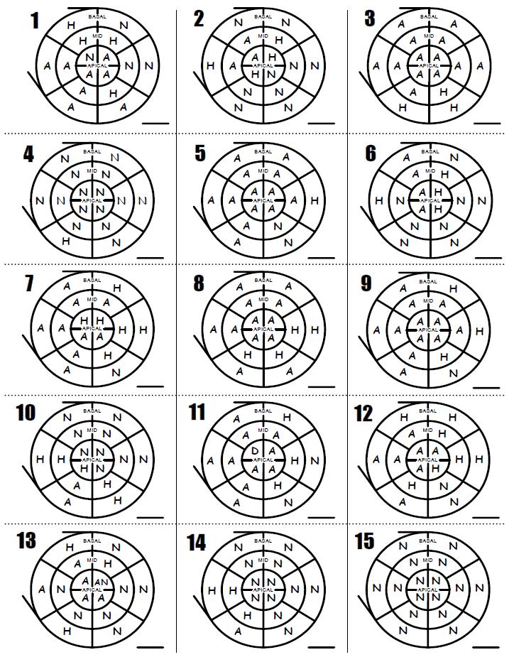
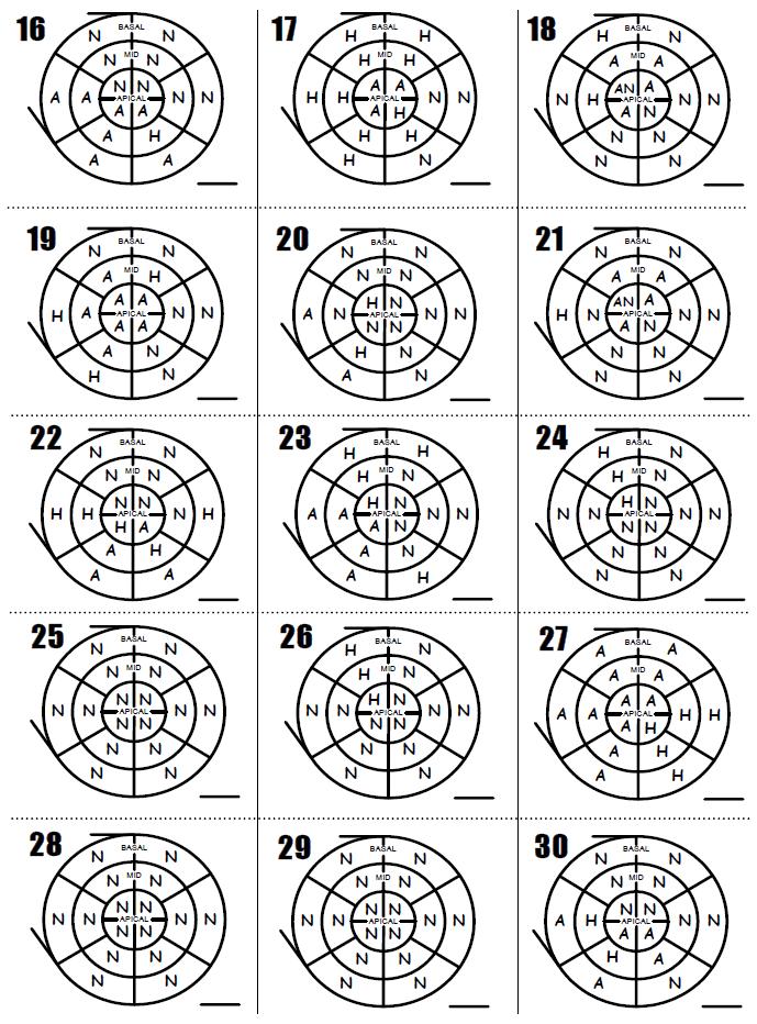
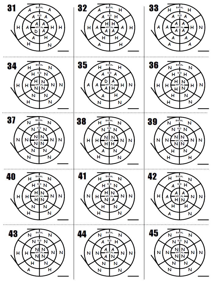
38 If we want to use a fast calculation system We begin our calculation at 16 then adding 1 for every HK segment and 2 for every akinetic segments going in a clockwise rotation. The normal segment get no additional points
39 Fast calculation Normal(N) = 1 Hypokinesia(H) = 2 Akinesia(A) = 3 Dyskinesia(D) = 4 Aneuvrysm(AN) = 5 Normal(N) = 0 Hypokinesia(H) = 1 Akinesia(A) = 2 Dyskinesia(D) = 3 Aneuvrysm(AN) = 4
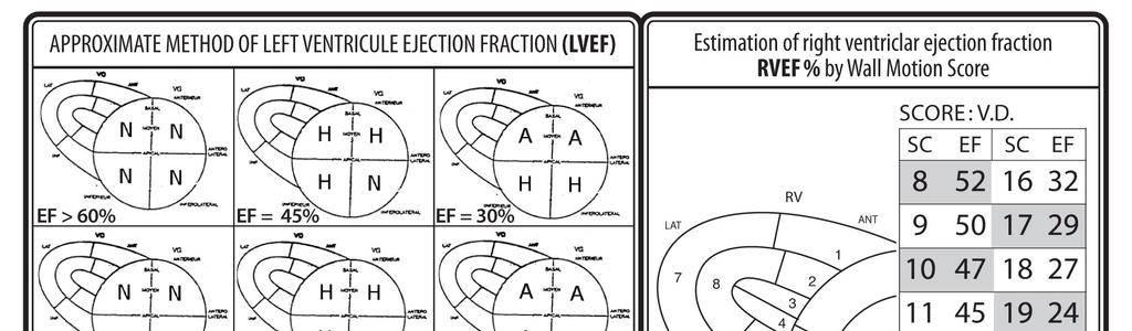
40 The total score is 32 / 16 The LVEF is 41% Try to do a Biplane Simpson in (4C-2C) in 5 seconds.
41 The goal of our study is to have a visual estimation not dependant of EF table.
42 Part 2 Semi quantitative visual estimation of LVEF from WMSI map - How to do it - Study and results
43 I developped a semiquantitative visual estimation of LVEF from the WMSI map even if we don t do the score calculation
44
45 This is done by pattern recognition and it is easy to do and is possible since there is a linear correlation between WMSI and LVEF obtained in nuclear medicine

46 Estimation of left ventricular ejection fraction according to wall motion score index
47 This linear correlation is confirm by the EF table. In this table of LVEF we can have 3 subgroup, those with high EF, those with intermediate or low EF The premise behind this rapid method is that if all segments are normal the LVEF is greater or equal to 60% If all are HK, it is 40% If all are AK, it is equal or less than 20%
48
49 This drawings represent several possibility For example, if all segment are HK the LVEF is 40% If half of the segments are NK and half HK the LVEF is 50% (mild hypokinesia) If half of the segments are HK and half AK the LVEF is 30% (severe hypokinesia)
50 Method A + N = H, H (3 + 1) = (2 + 2) D + H = A, A (4 + 2) = (3 + 3) D + N + N = H, H, H AN + N = A, A (5+1) = (3+3) AN + H + H = A, A, A AN + N + N + N = H, H, H, H
51 The way to do this simplification. If you have one normal segment and one akinetic segment, it is equal to 2 Hk segments. A dyskinetic segment and one hypokinetic segments equal 2 akinetic segments.
52 Normal(N) = 1 Hypokinesia(H) = 2 Akinesia(A) = 3 Dyskinesia(D) = 4 Aneuvrysm(AN) = 5 WMS-EF = 41%
53 Normal(N) = 1 Hypokinesia(H) = 2 Akinesia(A) = 3 Dyskinesia(D) = 4 Aneuvrysm(AN) = 5 WMS-EF = 41%
54 Normal(N) = 1 Hypokinesia(H) = 2 Akinesia(A) = 3 Dyskinesia(D) = 4 Aneuvrysm(AN) = 5 WMS-EF = 41%
55 This map represent a typical anterior. If we have one akinetic and one normal segment we can scratch them off and replace them by 2 HK segments.
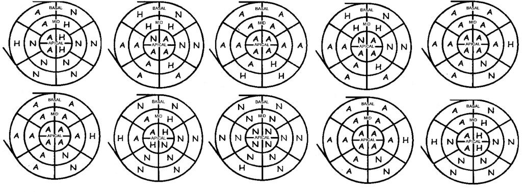
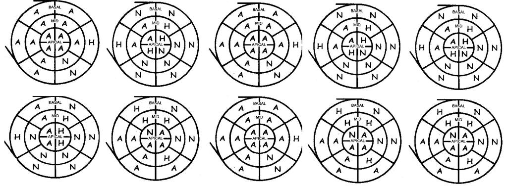
56 Normal(N) = 1 Hypokinesia(H) = 2 Akinesia(A) = 3 Dyskinesia(D) = 4 Aneuvrysm(AN) = 5 APPR = 40%
57 In this graph we would end up with all 16 segments being HK and a LVEF of 40% The LVEF by the standard method is 41% and 40% by semi quantitative visual estimate.
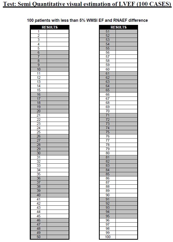
58 Normal(N) = 1 Hypokinesia(H) = 2 Akinesia(A) = 3 Dyskinesia(D) = 4 Aneuvrysm(AN) = 5 WMS-EF = 53%
59 This map represent a typical inferior infarct. Again we combine one akinetic and one normal segment and replace them with 2 HK segments.
60 Normal(N) = 1 Hypokinesia(H) = 2 Akinesia(A) = 3 Dyskinesia(D) = 4 Aneuvrysm(AN) = 5 APPR = 50%
61 In this graph we would end up with all 8 normal segments and 8 HK segments a LVEF of 50% The LVEF by the standard method is 53% and 50% by semi quantitative visual estimate.
62
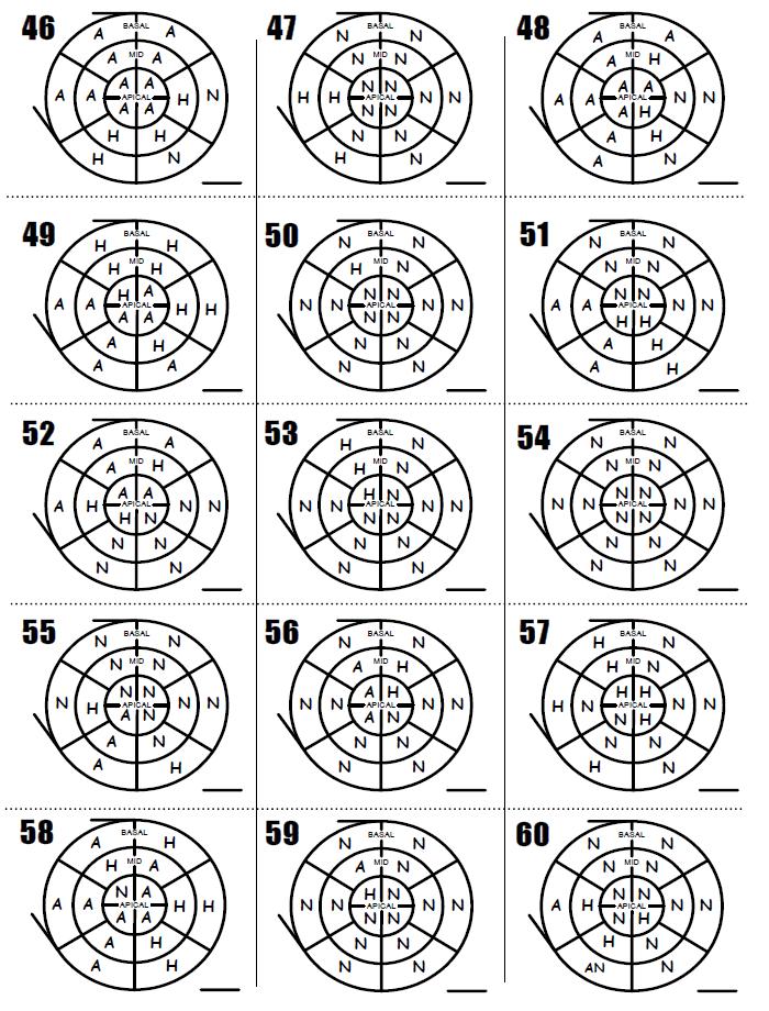
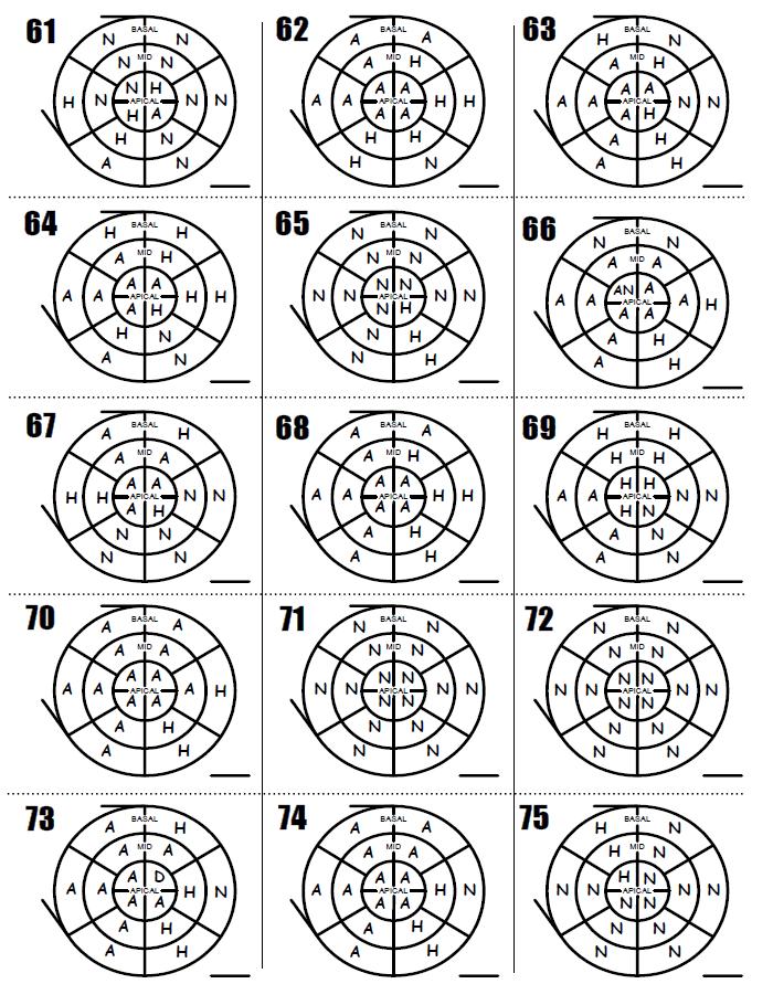
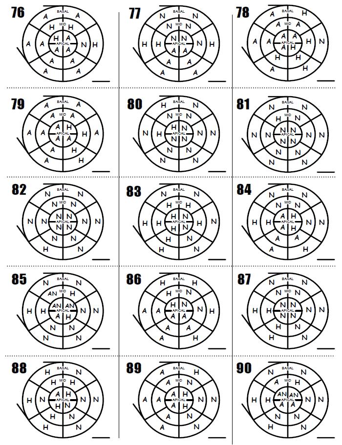
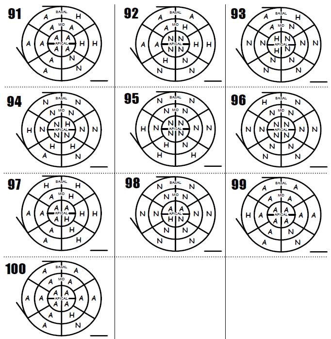
63 We gave 243 polar map to our participants and asked them to derive the LVEF.
64
65 It was also done in 100 polars maps with the 17 segments model.
66 16 segments model (243pts) observers RNA Echocardiographers (2) r: 0,87 S.D.: 12 Sonographers (3) r: 0,86 S.D.: 13 Fellows (3) r: 0,85 S.D.: 14 Outsiders (3) r: 0,86 S.D.: segments model (100pts) observers RNA Echocardiographers (2) r: 0,88 S.D.: 8 Outsiders (3) r: 0,88 S.D.: 8
67 The results are in the table The echocardiographer had a correlation of 0.87 compared to RNA The sonographers 0.86 The fellows 0.85 The outsider, one 16 y.o high school student, a university student in business and one in psychology also have a good correlation with RNA showing that even without medical formation you can succes in this test.
68 Conclusion Semiquantitative visual estimation of LVEF is a simple, rapid and safe method to evaluate and teach to fellows in cardiology
69 In conclusion, the semivisual estimation of LVEF is simple, rapid, accurate and easy method to teach to residents and technicians in transthoracic echocardiography.
70 If you want to test you ability to do this rapid semiquantitative evaluation of LVEF, you will find 100 cases on our internet site Thank you!
71
72
73
74
75
76
77
78
79
80
81
82
83
84 Comments 1. If you use mild hypokinesia, you can use a score of 1.5. If you use severe hypokinesia, you can use a score of 2.5. The maximal LVEF with the score technique is 67 % and the minimal is 15 %. You can use the biplane Simpson method for hyperkinetic state. If you use the 17 segment model, use only the first 16 segments in the calculation. 2. Lebeau Score D : for our anesthesiologists we compared a third method to RNA. Using 243 patients from our study A new tool for estimating LVEF derived from WMSI ( Lebeau. R. and all, Canadian Journal of Cardiology Vol 19, no4, mars 2003 (p.397) we found a new score derived from the WMSI regression formula (score-d).: Normal = 4 %, Mild Hypokinesia= 3.3 %, Moderate Hypokinesia=2.5 %, Severe Hypokinesia=1.8 % and Akinesia=1 % for each of the 16 segments model (R=0.89) and 17 segments model (R=0.90) to evaluated the LVEF. We simply add the total % score of the 16 or 17 segments model. N.B. Dyskinetic segment = -1% and aneurysmal segment = -2%. The lowest LVEF reported should be 15%. 3. Test yourself : compare your visual, Simpson Biplane and score method ( WMSI and score D) in ten of your patients.
85 References LEBEAU R, SERRI K, MORICE MC, HOVASSE T, UNTERSEEH T, PIÉCHAUD JF, GAROT J., Assessment of left ventricular ejection fraction using the wall motion score index in cardiac magnetic resonance imaging, Arch Cardiovasc Dis Feb;105(2):91-8. Epub 2012 Feb 22. Lebeau R, Potter BJ, Sas G, et al. Performance of a simplified wall motion score index method for non-cardiologists to assess left ventricular ejection fraction. ISRN Emergency Medicine Volume 2012: article ID Lebeau R, Di Lorenzo M, Sauvé C et al. Two-dimensionnel echocardiographic estimation of right ventricular ejection fraction by wall motion score index. Canadian Journal of Cardiology. Pulsus Group Inc. Volume 20,no2, February 2004 p
Questions on Chamber Quantitation
 Questions on Chamber Quantitation @RobertoMLang Which of the following statements is true? 1. The aortic annulus should be measured in midsystole. 2. The aortic annulus should be measured in enddiastole.
3/27/2014. Introduction.
 Introduction. Myocardial perfusion & contractility becomes abnormal immediately after the onset of ischaemia, even before the development of the symptoms & ST segment changes. 1 Myocardial Wall Motion
Stephen Glen ISCHAEMIC HEART DISEASE AND LEFT VENTRICULAR FUNCTION
 Stephen Glen ISCHAEMIC HEART DISEASE AND LEFT VENTRICULAR FUNCTION Overview Coronary arteries Terminology to describe contractility Measuring ventricular function Systolic dysfunction Practice cases- LV
10/7/2013. Systolic Function How to Measure, How Accurate is Echo, Role of Contrast. Thanks to our Course Director: Neil J.
 Systolic Function How to Measure, How Accurate is Echo, Role of Contrast Neil J. Weissman, MD MedStar Health Research Institute & Professor of Medicine Georgetown University Washington, D.C. No Disclosures
Research Article Performance of a Simplified Wall Motion Score Index Method for Noncardiologists to Assess Left Ventricular Ejection Fraction
 International Scholarly Research Network ISRN Emergency Medicine olume 2012, Article ID 309470, pages doi:.402/2012/309470 Research Article Performance of a Simplified Wall Motion Score Index Method for
LV FUNCTION ASSESSMENT: WHAT IS BEYOND EJECTION FRACTION
 LV FUNCTION ASSESSMENT: WHAT IS BEYOND EJECTION FRACTION Jamilah S AlRahimi Assistant Professor, KSU-HS Consultant Noninvasive Cardiology KFCC, MNGHA-WR Introduction LV function assessment in Heart Failure:
Rotation: Echocardiography: Transthoracic Echocardiography (TTE)
") Rotation: Echocardiography: Transthoracic Echocardiography (TTE) Rotation Format and Responsibilities: Fellows rotate in the echocardiography laboratory in each clinical year. Rotations during the first
Rotation: Echocardiography: Transthoracic Echocardiography (TTE) Rotation Format and Responsibilities: Fellows rotate in the echocardiography laboratory in each clinical year. Rotations during the first
Correlation Between Regional Wall Motion Abnormalities via 2-Dimensional Echocardiography, and Coronary Angiographic Findings
 THE ECHOCARDIOGRAPHY, IRAQI POSTGRADUATE MEDICAL AND CORONARY JOURNAL ANGIOGRAPHIC FINDINGS VOL.11, SUPPLEMENT,2012 Correlation Between Regional Wall Motion Abnormalities via 2-Dimensional Echocardiography,
Role of echocardiography in the assessment of ischemic heart disease 분당서울대학교병원윤연이
 Role of echocardiography in the assessment of ischemic heart disease 분당서울대학교병원윤연이 Outline Evaluation of Chest pain Evaluation of MI complications Prediction of Outcomes Evaluation of Chest pain Evaluation
Chamber Quantitation Guidelines: What is New?
 Chamber Quantitation Guidelines: What is New? Roberto M Lang, MD J AM Soc Echocardiogr 2005; 18:1440-1463 1 Approximately 10,000 citations iase in itune Cardiac Chamber Quantification: What is New? Database
Contrast-enhanced echocardiography improves agreement on the assessment of ejection fraction and left ventricular function. A multicentre study
 Eur J Echocardiography 7 Suppl. 2 (2006) S16 S21 Contrast-enhanced echocardiography improves agreement on the assessment of ejection fraction and left ventricular function. A multicentre study Rainer Hoffmann*
좌심실수축기능평가 Cardiac Function
 Basic Echo Review Course 좌심실수축기능평가 Cardiac Function Seonghoon Choi Cardiology Hallym university LV systolic function Systolic function 좌심실수축기능 - 심근의수축으로심실에서혈액을대동맥으로박출하는기능 실제임상에서 LV function 의의미 1Diagnosis
Echo in CAD: Wall Motion Assessment
 Echo in CAD: Wall Motion Assessment Joe M. Moody, Jr, MD UTHSCSA and STVHCS October 2007 Relevant References ACC/AHA/ASE 2003 Guideline Update for the Clinical Application of Echocardiography Bayes de
Basic Assessment of Left Ventricular Systolic Function
 WINFOCUS BASIC ECHO (WBE) Basic Assessment of Left Ventricular Systolic Function Ritesh Dhar, MD Director, Echocardiography Lab and Staff Cardiologist Intermountain Medical Center Murray, Utah Outline
Role of Early 2D Echocardiography in Patient with Acute Myocardial Infarction in Correlation with Electrocardiography and Clinical Presentation
 MVP Journal of Medical Sciences, Vol 1(2), 51 55, July 2014 Role of Early 2D Echocardiography in Patient with Acute Myocardial Infarction in Correlation with Electrocardiography and Clinical Presentation
Noncoronary Cardiac MDCT
 Noncoronary Cardiac MDCT David A. Bluemke, M.D., Ph.D. Professor, of Radiology and Medicine Johns Hopkins University School of Medicine Baltimore, Maryland Toshiba Disclosures Grant support Noncoronary
Global left ventricular circumferential strain is a marker for both systolic and diastolic myocardial function
 Global left ventricular circumferential strain is a marker for both systolic and diastolic myocardial function Toshinari Onishi 1, Samir K. Saha 2, Daniel Ludwig 1, Erik B. Schelbert 1, David Schwartzman
MAYON VOLCANO: FAST FACTS
 MAYON VOLCANO: FAST FACTS Type of Volcano: Stratovolcano Elevation: 2.46 km Base Diameter: 20 km Base Circumference: 62.8 km Area: 314.1 km 2 Reference: http://www.phivolcs.dost.gov.ph/html/update_vmepd/volcano/volcanolist/mayon.htm
Radiologic Assessment of Myocardial Viability
 November 2001 Radiologic Assessment of Myocardial Viability Joshua Moss, Harvard Medical School Year III Patient EF 66yo female with a 3-year history of intermittent chest pain previously relieved by sublingual
Title:Relation Between E/e' ratio and NT-proBNP Levels in Elderly Patients with Symptomatic Severe Aortic Stenosis
 Author's response to reviews Title:Relation Between E/e' ratio and NT-proBNP Levels in Elderly Patients with Symptomatic Severe Aortic Stenosis Authors: Mihai Strachinaru (m.strachinaru@erasmusmc.nl) Bas
2/2/2011. Strain and Strain Rate Imaging How, Why and When? Movement vs Deformation. Doppler Myocardial Velocities. Movement. Deformation.
 Strain and Strain Rate Imaging How, Why and When? João L. Cavalcante, MD Advanced Cardiac Imaging Fellow Cleveland Clinic Foundation Disclosures: No conflicts of interest Movement vs Deformation Movement
Quantification of Cardiac Chamber Size
 2017 KSE 2017-11-25 Quantification of Cardiac Chamber Size Division of Cardiology Keimyung University Dongsan Medical Center In-Cheol Kim M.D., Ph.D. LV size and function Internal linear dimensions PLX
Certificate in Clinician Performed Ultrasound (CCPU) Syllabus. Rapid Cardiac Echo (RCE)
 Syllabus. Rapid Cardiac Echo (RCE)") Certificate in Clinician Performed Ultrasound (CCPU) Syllabus Rapid Cardiac Echo (RCE) Purpose: Rapid Cardiac Echocardiography (RCE) This unit is designed to cover the theoretical and practical curriculum
Certificate in Clinician Performed Ultrasound (CCPU) Syllabus Rapid Cardiac Echo (RCE) Purpose: Rapid Cardiac Echocardiography (RCE) This unit is designed to cover the theoretical and practical curriculum
Manuscript submitted for review to Echo Research and Practice
 Manuscript submitted for review to Echo Research and Practice Left ventricular ejection fraction assessment by noncardiologists using a simplified wall motion score index Journal: Echo Research and Practice
Heart Failure in Women: Dr Goh Ping Ping Cardiologist Asian Heart & Vascular Centre
 Heart Failure in Women: More than EF? Dr Goh Ping Ping Cardiologist Asian Heart & Vascular Centre Overview Review pathophysiology as it relates to diagnosis and management Rational approach to workup:
Top 10 Facts in Contrast Echocardiography. Pamela R. Burgess, BS, RDCS, RDMS, RVT, FASE
 Top 10 Facts in Contrast Echocardiography Pamela R. Burgess, BS, RDCS, RDMS, RVT, FASE Presenter Disclosure The following relationship exist related to this presentation: Pamela R. Burgess, BS, RDCS, RDMS,
Velocity Vector Imaging as a new approach for cardiac magnetic resonance: Comparison with echocardiography
 Velocity Vector Imaging as a new approach for cardiac magnetic resonance: Comparison with echocardiography Toshinari Onishi 1, Samir K. Saha 2, Daniel Ludwig 1, Erik B. Schelbert 1, David Schwartzman 1,
Imaging and heart failure
 Imaging and heart failure Jeroen J Bax Dept of Cardiology Leiden Univ Medical Center The Netherlands Davos, feb 2013 Research grants: Medtronic, Biotronik, Boston, St Jude, BMS imaging, GE Healthcare,
Use of Nuclear Cardiology in Myocardial Viability Assessment and Introduction to PET and PET/CT for Advanced Users
 Use of Nuclear Cardiology in Myocardial Viability Assessment and Introduction to PET and PET/CT for Advanced Users February 1 5, 2011 University of Santo Tomas Hospital Angelo King A-V Auditorium Manila,
Strain and Strain Rate Imaging How, Why and When?
 Strain and Strain Rate Imaging How, Why and When? João L. Cavalcante, MD Advanced Cardiac Imaging Fellow Cleveland Clinic Foundation Disclosures: No conflicts of interest Movement vs Deformation Movement
Echocardiographic Cardiovascular Risk Stratification: Beyond Ejection Fraction
 Echocardiographic Cardiovascular Risk Stratification: Beyond Ejection Fraction October 4, 2014 James S. Lee, M.D., F.A.C.C. Associates in Cardiology, P.A. Silver Spring, M.D. Disclosures Financial none
Evaluation of the Right Ventricle and Risk Stratification for Sudden Cardiac Death
 Evaluation of the Right Ventricle and Risk Stratification for Sudden Cardiac Death Presenters: Sabrina Phillips, MD FACC FASE Director, Adult Congenital Heart Disease Services The University of Oklahoma
2019 Qualified Clinical Data Registry (QCDR) Performance Measures
 Performance Measures") 2019 Qualified Clinical Data Registry (QCDR) Performance Measures Description: This document contains the 18 performance measures approved by CMS for inclusion in the 2019 Qualified Clinical Data Registry
2019 Qualified Clinical Data Registry (QCDR) Performance Measures Description: This document contains the 18 performance measures approved by CMS for inclusion in the 2019 Qualified Clinical Data Registry
Degenerative Mitral Regurgitation: Etiology and Natural History of Disease and Triggers for Intervention
 Degenerative Mitral Regurgitation: Etiology and Natural History of Disease and Triggers for Intervention John N. Hamaty D.O. FACC, FACOI November 17 th 2017 I have no financial disclosures Primary Mitral
Evaluation of Systolic Function of the Left Ventricle
 Evaluation of Systolic Function of the Left Ventricle Roxy Senior MD DM FRCP FESC FACC and Vinay Kumar Bhatia PhD MRCP Department of Cardiovascular Medicine, Northwick Park Hospital and Institute for Medical
Cardiac Chamber Quantification by Echocardiography
 Cardiac Chamber Quantification by Echocardiography Maryam Bokhamseen, RCS, RCDS, EACVI Echotechnologist ǁ, Non invasive Cardiac Laboratory King Abdulaziz Cardiac Center. Outline: Introduction. Background
Echocardiographic assessment of left ventricular function in patients of acute myocardial infarction
 International Journal of Advances in Medicine Anjali VS et al. Int J Adv Med. 2017 Aug;4(4):926-931 http://www.ijmedicine.com pissn 2349-3925 eissn 2349-3933 Original Research Article DOI: http://dx.doi.org/10.18203/2349-3933.ijam20173070
Coronary artery disease (CAD) risk factors
 risk factors") Background Coronary artery disease (CAD) risk factors CAD Risk factors Hypertension Insulin resistance /diabetes Dyslipidemia Smoking /Obesity Male gender/ Old age Atherosclerosis Arterial stiffness precedes
Background Coronary artery disease (CAD) risk factors CAD Risk factors Hypertension Insulin resistance /diabetes Dyslipidemia Smoking /Obesity Male gender/ Old age Atherosclerosis Arterial stiffness precedes
Case 47 Clinical Presentation
 93 Case 47 C Clinical Presentation 45-year-old man presents with chest pain and new onset of a murmur. Echocardiography shows severe aortic insufficiency. 94 RadCases Cardiac Imaging Imaging Findings C
Segmental Tissue Doppler Image-Derived Tei Index in Patients With Regional Wall Motion Abnormalities
 ORIGINAL ARTICLE DOI 10.4070 / kcj.2010.40.3.114 Print ISSN 1738-5520 / On-line ISSN 1738-5555 Copyright c 2010 The Korean Society of Cardiology Open Access Segmental Tissue Doppler Image-Derived Tei Index
Echocardiography as a diagnostic and management tool in medical emergencies
 Echocardiography as a diagnostic and management tool in medical emergencies Frank van der Heusen MD Department of Anesthesia and perioperative Care UCSF Medical Center Objective of this presentation Indications
Contemporary Echocardiography. Non-ST Elevation. Myocardial Infarction
 Contemporary Echocardiography In Non-ST Elevation Myocardial Infarction Nicola Jayne Smith Student ID: 1379396 Master of Health Science (MHSc) Unitec Institute of Technology 2013 1 Abstract Background:
Feasibility and limitations of 2D speckle tracking echocardiography
 ORIGINAL ARTICLE 204 A prospective study in daily clinical practice Feasibility and limitations of 2D speckle tracking echocardiography Lina Melzer, Anja Faeh-Gunz, Barbara Naegeli, Burkhardt Seifert*,
Adult Echocardiography Examination Content Outline
 Adult Echocardiography Examination Content Outline (Outline Summary) # Domain Subdomain Percentage 1 2 3 4 5 Anatomy and Physiology Pathology Clinical Care and Safety Measurement Techniques, Maneuvers,
Research Presentation June 23, Nimish Muni Resident Internal Medicine
 Research Presentation June 23, 2009 Nimish Muni Resident Internal Medicine Research Question In adult patients with repaired Tetralogy of Fallot, how does Echocardiography compare to MRI in evaluating
VECTORS OF CONTRACTION
 1/3/216 Strain, Strain Rate, and Torsion: Myocardial Mechanics Simplified and Applied VECTORS OF CONTRACTION John Gorcsan, MD University of Pittsburgh, Pittsburgh, PA Shortening Thickening Twisting No
Evaluation of Left Ventricular Function and Hypertrophy Gerard P. Aurigemma MD
 Evaluation of Left Ventricular Function and Hypertrophy Gerard P. Aurigemma MD Board Review Course 2017 43 year old health assistant Severe resistant HTN LT BSA 2 Height 64 1 Here is the M mode echocardiogram
Coronary Artery Bypass Graft: Monitoring Patients and Detecting Complications
 Coronary Artery Bypass Graft: Monitoring Patients and Detecting Complications Madhav Swaminathan, MD, FASE Professor of Anesthesiology Division of Cardiothoracic Anesthesia & Critical Care Duke University
Typical chest pain with normal ECG
 Typical chest pain with normal ECG F. Mut, C. Bentancourt, M. Beretta Nuclear Medicine Service, Asociacion Española Montevideo, Uruguay Clinical history Male 41 y.o. Overweight, hypertension, high cholesterol,
Abnormal, Autoquant Adenosine Myocardial Perfusion Heart Imaging. ID: GOLD Date: Age: 46 Sex: M John Doe Phone (310)
") Background: Reason: preoperative assessment of CAD, Shortness of Breath Symptom: atypical chest pain Risk factors: hypertension Under influence: a beta blocker Medications: digoxin Height: 66 in. Weight:
Background: Reason: preoperative assessment of CAD, Shortness of Breath Symptom: atypical chest pain Risk factors: hypertension Under influence: a beta blocker Medications: digoxin Height: 66 in. Weight:
The new Guidelines: Focus on Chronic Heart Failure
 The new Guidelines: Focus on Chronic Heart Failure Petros Nihoyannopoulos MD, FRCP, FESC Professor of Cardiology Imperial College London and National & Kapodistrian University of Athens 2 3 4 The principal
Impaired Regional Myocardial Function Detection Using the Standard Inter-Segmental Integration SINE Wave Curve On Magnetic Resonance Imaging
 Original Article Impaired Regional Myocardial Function Detection Using the Standard Inter-Segmental Integration Ngam-Maung B, RT email : chaothawee@yahoo.com Busakol Ngam-Maung, RT 1 Lertlak Chaothawee,
ORIGINAL. Keywords : multidetector-row computed tomography, myocardial infarction, cardiac function
 ORIGINAL Department of Digestive and Cardiovascular Medicine, Institute of Health Biosciences, The University of Tokushima Graduate School, Tokushima, Japan, Faculty of Integrated Art and Sciences, Department
CABG Surgery following STEMI
 CABG Surgery following STEMI Susana Harrington, MS,APRN-NP Cardio-Thoracic Surgery Nebraska Methodist Hospital February 15, 2018 2013 ACCF/AHA Guideline for the Management of ST-Elevation Myocardial Infarction:
Index. K Knobology, TTE artifact, image resolution, ultrasound, 14
 A Acute aortic regurgitation (AR), 124 128 Acute aortic syndrome (AAS) classic aortic dissection diagnosis, 251 263 evolutive patterns, 253 255 pathology, 250 251 classifications, 247 248 incomplete aortic
Revealing new insights. irotate electronic rotation and xplane adjustable biplane imaging. Ultrasound cardiology. irotate and xplane
 Ultrasound cardiology irotate and xplane Revealing new insights irotate electronic rotation and xplane adjustable biplane imaging Annemien van den Bosch and Jackie McGhie Department of Cardiology, Erasmus
Quantifying LV function how good are we?
 Quantifying LV function how good are we? Professor Alan G Fraser Wales Heart Research Institute Cardiff University, U.K. Support for research from Hitachi Aloka, & GE Ultrasound Visual assessment of synchronicity
Left atrial function. Aliakbar Arvandi MD
 In the clinic Left atrial function Abstract The left atrium (LA) is a left posterior cardiac chamber which is located adjacent to the esophagus. It is separated from the right atrium by the inter-atrial
Cardiac MRI in ACHD What We. ACHD Patients
 Cardiac MRI in ACHD What We Have Learned to Apply to ACHD Patients Faris Al Mousily, MBChB, FAAC, FACC Consultant, Pediatric Cardiology, KFSH&RC/Jeddah Adjunct Faculty, Division of Pediatric Cardiology
How to Approach the Patient with CRT and Recurrent Heart Failure
 How to Approach the Patient with CRT and Recurrent Heart Failure Byron K. Lee MD Associate Professor of Medicine Electrophysiology and Arrhythmia Section UCSF Update in Electrocardiography and Arrhythmias
Common Codes for ICD-10
 Common Codes for ICD-10 Specialty: Cardiology *Always utilize more specific codes first. ABNORMALITIES OF HEART RHYTHM ICD-9-CM Codes: 427.81, 427.89, 785.0, 785.1, 785.3 R00.0 Tachycardia, unspecified
Cardiovascular hemodynamics in the stress echo lab with open-source software
 Cardiovascular hemodynamics in the stress echo lab with open-source software T. Bombardini, D. Cini, E. Picano Institute of Clinical Physiology of CNR, Pisa, Italy no conflict of interest Background Stress
Cardiac Magnetic Resonance in pregnant women
 Cardiac Magnetic Resonance in pregnant women Chen SSM, Leeton L, Dennis AT Royal Women s Hospital and The University of Melbourne, Parkville, Australia alicia.dennis@thewomens.org.au Quantification of
Diagnostic Imaging Utilization Management and Consultation Management Programs Imaging Code Listing for Connecticut, Maine and New Hampshire
 Diagnostic Imaging Utilization Management and Consultation Management Programs Imaging Code Listing for Connecticut, Maine and New Hampshire The grid below contains the CPT * codes that are subject to
Appendix II: ECHOCARDIOGRAPHY ANALYSIS
 Appendix II: ECHOCARDIOGRAPHY ANALYSIS Two-Dimensional (2D) imaging was performed using the Vivid 7 Advantage cardiovascular ultrasound system (GE Medical Systems, Milwaukee) with a frame rate of 400 frames
Pathophysiology and Current Evidence for Detection of Dyssynchrony
 Editorial Cardiol Res. 2017;8(5):179-183 Pathophysiology and Current Evidence for Detection of Dyssynchrony Michael Spartalis a, d, Eleni Tzatzaki a, Eleftherios Spartalis b, Christos Damaskos b, Antonios
Outline. EuroScore II. Society of Thoracic Surgeons Score. EuroScore II
 SURGICAL RISK IN VALVULAR HEART DISEASE: WHAT 2D AND 3D ECHO CAN TELL YOU AND WHAT THEY CAN'T Ernesto E Salcedo, MD Professor of Medicine University of Colorado School of Medicine Director of Echocardiography
Quantitation of right ventricular dimensions and function
 SCCS Basics of cardiac assessment Quantitation of right ventricular dimensions and function Tomasz Kukulski, MD PhD Dept of Cardiology, Congenital Heart Disease and Electrotherapy Silesian Medical University
Strain/Untwisting/Diastolic Suction
 What Is Diastole and How to Assess It? Strain/Untwisting/Diastolic Suction James D. Thomas, M.D., F.A.C.C. Cardiovascular Imaging Center Department of Cardiology Cleveland Clinic Foundation Cleveland,
A STUDY OF LEFT VENTRICULAR DIASTOLIC DYSFUNCTION IN HYPERTENSION Ravi Keerthy M 1
 A STUDY OF LEFT VENTRICULAR DIASTOLIC DYSFUNCTION IN HYPERTENSION Ravi Keerthy M 1 HOW TO CITE THIS ARTICLE: Ravi Keerthy M. A Study of Left Ventricular Diastolic Dysfunction in Hypertension. Journal of
Assessment of LV systolic function
 Tutorial 5 - Assessment of LV systolic function Assessment of LV systolic function A knowledge of the LV systolic function is crucial in the undertanding of and management of unstable hemodynamics or a
Value of echocardiography in chronic dyspnea
 Value of echocardiography in chronic dyspnea Jahrestagung Schweizerische Gesellschaft für /Schweizerische Gesellschaft für Pneumologie B. Kaufmann 16.06.2016 Chronic dyspnea Shortness of breath lasting
EDITOR S PICK CURRENT STATUS OF FULLY AUTOMATED SOFTWARE WITH THREE-DIMENSIONAL ECHOCARDIOGRAPHY FOR THE QUANTIFICATION OF LEFT VENTRICULAR FUNCTION
 ITOR S PICK This paper, courtesy of Yang and Takeuchi, provides a timely and well-considered update on the current status of fully-automated software with three-dimensional echocardiography for quantifying
Basics of Contrast Echocardiography Echo Hawaii 2017
 Basics of Contrast Echocardiography Echo Hawaii 2017 Maryellen Orsinelli, RN, RDCS, FASE Lead Cardiac Sonographer The Ohio State University The Ross Heart Hospital 1 DISCLOSURES NONE 1 OBJECTIVES Indications
Abstract ESC Pisa
 Abstract ESC 82441 Maximal left ventricular mass-to-power output: A novel index to assess left ventricular performance and to predict outcome in patients with advanced heart failure FL. Dini 1, D. Mele
Left Ventricular Wall Resection for Aneurysm and Akinesia due to Coronary Artery Disease: Fifty Consecutive Patients
 Left Ventricular Wall Resection for Aneurysm and Akinesia due to Coronary Artery Disease: Fifty Consecutive Patients Armand A. Lefemine, M.D., Rajagopalan Govindarajan, M.D., K. Ramaswamy, M.D., Harrison
Imaging in Heart Failure: A Multimodality Approach. Thomas Ryan, MD
 Imaging in Heart Failure: A Multimodality Approach Thomas Ryan, MD Heart Failure HFrEF HFpEF EF50% Lifetime risk 20% Prevalence 6M Americans Societal costs - $30B 50% 5-year survival 1 Systolic
Acute impairment of basal left ventricular rotation but not twist and untwist are involved in the pathogenesis of acute hypertensive pulmonary oedema
 Acute impairment of basal left ventricular rotation but not twist and untwist are involved in the pathogenesis of acute hypertensive pulmonary oedema A.D. Margulescu 1,2, R.C. Sisu 1,2, M. Florescu 2,
Title: Second-Opinion Stress Tele-Echocardiography for Aged Donor Heart Selection
 Author's response to reviews Title: Second-Opinion Stress Tele-Echocardiography for Aged Donor Heart Selection Authors: Daniele Franchi (franchid@ifc.cnr.it) Davide Cini (davide.cini@cnr.it) Giorgio Arpesella
Left ventricle pseudoaneurysm as late postoperative complication of a large apical aneurysm
 CASE REPORT Left ventricle pseudoaneurysm as late postoperative complication of a large apical aneurysm Mariana M. Floria 1, 4, Carmen Elena Pleșoianu 2, 4, Michel Buche 3, Baudouin Marchandise 4, Erwin
Echocardiography for the Electrophysiologist: Day-to-day practice. Emmanuel Fares, MD
 Echocardiography for the Electrophysiologist: Day-to-day practice Emmanuel Fares, MD EP and pacing service, Department of Cardiovascular Medicine, Cairo University Agenda Role of echo in arrhythmia management:
AMERICAN IMAGING MANAGEMENT
 2012 CPT Codes Computerized Tomography (CT) CPT Description Abdomen 74150 CT abdomen; w/o 74160 CT abdomen; with 74170 CT abdomen; w/o followed by Chest 71250 CT thorax; w/o 71260 CT thorax; with 71270
Aortic Stenosis Steven F. Bolling, M.D. Professor of Cardiac Surgery University of Michigan
 Aortic Stenosis - 2011 Steven F. Bolling, M.D. Professor of Cardiac Surgery University of Michigan Aortic Surgery Aortic Stenosis EB CT - Ca++ everywhere! Surgery for Aortic Stenosis 100,000 USA + 100,000
AMERICAN IMAGING MANAGEMENT
 2010 BCBS of Georgia CPT Codes With Grouper Numbers Computerized Tomography (CT) CPT Description Abdomen 74150 CT abdomen; w/o contrast 6 74160 CT abdomen; with contrast 74170 CT abdomen; w/o contrast
LV Function Cardiac Output EPSS
 LV Function Cardiac Output EPSS Mike Mallin, MD Why is LV function important? Systolic Dysfunction is bad... Is it worse? Is it the cause of my patients dyspnea? Does my patient need a inotrope? Why is
Echocardiographic Assessment of the Left Ventricle
 Echocardiographic Assessment of the Left Ventricle Theodora Zaglavara, MD, PhD, BSCI/BSCCT Department of Cardiovascular Imaging INTERBALKAN EUROPEAN MEDICAL CENTER 2015 The quantification of cardiac chamber
THE LEFT ATRIUM HOW CAN ECHO HELP US?
 THE LEFT ATRIUM HOW CAN ECHO HELP US? Dr. Dragos COZMA BACKGROUND Left atrium (LA) dilation can occur in a broad spectrum of cardiovascular diseases including hypertension, left ventricular dysfunction,
Patterns of Left Ventricular Remodeling in Chronic Heart Failure: The Role of Inadequate Ventricular Hypertrophy
 Abstract ESC 82445 Patterns of Left Ventricular Remodeling in Chronic Heart Failure: The Role of Inadequate Ventricular Hypertrophy FL. Dini 1, P. Capozza 1, P. Fontanive 2, MG. Delle Donne 1, V. Santonato
Aortic Regurgitation and Aortic Aneurysm - Epidemiology and Guidelines -
 Reconstruction of the Aortic Valve and Root - A Practical Approach - Aortic Regurgitation and Aortic Aneurysm Wednesday 14 th September - 9.45 Practice must always be founded on sound theory. Leonardo
LA Function analysis Marcia Barbosa Vice Presidente - Brazilian Soc of Cardiology President-elect - Interamerican Soc of Cardiology
 LA Function analysis Marcia Barbosa Vice Presidente - Brazilian Soc of Cardiology President-elect - Interamerican Soc of Cardiology Belo Horizonte Brazil DECLARATION OF CONFLICT OF INTEREST Nothing to
ECHOCARDIOGRAPHY. Patient Care. Goals and Objectives PF EF MF LF Aspirational
 Patient Care Be able to: Perform and interpret basic TTE and X cardiac Doppler examinations Perform and interpret a comprehensive X TTE and cardiac Doppler examination Perform and interpret a comprehensive
PROSTHETIC VALVE BOARD REVIEW
 PROSTHETIC VALVE BOARD REVIEW The correct answer D This two chamber view shows a porcine mitral prosthesis with the typical appearance of the struts although the leaflets are not well seen. The valve
NT-proBNP: Evidence-based application in primary care
 NT-proBNP: Evidence-based application in primary care Associate Professor Rob Doughty The University of Auckland, Auckland City Hospital, Auckland Heart Group NT-proBNP: Evidence in Primary Care The problem
Acute Myocardial Infarction
 Acute Myocardial Infarction Hafeza Shaikh, DO, FACC, RPVI Lourdes Cardiology Services Asst.Program Director, Cardiology Fellowship Associate Professor, ROWAN-SOM Acute Myocardial Infarction Definition:
Cardiovascular Listings. August 25, 2009 Institute of Medicine
 Cardiovascular Listings August 25, 2009 Institute of Medicine Updating the Cardiovascular Listings Laurence Desi, Sr., M.D., M.P.H. Medical Officer Office of Medical Listings Improvement 2 IOM General
E S A O T E. MyLab. Rev.E March Doc # 29B63EN05 STRESS ECHO OPTION
 E S A O T E Rev.E March 2010 MyLab STRESS ECHO OPTION Doc # 29B63EN05 M y L a b A D V A N C E D O P E R A T I O N S S T R E S S E C H O 2 M y L a b A D V A N C E D O P E R A T I O N S STRESS ECHO OPTION
QCVC Committees Scientific Activities Central Hall General Information FAC. SPECT tomography has the advantage of quantifying biventricular volumes.
 QCVC Committees Scientific Activities Central Hall General Information FAC Thematic Units Arrhythmias and Electrophysiology Basic Research Bioengineering and Medical Informatics Cardiac Surgical Intensive
Multiple Gated Acquisition (MUGA) Scanning
 Scanning") Multiple Gated Acquisition (MUGA) Scanning Dmitry Beyder MPA, CNMT Nuclear Medicine, Radiology Barnes-Jewish Hospital / Washington University St. Louis, MO Disclaimers/Relationships Standard of care research
Multiple Gated Acquisition (MUGA) Scanning Dmitry Beyder MPA, CNMT Nuclear Medicine, Radiology Barnes-Jewish Hospital / Washington University St. Louis, MO Disclaimers/Relationships Standard of care research
Three-dimensional Wall Motion Tracking:
 Three-dimensional Wall Motion Tracking: A Novel Echocardiographic Method for the Assessment of Ventricular Volumes, Strain and Dyssynchrony Jeffrey C. Hill, BS, RDCS, FASE Jennifer L. Kane, RCS Gerard
Ref 1. Ref 2. Ref 3. Ref 4. See graph
 Ref 1 Ref 2 Ref 3 1. Ages 6-23 y/o 2. Significant LVM differences by gender 3. For males 95 th percentiles: a. LVM/BSA = 103 b. LVM/height = 100 4. For females 95 th percentiles: a. LVM/BSA = 84 b. LVM/height