ORIGINAL. Keywords : multidetector-row computed tomography, myocardial infarction, cardiac function
|
|
|
- Shon Nichols
- 5 years ago
- Views:
Transcription
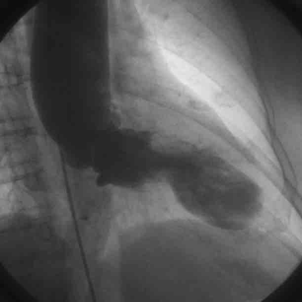
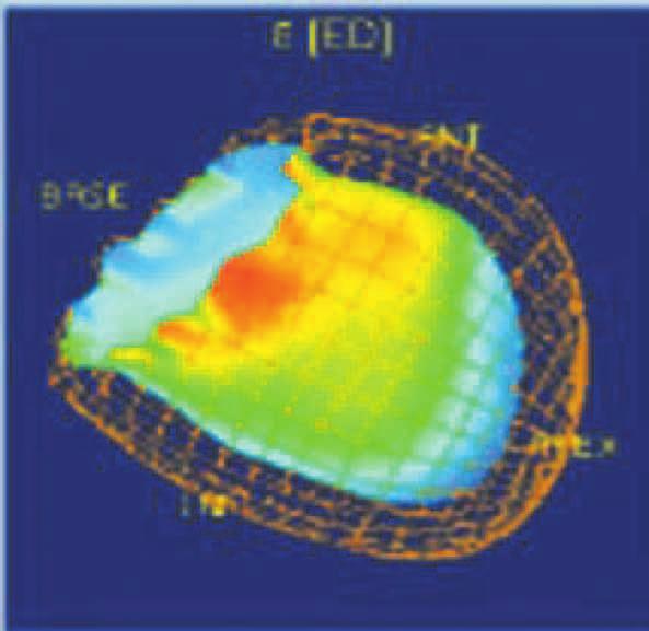
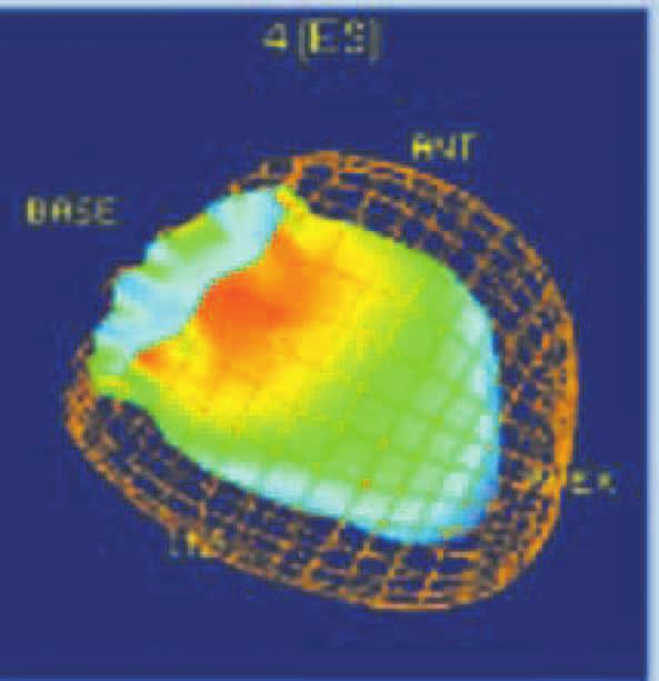
1 ORIGINAL Department of Digestive and Cardiovascular Medicine, Institute of Health Biosciences, The University of Tokushima Graduate School, Tokushima, Japan, Faculty of Integrated Art and Sciences, Department of Human and Social Sciences, The University of Tokushima, Tokushima, Japan ;and Department of Radiology, Institute of Health Biosciences, The University of Tokushima Graduate School, Tokushima, Japan Abstract : Recently we can evaluate cardiac function by multidetector-row computed tomography (MDCT) and quantitative gated SPECT(QGS) as well as left ventriculography (LVG). We evaluated regional and global cardiac function using MDCT and QGS, compared to LVG, and also evaluated parameters of left ventricular (LV) diastolic function using MDCT. Regional cardiac function was evaluated using shortening fraction(sf). Global cardiac function was evaluated using ejection fraction(ef). The peak filling rate(pfr) and the ratio of time to peak filling rate to RR interval(tpfr/rr) on MDCT were measured as parameters of LV diastolic function. The SFs by MDCT and LVG were correlated in almost each segment, but those by QGS and LVG were not correlated in some each segment. The SFs by QGS and LVG were not correlated in the myocardial infarcted segments, but those by QGS and LVG were correlated in the non-infarcted segments. Except for patients who had wall motion abnormalities at the ventricular septum or posterolateral wall, the EFs by MDCT and LVG were correlated, but those by QGS and LVG were not correlated. MDCT was more useful in detecting regional and global cardiac function compared to QGS, and parameters of LV diastolic function could be also measured by MDCT. J. Med. Invest. 54 : 72-82, February, 2007 Keywords : multidetector-row computed tomography, myocardial infarction, cardiac function The Journal of Medical Investigation Vol
2 The Journal of Medical Investigation Vol. 54 February ) Patients 2) Left ventriculography (LVG) et al. et al. 3) Protocol of MDCT scan
3 K. Yamaguchi, et al. Cardiac function by MDCT 4) Quantitative gated SPECT ; QGS method et al. 5) Regional cardiac function 6) Ultrasound echocardiography (UCG)
4 The Journal of Medical Investigation Vol. 54 February ) Statistical analyses 1) Regional cardiac function determined by LVG and MDCT using SF
5 K. Yamaguchi, et al. Cardiac function by MDCT 2) Regional cardiac function determined by LVG and QGS using SF 3) Regional cardiac function for infarcted segments and non-infarcted segments determined by LVG and QGS using SF 4) Representative anterior OMI case 5) Global cardiac function determined by LVG, MDCT and QGS using LVEF


6 The Journal of Medical Investigation Vol. 54 February 2007


7 K. Yamaguchi, et al. Cardiac function by MDCT 6) Representative lateral OMI case


8 The Journal of Medical Investigation Vol. 54 February ) Parameters of LV diastolic function determined by MDCT and UCG
9 K. Yamaguchi, et al. Cardiac function by MDCT of LV diastolic function using MDCT, compared to UCG. 1) Assessment of regional cardiac function by MDCT and QGS, compared to LVG Assessment of global cardiac function by MDCT and QGS, compared to LVG 2) Assessment of parameter of LV diastolic function by MDCT, compared to UCG
10 The Journal of Medical Investigation Vol. 54 February ) The merits of cardiac diagnosis with MDCT
11 K. Yamaguchi, et al. Cardiac function by MDCT
SPECT. quantitative gated SPECT (QGS) II. viability RH-2 QGS. Butterworth. 14% 10% 0.43 cycles/cm ( 39: 21 27, 2002) ( )
 II. viability RH-2 QGS. Butterworth. 14% 10% 0.43 cycles/cm ( 39: 21 27, 2002) ( )") 21 201 Tl SPECT * * * * * * * * SPECT QGS 99m Tc 201 Tl QGS 20 QGS Butterworth Butterworth 0.39 cycles/cm 14% 10% 0.43 cycles/cm ( r = 0.80 r = 0.86 r = 0.80) 201 Tl QGS ( 39: 21 27, 2002) I. quantitative
21 201 Tl SPECT * * * * * * * * SPECT QGS 99m Tc 201 Tl QGS 20 QGS Butterworth Butterworth 0.39 cycles/cm 14% 10% 0.43 cycles/cm ( r = 0.80 r = 0.86 r = 0.80) 201 Tl QGS ( 39: 21 27, 2002) I. quantitative
Impaired Regional Myocardial Function Detection Using the Standard Inter-Segmental Integration SINE Wave Curve On Magnetic Resonance Imaging
 Original Article Impaired Regional Myocardial Function Detection Using the Standard Inter-Segmental Integration Ngam-Maung B, RT email : chaothawee@yahoo.com Busakol Ngam-Maung, RT 1 Lertlak Chaothawee,
Abnormal, Autoquant Adenosine Myocardial Perfusion Heart Imaging. ID: GOLD Date: Age: 46 Sex: M John Doe Phone (310)
") Background: Reason: preoperative assessment of CAD, Shortness of Breath Symptom: atypical chest pain Risk factors: hypertension Under influence: a beta blocker Medications: digoxin Height: 66 in. Weight:
Background: Reason: preoperative assessment of CAD, Shortness of Breath Symptom: atypical chest pain Risk factors: hypertension Under influence: a beta blocker Medications: digoxin Height: 66 in. Weight:
Multiple Gated Acquisition (MUGA) Scanning
 Scanning") Multiple Gated Acquisition (MUGA) Scanning Dmitry Beyder MPA, CNMT Nuclear Medicine, Radiology Barnes-Jewish Hospital / Washington University St. Louis, MO Disclaimers/Relationships Standard of care research
Multiple Gated Acquisition (MUGA) Scanning Dmitry Beyder MPA, CNMT Nuclear Medicine, Radiology Barnes-Jewish Hospital / Washington University St. Louis, MO Disclaimers/Relationships Standard of care research
General Cardiovascular Magnetic Resonance Imaging
 2 General Cardiovascular Magnetic Resonance Imaging 19 Peter G. Danias, Cardiovascular MRI: 150 Multiple-Choice Questions and Answers Humana Press 2008 20 Cardiovascular MRI: 150 Multiple-Choice Questions
Noncoronary Cardiac MDCT
 Noncoronary Cardiac MDCT David A. Bluemke, M.D., Ph.D. Professor, of Radiology and Medicine Johns Hopkins University School of Medicine Baltimore, Maryland Toshiba Disclosures Grant support Noncoronary
Gated blood pool ventriculography: Is there still a role in myocardial viability?
 Gated blood pool ventriculography: Is there still a role in myocardial viability? Oliver C. Alix, MD Adult Clinical and Nuclear Cardiology St. Luke s Medical Centre - Global City Case Presentation A 62-year-old
QCVC Committees Scientific Activities Central Hall General Information FAC. SPECT tomography has the advantage of quantifying biventricular volumes.
 QCVC Committees Scientific Activities Central Hall General Information FAC Thematic Units Arrhythmias and Electrophysiology Basic Research Bioengineering and Medical Informatics Cardiac Surgical Intensive
Normal LV Ejection Fraction Limits Using 4D-MSPECT: Comparisons of Gated Perfusion and Gated Blood Pool SPECT Data with Planar Blood Pool
 Normal LV Ejection Fraction Limits Using 4D-MSPECT: Comparisons of Gated Perfusion and Gated Blood Pool SPECT Data with Planar Blood Pool EP Ficaro, JN Kritzman, JR Corbett University of Michigan Health
3/27/2014. Introduction.
 Introduction. Myocardial perfusion & contractility becomes abnormal immediately after the onset of ischaemia, even before the development of the symptoms & ST segment changes. 1 Myocardial Wall Motion
2019 Qualified Clinical Data Registry (QCDR) Performance Measures
 Performance Measures") 2019 Qualified Clinical Data Registry (QCDR) Performance Measures Description: This document contains the 18 performance measures approved by CMS for inclusion in the 2019 Qualified Clinical Data Registry
2019 Qualified Clinical Data Registry (QCDR) Performance Measures Description: This document contains the 18 performance measures approved by CMS for inclusion in the 2019 Qualified Clinical Data Registry
evicore cardiology procedures and services requiring prior authorization
 evicore cardiology procedures and services requiring prior authorization Moda Health Commercial Group and Individual Members* *Check EBT to verify member enrollment in evicore program Radiology Advanced
Typical chest pain with normal ECG
 Typical chest pain with normal ECG F. Mut, C. Bentancourt, M. Beretta Nuclear Medicine Service, Asociacion Española Montevideo, Uruguay Clinical history Male 41 y.o. Overweight, hypertension, high cholesterol,
DOWNLOAD PDF MYOCARDIAL CONTRAST TWO DIMENSIONAL ECHOCARDIOGRAPHY (DEVELOPMENTS IN CARDIOVASCULAR MEDICINE)
") Chapter 1 : Imaging Cardiovascular Medicine Stanford Medicine contrast two-dimensional echocardiography (MC-2DE), a new and exciting diagnostic methodology for assessment of myocardial perfusion, which
Chapter 1 : Imaging Cardiovascular Medicine Stanford Medicine contrast two-dimensional echocardiography (MC-2DE), a new and exciting diagnostic methodology for assessment of myocardial perfusion, which
Prognostic Value of Cardiopulmonary Exercise Testing in Patients with Atrial Fibrillation
 Prognostic Value of Cardiopulmonary Exercise Testing in Patients with Atrial Fibrillation Hidekazu Tsuneoka 1)2), Akira Koike 2), Osamu Nagayama 2), Koji Sakurada 2), Hitoshi Sawada 2), Kazutaka Aonuma
Gated SPECT SPECT. small heart small heart 2. R-R SPECT. MBq Marconi/Shimadzu 3 R-R SPECT R-R R-R. (OdysseyFX) 16 8 QGS (Quantitative Gated SPECT)
 16 8 QGS (Quantitative Gated SPECT)") 97 Gated SPECT * * SPECT R-R R-R 20 ml small heart SPECT ( 39: 97 102, 2002) 1. SPECT SPECT R-R R-R * 13 11 21 1715 (0 270 1694) small heart small heart 2. R-R 12 48 99m Tc-tetrofosmin 740 MBq Marconi/Shimadzu
97 Gated SPECT * * SPECT R-R R-R 20 ml small heart SPECT ( 39: 97 102, 2002) 1. SPECT SPECT R-R R-R * 13 11 21 1715 (0 270 1694) small heart small heart 2. R-R 12 48 99m Tc-tetrofosmin 740 MBq Marconi/Shimadzu
Cigna - Prior Authorization Procedure List Cardiology
 Cigna - Prior Authorization Procedure List Cardiology Category CPT Code CPT Code Description 33206 Insertion of new or replacement of permanent pacemaker with transvenous electrode(s); atrial 33207 Insertion
Patterns of Left Ventricular Remodeling in Chronic Heart Failure: The Role of Inadequate Ventricular Hypertrophy
 Abstract ESC 82445 Patterns of Left Ventricular Remodeling in Chronic Heart Failure: The Role of Inadequate Ventricular Hypertrophy FL. Dini 1, P. Capozza 1, P. Fontanive 2, MG. Delle Donne 1, V. Santonato
Improvement of Image Quality with ß-Blocker Premedication on ECG-Gated 16-MDCT Coronary Angiography
 16-MDCT Coronary Angiography Shim et al. 16-MDCT Coronary Angiography Sung Shine Shim 1 Yookyung Kim Soo Mee Lim Received December 1, 2003; accepted after revision June 1, 2004. 1 All authors: Department
Velocity Vector Imaging as a new approach for cardiac magnetic resonance: Comparison with echocardiography
 Velocity Vector Imaging as a new approach for cardiac magnetic resonance: Comparison with echocardiography Toshinari Onishi 1, Samir K. Saha 2, Daniel Ludwig 1, Erik B. Schelbert 1, David Schwartzman 1,
Department of Radiology Ehime University Graduate School of Medicine, Japan
 Detection of Left Atrial Appendage Thrombus by Cardiac Computed Tomography in Patients with Atrial Fibrillation and Initial Report about Feasibility of Scanning with Left Lateral Decubitus Position Department
Codes Requiring Authorization from MedSolutions (MSI): Updated 3/2014
: Updated 3/2014") s Requiring Authorization from MedSolutions (): Updated 3/2014 0042T Cerebral Perfusion Analysis using CT with contrast 0159T CAD, including computer algorithm analysis, BREAST MRI 0195T prepare interspace,
s Requiring Authorization from MedSolutions (): Updated 3/2014 0042T Cerebral Perfusion Analysis using CT with contrast 0159T CAD, including computer algorithm analysis, BREAST MRI 0195T prepare interspace,
좌심실수축기능평가 Cardiac Function
 Basic Echo Review Course 좌심실수축기능평가 Cardiac Function Seonghoon Choi Cardiology Hallym university LV systolic function Systolic function 좌심실수축기능 - 심근의수축으로심실에서혈액을대동맥으로박출하는기능 실제임상에서 LV function 의의미 1Diagnosis
Prior Authorization for Non-emergency Cardiac Imaging Procedures
 Attention: All Providers Prior Authorization for Non-emergency Cardiac Imaging Procedures The N.C. Medicaid Program is considering implementation of a prior authorization (PA) program for non-emergency
Multimodality Imaging of Anomalous Left Coronary Artery from the Pulmonary
 1 IMAGES IN CARDIOVASCULAR ULTRASOUND 2 3 4 Multimodality Imaging of Anomalous Left Coronary Artery from the Pulmonary Artery 5 6 7 Byung Gyu Kim, MD 1, Sung Woo Cho, MD 1, Dae Hyun Hwang, MD 2 and Jong
ORIGINAL ARTICLE. Koichi Okuda, PhD 1) and Kenichi Nakajima, MD 2) Annals of Nuclear Cardiology Vol. 3 No
 and Kenichi Nakajima, MD 2) Annals of Nuclear Cardiology Vol. 3 No") Annals of Nuclear Cardiology Vol. 3 No. 1 29-33 ORIGINAL ARTICLE Normal Values and Gender Differences of Left Ventricular Functional Parameters with CardioREPO Software: Volume, Diastolic Function, and
Annals of Nuclear Cardiology Vol. 3 No. 1 29-33 ORIGINAL ARTICLE Normal Values and Gender Differences of Left Ventricular Functional Parameters with CardioREPO Software: Volume, Diastolic Function, and
Global left ventricular circumferential strain is a marker for both systolic and diastolic myocardial function
 Global left ventricular circumferential strain is a marker for both systolic and diastolic myocardial function Toshinari Onishi 1, Samir K. Saha 2, Daniel Ludwig 1, Erik B. Schelbert 1, David Schwartzman
Cardiac Chamber Quantification by Echocardiography
 Cardiac Chamber Quantification by Echocardiography Maryam Bokhamseen, RCS, RCDS, EACVI Echotechnologist ǁ, Non invasive Cardiac Laboratory King Abdulaziz Cardiac Center. Outline: Introduction. Background
Abstract ESC Pisa
 Abstract ESC 82441 Maximal left ventricular mass-to-power output: A novel index to assess left ventricular performance and to predict outcome in patients with advanced heart failure FL. Dini 1, D. Mele
A New Method to Rapidly Evaluate LVEF from a Contractility Polar Map. Lebeau et al.
 A New Method to Rapidly Evaluate LVEF from a Contractility Polar Map Lebeau et al. Good afternoon It is my pleasure to present to you a new method to rapidly evaluate LVEF from a contractility polar map
Previous MI with no intervention
 Previous MI with no intervention F. Mut, M. Beretta Nuclear Medicine Service, Asociacion Española Montevideo, Uruguay Clinical history Woman 68 y.o. Recent acute MI (3 weeks) with no intervention. Discharged
Introduction Myocardial perfusion single photon emission tomography (SPET), (MPS) has been
, (MPS) has been") Application of the R0-R3 formulas using the ECToolbox software to calculate left ventricular ejection fraction in myocardial perfusion SPET and comparison with equilibrium radionuclide ventriculography.
Application of the R0-R3 formulas using the ECToolbox software to calculate left ventricular ejection fraction in myocardial perfusion SPET and comparison with equilibrium radionuclide ventriculography.
Cardiovascular Imaging
 Cardiovascular Imaging Cardiovascular Imaging Cardio and Vascular Imaging Vascularization / Angiogenesis Cardiovascular Imaging metabolic imaging of the heart myocardial perfusion imaging Cardiac CT Vascularization
AIM 2014 CPT Radiology & Cardiac Codes Requiring Review
 AIM 2014 CPT Radiology & Cardiac Codes Requiring Review Modality Body Part CT Head 1 70480 CT orbit, sella or posterior fossa; w/o contrast 1 CT Head 1 70481 CT orbit, sella or posterior fossa; with CT
Cardiac Magnetic Resonance in pregnant women
 Cardiac Magnetic Resonance in pregnant women Chen SSM, Leeton L, Dennis AT Royal Women s Hospital and The University of Melbourne, Parkville, Australia alicia.dennis@thewomens.org.au Quantification of
Fellows on this rotation are expected to attend nuclear conferences and multimodality imaging conference.
 Rotation: Imaging 1 Imaging 1 provides COCATS Level 1 experience for nuclear cardiology (including SPECT and PET) and cardiac CT. Fellows will administer, process, and read cardiac nuclear studies with
05/02/ CPT Preauthorization Groupings Effective May 2, Computerized Tomography (CT) Abdomen 6. CPT Description SEGR CT01
 Abdomen 6. CPT Description SEGR CT01") Computerized Tomography (CT) 6 & 101 5 Upper Extremity 11 Lower Extremity 12 Head 3 Orbit 1 Sinus 2 Neck 4 7 Cervical Spine 8 Thoracic Spine 9 Lumbar Spine 10 Colon 13 CPT Preauthorization Groupings CPT
Computerized Tomography (CT) 6 & 101 5 Upper Extremity 11 Lower Extremity 12 Head 3 Orbit 1 Sinus 2 Neck 4 7 Cervical Spine 8 Thoracic Spine 9 Lumbar Spine 10 Colon 13 CPT Preauthorization Groupings CPT
Cigna - Prior Authorization Procedure List: Radiology & Cardiology
 Cigna - Prior Authorization Procedure List: Radiology & Cardiology Product Category CPT Code CPT Code Description Radiology MR 70336 MRI Temporomandibular Joint(s), (TMJ) Radiology CT 70450 CT Head or
2014 CPT Radiology Codes Requiring Review
 CT Head 1 70480 CT orbit, sella or posterior fossa; w/o contrast 1 CT Head 1 70481 CT orbit, sella or posterior fossa; with CT orbit, sella or posterior fossa; w/o contrast CT Head 1 70482 followed by
2012 CPT Radiology Codes Requiring Review Blue Cross and Blue Shield of Louisiana
 2012 CPT Radiology Codes Requiring Review Blue Cross and Blue Shield of Louisiana CT Head 70480 CT orbit, sella or posterior fossa; w/o CT Head 70481 CT orbit, sella or posterior fossa; with CT Head 70482
Stephen Glen ISCHAEMIC HEART DISEASE AND LEFT VENTRICULAR FUNCTION
 Stephen Glen ISCHAEMIC HEART DISEASE AND LEFT VENTRICULAR FUNCTION Overview Coronary arteries Terminology to describe contractility Measuring ventricular function Systolic dysfunction Practice cases- LV
Current Guidelines for Diagnosis of AMI Chest pain ST change on EKG Cardiac Enzymes
 Noninvasive Cardiac Imaging in Myocardial Infarction Sangchol Lee Sungkyunkwan University Samsung Medical Center Current Guidelines for Diagnosis of AMI Chest pain ST change on EKG Cardiac Enzymes Do We
Tissue Doppler Imaging in Congenital Heart Disease
 Tissue Doppler Imaging in Congenital Heart Disease L. Youngmin Eun, M.D. Department of Pediatrics, Division of Pediatric Cardiology, Kwandong University College of Medicine The potential advantage of ultrasound
Comparison of Cardiac MDCT with MRI and Echocardiography in the Assessement of Left Ventricular Function
 Comparison of Cardiac MDCT with MRI and Echocardiography in the Assessement of Left Ventricular Function Poster No.: C-0969 Congress: ECR 2012 Type: Scientific Exhibit Authors: B. Kara, Y. Paksoy, C. Erol,
Cigna - Prior Authorization Procedure List: Radiology & Cardiology
 Cigna - Prior Authorization Procedure List: Radiology & Cardiology Category CPT Code CPT Code Description 93451 Right heart catheterization 93452 Left heart catheterization 93453 Combined right and left
BEDSIDE ASSESSMENT OF PATIENTS WITH STEMI
 BEDSIDE ASSESSMENT OF PATIENTS WITH STEMI Prof. Maria Dorobantu, PhD, FESC, FACC Emergency Hospital of Bucharest, Romania Presenter Disclosures There are no conflicts/ grants/ disclosures for this presentation.
Quantification of left ventricular regional functions using ECG-gated myocardial perfusion SPECT Validation of left ventricular systolic functions
 ORIGINAL ARTICLE Annals of Nuclear Medicine Vol. 20, No. 7, 449 456, 2006 Quantification of left ventricular regional functions using ECG-gated myocardial perfusion SPECT Validation of left ventricular
INTRODUCTION. Key Words:
 Original Article Acta Cardiol Sin 2013;29:243 250 Coronary Artery Disease Prognostic Value of Functional Variables as Assessed by Gated Thallium-201 Myocardial Perfusion Single Photon Emission Computed
XVth Balkan Congress of Radiology Danubius Hotel Helia, October 2017, Budapest, Hungary
 XVth Balkan Congress of Radiology Danubius Hotel Helia, 12-14 October 2017, Budapest, Hungary Ružica Maksimović MRI in Myocarditis Faculty of Medicine, University of Belgrade, Centre for Radiology and
Appendix II: ECHOCARDIOGRAPHY ANALYSIS
 Appendix II: ECHOCARDIOGRAPHY ANALYSIS Two-Dimensional (2D) imaging was performed using the Vivid 7 Advantage cardiovascular ultrasound system (GE Medical Systems, Milwaukee) with a frame rate of 400 frames
Study of Left Ventricular Myocardial Function in Hemodialysis Patients using Transthoracic Echocardiography
 EUROPEAN ACADEMIC RESEARCH Vol. VI, Issue 4/ July 2018 ISSN 2286-4822 www.euacademic.org Impact Factor: 3.4546 (UIF) DRJI Value: 5.9 (B+) Study of Left Ventricular Myocardial Function in Hemodialysis Patients
Form 4: Coronary Evaluation
 Form : Coronary Evaluation Print this Form t Started Date of Coronary Evaluation Coronary Evaluation Indication for Coronary Evaluation Check only one. Angio NOT DONE: n invasive test performed Followup
Pushing the limits of cardiac CT. Steven Dymarkowski Radiology / Medical Imaging Research Centre
 Pushing the limits of cardiac CT Steven Dymarkowski Radiology / Medical Imaging Research Centre 5 X 2013 Introduction Rapid technological advances and new clinical applications in cardiovascular imaging
Rotation: Echocardiography: Transthoracic Echocardiography (TTE)
") Rotation: Echocardiography: Transthoracic Echocardiography (TTE) Rotation Format and Responsibilities: Fellows rotate in the echocardiography laboratory in each clinical year. Rotations during the first
Rotation: Echocardiography: Transthoracic Echocardiography (TTE) Rotation Format and Responsibilities: Fellows rotate in the echocardiography laboratory in each clinical year. Rotations during the first
Click here for Link to References: CMS Website HOPPS CY 2018 Final Rule. CMS Website HOPPS CY2018 Final Rule Updated November 2017.
 Final Compared to 3Q 2017 Rates Medicare Hospital Outpatient Prospective Payment System HOPPS () Nuclear Cardiology Procedures, Radiopharmaceuticals, and Drugs Click here for Link to References: CMS Website
Cardiac MR Cardiac CT
 Cardiac MR Cardiac CT A selection of recent articles JN Dacher 1, A Manrique 2 and J Caudron 1 1 Rouen University Hospital - Department of Radiology 2 Caen University Hospital - Cyceron Imaging Centre
Cardiovascular hemodynamics in the stress echo lab with open-source software
 Cardiovascular hemodynamics in the stress echo lab with open-source software T. Bombardini, D. Cini, E. Picano Institute of Clinical Physiology of CNR, Pisa, Italy no conflict of interest Background Stress
Assessment of L.V. Function by Multislice Cardiac Ct As Compared to 2d_Echocardiography
 THE MULTISLICE IRAQI POSTGRADUATE CARDIAC MEDICAL CT JOURNAL VOL. 14,NO.1, 2015 Assessment of L.V. Function by Multislice Cardiac Ct As Compared to 2d_Echocardiography Hayder Ryaid Abdul Satar*,Hasan Ali
ASSOCIATION BETWEEN LEFT VENTRICULAR EJECTION FRACTION (LVEF) IN PATIENTS WITH REGIONAL ISCHEMIA AND INFARCTION ON MYOCARDIAL PERFUSION IMAGES
 IN PATIENTS WITH REGIONAL ISCHEMIA AND INFARCTION ON MYOCARDIAL PERFUSION IMAGES") University of New Mexico UNM Digital Repository Biomedical Sciences ETDs Electronic Theses and Dissertations 7-1-2015 ASSOCIATION BETWEEN LEFT VENTRICULAR EJECTION FRACTION (LVEF) IN PATIENTS WITH REGIONAL
University of New Mexico UNM Digital Repository Biomedical Sciences ETDs Electronic Theses and Dissertations 7-1-2015 ASSOCIATION BETWEEN LEFT VENTRICULAR EJECTION FRACTION (LVEF) IN PATIENTS WITH REGIONAL
Cardiomyopathy. Cardiomyopathies HOCM. Hypertrophic Obstructive Cardiomyopathy. Systolic Anterior Movement (SAM) of Mitral Valve (Venturi Effect) Cine
 of Mitral Valve (Venturi Effect) Cine") Jens Bremerich Radiology University Hospital Basel Hypertrophic Obstructive Cine VENC Cine (5m/s) Modified Bernoulli Equation: P (in mmhg) = 4 x (Vmax)2 Vmax= 4.2 m/s, P = 70mm Hg Hydrodynamica 738 HOCM
Jens Bremerich Radiology University Hospital Basel Hypertrophic Obstructive Cine VENC Cine (5m/s) Modified Bernoulli Equation: P (in mmhg) = 4 x (Vmax)2 Vmax= 4.2 m/s, P = 70mm Hg Hydrodynamica 738 HOCM
Accuracy of ECG in assessment of postmyocardial infarction left ventricular function & its comparison with echocardiography
 IOSR Journal of Dental and Medical Sciences (IOSR-JDMS) e-issn: 2279-0853, p-issn: 2279-0861.Volume 13, Issue 10 Ver. V (Oct. 2014), PP 80-84 Accuracy of ECG in assessment of postmyocardial infarction
DELAYED ENHANCEMENT IMAGING IN CHILDREN
 NASCI 38 TH ANNUAL MEENG, SEATLE October 3-5, 21 1. DELAYED ENHANCEMENT IN CHILDREN Shi-Joon Yoo, MD Lars Grosse-Wortmann, MD University of Toronto Canada -1. 1. 1. Magnitude image Magnitude images -1.
411.1 INTERMED CORONARY SYNDROME 412 OLD MYOCARDIAL INFARCT ANGINA PECTORIS OT/UNSPEC CORONARY ATHRSCL UNS VESSEL
 78451 Myocardial perfusion imaging, tomographic (SPECT) (including attenuation correction, qualitative NUC 1073 410.91 AMI UNSP INITIAL EPISODE 410.92 AMI UNSP SUBSEQUENT EPISODE 414.8 CHR ISCHEMIC HRT
Radiologic Assessment of Myocardial Viability
 November 2001 Radiologic Assessment of Myocardial Viability Joshua Moss, Harvard Medical School Year III Patient EF 66yo female with a 3-year history of intermittent chest pain previously relieved by sublingual
Evaluation of Left Ventricular Diastolic Dysfunction by Doppler and 2D Speckle-tracking Imaging in Patients with Primary Pulmonary Hypertension
 ESC Congress 2011.No 85975 Evaluation of Left Ventricular Diastolic Dysfunction by Doppler and 2D Speckle-tracking Imaging in Patients with Primary Pulmonary Hypertension Second Department of Internal
Aortic Stenosis Steven F. Bolling, M.D. Professor of Cardiac Surgery University of Michigan
 Aortic Stenosis - 2011 Steven F. Bolling, M.D. Professor of Cardiac Surgery University of Michigan Aortic Surgery Aortic Stenosis EB CT - Ca++ everywhere! Surgery for Aortic Stenosis 100,000 USA + 100,000
Form 4: Coronary Evaluation
 Patient Details Hidden Show Show/Hide Annotations Form : Coronary Evaluation Print this Form t Started Date of Coronary Evaluation Coronary Evaluation Indication for Coronary Evaluation Check only one.
Novel echocardiographic modalities: 3D echo, speckle tracking and strain rate imaging. Potential roles in sports cardiology. Stefano Caselli, MD, PhD
 Novel echocardiographic modalities: 3D echo, speckle tracking and strain rate imaging. Potential roles in sports cardiology. Stefano Caselli, MD, PhD Ospedale San Pietro Fatebenefratelli Rome, Italy Differential
Improved accuracy in estimation of left ventricular function parameters from QGS software with Tc-99m tetrofosmin gated-spect: a multivariate analysis
 ORIGINAL ARTICLE Annals of Nuclear Medicine Vol. 17, No. 7, 575 582, 2003 Improved accuracy in estimation of left ventricular function parameters from QGS software with Tc-99m tetrofosmin gated-spect:
Entered: / / 20 Initials: Verified: / / 20 Initials: For office use only.
 Entered: / / 20 Initials: Verified: / / 20 Initials: For office use only. Research Coordinator Assessment Baseline (RCAB) Version: 06/15/2006 Patient ID - - Form Completion Date / / 20 mm dd yy Certification
Pearls & Pitfalls in nuclear cardiology
 Pearls & Pitfalls in nuclear cardiology Maythinee Chantadisai, MD., NM physician Division of Nuclear Medicine, Department of radiology, KCMH Principle of myocardial perfusion imaging (MPI) Radiotracer
Ischemic heart disease is the leading cause of morbidity and
 REVIEW ARTICLE Assessment of Cardiac Function Using Multidetector Row Computed Tomography Sarwar H. Orakzai, MD,* Raza H. Orakzai, MD,* Khurram Nasir, MD, MPH,*Þ and Matthew J. Budoff, MDþ Abstract: In
Mechanisms of False Positive Exercise Electrocardiography: Is False Positive Test Truly False?
 Mechanisms of False Positive Exercise Electrocardiography: Is False Positive Test Truly False? Masaki Izumo a, Kengo Suzuki b, Hidekazu Kikuchi b, Seisyo Kou b, Keisuke Kida b, Yu Eguchi b, Nobuyuki Azuma
Prognostic Value of Left Ventricular Dyssynchrony by Phase Analysis of Gated SPECT in Patients Undergoing Myocardial Perfusion Imaging
 Prognostic Value of Left Ventricular Dyssynchrony by Phase Analysis of Gated SPECT in Patients Undergoing Myocardial Perfusion Imaging Nili Zafrir, Tamir Bental, Ariel Gutstein, Israel Mats, Doron Belzer,
Septal Myectomy, Papillary Muscle Resection, and Mitral Valve Replacement for Hypertrophic Obstructive Cardiomyopathy: A Case Report
 Case Report Septal Myectomy, Papillary Muscle Resection, and Mitral Valve Replacement for Hypertrophic Obstructive Cardiomyopathy: A Case Report Junichiro Takahashi, MD, 1 Yutaka Wakamatsu, MD, 1 Jun Okude,
Viability Testing Using Dynamic Echocardiography
 Viability Testing Using Dynamic Echocardiography Theodora A Zaglavara, MD, PhD Director of Echocardiography EUROMEDICA KYANOUS STAVROS HOSPITAL Thessaloniki GREECE Goals of Cardiac Imaging in Coronary
How to Assess Dyssynchrony
 How to Assess Dyssynchrony Otto A. Smiseth, Professor, MD, PhD Oslo University Hospital None Conflicts of interest Cardiac resynchronization therapy effect on mortality Cleland JG et al, N Engl J Med
Cardiac Imaging Tests
 Cardiac Imaging Tests http://www.medpagetoday.com/upload/2010/11/15/23347.jpg Standard imaging tests include echocardiography, chest x-ray, CT, MRI, and various radionuclide techniques. Standard CT and
Nancy Goldman Cutler, MD Beaumont Children s Hospital Royal Oak, Mi
 Nancy Goldman Cutler, MD Beaumont Children s Hospital Royal Oak, Mi Identify increased LV wall thickness (WT) Understand increased WT in athletes Understand hypertrophic cardiomyopathy (HCM) Enhance understanding
Stress, strain and contrast. UK available agents. Safety 13/06/2018. Which enhancing agent do you use? Ultrasound enhancing agents.
 Stress, strain and contrast Stephen Glen Ultrasound enhancing agents Safety Effectiveness during stress Perfusion / myocardial contrast UK available agents Which enhancing agent do you use? Name Bubble
10/7/2013. Systolic Function How to Measure, How Accurate is Echo, Role of Contrast. Thanks to our Course Director: Neil J.
 Systolic Function How to Measure, How Accurate is Echo, Role of Contrast Neil J. Weissman, MD MedStar Health Research Institute & Professor of Medicine Georgetown University Washington, D.C. No Disclosures
Role of echocardiography in the assessment of ischemic heart disease 분당서울대학교병원윤연이
 Role of echocardiography in the assessment of ischemic heart disease 분당서울대학교병원윤연이 Outline Evaluation of Chest pain Evaluation of MI complications Prediction of Outcomes Evaluation of Chest pain Evaluation
High Tech Imaging Quick Reference Guide
 High Tech Imaging Quick Reference Guide 1 High Tech Imaging Authorizations may now be requested through our secure provider portal, BlueAccess. Getting Started Step 1: Log into BlueAccess from www.bcbst.com
ENVIRONMENT Operating Room, Simulation Suite, Echo Lab. Operating Room, Simulation Suite. Simulation Suite, Echo Lab.
 Goals and Objectives, Perioperative Transesophageal Echocardiography, CA-3 year UCSD DEPARTMENT OF ANESTHESIOLOGY PERIOPERATIVE TRANSESOPHAGEAL ECHOCARDIOGRAPHY GOALS AND OBJECTIVES, CA-3 YEAR PATIENT
Incorporating the New Echo Guidelines Into Everyday Practice
 Incorporating the New Echo Guidelines Into Everyday Practice Clinical Case RIGHT VENTRICULAR FAILURE Gustavo Restrepo MD President Elect Interamerican Society of Cardiology Director Fellowship Training
BIOAUTOMATION, 2009, 13 (4), 89-96
, 89-96") Preliminary Results оf Assessment of Systolic and Diastolic Function in Patients with Cardiac Syndrome X Using SPECT CT Tsonev Sv. 1, Donova T. 1, Garcheva M. 1, Matveev M. 2 1 Medical University Sofia
Preliminary Results оf Assessment of Systolic and Diastolic Function in Patients with Cardiac Syndrome X Using SPECT CT Tsonev Sv. 1, Donova T. 1, Garcheva M. 1, Matveev M. 2 1 Medical University Sofia
Three-dimensional Wall Motion Tracking:
 Three-dimensional Wall Motion Tracking: A Novel Echocardiographic Method for the Assessment of Ventricular Volumes, Strain and Dyssynchrony Jeffrey C. Hill, BS, RDCS, FASE Jennifer L. Kane, RCS Gerard
Advanced Multi-Layer Speckle Strain Permits Transmural Myocardial Function Analysis in Health and Disease:
 Advanced Multi-Layer Speckle Strain Permits Transmural Myocardial Function Analysis in Health and Disease: Clinical Case Examples Jeffrey C. Hill, BS, RDCS Echocardiography Laboratory, University of Massachusetts
Acute Myocarditis Mimicking ST-segment Elevation Myocardial Infarction: Relation Between ECG Changes And Myocardial Damage As Assessed By CMR
 Acute Myocarditis Mimicking ST-segment Elevation Myocardial Infarction: Relation Between ECG Changes And Myocardial Damage As Assessed By CMR G. Nucifora 1, A. Di Chiara 2, D. Miani 1, G. Piccoli 3, M.
Unusual Serial Electrocardiographic Changes which Progressed to Arrhythmogenic Right Ventricular Cardiomyopathy
 CASE REPORT Unusual Serial Electrocardiographic Changes which Progressed to Arrhythmogenic Right Ventricular Cardiomyopathy Shu Yoshihara 1,2, Masaki Matsunaga 2, Taku Yaegashi 3, Shioto Suzuki 4, Masaaki
Diagnostic Imaging Utilization Management and Consultation Management Programs Imaging Code Listing for Connecticut, Maine and New Hampshire
 Diagnostic Imaging Utilization Management and Consultation Management Programs Imaging Code Listing for Connecticut, Maine and New Hampshire The grid below contains the CPT * codes that are subject to
Imaging of Coronary Artery Disease: II
 Acta Radiológica Portuguesa, Vol.XIX, nº 74, pág. 45-51, Abr.-Jun., 2007 Imaging of Coronary Artery Disease: II Jean Jeudy University of Maryland School of Medicine Department of Diagnostic Radiology Armed
Cardiovascular Imaging Stress Echo
 Cardiovascular Imaging Stress Echo Theodora A Zaglavara, MD, PhD Cardiac Imaging Department INTERBALKAN MEDICAL CENTER Thessaloniki GREECE Evolution of Stress Echo: From Innovation to a Widely Established
TW Hamilton, EP Ficaro, TA Mitchell, JN Kritzman, JR Corbett University of Michigan Health System, Ann Arbor, MI
 Accuracy and Variability of of 3D-MSPECT for Estimating the Left Ventricular Ejection Fraction as as a Function of of Gating Frames and Reconstruction Filters TW Hamilton, EP Ficaro, TA Mitchell, JN Kritzman,
Aims and objectives. Page 2 of 13
 Evaluation of right heart function with use of retrospectively ECG- gated 64-slice low dose computed tomography in subjects with and without pulmonary hypertension Poster No.: C-2562 Congress: ECR 2015
Stable Angina: Indication for revascularization and best medical therapy
 Stable Angina: Indication for revascularization and best medical therapy Cardiology Basics and Updated Guideline 2018 Chang-Hwan Yoon, MD/PhD Cardiovascular Center, Department of Internal Medicine Bundang
MR Assessment of Myocardial Viability
 MR Assessment of Myocardial Viability Definition of Viability Clinical Metabolism: Presence of glucose uptake Perfusion / Perfusion reserve Morphology: Wall thickness, wall thickening Contractility: Recovery
Detection and Assessment of MI: Use of Imaging Methods. Robert O. Bonow, M.D.
 Detection and Assessment of MI: Use of Imaging Methods Robert O. Bonow, M.D. Detection and Assessment of MI: Use of Imaging Methods Robert O. Bonow, M.D. No Relationships to Disclose Expert Consensus Document
NUCLEAR CARDIOLOGY AND ADVANCED VASCULAR IMAGING. Joel Kahn, MD, FACC
 NUCLEAR CARDIOLOGY AND ADVANCED VASCULAR IMAGING Joel Kahn, MD, FACC Short History Nuclear Cardiology Hermann blumgart-1927-injected radon to measure circulation time Hal Anger-1952-gamma camera-beginning
3D-stress echocardiography Bernard Cosyns, MD, PhD
 3D-stress echocardiography Bernard Cosyns, MD, PhD No Disclosure The Pro-Technology bias Sicari et al. Cardiovascular Ultrasound 2006, 4:11 Overview 2D stress echocardiography: main limitations 3D echocardiography: