Chamber Quantitation Guidelines: What is New?
|
|
|
- Robert McLaughlin
- 6 years ago
- Views:
Transcription


1 Chamber Quantitation Guidelines: What is New? Roberto M Lang, MD J AM Soc Echocardiogr 2005; 18:


2 Approximately 10,000 citations iase in itune Cardiac Chamber Quantification: What is New? Database Deformation Imaging RT3DE 2


3 Eur Heart J Cardiovasc Imaging Mar;16(3): J Am Soc Echocardiogr 2015;28:1-39 In Chinese.. 3

4 Partition Values for Severity of Abnormalities Cutoffs based on SD Data readily exist Echo parameters are not normally distributed Asymmetric distribution Cutoffs based on percentile values (95 th ) Cutoffs based on outcomes or prognosis Cutoffs based on consensus LV EF, LA, LA size and LV mass Normal Reference Values for 2DE Seven data bases (Asklepios, Flemengho, Cardia5, Cardia 25, Padua 3D Echo Normal, Norre Study) No contrast studies Age, gender, ethnicity, height and weight Nl BP, no diabetes, nl BMI, creatinine, glomerular filtration rate, cholesterol, LDL and triglicerides 4


5 Left Ventricle and Left Atrium Eye ball How do we Assess LV Function? Qualitative Assessment Subjective Experience dependent Lack of standardization Large inter- and intraobserver variability 5


6 Left Ventricular Linear Measurement Left Ventricular Volumetric Measurement TEICHHOLZ Formula 6


7 Left Ventricular Volumetric Measurement 1 Biplane Disk Summation Area Length Method 2 7


8 Left Ventricular Ejection Fraction Normal Mild Moderate Severe 2015 > < > <30 8


9 LV Ejection Fraction Male Normal Mildly Moderately Severely LVEF <30 Female Normal Mildly Moderately Severely LVEF <30 3 9
 11 10 9 8 A4C * 7 2D 3D")


10 Why is 3D More Accurate? long axis (cm) A4C * 7 2D 3D Mor-Avi V, Lang RM et al., Circulation : Validation by MRI EDV, ESV Excellent correlation (r²>0.85) but RT3DE underestimates volumes Jacobs LD, et al. Eur Heart J 2005; 27:460-8 Sugeng L, et al. Circulation 2006; 114: Jenkins C, et al. J Am Soc Echocardiogr 2007; 20:962-8 Soliman OI, et al. Am Soc Echocardiogr 2007; 20:


11 Sources of error Latex balloon: Human ventricles: Patient A Patient B True volume: 150 ml Tracing error is the most important factor contributing to LV volume underestimation Mor-Avi V. et al, JACC Cardiovasc Img 2008: 1: LV Volumes: 3DE Advantages Avoid image foreshortening No geometric assumptions More accurate and reproducible Disadvantages Low temporal resolution Less data on normals 11


12 2D Methods Linear Method Cubed Formula Area Length Truncated ellipsoid Men Women LV Mass/BSA RWT, cm Septal WT, cm PWT, cm Men Women LV mass/bsa, g/m Direct measurement without geometrical assumptions about cavity shape and hypertrophy distribution More accurate that the linear or the 2D measurements Higher inter-measurement and test/retest reproducibility Better discriminates small changes within a patient 3D Methods Normal values less well established Dependent on image quality Patients cooperation required 12


13 Lang RM, JASE 2005; 18: RWT = 2PW TH / LVIDd LV Global Longitudinal Strain Peak GLS in the range of -20% can be expected in a healthy person Low Flow AS Cardio-oncology Valvular Regurgitation 13


14 LV Global Longitudinal Strain LV Segmentation: Regional Deformation Quantitative assessment of the magnitude of regional LV deformation is not recommended lack of reference values suboptimal reproducibility considerable intervendor measurement variability


15 1. Normal or Hyperkinetic 2. Hypokinetic (reduced thickening) 3. Akinetic (absent or negligible thickening 4. Dyskinetic (systolic thinning or stretching) 15


16 LV Volume 3/30/2017 The Left Atrium Reservoir Conduit Booster Pump 15-30% LV SV Mehrzad et al. Int. J. Mol. Sci. 2014, 15, Left atrial function 3DE Conduit Reservoir Booster 16
17 Left atrial function Conduit volume = LV SV LA max LA min Max = End-systole, just before mitral valve opening Min = End-diastole, when the mitral valve closes Pre-A = Immediately before atrial systole (p-wave) Hoit BD. J Am Coll Cardiol 2014;63: Left atrial function 2DE 2D Speckle-tracking analysis Reservoir function Conduit function Booster function Singh A, Addetia K Lang RM ASE


18 Diastolic Dysfunction Hypertension Ischemia Sleep Apnea Mitral /aortic valve disease Volume/Pressure Overload LA Enlargement Clinical Outcomes 3D Echo for Assessing the Left Atrium LA size has a powerful prognostic value in a variety of clinical conditions: atrial fibrillation systolic heart failure diastolic dysfunction chronic coronary artery disease myocardial infarction mitral regurgitation systemic hypertension stroke hypertrophic cardiomyopathy renal failure Tsang, T.S.M. et al. J Am Coll Cardiol


19 Assesment of Left Atrial Size/Volumes Diameters - M-mode - 2D guided Area - 4Ch TIME EVOLUTION Volume - Calculated from 2D - Measured by 3D 19


20 3D Echo for Assessing the Left Atrium Assymmetrical LA Remodelling LA enlargement does not occur uniformly in all directions Time LA Linear Dimension 20


21 LA Volume Left atrial volume on 2DE Accuracy of 2DE is limited: View-dependent Geometrical assumptions Measured on apical views optimized for LV 21


22 Left atrial volume on 2DE LA axis LV axis View optimized for LV View optimized for LA LAVi 34.0 ml/m 2 LAVi 38.4 ml/m 2 LA volume assessment on 2DE Single plane method of disks Single plane area-length Biplane area-length Biplane method of disks ASE/EACVI Chamber Quantification Guidelines D Echo 22


, but were larger (Bland")
. V.")
23 Left atrial volume on 2DE Biplane volume: 82 ml 3D volume: 88 ml A4C Standard views A2C 3DE-derived views Biplane volume: 87 ml A4C Atrial-focused views A2C Left atrial volume on 2DE LA volumes obtained from non-foreshortened LAfocused views correlated highly with those obtained from conventional A4C views (r=0.94), but were larger (Bland Altman bias 7 ml, limits of agreement ±19 ml). V. Mor-Avi, Addetia K and Lang RML work in progress 23
24 LA Volume LA Vol/BSA Normal Mildly Moderately Severely >40 Lang RM et al; J Am Soc Echocardiogr 2005; 18: LA Vol/BSA 34 Normal Mildly Moderately Severely >48 Lang RM et al; J Am Soc Echocardiogr 2015; 28:1-39 2DE vs. 3DE for LA Volume Quantification 3DE Mor-Avi V,Lang RM et al.: Real-time 3D echocardiographic quantification of left atrial volume: Multicenter study for validation with magnetic resonance imaging. JACC Imaging


25 Left atrial function Conduit volume = LV SV LA max LA min Max = End-systole, just before mitral valve opening Min = End-diastole, when the mitral valve closes Pre-A = Immediately before atrial systole (p-wave) Hoit BD. J Am Coll Cardiol 2014;63: Left atrial function 2DE 2D Speckle-tracking analysis Reservoir function Conduit function Booster function Singh A, Addetia K Lang RM ASE


26 Aorta Aortic Annulus Measurements NCC LCC RCC When: mid-systole: slightly larger and rounder Where: mid right coronary cusp and the edge of the commissures between the LCC and NCC from inner edge to inner edge 26
")


27 Sinuses of Valsalva (Enddiastole) Sino-tubular junction (Enddiastole) Maximal diameter of the proximal Asc Ao (Enddiastole) Leading edge to leading edge 27

28 Aortic Root Measurements (Sinus of Valsalva) 28
29 Summary 1. Reference ranges for left ventricular volumes and ejection fraction as well as LA volumes have changed in the recent guidelines due to the use of large echo databases. 2. Left ventricular wall motion scoring has changed to a 4-grade system. 3. Three-dimensional echocardiography is recommended for measurement of left and right ventricular volumes if possible. Summary 4. If global longitudinal strain is being used to follow patients, it should be using the same vendors machine and analysis package. Lang et al. Recommendations for Cardiac Chamber Quantification by Echocardiography in Adults: An Update from the American Society of Echocardiography and the European Association of Cardiovascular Imaging. J Am. Soc. Echocardiogr. 2015;28:


30 30
Quantification of Cardiac Chamber Size
 2017 KSE 2017-11-25 Quantification of Cardiac Chamber Size Division of Cardiology Keimyung University Dongsan Medical Center In-Cheol Kim M.D., Ph.D. LV size and function Internal linear dimensions PLX
LV FUNCTION ASSESSMENT: WHAT IS BEYOND EJECTION FRACTION
 LV FUNCTION ASSESSMENT: WHAT IS BEYOND EJECTION FRACTION Jamilah S AlRahimi Assistant Professor, KSU-HS Consultant Noninvasive Cardiology KFCC, MNGHA-WR Introduction LV function assessment in Heart Failure:
Conflict of Interests
 The Left Ventricle: How Should We Quantify Its Size and Function; Is It Time for 3D in Everyone? Roberto M Lang, MD Conflict of Interests Philips Medical Imaging Research Grants Speakers bureau Advisory
MAYON VOLCANO: FAST FACTS
 MAYON VOLCANO: FAST FACTS Type of Volcano: Stratovolcano Elevation: 2.46 km Base Diameter: 20 km Base Circumference: 62.8 km Area: 314.1 km 2 Reference: http://www.phivolcs.dost.gov.ph/html/update_vmepd/volcano/volcanolist/mayon.htm
Assessment of cardiac function with 3D echocardiography. Đánh giá chức năng tim bằng siêu âm tim 3D
 Assessment of cardiac function with 3D echocardiography Đánh giá chức năng tim bằng siêu âm tim 3D TS. BS. Nguyễn Thị Thu Hoài Viện Tim Mạch Quốc Gia Việt Nam TỪ SIÊU ÂM M-mode ĐẾN SIÊU ÂM 3D TỪ SIÊU ÂM
Left atrial function. Aliakbar Arvandi MD
 In the clinic Left atrial function Abstract The left atrium (LA) is a left posterior cardiac chamber which is located adjacent to the esophagus. It is separated from the right atrium by the inter-atrial
When Does 3D Echo Make A Difference?
 When Does 3D Echo Make A Difference? Wendy Tsang, MD, SM Assistant Professor, University of Toronto Toronto General Hospital, University Health Network 1 Practical Applications of 3D Echocardiography Recommended
Value of echocardiography in chronic dyspnea
 Value of echocardiography in chronic dyspnea Jahrestagung Schweizerische Gesellschaft für /Schweizerische Gesellschaft für Pneumologie B. Kaufmann 16.06.2016 Chronic dyspnea Shortness of breath lasting
10/7/2013. Systolic Function How to Measure, How Accurate is Echo, Role of Contrast. Thanks to our Course Director: Neil J.
 Systolic Function How to Measure, How Accurate is Echo, Role of Contrast Neil J. Weissman, MD MedStar Health Research Institute & Professor of Medicine Georgetown University Washington, D.C. No Disclosures
Questions on Chamber Quantitation
 Questions on Chamber Quantitation @RobertoMLang Which of the following statements is true? 1. The aortic annulus should be measured in midsystole. 2. The aortic annulus should be measured in enddiastole.
RIGHT VENTRICULAR SIZE AND FUNCTION
 RIGHT VENTRICULAR SIZE AND FUNCTION Edwin S. Tucay, MD, FPCC, FPCC, FPSE Philippine Society of Echocardiography Quezon City, Philippines Echo Mission, BRTTH, Legaspi City, July 1-2, 2016 NO DISCLOSURE
Echocardiographic Assessment of the Left Ventricle
 Echocardiographic Assessment of the Left Ventricle Theodora Zaglavara, MD, PhD, BSCI/BSCCT Department of Cardiovascular Imaging INTERBALKAN EUROPEAN MEDICAL CENTER 2015 The quantification of cardiac chamber
Cardiac Chamber Quantification by Echocardiography
 Cardiac Chamber Quantification by Echocardiography Maryam Bokhamseen, RCS, RCDS, EACVI Echotechnologist ǁ, Non invasive Cardiac Laboratory King Abdulaziz Cardiac Center. Outline: Introduction. Background
THE LEFT ATRIUM HOW CAN ECHO HELP US?
 THE LEFT ATRIUM HOW CAN ECHO HELP US? Dr. Dragos COZMA BACKGROUND Left atrium (LA) dilation can occur in a broad spectrum of cardiovascular diseases including hypertension, left ventricular dysfunction,
Evaluation of Left Ventricular Function and Hypertrophy Gerard P. Aurigemma MD
 Evaluation of Left Ventricular Function and Hypertrophy Gerard P. Aurigemma MD Board Review Course 2017 43 year old health assistant Severe resistant HTN LT BSA 2 Height 64 1 Here is the M mode echocardiogram
3D-stress echocardiography Bernard Cosyns, MD, PhD
 3D-stress echocardiography Bernard Cosyns, MD, PhD No Disclosure The Pro-Technology bias Sicari et al. Cardiovascular Ultrasound 2006, 4:11 Overview 2D stress echocardiography: main limitations 3D echocardiography:
Advanced imaging of the left atrium - strain, CT, 3D, MRI -
 Advanced imaging of the left atrium - strain, CT, 3D, MRI - Monica Rosca, MD Carol Davila University of Medicine and Pharmacy, Bucharest, Romania Declaration of interest: I have nothing to declare Case
Martin G. Keane, MD, FASE Temple University School of Medicine
 Martin G. Keane, MD, FASE Temple University School of Medicine Measurement of end-diastolic LV internal diameter (LVIDd) made by properly-oriented M-Mode techniques in the Parasternal Long Axis View (PLAX):
Prognostic Value of Left Atrial Size and Function
 Prognostic Value of Left Atrial Size and Function James D. Thomas, M.D., F.A.C.C. Cardiovascular Imaging Center Department of Cardiology Cleveland Clinic Foundation Cleveland, Ohio, USA Conflicts: None
좌심실수축기능평가 Cardiac Function
 Basic Echo Review Course 좌심실수축기능평가 Cardiac Function Seonghoon Choi Cardiology Hallym university LV systolic function Systolic function 좌심실수축기능 - 심근의수축으로심실에서혈액을대동맥으로박출하는기능 실제임상에서 LV function 의의미 1Diagnosis
Three-dimensional Wall Motion Tracking:
 Three-dimensional Wall Motion Tracking: A Novel Echocardiographic Method for the Assessment of Ventricular Volumes, Strain and Dyssynchrony Jeffrey C. Hill, BS, RDCS, FASE Jennifer L. Kane, RCS Gerard
Tissue Doppler and Strain Imaging
 Tissue Doppler and Strain Imaging Steven J. Lester MD, FRCP(C), FACC, FASE Relevant Financial Relationship(s) None Off Label Usage None 1 Objective way with which to quantify the minor amplitude and temporal
Beginner s Guide to Strain: What should be in your lab in Disclosures
 Beginner s Guide to Strain: What should be in your lab in 2018 Bonita Anderson DMU (Cardiac), MApplSc (Med Ultrasound), ACS, AMS, FASE None Disclosures Calculation of Strain Strain can be Positive Strain
The importance of left atrium in LV diastolic function
 II Baltic Heart Failure Meeting and Congress of Latvian Society of Cardiology The importance of left atrium in LV diastolic function Dr. Artem Kalinin Eastern Clinical University Hospital Riga 30.09.2010.
Echocardiographic Cardiovascular Risk Stratification: Beyond Ejection Fraction
 Echocardiographic Cardiovascular Risk Stratification: Beyond Ejection Fraction October 4, 2014 James S. Lee, M.D., F.A.C.C. Associates in Cardiology, P.A. Silver Spring, M.D. Disclosures Financial none
POSITION PAPER. Keywords Adult echocardiography Transthoracic echocardiography Ventricular function Normal values
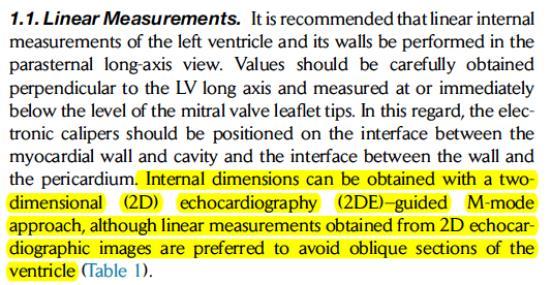
 European Heart Journal Cardiovascular Imaging (2015) 16, 233 271 doi:10.1093/ehjci/jev014 POSITION PAPER Recommendations for Cardiac Chamber Quantification by Echocardiography in Adults: An Update from
Tissue Doppler and Strain Imaging
 Tissue Doppler and Strain Imaging Steven J. Lester MD, FRCP(C), FACC, FASE Relevant Financial Relationship(s) None Off Label Usage None 1 Objective way with which to quantify the minor amplitude and temporal
Tissue Doppler and Strain Imaging. Steven J. Lester MD, FRCP(C), FACC, FASE
, FACC, FASE") Tissue Doppler and Strain Imaging Steven J. Lester MD, FRCP(C), FACC, FASE Relevant Financial Relationship(s) None Off Label Usage None a. Turn the wall filters on and turn down the receiver gain. b. Turn
Tissue Doppler and Strain Imaging Steven J. Lester MD, FRCP(C), FACC, FASE Relevant Financial Relationship(s) None Off Label Usage None a. Turn the wall filters on and turn down the receiver gain. b. Turn
Alicia Armour, MA, BS, RDCS
 Alicia Armour, MA, BS, RDCS No disclosures Review 2D Speckle Strain (briefly) Discuss some various patient populations & disease pathways where Strain can be helpful Discuss how to acquire images for Strain
Conflict of Interests
 New Approaches to Systolic Function: 4D Roberto M Lang, MD Conflict of Interests Philips Medical Imaging Research Grants Speakers bureau Advisory bureau Tomtec Research Grants Epsilon Research Grants 1
Quantifying LV function how good are we?
 Quantifying LV function how good are we? Professor Alan G Fraser Wales Heart Research Institute Cardiff University, U.K. Support for research from Hitachi Aloka, & GE Ultrasound Visual assessment of synchronicity
LV geometric and functional changes in VHD: How to assess? Mi-Seung Shin M.D., Ph.D. Gachon University Gil Hospital
 LV geometric and functional changes in VHD: How to assess? Mi-Seung Shin M.D., Ph.D. Gachon University Gil Hospital LV inflow across MV LV LV outflow across AV LV LV geometric changes Pressure overload
Comprehensive Echo Assessment of Aortic Stenosis
 Comprehensive Echo Assessment of Aortic Stenosis Smonporn Boonyaratavej, MD, MSc King Chulalongkorn Memorial Hospital Bangkok, Thailand Management of Valvular AS Medical and interventional approaches to
ECHO HAWAII. Role of Stress Echo in Valvular Heart Disease. Not only ischemia! Cardiomyopathy. Prosthetic Valve. Diastolic Dysfunction
 Role of Stress Echo in Valvular Heart Disease ECHO HAWAII January 15 19, 2018 Kenya Kusunose, MD, PhD, FASE Tokushima University Hospital Japan Not only ischemia! Cardiomyopathy Prosthetic Valve Diastolic
Feasibility and limitations of 2D speckle tracking echocardiography
 ORIGINAL ARTICLE 204 A prospective study in daily clinical practice Feasibility and limitations of 2D speckle tracking echocardiography Lina Melzer, Anja Faeh-Gunz, Barbara Naegeli, Burkhardt Seifert*,
HYPERTROPHY: Behind the curtain. V. Yotova St. Radboud Medical University Center, Nijmegen
 HYPERTROPHY: Behind the curtain V. Yotova St. Radboud Medical University Center, Nijmegen Disclosure of interest: none Relative wall thickness (cm) M 0.22 0.42 0.43 0.47 0.48 0.52 0.53 F 0.24 0.42 0.43
Prof. JL Zamorano Hospital Universitario Ramón y Cajal
 Prof. JL Zamorano Hospital Universitario Ramón y Cajal Fully Automated Quantification Software Adaptive analytical algorithm consists in knowledge-based identification of global shape and specific adaptation
Evalua&on)of)Le-)Ventricular)Diastolic) Dysfunc&on)by)Echocardiography:) Role)of)Ejec&on)Frac&on)
of)Le-)Ventricular)Diastolic) Dysfunc&on)by)Echocardiography:) Role)of)Ejec&on)Frac&on)") Evalua&on)of)Le-)Ventricular)Diastolic) Dysfunc&on)by)Echocardiography:) Role)of)Ejec&on)Frac&on) N.Koutsogiannis) Department)of)Cardiology) University)Hospital)of)Patras)! I have no conflicts of interest
Evalua&on)of)Le-)Ventricular)Diastolic) Dysfunc&on)by)Echocardiography:) Role)of)Ejec&on)Frac&on) N.Koutsogiannis) Department)of)Cardiology) University)Hospital)of)Patras)! I have no conflicts of interest
Aortic Regurgitation and Aortic Aneurysm - Epidemiology and Guidelines -
 Reconstruction of the Aortic Valve and Root - A Practical Approach - Aortic Regurgitation and Aortic Aneurysm Wednesday 14 th September - 9.45 Practice must always be founded on sound theory. Leonardo
Conflict of Interests
 Introduction to Interventional Echocardiography Roberto M Lang, MD Tomtec Conflict of Interests Research Grants Philips Medical Imaging Research Grants Speakers bureau Advisory bureau 1 Structural Heart
LA Function analysis Marcia Barbosa Vice Presidente - Brazilian Soc of Cardiology President-elect - Interamerican Soc of Cardiology
 LA Function analysis Marcia Barbosa Vice Presidente - Brazilian Soc of Cardiology President-elect - Interamerican Soc of Cardiology Belo Horizonte Brazil DECLARATION OF CONFLICT OF INTEREST Nothing to
Incorporating the New Echo Guidelines Into Everyday Practice
 Incorporating the New Echo Guidelines Into Everyday Practice Clinical Case RIGHT VENTRICULAR FAILURE Gustavo Restrepo MD President Elect Interamerican Society of Cardiology Director Fellowship Training
Adel Hasanin Ahmed 1 LV MORPHOLOGY
 Adel Hasanin Ahmed 1 LV MORPHOLOGY The left ventricular wall comprises three layers- middle circumferential layer and superficial and deep longitudinal layers: 1. Subepicardial longitudinal layer (25%
Heart Failure in Women: Dr Goh Ping Ping Cardiologist Asian Heart & Vascular Centre
 Heart Failure in Women: More than EF? Dr Goh Ping Ping Cardiologist Asian Heart & Vascular Centre Overview Review pathophysiology as it relates to diagnosis and management Rational approach to workup:
Three-dimensional echocardiography in the clinical world
 Three-dimensional echocardiography in the clinical world Dr. JL Zamorano Director CV Institute University Clinic SC, Madrid Advantages of 3D. Spatial manipulation. Optimal alineation of structures. Views
Athlete s Heart vs. Cardiomyopathy
 Athlete s Heart vs. Cardiomyopathy Linda D. Gillam, MD, MPH, FASE Chair, Department of Cardiovascular Medicine Medical Director, Cardiovascular Service Line Former Team Cardiologist to the New York Jets
A New Method to Rapidly Evaluate LVEF from a Contractility Polar Map. Lebeau et al.
 A New Method to Rapidly Evaluate LVEF from a Contractility Polar Map Lebeau et al. Good afternoon It is my pleasure to present to you a new method to rapidly evaluate LVEF from a contractility polar map
Novel echocardiographic modalities: 3D echo, speckle tracking and strain rate imaging. Potential roles in sports cardiology. Stefano Caselli, MD, PhD
 Novel echocardiographic modalities: 3D echo, speckle tracking and strain rate imaging. Potential roles in sports cardiology. Stefano Caselli, MD, PhD Ospedale San Pietro Fatebenefratelli Rome, Italy Differential
LV function in ischemic heart failure - decreased correlation between Echo and CMR
 LV function in ischemic heart failure - decreased correlation between Echo and CMR Poster No.: C-0590 Congress: ECR 2011 Type: Scientific Exhibit Authors: K. Gruszczy#ska, L. Krzych, K. Golba, P. Ulbrych,
Nancy Goldman Cutler, MD Beaumont Children s Hospital Royal Oak, Mi
 Nancy Goldman Cutler, MD Beaumont Children s Hospital Royal Oak, Mi Identify increased LV wall thickness (WT) Understand increased WT in athletes Understand hypertrophic cardiomyopathy (HCM) Enhance understanding
EDITOR S PICK CURRENT STATUS OF FULLY AUTOMATED SOFTWARE WITH THREE-DIMENSIONAL ECHOCARDIOGRAPHY FOR THE QUANTIFICATION OF LEFT VENTRICULAR FUNCTION
 ITOR S PICK This paper, courtesy of Yang and Takeuchi, provides a timely and well-considered update on the current status of fully-automated software with three-dimensional echocardiography for quantifying
Cardiac hypertrophy : differentiating disease from athlete
 Cardiac hypertrophy : differentiating disease from athlete Ario Soeryo Kuncoro, MD, Cardiologist Echocardiography Division, National Cardiovascular Centre Harapan Kita-Jakarta Departement of Cardiology
Role of echocardiography in the assessment of ischemic heart disease 분당서울대학교병원윤연이
 Role of echocardiography in the assessment of ischemic heart disease 분당서울대학교병원윤연이 Outline Evaluation of Chest pain Evaluation of MI complications Prediction of Outcomes Evaluation of Chest pain Evaluation
An Integrated Approach to Study LV Diastolic Function
 An Integrated Approach to Study LV Diastolic Function Assoc. Prof. Adriana Ilieşiu, FESC University of Medicine Carol Davila Bucharest, Romania LV Diastolic Dysfunction impaired relaxation (early diastole)
Coronary artery disease (CAD) risk factors
 risk factors") Background Coronary artery disease (CAD) risk factors CAD Risk factors Hypertension Insulin resistance /diabetes Dyslipidemia Smoking /Obesity Male gender/ Old age Atherosclerosis Arterial stiffness precedes
Background Coronary artery disease (CAD) risk factors CAD Risk factors Hypertension Insulin resistance /diabetes Dyslipidemia Smoking /Obesity Male gender/ Old age Atherosclerosis Arterial stiffness precedes
Cardiology for the Practitioner Advanced Cardiac Imaging: Worth the pretty pictures?
 Keenan Research Centre Li Ka Shing Knowledge Institute Cardiology for the Practitioner Advanced Cardiac Imaging: Worth the pretty pictures? Howard Leong-Poi, MD, FRCPC Associate Professor of Medicine St.
Strain Imaging: Myocardial Mechanics Simplified and Applied
 9/28/217 Strain Imaging: Myocardial Mechanics Simplified and Applied John Gorcsan III, MD Professor of Medicine Director of Clinical Research Division of Cardiology VECTORS OF CONTRACTION Shortening Thickening
Normal values for cardiovascular magnetic resonance in adults and children
 Kawel-Boehm et al. Journal of Cardiovascular Magnetic Resonance (2015) 17:29 DOI 10.1186/s12968-015-0111-7 REVIEW Normal values for cardiovascular magnetic resonance in adults and children Nadine Kawel-Boehm
Strain/Untwisting/Diastolic Suction
 What Is Diastole and How to Assess It? Strain/Untwisting/Diastolic Suction James D. Thomas, M.D., F.A.C.C. Cardiovascular Imaging Center Department of Cardiology Cleveland Clinic Foundation Cleveland,
Global left ventricular circumferential strain is a marker for both systolic and diastolic myocardial function
 Global left ventricular circumferential strain is a marker for both systolic and diastolic myocardial function Toshinari Onishi 1, Samir K. Saha 2, Daniel Ludwig 1, Erik B. Schelbert 1, David Schwartzman
Comparison of Cardiac MDCT with MRI and Echocardiography in the Assessement of Left Ventricular Function
 Comparison of Cardiac MDCT with MRI and Echocardiography in the Assessement of Left Ventricular Function Poster No.: C-0969 Congress: ECR 2012 Type: Scientific Exhibit Authors: B. Kara, Y. Paksoy, C. Erol,
Basic Assessment of Left Ventricular Systolic Function
 WINFOCUS BASIC ECHO (WBE) Basic Assessment of Left Ventricular Systolic Function Ritesh Dhar, MD Director, Echocardiography Lab and Staff Cardiologist Intermountain Medical Center Murray, Utah Outline
Diastology State of The Art Assessment
 Diastology State of The Art Assessment Dr. Mohammad AlGhamdi Assistant professor, KSAU-HS Consultant Cardiologist King AbdulAziz Cardiac Center Ministry of National Guard Health Affairs Diagnostic Clinical
Advanced Multi-Layer Speckle Strain Permits Transmural Myocardial Function Analysis in Health and Disease:
 Advanced Multi-Layer Speckle Strain Permits Transmural Myocardial Function Analysis in Health and Disease: Clinical Case Examples Jeffrey C. Hill, BS, RDCS Echocardiography Laboratory, University of Massachusetts
Recommendations for chamber quantification *
 Eur J Echocardiography (2006) 7, 79e108 GUIDELINES Recommendations for chamber quantification * Roberto M. Lang, Michelle Bierig, Richard B. Devereux, Frank A. Flachskampf *, Elyse Foster, Patricia A.
Københavns Universitet
 university of copenhagen Københavns Universitet Total average diastolic longitudinal displacement by colour tissue doppler imaging as an assessment of diastolic function de Knegt, Martina Chantal; Biering-Sørensen,
Evaluation of Left Ventricular Diastolic Dysfunction by Doppler and 2D Speckle-tracking Imaging in Patients with Primary Pulmonary Hypertension
 ESC Congress 2011.No 85975 Evaluation of Left Ventricular Diastolic Dysfunction by Doppler and 2D Speckle-tracking Imaging in Patients with Primary Pulmonary Hypertension Second Department of Internal
Restrictive Cardiomyopathy
 ESC Congress 2011, Paris Imaging Unusual Causes of Cardiomyopathy Restrictive Cardiomyopathy Kazuaki Tanabe, MD, PhD Professor of Medicine Chair, Division of Cardiology Izumo, Japan I Have No Disclosures
DECLARATION OF CONFLICT OF INTEREST. None
 DECLARATION OF CONFLICT OF INTEREST None Hot Topics in Echocardiography: The position of the EAE EAE / ASE recommendation about Echo Assessment of Cardiac Mechanics Jens-Uwe Voigt Dpt. of Cardiovascular
Η ηχωκαρδιολογία στην διάγνωση κα πρόγνωση της καρδιακής ανεπάρκειας µε µειωµένο και φυσιολογικό κλάσµα εξώθησης
 Η ηχωκαρδιολογία στην διάγνωση κα πρόγνωση της καρδιακής ανεπάρκειας µε µειωµένο και φυσιολογικό κλάσµα εξώθησης Βασίλειος Σαχπεκίδης Επιµελητής Β Καρδιολογίας Γ.Ν. Παπαγεωργίου Θεσσαλονίκη ESC Guidelines
Hemodynamic Assessment. Assessment of Systolic Function Doppler Hemodynamics
 Hemodynamic Assessment Matt M. Umland, RDCS, FASE Aurora Medical Group Milwaukee, WI Assessment of Systolic Function Doppler Hemodynamics Stroke Volume Cardiac Output Cardiac Index Tei Index/Index of myocardial
Little is known about the degree and time course of
 Differential Changes in Regional Right Ventricular Function Before and After a Bilateral Lung Transplantation: An Ultrasonic Strain and Strain Rate Study Virginija Dambrauskaite, MD, Lieven Herbots, MD,
Conflict of interest: none declared
 The value of left ventricular global longitudinal strain assessed by three-dimensional strain imaging in the early detection of anthracycline-mediated cardiotoxicity C. Mornoş, A. Ionac, D. Cozma, S. Pescariu,
THE RIGHT VENTRICLE IN PULMONARY HYPERTENSION R. DRAGU
 THE RIGHT VENTRICLE IN PULMONARY HYPERTENSION R. DRAGU Cardiology Dept. Rambam Health Care Campus Rappaport Faculty of Medicine Technion, Israel Why the Right Ventricle? Pulmonary hypertension (PH) Right
The athlete s heart: Different training responses in African and Caucasian male elite football players
 The athlete s heart: Different training responses in African and Caucasian male elite football players Gard Filip Gjerdalen Oslo University Hospital, Aker. Bjørknes College Co-writers: Hisdal J, Solberg
Assessment of LV systolic function
 Tutorial 5 - Assessment of LV systolic function Assessment of LV systolic function A knowledge of the LV systolic function is crucial in the undertanding of and management of unstable hemodynamics or a
OPTIMIZING ECHO ACQUISTION FOR STRAIN AND DIASTOLOGY
 OPTIMIZING ECHO ACQUISTION FOR STRAIN AND DIASTOLOGY October 8, 2017 Deborah Agler, ACS, RDCS, FASE Coordinator of Education and Training Cleveland Clinic General Principles Diastology Clinical Data Heart
Νεότερα ςτην Υπερηχοκαρδιογραφία. Βαςίλειοσ Καμπερίδησ Clinical research fellow in Cardiology
 Νεότερα ςτην Υπερηχοκαρδιογραφία Βαςίλειοσ Καμπερίδησ Clinical research fellow in Cardiology Disclosures ESC training grant EACVI research grant HCS training grant ELIKAR research grant Evolution of Echocardiography
Mechanisms of False Positive Exercise Electrocardiography: Is False Positive Test Truly False?
 Mechanisms of False Positive Exercise Electrocardiography: Is False Positive Test Truly False? Masaki Izumo a, Kengo Suzuki b, Hidekazu Kikuchi b, Seisyo Kou b, Keisuke Kida b, Yu Eguchi b, Nobuyuki Azuma
VECTORS OF CONTRACTION
 1/3/216 Strain, Strain Rate, and Torsion: Myocardial Mechanics Simplified and Applied VECTORS OF CONTRACTION John Gorcsan, MD University of Pittsburgh, Pittsburgh, PA Shortening Thickening Twisting No
Back to Basics: Common Errors In Quantitation In Everyday Practice
 Back to Basics: Common Errors In Quantitation In Everyday Practice Deborah Agler, ACS, RDCS, FASE October 9, 2017 ASE: Echo Florida Rebecca T. Hahn, MD Director of Interventional Echocardiography Professor
HFpEF. April 26, 2018
 HFpEF April 26, 2018 (J Am Coll Cardiol 2017;70:2476 86) HFpEF 50% or more (40-71%) of patients with CHF have preserved LV systolic function. HFpEF is an increasingly frequent hospital discharge. Outcomes
Pediatric Echocardiographic Normal values. SIEC Firenze Febbraio 2016
 Pediatric Echocardiographic Normal values Massimiliano Cantinotti MD Fondazione Toscana G. Monasterio and Institute of Clinical Physiology (CNR) Massa and Pisa SIEC Firenze 18-20 Febbraio 2016 Background
Evaluation of Ejection Fraction in Patients with Cardiac Resynchronization Therapy by Two and Three Dimensional Echocardiography
 58 Original article Evaluation of Ejection Fraction in Patients with Cardiac Resynchronization Therapy by Two and Three Dimensional Echocardiography Anil OM Department of cardiology, Manmohan Cardiothoracic
Appendix II: ECHOCARDIOGRAPHY ANALYSIS
 Appendix II: ECHOCARDIOGRAPHY ANALYSIS Two-Dimensional (2D) imaging was performed using the Vivid 7 Advantage cardiovascular ultrasound system (GE Medical Systems, Milwaukee) with a frame rate of 400 frames
Ref 1. Ref 2. Ref 3. Ref 4. See graph
 Ref 1 Ref 2 Ref 3 1. Ages 6-23 y/o 2. Significant LVM differences by gender 3. For males 95 th percentiles: a. LVM/BSA = 103 b. LVM/height = 100 4. For females 95 th percentiles: a. LVM/BSA = 84 b. LVM/height
Athlete s Heart vs. Cardiomyopathy
 Athlete s Heart vs. Cardiomyopathy Linda D. Gillam, MD, MPH, FASE Chair, Department of Cardiovascular Medicine Medical Director, Cardiovascular Service Line Former Team Cardiologist to the New York Jets
Adult Echocardiography Examination Content Outline
 Adult Echocardiography Examination Content Outline (Outline Summary) # Domain Subdomain Percentage 1 2 3 4 5 Anatomy and Physiology Pathology Clinical Care and Safety Measurement Techniques, Maneuvers,
Left atrial Function In Echocardiography
 Left atrial Function In Echocardiography Pr Erwan DONAL erwan.donal@chu-rennes.fr Inserm Diastole LA Pressure (mmhg) -t/ 17 LA function 15 A-Loop 13 V-Loop 11 9 7 5 1 2 3 4 20 30 40 50 60 70 LA Volume
Echocardiography. Guidelines for Valve and Chamber Quantification. In partnership with
 Echocardiography Guidelines for Valve and Chamber Quantification In partnership with Explanatory note & references These guidelines have been developed by the Education Committee of the British Society
New ASE Guidelines: What you must know
 New ASE Guidelines: What you must know Federico M Asch MD, FASE, FACC Chair, ASE Guidelines and Standards Committee Medstar Washington Hospital Center Medstar Health Research Institute Georgetown University
Degenerative Mitral Regurgitation: Etiology and Natural History of Disease and Triggers for Intervention
 Degenerative Mitral Regurgitation: Etiology and Natural History of Disease and Triggers for Intervention John N. Hamaty D.O. FACC, FACOI November 17 th 2017 I have no financial disclosures Primary Mitral
Index. K Knobology, TTE artifact, image resolution, ultrasound, 14
 A Acute aortic regurgitation (AR), 124 128 Acute aortic syndrome (AAS) classic aortic dissection diagnosis, 251 263 evolutive patterns, 253 255 pathology, 250 251 classifications, 247 248 incomplete aortic
How NOT to miss Hypertrophic Cardiomyopathy? Adaya Weissler-Snir, MD University Health Network, University of Toronto
 How NOT to miss Hypertrophic Cardiomyopathy? Adaya Weissler-Snir, MD University Health Network, University of Toronto Introduction Hypertrophic cardiomyopathy is the most common genetic cardiomyopathy,
ATRIAL FIBRILLATION AND LEFT ATRIUM: STRUCTURAL REMODELING AND EVOLUTION OF ARRHYTHMIA PATTERN
 Atrial Fibrillation and Heart Failure: the ugly and the nasty 13 International Meeting February 14-15 th 2019, Bologna ATRIAL FIBRILLATION AND LEFT ATRIUM: STRUCTURAL REMODELING AND EVOLUTION OF ARRHYTHMIA
Imaging in Heart Failure: A Multimodality Approach. Thomas Ryan, MD
 Imaging in Heart Failure: A Multimodality Approach Thomas Ryan, MD Heart Failure HFrEF HFpEF EF50% Lifetime risk 20% Prevalence 6M Americans Societal costs - $30B 50% 5-year survival 1 Systolic
Outline. EuroScore II. Society of Thoracic Surgeons Score. EuroScore II
 SURGICAL RISK IN VALVULAR HEART DISEASE: WHAT 2D AND 3D ECHO CAN TELL YOU AND WHAT THEY CAN'T Ernesto E Salcedo, MD Professor of Medicine University of Colorado School of Medicine Director of Echocardiography
Quantitation of right ventricular dimensions and function
 SCCS Basics of cardiac assessment Quantitation of right ventricular dimensions and function Tomasz Kukulski, MD PhD Dept of Cardiology, Congenital Heart Disease and Electrotherapy Silesian Medical University
Mechanisms of heart failure with normal EF Arterial stiffness and ventricular-arterial coupling. What is the pathophysiology at presentation?
 Mechanisms of heart failure with normal EF Arterial stiffness and ventricular-arterial coupling What is the pathophysiology at presentation? Ventricular-arterial coupling elastance Central arterial pressure
Cardiac Risk Assessment Using 2D and 3D Transthoracic Echocardiography in Patients Undergoing Haemodialysis
 Cardiac Risk Assessment Using 2D and 3D Transthoracic Echocardiography in Patients Undergoing Haemodialysis A thesis submitted to The University of Manchester for the degree of Doctor of Philosophy (phd)