Echocardiography for the Electrophysiologist: Day-to-day practice. Emmanuel Fares, MD
|
|
|
- Godfrey Sparks
- 5 years ago
- Views:
Transcription
1 Echocardiography for the Electrophysiologist: Day-to-day practice Emmanuel Fares, MD EP and pacing service, Department of Cardiovascular Medicine, Cairo University Agenda Role of echo in arrhythmia management: Atrial Fibrillation: Evaluation of LA size Assessment of LV diastolic function Risk stratification (LAA thrombi detection) Role of intracardiac echo Ventricular arrhythmias and SCD: Ischemic Cardiomyopathy Non-ischemic cardiomyopathy Inheritable arrhythmogenic diseases: HCM, ARVD Role of echo in CRT optimization 1
2 Introduction In the setting of arrhythmias, the utility of echocardiography lies primarily in the identification of associated heart disease, the knowledge of which will influence treatment of the arrhythmia or provide prognostic information Atrial Fibrillation AF is the most frequently encountered arrhythmia in clinical practice, with an overall prevalence of 0.4% in the general population. 2


:e28-e292.")
3 AF complications Go AS, Mozaffarian D, Roger VL, et al. Heart disease and stroke statistics 2014 update: a report from the American Heart Association. Circulation. 2014;129(3):e28-e292. Assessment of LA dimensions Left atrial dimensions: AP, transverse and longitudinal 3
 views at ventricular endsystole (maximum volume) Reference ranges for LA size Women Men Reference range Mildly abnormal Moderately")

4 Assessment of LA volume Measurement of left atrial volume from biplane method of disks (modified Simpson s rule) using apical 4-chamber (a) and apical 2-chamber (b) views at ventricular endsystole (maximum volume) Reference ranges for LA size Women Men Reference range Mildly abnormal Moderately abnormal Severely abnormal Reference range Mildly abnormal Moderately abnormal Severely abnormal Atrial dimensions LA diameter, cm Atrial volumes LA volume/bs A, ml/m 2 22 ± ± BSA, Body surface area; LA, left atrial 4
5 Clinical perspectives LA size has been established as a prognostic marker for cardiovascular events: According to the data from the Framingham study, a 5-mm increase in LA dimension was associated with a 39% increased risk for development of AF. Subjects in sinus rhythm with a LA dimension over 5.0 cm had about 4X the risk of developing AF during the following period of surveillance. LA enlargement is associated with a low probability of successful cardioversion for chronic AF or maintenance of sinus rhythm An increased LA volume index can predict recurrence of AF after PVI Although AF promotes further LA enlargement, cardioversion and maintenance of sinus rhythm may reverse this process. Kaplan Meier survival curves free of new-onset atrial fibrillation. Patients are stratified according to left atrial (LA) diameter (A), LA volume (B) Philippe Debonnaire et al. Circ Arrhythm Electrophysiol. 2017;10:e
6 Association between left atrial size and the incidence of AF in the Cardiovascular Health Study. LAD indicates left atrial dimension; RR, relative risk. Agenda Role of echo in arrhythmia management: Atrial Fibrillation: Evaluation of LA size Assessment of LV diastolic function Risk stratification (LAA thrombi detection) Role of intracardiac echo Ventricular arrhythmias and SCD: Ischemic Cardiomyopathy Non-ischemic cardiomyopathy Inheritable arrhythmogenic diseases: HCM, ARVD Role of echo in CRT optimization 6
.")
7 Assessment of LV Diastolic Function The evaluation of diastolic function in patients with AF has remained a clinical challenge: The impairment of the LA mechanical function The variable cycle lengths All echocardiographic parameters, should be measured and averaged from 5 to 10 cardiac cycles PWD study of transmitral inflow Arrow indicates early transmitral flow deceleration time (DT). E represents peak LV early diastolic filling velocity. Peak E velocity and DT vary depending on cardiac cycle length. 7


8 TDI Tissue Doppler imaging of the septal mitral annular velocities (e') Color M-mode Mitral inflow propagation velocity, is acquired from the apical 4-chamber view by placing a cursor over the center of mitral inflow and reducing the Nyquist limit for flow toward apex to a point when the central color turns blue. 8
9 Assessment of the diastolic function PWD: Mitral DT 100 ms predicted a mean of pulmonary capillary wedge pressure (PCWP) 18 mmhg TDI: E/e' (septal) > 15 and E/e' (lateral) > 10 have been shown to be highly specific for elevated LV filling pressure Color M-mode: A Vp < 45 cm/s is consistent with the presence of diastolic dysfunction Agenda Role of echo in arrhythmia management: Atrial Fibrillation: Evaluation of LA size Assessment of LV diastolic function Risk stratification (LAA thrombi detection) Role of intracardiac echo Ventricular arrhythmias and SCD: Ischemic Cardiomyopathy Non-ischemic cardiomyopathy Inheritable arrhythmogenic diseases: HCM, ARVD Role of echo in CRT optimization 9
 within the left atrial")
10 Detection of LA thrombi TEE provides excellent visualization of posterior cardiac structures and is the modality of choice for detecting LA or LAA thrombi with a sensitivity and specificity of approximately 95% to 100% Thrombus (arrow) within the left atrial appendage 10


11 Spontaneous echo contrast (*) swirling detected by transesophageal echocardiography in a patient with AF It is difficult to distinguish small thrombi from artifacts, including prominent trabecular structures, duplication artifacts, coumadin ridge 11

12 Echocardiographic predictors of systemic thromboembolism TEE-guided early cardioversion 12
13 Advantages and disadvantages of TEE-guided early cardioversion Agenda Role of echo in arrhythmia management: Atrial Fibrillation: Evaluation of LA size Assessment of LV diastolic function Risk stratification (LAA thrombi detection) Role of intracardiac echo Ventricular arrhythmias and SCD: Ischemic Cardiomyopathy Non-ischemic cardiomyopathy Inheritable arrhythmogenic diseases: HCM, ARVD Role of echo in CRT optimization 13
 for transseptal puncture guided by ICE ICE advantages Detect anatomical variations of interatrial septum such as aneurismal")

14 ICE during PVE Two-dimensional ICE image that shows the Brockenbrough-curve needle is advanced from the right atrium (RA) to the fossa ovalis (FO) for transseptal puncture guided by ICE ICE advantages Detect anatomical variations of interatrial septum such as aneurismal septum, double membrane septum, PFO The use of ICE helps in determining the exact position of tip of the transseptal sheath by looking for tenting of the interatrial septum. It enables avoidance of the life-threatening complication, such as perforation of lateral wall of LA or aortic root. 14
15 ICE advanatges ICE is useful as a monitoring tool for titration of energy power and duration. It can control lesion formation The appearance of microbubbles resulting from overheating can precede increased impedance Ablation needs to be terminated immediately if a sudden appearance of microbubbles is observed. This can prevent tissue damage and scar formation which may promote PV stenosis, LA perforation, or pericardial effusion Agenda Role of echo in arrhythmia management: Atrial Fibrillation: Evaluation of LA size Assessment of LV diastolic function Risk stratification (LAA thrombi detection) Role of intracardiac echo Ventricular arrhythmias and SCD: Ischemic Cardiomyopathy Non-ischemic cardiomyopathy Inheritable arrhythmogenic diseases: HCM, ARVD Role of echo in CRT optimization 15
16 Role of echo in prediction of SCD Echocardiography remains the imaging technique of first choice to rule out the presence of structural heart disease and assess ventricular functions LVEF has consistently shown an association with increased risk of ventricular arrhythmias and therefore, this variable is included in the recommendations for ICD as primary prevention (class I) New echo indices may be associated with increased arrhythmogenicity ICD trials in heart failure EF < 35% is an indication for 1ry prevention ICD 16
17 Echocardiographic parameters (beyond LVEF) associated with ventricular arrhythmias in ischemic heart failure patients Imaging technique Parameter Evidence Echocardiography Speckle tracking LV GLS Reduced magnitude of LV GLS was associated with 1.24-fold increased risk of ventricular arrhythmias (95% CI 1.10 to 1.40; P = ) in 988 patients with acute STEMI LV mechanical dispersion Each 10 ms increase in LV mechanical dispersion has been associated with increased risk of arrhythmias (HR 1.7) Dobutamine stress echocardiography Inducible ischemia The presence of inducible ischemia was associated with ventricular arrhythmias (HR 2.1, 95% CI ; P < 0.001) in 90 patients with chronic IHD Echocardiographic parameters (beyond LVEF) associated with ventricular arrhythmias in non-ischemic heart failure patients Imaging technique No. Cardiomyopathy Parameter Evidence Speckle tracking echocardiography 100 Sarcoidosis GLS GLS was independently associated with 1.4-fold increased risk of composite end point (including arrhythmias) 94 DCM GLS Mechanical dispersion Each 1% worsening in GLS was independently associated with ventricular arrhythmias, SCD and appropriate ICD therapy (HR 1.26, 95% CI ;P = 0.02) Each 10 ms increment in mechanical dispersion was associated with a 1.20 increased risk for ventricular arrhythmias, SCD, and appropriate ICD therapy (95% CI ; P = 0.02) 17

18 Mechanical dispersion Left panel shows synchronous contraction by longitudinal strain in a patient after myocardial infarction. Mid panel shows heterogeneous timing of contraction and pronounced mechanical dispersion in a patient after myocardial infarction with ventricular arrhythmias. Agenda Role of echo in arrhythmia management: Atrial Fibrillation: Evaluation of LA size Assessment of LV diastolic function Risk stratification (LAA thrombi detection) Role of intracardiac echo Ventricular arrhythmias and SCD: Ischemic Cardiomyopathy Non-ischemic cardiomyopathy Inheritable arrhythmogenic diseases: HCM, ARVD Role of echo in CRT optimization 18

19 HCM The diagnosis is based on the presence of abnormally increased LV wall thickness ( 15 mm) by any imaging modality not explained by loading conditions and in the absence of other diseases associated with LV hypertrophy HCM The most frequent genetic heart disease, usually caused by mutations of genes encoding sarcomeric proteins, transmitted with an autosomal dominant inheritance The annual incidence of SCD is 0.8%, with the highest prevalence among young patients 19

20 Estimation of SCD risk in HCM Risk stratification of patients with HCM using 2D speckle tracking echocardiography Speckle tracking echocardiographic LV strain analysis has demonstrated to correlate with the amount of replacement fibrosis in hypertrophic cardiomyopathy patients, and therefore it could be hypothesized that the assessment of LV strain may be also associated with increased risk of ventricular arrhythmias 20
 (C) ARVC")
21 Risk stratification of patients with HCM using 2D speckle tracking echocardiography On echocardiographic speckle tracking analysis, the magnitude of GLS is 13.8% (B). The study by Debonnaire et al showed that patients with a left ventricular GLS 14% had higher rates of appropriate ICD therapy compared with patients with more preserved GLS (< 14%) (C) ARVC Autosomal dominant inheritance disease with variable penetrance and phenotype expression characterized by replacement of right ventricular myocardium by fibrous and fatty tissue 21
, or - PSAX RVOT 36 mm (PSAX/BSA 21 mm/m 2 ), or - Fractional area change 33% CMR criteria Regional RV akinesia or dyskinesia or dyssynchronous RV contraction AND 1 of the")
, or - PSAX RVOT 32 to <36 mm (PSAX/BSA 18 to <21")
22 1. Global or regional dysfunction and structural alterations Major 2D Echo Criteria Regional RV akinesia, dyskinesia, or aneurysm AND 1 of the following measured at end diastole: Minor - PLAX RVOT 32 mm (PLAX/BSA 19 mm/m 2 ), or - PSAX RVOT 36 mm (PSAX/BSA 21 mm/m 2 ), or - Fractional area change 33% CMR criteria Regional RV akinesia or dyskinesia or dyssynchronous RV contraction AND 1 of the following: - RV EDV/BSA 110 ml/m 2 (male) or 100 ml/m 2 (female) - RV ejection fraction 40% RV angiography criteria Regional RV akinesia, dyskinesia, or aneurysm 2D Echo Criteria Regional RV akinesia or dyskinesia or dyssynchronous RV contraction AND 1 of the following measured at end diastole: - PLAX RVOT 29 to <32 mm (PLAX/BSA 16 to <19 mm/m 2 ), or - PSAX RVOT 32 to <36 mm (PSAX/BSA 18 to <21 mm/m 2 ), or - Fractional area change > 33% 40% CMR criteria Regional RV akinesia or dyskinesia or dyssynchronous RV contraction AND 1 of the following: - RV EDV/BSA 100 to 110 ml/m 2 (male) or 90 to 100 ml/m 2 (female) - RV ejection fraction >40 to 45% Echo in ARVC 22
23 LV involvement in ARVC Clinical course of patients with ARVD/C with LV involvement versus isolated RV disease. Kaplan-Meier survival plots for LVEF(left) and LV involvement deformation imaging (right). Signs of LV involvement are associated with adverse outcome. Agenda Role of echo in arrhythmia management: Atrial Fibrillation: Evaluation of LA size Assessment of LV diastolic function Risk stratification (LAA thrombi detection) Role of intracardiac echo Ventricular arrhythmias and SCD: Ischemic Cardiomyopathy Non-ischemic cardiomyopathy Inheritable arrhythmogenic diseases: HCM, ARVD Role of echo in CRT optimization 23

24 CRT optimization CRT optimization Multiple methods, no gold standard for optimization The guidelines do not provide any specific recommendation 24


25 Methods Utilizing the MV inflow: The Ritter method The Iterative method Utilizing the LV outflow VTI The Ritter method The interval from the pacing spike to the end of the A wave on mitral inflow is measured at short and long AV delays. The difference in these time intervals is then subtracted from the long AV delay to calculate the optimal AV delay 25

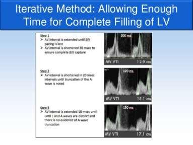
26 The Iterative method 26

27 LVOT VTI method AV delay optimized to achieve the maximum stroke volume based on the aortic outflow tract VTI. In this case, the VTI increased from 16 to 23 cm with an increase in the AV delay from 80 to 150 ms. This can also be used in VV interval optimization Areas of uncertainty in AV optimization While the majority of the landmark trials of CRT incorporated some form of AV delay optimization at the time of implantation, definitive data supporting their superiority over an empiric, fixed AV delay are lacking Most available data involve hemodynamic studies demonstrating an acute improvement with optimization Most studies of routine optimization vs. fixed intervals have had mixed results 27
28 Take home messages Echocardiography can play a key role in risk stratification and management of patients with AF Transesophageal echocardiography provides accurate information about the presence of a thrombus in the atria and thromboembolic risk. The novel technique of intracardiac echocardiography has emerged as a popular and useful tool in the practice of interventional electrophysiology. Take home messages Left ventricular ejection fraction assessed with any imaging technique remains as an important criterion to identify the patients at risk of having ventricular arrhythmias or SCD Advanced echocardiographic imaging techniques evaluating the active deformation of the myocardium provided the incremental value for risk stratification in a variety of patients with cardiac disease. 28
29 Take home messages CRT optimization remains an important tool for non-responders Echocardiography based methods are available and diverse The choice of method used should be individualized as no clear data about the superior efficacy of any of the described methods exist to date Thank You 29
Echocardiographic Evaluation of the Cardiomyopathies. Stephanie Coulter, MD, FACC, FASE April, 2016
 Echocardiographic Evaluation of the Cardiomyopathies Stephanie Coulter, MD, FACC, FASE April, 2016 Cardiomyopathies (CMP) primary disease intrinsic to cardiac muscle Dilated CMP Hypertrophic CMP Infiltrative
LV FUNCTION ASSESSMENT: WHAT IS BEYOND EJECTION FRACTION
 LV FUNCTION ASSESSMENT: WHAT IS BEYOND EJECTION FRACTION Jamilah S AlRahimi Assistant Professor, KSU-HS Consultant Noninvasive Cardiology KFCC, MNGHA-WR Introduction LV function assessment in Heart Failure:
Highlights from EuroEcho 2009 Echo in cardiomyopathies
 Highlights from EuroEcho 2009 Echo in cardiomyopathies Bogdan A. Popescu University of Medicine and Pharmacy, Bucharest, Romania ESC Congress 2010 Hypertrophic cardiomyopathy To determine the differences
Heart Failure in Women: Dr Goh Ping Ping Cardiologist Asian Heart & Vascular Centre
 Heart Failure in Women: More than EF? Dr Goh Ping Ping Cardiologist Asian Heart & Vascular Centre Overview Review pathophysiology as it relates to diagnosis and management Rational approach to workup:
Managing Hypertrophic Cardiomyopathy with Imaging. Gisela C. Mueller University of Michigan Department of Radiology
 Managing Hypertrophic Cardiomyopathy with Imaging Gisela C. Mueller University of Michigan Department of Radiology Disclosures Gadolinium contrast material for cardiac MRI Acronyms Afib CAD Atrial fibrillation
Adel Hasanin Ahmed 1
 Adel Hasanin Ahmed 1 PERICARDIAL DISEASE The pericardial effusion ends anteriorly to the descending aorta and is best visualised in the PLAX. PSAX is actually very useful sometimes for looking at posterior
Role of echocardiography in the assessment of ischemic heart disease 분당서울대학교병원윤연이
 Role of echocardiography in the assessment of ischemic heart disease 분당서울대학교병원윤연이 Outline Evaluation of Chest pain Evaluation of MI complications Prediction of Outcomes Evaluation of Chest pain Evaluation
DIAGNOSIS AND MANAGEMENT OF ARRHYTHMOGENIC CARDIOMYOPATHY. David SIU MD ( 蕭頌華醫生 ) Division of Cardiology The University of Hong Kong
 Division of Cardiology The University of Hong Kong") APHRS Summit 2018 in conjunction with HKCC Heart Rhythm Refresher Course DIAGNOSIS AND MANAGEMENT OF ARRHYTHMOGENIC CARDIOMYOPATHY David SIU MD ( 蕭頌華醫生 ) Division of Cardiology The University of Hong Kong
APHRS Summit 2018 in conjunction with HKCC Heart Rhythm Refresher Course DIAGNOSIS AND MANAGEMENT OF ARRHYTHMOGENIC CARDIOMYOPATHY David SIU MD ( 蕭頌華醫生 ) Division of Cardiology The University of Hong Kong
Left atrial function. Aliakbar Arvandi MD
 In the clinic Left atrial function Abstract The left atrium (LA) is a left posterior cardiac chamber which is located adjacent to the esophagus. It is separated from the right atrium by the inter-atrial
LV geometric and functional changes in VHD: How to assess? Mi-Seung Shin M.D., Ph.D. Gachon University Gil Hospital
 LV geometric and functional changes in VHD: How to assess? Mi-Seung Shin M.D., Ph.D. Gachon University Gil Hospital LV inflow across MV LV LV outflow across AV LV LV geometric changes Pressure overload
Strain and Strain Rate Imaging How, Why and When?
 Strain and Strain Rate Imaging How, Why and When? João L. Cavalcante, MD Advanced Cardiac Imaging Fellow Cleveland Clinic Foundation Disclosures: No conflicts of interest Movement vs Deformation Movement
Nancy Goldman Cutler, MD Beaumont Children s Hospital Royal Oak, Mi
 Nancy Goldman Cutler, MD Beaumont Children s Hospital Royal Oak, Mi Identify increased LV wall thickness (WT) Understand increased WT in athletes Understand hypertrophic cardiomyopathy (HCM) Enhance understanding
BMR Medicine. Case Study YOUNG PATIENT WITH RECURRENT PRESYNCOPE: A CASE REPORT
 www.bmrjournals.com Open Access Scientific Publisher Case Study YOUNG PATIENT WITH RECURRENT PRESYNCOPE: A CASE REPORT ABSTRACT Manish Ruhela *, Vijay Pathak, Anoop Jain, Department of Cardiology, Sawai
Basic Approach to the Echocardiographic Evaluation of Ventricular Diastolic Function
 Basic Approach to the Echocardiographic Evaluation of Ventricular Diastolic Function J A F E R A L I, M D U N I V E R S I T Y H O S P I T A L S C A S E M E D I C A L C E N T E R S T A F F C A R D I O T
Index of subjects. effect on ventricular tachycardia 30 treatment with 101, 116 boosterpump 80 Brockenbrough phenomenon 55, 125
 145 Index of subjects A accessory pathways 3 amiodarone 4, 5, 6, 23, 30, 97, 102 angina pectoris 4, 24, 1l0, 137, 139, 140 angulation, of cavity 73, 74 aorta aortic flow velocity 2 aortic insufficiency
Global left ventricular circumferential strain is a marker for both systolic and diastolic myocardial function
 Global left ventricular circumferential strain is a marker for both systolic and diastolic myocardial function Toshinari Onishi 1, Samir K. Saha 2, Daniel Ludwig 1, Erik B. Schelbert 1, David Schwartzman
DON T FORGET TO OPTIMISE DEVICE PROGRAMMING
 CRT:NON-RESPONDERS OR NON-PROGRESSORS? DON T FORGET TO OPTIMISE DEVICE PROGRAMMING Prof. ALİ OTO,MD,FESC,FACC,FHRS Chairman,Department of Cardiology Hacettepe University Faculty of Medicine,Ankara Causes
Evalua&on)of)Le-)Ventricular)Diastolic) Dysfunc&on)by)Echocardiography:) Role)of)Ejec&on)Frac&on)
of)Le-)Ventricular)Diastolic) Dysfunc&on)by)Echocardiography:) Role)of)Ejec&on)Frac&on)") Evalua&on)of)Le-)Ventricular)Diastolic) Dysfunc&on)by)Echocardiography:) Role)of)Ejec&on)Frac&on) N.Koutsogiannis) Department)of)Cardiology) University)Hospital)of)Patras)! I have no conflicts of interest
Evalua&on)of)Le-)Ventricular)Diastolic) Dysfunc&on)by)Echocardiography:) Role)of)Ejec&on)Frac&on) N.Koutsogiannis) Department)of)Cardiology) University)Hospital)of)Patras)! I have no conflicts of interest
RIGHT VENTRICULAR SIZE AND FUNCTION
 RIGHT VENTRICULAR SIZE AND FUNCTION Edwin S. Tucay, MD, FPCC, FPCC, FPSE Philippine Society of Echocardiography Quezon City, Philippines Echo Mission, BRTTH, Legaspi City, July 1-2, 2016 NO DISCLOSURE
Implications of the new diagnostic criteria for ARVC
 EUROECHO 2010 Echocardiographic assessment of cardiomyopathies Implications of the new diagnostic criteria for ARVC Barbara Bauce, MD, PhD Department of Cardiac, Thoracic and Vascular Sciences University
PROSTHETIC VALVE BOARD REVIEW
 PROSTHETIC VALVE BOARD REVIEW The correct answer D This two chamber view shows a porcine mitral prosthesis with the typical appearance of the struts although the leaflets are not well seen. The valve
3/27/2014. Introduction.
 Introduction. Myocardial perfusion & contractility becomes abnormal immediately after the onset of ischaemia, even before the development of the symptoms & ST segment changes. 1 Myocardial Wall Motion
Echocardiographic Cardiovascular Risk Stratification: Beyond Ejection Fraction
 Echocardiographic Cardiovascular Risk Stratification: Beyond Ejection Fraction October 4, 2014 James S. Lee, M.D., F.A.C.C. Associates in Cardiology, P.A. Silver Spring, M.D. Disclosures Financial none
WHAT DO ELECTROPHYSIOLOGISTS WANT TO KNOW FROM ECHOCARDIOGRAPHERS BEFORE, DURING&AFTER CARDIAC RESYNCHRONIZATION THERAPY?
 WHAT DO ELECTROPHYSIOLOGISTS WANT TO KNOW FROM ECHOCARDIOGRAPHERS BEFORE, DURING&AFTER CARDIAC RESYNCHRONIZATION THERAPY? Mary Ong Go, MD, FPCP, FPCC, FACC OUTLINE What is CRT Who needs CRT What does the
The new Guidelines: Focus on Chronic Heart Failure
 The new Guidelines: Focus on Chronic Heart Failure Petros Nihoyannopoulos MD, FRCP, FESC Professor of Cardiology Imperial College London and National & Kapodistrian University of Athens 2 3 4 The principal
Stephen Glen ISCHAEMIC HEART DISEASE AND LEFT VENTRICULAR FUNCTION
 Stephen Glen ISCHAEMIC HEART DISEASE AND LEFT VENTRICULAR FUNCTION Overview Coronary arteries Terminology to describe contractility Measuring ventricular function Systolic dysfunction Practice cases- LV
Imaging in Heart Failure: A Multimodality Approach. Thomas Ryan, MD
 Imaging in Heart Failure: A Multimodality Approach Thomas Ryan, MD Heart Failure HFrEF HFpEF EF50% Lifetime risk 20% Prevalence 6M Americans Societal costs - $30B 50% 5-year survival 1 Systolic
2/2/2011. Strain and Strain Rate Imaging How, Why and When? Movement vs Deformation. Doppler Myocardial Velocities. Movement. Deformation.
 Strain and Strain Rate Imaging How, Why and When? João L. Cavalcante, MD Advanced Cardiac Imaging Fellow Cleveland Clinic Foundation Disclosures: No conflicts of interest Movement vs Deformation Movement
Index. K Knobology, TTE artifact, image resolution, ultrasound, 14
 A Acute aortic regurgitation (AR), 124 128 Acute aortic syndrome (AAS) classic aortic dissection diagnosis, 251 263 evolutive patterns, 253 255 pathology, 250 251 classifications, 247 248 incomplete aortic
THE LEFT ATRIUM HOW CAN ECHO HELP US?
 THE LEFT ATRIUM HOW CAN ECHO HELP US? Dr. Dragos COZMA BACKGROUND Left atrium (LA) dilation can occur in a broad spectrum of cardiovascular diseases including hypertension, left ventricular dysfunction,
How NOT to miss Hypertrophic Cardiomyopathy? Adaya Weissler-Snir, MD University Health Network, University of Toronto
 How NOT to miss Hypertrophic Cardiomyopathy? Adaya Weissler-Snir, MD University Health Network, University of Toronto Introduction Hypertrophic cardiomyopathy is the most common genetic cardiomyopathy,
Normal TTE/TEE Examinations
 Normal TTE/TEE Examinations Geoffrey A. Rose, MD FACC FASE Sanger Heart & Vascular Institute Before you begin imaging... Obtain the patient s Height Weight BP PLAX View PLAX View Is apex @ 9-10 o clock?
What s New in Cardiac MRI
 What s New in Cardiac MRI Katie M. Hawthorne, MD Director, Cardiac MRI Main Line Health Philadelphia Cardiovascular Summit November 18, 2017 Cardiac MRI: Disclosure 2 Disclosures No financial disclosures
ECHOCARDIOGRAPHY DATA REPORT FORM
 Patient ID Patient Study ID AVM - - Date of form completion / / 20 Initials of person completing the form mm dd yyyy Study period Preoperative Postoperative Operative 6-month f/u 1-year f/u 2-year f/u
The importance of left atrium in LV diastolic function
 II Baltic Heart Failure Meeting and Congress of Latvian Society of Cardiology The importance of left atrium in LV diastolic function Dr. Artem Kalinin Eastern Clinical University Hospital Riga 30.09.2010.
OPTIMIZING ECHO ACQUISTION FOR STRAIN AND DIASTOLOGY
 OPTIMIZING ECHO ACQUISTION FOR STRAIN AND DIASTOLOGY October 8, 2017 Deborah Agler, ACS, RDCS, FASE Coordinator of Education and Training Cleveland Clinic General Principles Diastology Clinical Data Heart
GENERAL PRINCIPLES FOR ECHO ASSESSMENT OF DIASTOLIC FUNCTION (For full recommendation refer to the Left Ventricular Diastolic Function Guideline)
") 1 THE AMERICAN SOCIETY OF ECHOCARDIOGRAPHY RECOMMENDATIONS FOR THE EVALUATION OF LEFT VENTRICULAR DIASTOLIC FUNCTION BY ECHOCARDIOGRAPHY: A QUICK REFERENCE GUIDE FROM THE ASE WORKFLOW AND LAB MANAGEMENT
1 THE AMERICAN SOCIETY OF ECHOCARDIOGRAPHY RECOMMENDATIONS FOR THE EVALUATION OF LEFT VENTRICULAR DIASTOLIC FUNCTION BY ECHOCARDIOGRAPHY: A QUICK REFERENCE GUIDE FROM THE ASE WORKFLOW AND LAB MANAGEMENT
British Society of Echocardiography
 British Society of Echocardiography Affiliated to the British Cardiac Society A Minimum Dataset for a Standard Adult Transthoracic Echocardiogram From the British Society of Echocardiography Education
Cardiac Chamber Quantification by Echocardiography
 Cardiac Chamber Quantification by Echocardiography Maryam Bokhamseen, RCS, RCDS, EACVI Echotechnologist ǁ, Non invasive Cardiac Laboratory King Abdulaziz Cardiac Center. Outline: Introduction. Background
FANS ARVC (Arrhythmogenic Right Ventricular Cardiomyopathy) Investigation Protocol
 Investigation Protocol") Clinical Features FANS ARVC (Arrhythmogenic Right Ventricular Cardiomyopathy) Investigation Protocol History: Progressive disease, characterised by the following clinical stages: o Early concealed phase
Clinical Features FANS ARVC (Arrhythmogenic Right Ventricular Cardiomyopathy) Investigation Protocol History: Progressive disease, characterised by the following clinical stages: o Early concealed phase
Athlete s Heart vs. Cardiomyopathy
 Athlete s Heart vs. Cardiomyopathy Linda D. Gillam, MD, MPH, FASE Chair, Department of Cardiovascular Medicine Medical Director, Cardiovascular Service Line Former Team Cardiologist to the New York Jets
Effect of Ventricular Pacing on Myocardial Function. Inha University Hospital Sung-Hee Shin
 Effect of Ventricular Pacing on Myocardial Function Inha University Hospital Sung-Hee Shin Contents 1. The effect of right ventricular apical pacing 2. Strategies for physiologically optimal ventricular
Three-dimensional Wall Motion Tracking:
 Three-dimensional Wall Motion Tracking: A Novel Echocardiographic Method for the Assessment of Ventricular Volumes, Strain and Dyssynchrony Jeffrey C. Hill, BS, RDCS, FASE Jennifer L. Kane, RCS Gerard
Screening of Children and Adolescents at Risk of Sudden Cardiac Arrest: What Is the Utility of Non-Invasive Imaging?
 Screening of Children and Adolescents at Risk of Sudden Cardiac Arrest: What Is the Utility of Non-Invasive Imaging? Beth F. Printz, M.D., Ph.D. Medical Director, Non-Invasive Imaging Rady Children s Hospital,
Evaluation of Left Ventricular Diastolic Dysfunction by Doppler and 2D Speckle-tracking Imaging in Patients with Primary Pulmonary Hypertension
 ESC Congress 2011.No 85975 Evaluation of Left Ventricular Diastolic Dysfunction by Doppler and 2D Speckle-tracking Imaging in Patients with Primary Pulmonary Hypertension Second Department of Internal
DELAYED ENHANCEMENT IMAGING IN CHILDREN
 NASCI 38 TH ANNUAL MEENG, SEATLE October 3-5, 21 1. DELAYED ENHANCEMENT IN CHILDREN Shi-Joon Yoo, MD Lars Grosse-Wortmann, MD University of Toronto Canada -1. 1. 1. Magnitude image Magnitude images -1.
Rest and Exercise Echocardiography in Hypertrophic Cardiomyopathy: Determinants of Exercise Peak Gradient and Predictors of Outcome
 Rest and Exercise Echocardiography in Hypertrophic Cardiomyopathy: Determinants of Exercise Peak Gradient and Predictors of Outcome G. Deswarte, AS. Polge, N. Lamblin, A. Millaire, M. Richardson, C. Bauters,
ECHOCARDIOGRAPHY. Patient Care. Goals and Objectives PF EF MF LF Aspirational
 Patient Care Be able to: Perform and interpret basic TTE and X cardiac Doppler examinations Perform and interpret a comprehensive X TTE and cardiac Doppler examination Perform and interpret a comprehensive
Echocardiography. Guidelines for Valve and Chamber Quantification. In partnership with
 Echocardiography Guidelines for Valve and Chamber Quantification In partnership with Explanatory note & references These guidelines have been developed by the Education Committee of the British Society
The Management of HOCM: What are the Surgical Options
 The Management of HOCM: What are the Surgical Options Konstadinos A Plestis, MD System Chief of Cardiac Thoracic and Vascular Surgery Main Line Health Care System Professor Sidney Kimmel Medical College
Congestive Heart Failure or Heart Failure
 Congestive Heart Failure or Heart Failure Dr Hitesh Patel Ascot Cardiology Group Heart Failure Workshop April, 2014 Question One What is the difference between congestive heart failure and heart failure?
Update on use of cardiac MRI in ARVC/D. Stefan L. Zimmerman, MD Johns Hopkins University Department of Radiology
 Update on use of cardiac MRI in ARVC/D Stefan L. Zimmerman, MD Johns Hopkins University Department of Radiology Outline Background Diagnosis Characteristic imaging findings Genetics of ARVC Genotype phenotype
Adult Echocardiography Examination Content Outline
 Adult Echocardiography Examination Content Outline (Outline Summary) # Domain Subdomain Percentage 1 2 3 4 5 Anatomy and Physiology Pathology Clinical Care and Safety Measurement Techniques, Maneuvers,
Coronary artery disease (CAD) risk factors
 risk factors") Background Coronary artery disease (CAD) risk factors CAD Risk factors Hypertension Insulin resistance /diabetes Dyslipidemia Smoking /Obesity Male gender/ Old age Atherosclerosis Arterial stiffness precedes
Background Coronary artery disease (CAD) risk factors CAD Risk factors Hypertension Insulin resistance /diabetes Dyslipidemia Smoking /Obesity Male gender/ Old age Atherosclerosis Arterial stiffness precedes
Role of CMR in heart failure and cardiomyopathy
 Role of CMR in heart failure and cardiomyopathy Hajime Sakuma Department of Radiology, Mie University Late gadolinium enhancement (LGE) LGE MRI can demonstrate site of necrosis, fibrosis or deposition
Dr. Dermot Phelan MB BCh BAO PhD European Society of Cardiology 2012
 Relative Apical Sparing of Longitudinal Strain Using 2- Dimensional Speckle-Tracking Echocardiography is Both Sensitive and Specific for the Diagnosis of Cardiac Amyloidosis. Dr. Dermot Phelan MB BCh BAO
Atrial dyssynchrony syndrome: An overlooked cause of heart failure with normal ejection fraction
 Atrial dyssynchrony syndrome: An overlooked cause of heart failure with normal ejection fraction JC Eicher, G Laurent, O Barthez, A Mathé, G Bertaux, JE Wolf Heart Failure Treatment Unit, Rhythmology and
Utility of Echocardiography
 Hypertrophic Cardiomyopathy and Beyond- Echo Hawaii 2018 Lawrence Rudski MD FRCPC FACC FASE Professor of Medicine Director, Division of Cardiology and Azrieli Heart Center Jewish General Hospital, McGill
Ruolo della ablazione della fibrillazione atriale nello scompenso cardiaco
 Ruolo della ablazione della fibrillazione atriale nello scompenso cardiaco Matteo Anselmino Division of Cardiology Città della Salute e della Scienza Hospital University of Turin, Italy Disclosure: Honoraria
Echocardiography Conference
 Echocardiography Conference David Stultz, MD Cardiology Fellow, PGY-6 September 20, 2005 Atrial Septal Aneurysm Bulging of Fossa Ovalis Associated commonly with Atrial septal defect or small perforations
Mechanisms of False Positive Exercise Electrocardiography: Is False Positive Test Truly False?
 Mechanisms of False Positive Exercise Electrocardiography: Is False Positive Test Truly False? Masaki Izumo a, Kengo Suzuki b, Hidekazu Kikuchi b, Seisyo Kou b, Keisuke Kida b, Yu Eguchi b, Nobuyuki Azuma
Evaluation of the Right Ventricle and Risk Stratification for Sudden Cardiac Death
 Evaluation of the Right Ventricle and Risk Stratification for Sudden Cardiac Death Presenters: Sabrina Phillips, MD FACC FASE Director, Adult Congenital Heart Disease Services The University of Oklahoma
Echocardiography: Guidelines for Valve Quantification
 Echocardiography: Guidelines for Echocardiography: Guidelines for Chamber Quantification British Society of Echocardiography Education Committee Richard Steeds (Chair), Gill Wharton (Lead Author), Jane
Adel Hasanin Ahmed 1 CARDIOMYOPATHIES
 Adel Hasanin Ahmed 1 CARDIOMYOPATHIES 1. DILATED CARDIOMYOPATHY DCM is characterized by dilatation and systolic impairment of the LV, usually accompanied by dilatation of the RV and the atria. Diagnostic
Left ventricular diastolic function and filling pressure in patients with dilated cardiomyopathy
 Left ventricular diastolic function and filling pressure in patients with dilated cardiomyopathy Bogdan A. Popescu University of Medicine and Pharmacy Bucharest, Romania My conflicts of interest: I have
Steel vs Alcohol. Or Neither. Management of Hypertrophic Cardiomyopathy. Josh Doll, MD January 24, 2015
 Steel vs Alcohol Or Neither Management of Hypertrophic Cardiomyopathy Josh Doll, MD January 24, 2015 47yo Male, Mr. L Severe progressive dyspnea on exertion and weight gain Previous avid Cross-Fit participant
ESC Guidelines on Hypertrophic Cardiomyopathy
 2014 version ES Guidelines on Hypertrophic ardiomyopathy Pr Michel KOMAJDA Dept of ardiology HU PTE SALPETRERE University Pierre et Marie urie PARS FRANE European Heart Journal (2014):doi:10.1093/eurheartj/ehu284
Νεότερα ςτην Υπερηχοκαρδιογραφία. Βαςίλειοσ Καμπερίδησ Clinical research fellow in Cardiology
 Νεότερα ςτην Υπερηχοκαρδιογραφία Βαςίλειοσ Καμπερίδησ Clinical research fellow in Cardiology Disclosures ESC training grant EACVI research grant HCS training grant ELIKAR research grant Evolution of Echocardiography
Restrictive Cardiomyopathy
 ESC Congress 2011, Paris Imaging Unusual Causes of Cardiomyopathy Restrictive Cardiomyopathy Kazuaki Tanabe, MD, PhD Professor of Medicine Chair, Division of Cardiology Izumo, Japan I Have No Disclosures
Cardiomyopathy. Jeff Grubbe MD FACP, Chief Medical Director, Allstate Life & Retirement
 Cardiomyopathy Jeff Grubbe MD FACP, Chief Medical Director, Allstate Life & Retirement Nebraska Home Office Life Underwriters Association March 20, 2018 1 Cardiomyopathy A myocardial disorder in which
HIGHLIGHT SESSION. Imaging. J. L. Zamorano Gomez (Madrid, ES) Disclosures: Speaker Philips
 Disclosures: Speaker Philips") Imaging. J. L. Zamorano Gomez (Madrid, ES) Disclosures: Speaker Philips Agenda ECHO Diagnosis & Prognosis : Functional MR Severity Aortic Stenosis CT How to select pts for TAVI Adding prognostic info to
Imaging. J. L. Zamorano Gomez (Madrid, ES) Disclosures: Speaker Philips Agenda ECHO Diagnosis & Prognosis : Functional MR Severity Aortic Stenosis CT How to select pts for TAVI Adding prognostic info to
HYPERTROPHY: Behind the curtain. V. Yotova St. Radboud Medical University Center, Nijmegen
 HYPERTROPHY: Behind the curtain V. Yotova St. Radboud Medical University Center, Nijmegen Disclosure of interest: none Relative wall thickness (cm) M 0.22 0.42 0.43 0.47 0.48 0.52 0.53 F 0.24 0.42 0.43
Advanced imaging of the left atrium - strain, CT, 3D, MRI -
 Advanced imaging of the left atrium - strain, CT, 3D, MRI - Monica Rosca, MD Carol Davila University of Medicine and Pharmacy, Bucharest, Romania Declaration of interest: I have nothing to declare Case
Diastology State of The Art Assessment
 Diastology State of The Art Assessment Dr. Mohammad AlGhamdi Assistant professor, KSAU-HS Consultant Cardiologist King AbdulAziz Cardiac Center Ministry of National Guard Health Affairs Diagnostic Clinical
Athlete s Heart vs. Cardiomyopathy
 Athlete s Heart vs. Cardiomyopathy Linda D. Gillam, MD, MPH, FASE Chair, Department of Cardiovascular Medicine Medical Director, Cardiovascular Service Line Former Team Cardiologist to the New York Jets
Fig.1 Normal appearance of RV in SAX:
 Tutorial 7 - Assessment of the right heart Assessment of the Right heart The right heart assessment clinically and echocardiographically is not a very important part of mainstream cardiology. In the ICU,
E/Ea is NOT an essential estimator of LV filling pressures
 Euroecho Kopenhagen Echo in Resynchronization in 2010 E/Ea is NOT an essential estimator of LV filling pressures Wilfried Mullens, MD, PhD December 10, 2010 Ziekenhuis Oost Limburg Genk University Hasselt
MAYON VOLCANO: FAST FACTS
 MAYON VOLCANO: FAST FACTS Type of Volcano: Stratovolcano Elevation: 2.46 km Base Diameter: 20 km Base Circumference: 62.8 km Area: 314.1 km 2 Reference: http://www.phivolcs.dost.gov.ph/html/update_vmepd/volcano/volcanolist/mayon.htm
How to Approach the Patient with CRT and Recurrent Heart Failure
 How to Approach the Patient with CRT and Recurrent Heart Failure Byron K. Lee MD Associate Professor of Medicine Electrophysiology and Arrhythmia Section UCSF Update in Electrocardiography and Arrhythmias
Prognostic Value of Left Atrial Size and Function
 Prognostic Value of Left Atrial Size and Function James D. Thomas, M.D., F.A.C.C. Cardiovascular Imaging Center Department of Cardiology Cleveland Clinic Foundation Cleveland, Ohio, USA Conflicts: None
Value of echocardiography in chronic dyspnea
 Value of echocardiography in chronic dyspnea Jahrestagung Schweizerische Gesellschaft für /Schweizerische Gesellschaft für Pneumologie B. Kaufmann 16.06.2016 Chronic dyspnea Shortness of breath lasting
An Integrated Approach to Study LV Diastolic Function
 An Integrated Approach to Study LV Diastolic Function Assoc. Prof. Adriana Ilieşiu, FESC University of Medicine Carol Davila Bucharest, Romania LV Diastolic Dysfunction impaired relaxation (early diastole)
Pulmonary Hypertension. Echocardiography: Pearls & Pitfalls
 Pulmonary Hypertension Echocardiography: Pearls & Pitfalls Αθανάσιος Γ. Κουτσάκης Ειδικευόμενος Καρδιολογίας Α Καρδιολογική Κλινική ΑΠΘ Σεμινάρια Ομάδων Εργασίας Ελληνικής Καρδιολογικής Εταιρείας Ιωάννινα,
PVCs: Do they cause Cardiomyopathy? Raed Abu Sham a, M.D.
 PVCs: Do they cause Cardiomyopathy? Raed Abu Sham a, M.D. Cardiologist and Electrophysiologist No conflict of interest related to this presentation Objectives 1. PVCs are benign. What is the Evidence?
The Patient with Atrial Fibrilation
 Assessment of Diastolic Function The Patient with Atrial Fibrilation Assoc. Prof. Adriana Ilieşiu, FESC University of Medicine Carol Davila Bucharest, Romania Associated Conditions with Atrial Fibrillation
NEW GUIDELINES MAJOR ECHOCARDIOGRAPHIC CRITERIA FOR ARVC MINOR ECHOCARDIOGRAPHIC CRITERIA FOR ARVC
 NEW GUIDELINES The Echocardiographic Assessment of the Right Ventricle with particular reference to Arrhythmogenic Right Ventricular Cardiomyopathy A Protocol of the British Society of Echocardiography
Tissue Doppler Imaging in Congenital Heart Disease
 Tissue Doppler Imaging in Congenital Heart Disease L. Youngmin Eun, M.D. Department of Pediatrics, Division of Pediatric Cardiology, Kwandong University College of Medicine The potential advantage of ultrasound
Cardiac Imaging in abnormal rhythm Role of MDCT
 Cardiac Imaging in abnormal rhythm Role of MDCT Cardiac Imaging in abnormal rhythm Role of MDCT Scope of the problem CT in Atrial Fibrillation CT and pacing Ventricular arrhythmia Other applications 1
좌심실수축기능평가 Cardiac Function
 Basic Echo Review Course 좌심실수축기능평가 Cardiac Function Seonghoon Choi Cardiology Hallym university LV systolic function Systolic function 좌심실수축기능 - 심근의수축으로심실에서혈액을대동맥으로박출하는기능 실제임상에서 LV function 의의미 1Diagnosis
Giovanni Di Salvo MD, PhD, FESC Second University of Naples Monaldi Hospital
 Giovanni Di Salvo MD, PhD, FESC Second University of Naples Monaldi Hospital VSD is one of the most common congenital cardiac abnormalities in the newborn. It can occur as an isolated finding or in combination
DOPPLER HEMODYNAMICS (1) QUANTIFICATION OF PRESSURE GRADIENTS and INTRACARDIAC PRESSURES
 QUANTIFICATION OF PRESSURE GRADIENTS and INTRACARDIAC PRESSURES") THORAXCENTRE DOPPLER HEMODYNAMICS (1) QUANTIFICATION OF PRESSURE GRADIENTS and INTRACARDIAC PRESSURES J. Roelandt DOPPLER HEMODYNAMICS Intracardiac pressures and pressure gradients Volumetric measurement
THORAXCENTRE DOPPLER HEMODYNAMICS (1) QUANTIFICATION OF PRESSURE GRADIENTS and INTRACARDIAC PRESSURES J. Roelandt DOPPLER HEMODYNAMICS Intracardiac pressures and pressure gradients Volumetric measurement
Strain/Untwisting/Diastolic Suction
 What Is Diastole and How to Assess It? Strain/Untwisting/Diastolic Suction James D. Thomas, M.D., F.A.C.C. Cardiovascular Imaging Center Department of Cardiology Cleveland Clinic Foundation Cleveland,
The Doppler Examination. Katie Twomley, MD Wake Forest Baptist Health - Lexington
 The Doppler Examination Katie Twomley, MD Wake Forest Baptist Health - Lexington OUTLINE Principles/Physics Use in valvular assessment Aortic stenosis (continuity equation) Aortic regurgitation (pressure
Echocardiography as a diagnostic and management tool in medical emergencies
 Echocardiography as a diagnostic and management tool in medical emergencies Frank van der Heusen MD Department of Anesthesia and perioperative Care UCSF Medical Center Objective of this presentation Indications
ESSA HEART AND VASCULAR INSTITUTE APR/MAY/JUNE 2009 CLINICAL LETTER
 CLINICAL LETTER Exciting things are happening at the ESSA Heart and Vascular Institute and the Pocono Medical Center! We are all proud of the stellar team of professionals who are working very hard to
ACUTE CENTRAL PERIFERALEMBOLISM
 EAE TEACHING COURSE 2010 Belgrade, Serbia October 22-23, 2010 ACUTE CENTRAL and PERIFERALEMBOLISM Maria João Andrade Lisbon, PT BACKGROUND Stroke is a leading cause of mortality and long-term disability
Cardiomyopathy: The Good, the Bad.and the Insurable?
 Cardiomyopathy: The Good, the Bad.and the Insurable? WAHLU Spring Seminar 2014 Joy Geiger, RN, BSN, ALMI Medical Consultant The Northwestern Mutual Life Insurance Company Milwaukee, WI Objectives Overview
Alicia Armour, MA, BS, RDCS
 Alicia Armour, MA, BS, RDCS No disclosures Review 2D Speckle Strain (briefly) Discuss some various patient populations & disease pathways where Strain can be helpful Discuss how to acquire images for Strain
Quantification of Cardiac Chamber Size
 2017 KSE 2017-11-25 Quantification of Cardiac Chamber Size Division of Cardiology Keimyung University Dongsan Medical Center In-Cheol Kim M.D., Ph.D. LV size and function Internal linear dimensions PLX
LA Function analysis Marcia Barbosa Vice Presidente - Brazilian Soc of Cardiology President-elect - Interamerican Soc of Cardiology
 LA Function analysis Marcia Barbosa Vice Presidente - Brazilian Soc of Cardiology President-elect - Interamerican Soc of Cardiology Belo Horizonte Brazil DECLARATION OF CONFLICT OF INTEREST Nothing to